Falet-JEM-Hartwig-2010 - University of Birmingham
Falet-JEM-Hartwig-2010 - University of Birmingham
Falet-JEM-Hartwig-2010 - University of Birmingham

Create successful ePaper yourself
Turn your PDF publications into a flip-book with our unique Google optimized e-Paper software.
Published August 16, <strong>2010</strong><br />
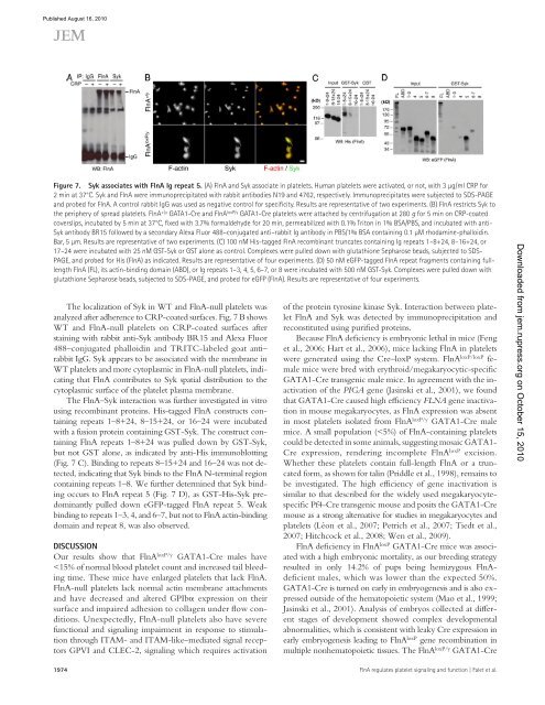
Figure 7. Syk associates with FlnA Ig repeat 5. (A) FlnA and Syk associate in platelets. Human platelets were activated, or not, with 3 µg/ml CRP for<br />
2 min at 37°C. Syk and FlnA were immunoprecipitated with rabbit antibodies N19 and 4762, respectively. Immunoprecipitates were subjected to SDS-PAGE<br />
and probed for FlnA. A control rabbit IgG was used as negative control for specificity. Results are representative <strong>of</strong> two experiments. (B) FlnA restricts Syk to<br />
the periphery <strong>of</strong> spread platelets. FlnA +/y GATA1-Cre and FlnA loxP/y GATA1-Cre platelets were attached by centrifugation at 280 g for 5 min on CRP-coated<br />
coverslips, incubated by 5 min at 37°C, fixed with 3.7% formaldehyde for 20 min, permeabilized with 0.1% Triton in 1% BSA/PBS, and incubated with anti-<br />
Syk antibody BR15 followed by a secondary Alexa Fluor 488–conjugated anti–rabbit Ig antibody in PBS/1% BSA containing 0.1 µM rhodamine-phalloidin.<br />
Bar, 5 µm. Results are representative <strong>of</strong> two experiments. (C) 100 nM His-tagged FlnA recombinant truncates containing Ig repeats 1–8+24, 8–16+24, or<br />
17–24 were incubated with 25 nM GST-Syk or GST alone as control. Complexes were pulled down with glutathione Sepharose beads, subjected to SDS-<br />
PAGE, and probed for His (FlnA) as indicated. Results are representative <strong>of</strong> four experiments. (D) 50 nM eGFP-tagged FlnA repeat fragments containing fulllength<br />
FlnA (FL), its actin-binding domain (ABD), or Ig repeats 1–3, 4, 5, 6–7, or 8 were incubated with 500 nM GST-Syk. Complexes were pulled down with<br />
glutathione Sepharose beads, subjected to SDS-PAGE, and probed for eGFP (FlnA). Results are representative <strong>of</strong> four experiments.<br />
The localization <strong>of</strong> Syk in WT and FlnA-null platelets was<br />
analyzed after adherence to CRP-coated surfaces. Fig. 7 B shows<br />
WT and FlnA-null platelets on CRP-coated surfaces after<br />
staining with rabbit anti-Syk antibody BR15 and Alexa Fluor<br />
488–conjugated phalloidin and TRITC-labeled goat anti–<br />
rabbit IgG. Syk appears to be associated with the membrane in<br />
WT platelets and more cytoplasmic in FlnA-null platelets, indicating<br />
that FlnA contributes to Syk spatial distribution to the<br />
cytoplasmic surface <strong>of</strong> the platelet plasma membrane.<br />
The FlnA–Syk interaction was further investigated in vitro<br />
using recombinant proteins. His-tagged FlnA constructs containing<br />
repeats 1–8+24, 8–15+24, or 16–24 were incubated<br />
with a fusion protein containing GST-Syk. The construct containing<br />
FlnA repeats 1–8+24 was pulled down by GST-Syk,<br />
but not GST alone, as indicated by anti-His immunoblotting<br />
(Fig. 7 C). Binding to repeats 8–15+24 and 16–24 was not detected,<br />
indicating that Syk binds to the FlnA N-terminal region<br />
containing repeats 1–8. We further determined that Syk binding<br />
occurs to FlnA repeat 5 (Fig. 7 D), as GST-His-Syk predominantly<br />
pulled down eGFP-tagged FlnA repeat 5. Weak<br />
binding to repeats 1–3, 4, and 6–7, but not to FlnA actin-binding<br />
domain and repeat 8, was also observed.<br />
DISCUSSION<br />
Our results show that FlnA loxP/y GATA1-Cre males have<br />


![Benyamin Asadipour-Farsani [EngD Conference abstract]](https://img.yumpu.com/51622940/1/184x260/benyamin-asadipour-farsani-engd-conference-abstract.jpg?quality=85)