MEIOFAUNA MARINA - Gastrotricha World Portal
MEIOFAUNA MARINA - Gastrotricha World Portal
MEIOFAUNA MARINA - Gastrotricha World Portal

You also want an ePaper? Increase the reach of your titles
YUMPU automatically turns print PDFs into web optimized ePapers that Google loves.
<strong>MEIOFAUNA</strong> <strong>MARINA</strong><br />
Biodiversity, morphology and ecology<br />
of small benthic organisms<br />
16<br />
pfeil
<strong>MEIOFAUNA</strong> <strong>MARINA</strong><br />
Biodiversity, morphology and ecology of small benthic organisms<br />
Volume 16 • March 2008<br />
pages 1-200, 190 fi gs., 14 tabs.<br />
Chief editors<br />
Andreas Schmidt-Rhaesa Zoological Museum, University Hamburg, Martin-Luther-King-Platz 3, D–20146 Hamburg, Germany<br />
Phone: +49 (0)40 42838-3921, Fax: +49 (0)40 42838-3937<br />
E-mail: andreas.schmidt-rhaesa@uni-hamburg.de<br />
Pedro Martinez Arbízu Deutsches Zentrum für Marine Biodiversitätsforschung, Forschungsinstitut Senckenberg, Südstrand 44,<br />
D–26382 Wilhelmshafen, Germany<br />
E-mail: pmartinez@senckenberg.de<br />
M. Antonio Todaro Dipartimento di Biologia Animale, Università di Modena e Reggio Emilia, Via Campi 213/d, I–41100<br />
Modena, Italia<br />
E-mail: todaro.antonio@unimore.it<br />
Editorial board<br />
Werner Armonies Alfred-Wegener-Institut für Polar- und Meeresforschung, Wattenmeerstation List auf Sylt<br />
Thomas Bartolomaeus Freie Universität Berlin, Germany<br />
Susan Bell University of South Florida, Tampa, FL, U.S.A.<br />
Marco Curini Galletti University of Sassari, Italy<br />
Nicole Dubilier Max-Planck-Institut für Molekulare Mikrobiologie, Bremen, Germany<br />
Peter Funch University of Åarhus, Denmark<br />
Gerhard Haszprunar Zoologische Staatssammlung, München, Germany<br />
Rony Huys Natural History Museum, London, England<br />
Ulf Jondelius University of Uppsala, Sweden<br />
Marianne K. Litvaitis University of New Hampshire, Durham, NH, U.S.A.<br />
Reinhardt Møbjerg Kristensen Zoological Museum, University of Copenhagen, Denmark<br />
Ken-Ichi Tajika Nihon University School of Medicine, Tokyo, Japan<br />
Seth Tyler University of Maine, Orono, ME, U.S.A.<br />
Magda Vincx University Gent, Belgium<br />
Wilfried Westheide Universität Osnabrück, Germany<br />
Meiofauna marina is published annually<br />
Subscriptions should be addressed to the Publisher:<br />
Verlag Dr. Friedrich Pfeil, Wolfratshauser Str. 27, D–81379 München, Germany<br />
PERSONAL SUBSCRIPTION: 48.– Euro<br />
INSTITUTIONAL SUBSCRIPTION: 96.– Euro<br />
Fees for mailing will be added<br />
Manuscripts should be addressed to the editors<br />
Bibliografi sche Information Der Deutschen Bibliothek<br />
Die Deutsche Bibliothek verzeichnet diese Publikation in der Deutschen Nationalbibliografi e;<br />
detaillierte bibliografi sche Daten sind im Internet über<br />
http://dnb.ddb.de abrufbar.<br />
Copyright © 2008 by Verlag Dr. Friedrich Pfeil, München, Germany<br />
All rights reserved.<br />
No part of this publication may be reproduced, stored in a retrieval system, or transmitted in any form or by any<br />
means, electronic, mechanical, photocopying or otherwise, without the prior permission of the copyright owner.<br />
Applications for such permission, with a statement of the purpose and extent of the reproduction, should be<br />
addressed to the Publisher, Verlag Dr. Friedrich Pfeil, Wolfratshauser Str. 27, D–81379 München, Germany.<br />
Printed by Advantage Printpool, Gilching<br />
ISSN 1611-7557<br />
Printed in the European Union<br />
Verlag Dr. Friedrich Pfeil, Wolfratshauser Str. 27, D–81379 München, Germany<br />
Phone: + 49 (0)89 742827-0 • Fax: + 49 (0)89 7242772 • E-mail: info@pfeil-verlag.de • www.pfeil-verlag.de
Meiofauna Marina, Vol. 16, pp. 3-20, 15 figs., March 2008<br />
© 2008 by Verlag Dr. Friedrich Pfeil, München, Germany – ISSN 1611-7557<br />
Meiofauna Marina, Vol. 16<br />
An overview and a dichotomous key to genera<br />
of the phylum <strong>Gastrotricha</strong><br />
M. Antonio Todaro* and William D. Hummon**<br />
Abstract<br />
<strong>Gastrotricha</strong> are microscopic (0.06-3.0 mm in body length) free-living, acoelomate, aquatic worms, characterised by<br />
a meiobenthic life style. In marine habitats they are mainly interstitial, whereas in fresh waters they are ubiquitous<br />
as a component of periphyton and benthos and to a more limited extend also of the plankton. The phylum is cosmopolitan<br />
with about 700 described species grouped into two orders: Macrodasyida, with some 250 strap-shaped<br />
species, all but two of which are marine or estuarine, and Chaetonotida with some 450 tenpin-shaped species,<br />
two thirds of which are freshwater. Macrodasyida include 7 families and 32 genera, whereas Chaetonotida counts<br />
8 families and 30 genera. This key includes several recently described taxa, namely Xenodasyidae, Muselliferidae,<br />
Chordodasiopsis and Diuronotus.<br />
Keywords: meiofauna, invertebrates, benthos, teaching, taxonomy<br />
Introduction<br />
<strong>Gastrotricha</strong> are microscopic (0.06-3.0 mm in<br />
body length) free-living, acoelomate, aquatic<br />
worms, characterised by a meiobenthic life style.<br />
In marine habitats they are mainly interstitial,<br />
whereas in fresh waters they are ubiquitous as<br />
a component of periphyton and benthos and<br />
to a more limited extend also of the plankton.<br />
In marine sediments, gastrotrich density may<br />
reach 364 individuals/10 cm 2 ; typically they rank<br />
third in abundance following the Nematoda and<br />
the harpacticoid Copepoda, although in several<br />
instances they have been found to be first or the<br />
second most abundant meiofaunal taxon (Coull<br />
1985, Todaro et al. 1995, Hochberg 1999).<br />
In freshwater ecosystems population density<br />
may reach 158 ind./10 cm 2 making the taxon<br />
rank among the top 5 most abundant groups. In<br />
aquatic environments the ecological role of the<br />
gastrotrichs is realised within the microphagous,<br />
detritivorous, benthic community. Like free-living<br />
nematodes, gastrotrichs swallow their food, which<br />
is made up of microalgae, bacteria and small<br />
protozoans, by means of the powerful sucking<br />
action of the triradiate muscular pharynx, and in<br />
turn they are preyed upon by turbellarians and<br />
small macrofauna.<br />
The phylum is cosmopolitan with about 700<br />
described species grouped into two orders: Macrodasyida,<br />
with some 250 strap-shaped species, all<br />
but two of which are marine or estuarine, and<br />
* Dipartimento di Biologia Animale, Università di Modena e Reggio Emilia, via Campi, 213/d, 41100 Modena,<br />
Italy; e-mail: todaro.antonio @unimore.it<br />
** Department of Biological Sciences, Ohio University, Athens, Ohio 45701, USA<br />
3
4<br />
Chaetonotida with some 450 tenpin-shaped species,<br />
two thirds of which live in freshwater. Macrodasyida<br />
include 7 families and 32 genera, whereas<br />
Chaetonotida counts 8 families and 30 genera.<br />
However due to the numerous species, and at least<br />
three new genera that wait to be described, these<br />
statistics should be considered as very conservative,<br />
particularly for the Chaetonotida. Despite<br />
their diversity and abundance, the phylogenetic<br />
relationships of the <strong>Gastrotricha</strong> are still unclear.<br />
Based on morphology, most researchers, though<br />
considering the evolutionary connections of these<br />
worms to be quite obscure, regard them as close<br />
allies of the Gnathostomulida, the Rotifera, or the<br />
Nematoda. On the other hand, a re-examination of<br />
the “Aschelminthes” phylogeny based on the SSU<br />
rRNA gene sequence analysis showed the <strong>Gastrotricha</strong><br />
as the sister taxon of the Platyhelminthes,<br />
while later studies placed them close to the<br />
Ecdysozoa, the Lophotrochozoa, or neither one.<br />
Such discrepancies between the traditional and<br />
the modern views on the gastrotrich phylogeny<br />
suggest that further research in this direction is<br />
necessary (see Todaro et al. 2006a). Unclear are<br />
also the in-group phylogenetic relationships;<br />
representative of the two orders Macrodasyida<br />
and Chaetonotida are so different to cast doubt<br />
about their affiliation to the same phylum (Todaro<br />
et al. 2003a). Fortunately, relationships among<br />
taxa belonging to different families are becoming<br />
less obscure (Todaro et al. 2006b, Leasi & Todaro<br />
2008).<br />
An introduction to the gastrotrichs and their<br />
morphology can be obtained from several sources,<br />
e.g. d’Hondt (1971), Hummon (1982), Ruppert<br />
(1988, 1991) and Balsamo & Todaro (2002).<br />
Materials and methods<br />
Sampling techniques in marine systems and<br />
freshwater habitats are generally similar; qualitative<br />
sampling involves the collection of sediment<br />
using a shovel, a jar or a corer, while quantitative<br />
work mostly uses a small corer (2-5 cm inner diameter),<br />
such as a syringe with the tip cut-off. The<br />
marine-estuarine species are mostly interstitial,<br />
living amid relatively clean, fine to coarse sands,<br />
although some are tolerant of high organic, sulfide<br />
or pollution loads, and a few even occur in mud<br />
and oozes (e.g. Hummon et al. 1990; Todaro &<br />
Rocha 2004, 2005; Hummon 2006; Todaro et al.<br />
2006c; Balsamo et al. 2007).<br />
Qualitative littoral samples are usually taken<br />
by digging holes in the beach and removing the<br />
sediment from the wall and the bottom of the hole<br />
with a scoop or spoon whereas bulk sublittoral<br />
sediments can be collected directly removing<br />
sediment from the top 10-cm layer with a 500-ml<br />
plastic jar. Replicated small samples are more<br />
representative of the community of a site than a<br />
single large sample, because the distribution of<br />
gastrotrichs, as most meiofaunal taxa, is patchy.<br />
Similar techniques apply to the interstitial forms<br />
of freshwater habitats. Periphytic and semipelagic<br />
freshwater species are collected by sampling<br />
clumps of vegetation mixed with sediment and<br />
by repeatedly filtering the water through a 30 μm<br />
mesh plankton net (Hummon 1981). Both marine<br />
and freshwater samples should be processed<br />
within a week to extract the living animals, which<br />
are generally more suitable than fixed specimens.<br />
For freshwater samples only, additional checks<br />
some time after collection are recommended, since<br />
as a result of resting eggs, species initially absent<br />
may be found later. Interstitial gastrotrichs can be<br />
extracted from the sediment by narcotisation with<br />
aqueous solution of MgCl 2 (7 % marine or 1 %<br />
freshwater); to this end place a spoonful of sand in<br />
a small jar, add enough narcotic solution to cover<br />
the sand, swirl, leave for 10 min, swirl, decant<br />
into a small Petri dish, add an equal amount of<br />
either seawater or freshwater and observe under<br />
a dissecting microscope at 40-50 × magnification.<br />
Periphytic species are extracted from the vegetation<br />
by repeatedly rinsing and squeezing the<br />
plants, and the supernatant is filtered through a<br />
30 μm mesh sieve. For the qualitative extraction<br />
of epibentic freshwater forms, either stirring the<br />
sediment into a suspension and decanting the water<br />
through a fine mesh net or centrifugation using<br />
a density gradient are suitable methods. To study<br />
living specimens, use a micropipette for transfer<br />
to a slide, then use modelling clay posts beneath<br />
the corners of 15-18 mm square coverslips, and<br />
observe under a compound microscope using<br />
differential interference contrast optics (DIC).<br />
Cuticular details may require SEM survey, for<br />
which specimens are prepared by critical point<br />
drying or the hesamethildysilazane technique<br />
(e.g. Todaro 1992, Hochberg & Litvaitis 2000).<br />
For quantitative studies, a treatment of the<br />
samples with an aqueous solution of MgCl 2<br />
for 10 minutes is highly recommended prior<br />
to fixation. Preservation may be carried out in<br />
10 % borax-buffered formalin with rose bengal<br />
Todaro & Hummon: Key to genera of the phylum <strong>Gastrotricha</strong>
(1 %) to facilitate sorting. The gastrotrichs of<br />
the quantitative sample can separated from the<br />
sediment, generally by flotation and multiple<br />
decantations. If samples are richer in detritus,<br />
extraction can be performed by using the silica<br />
gel gradient centrifugation technique (LUDOX<br />
AM, d = 1.210; Pfannkuche & Thiel 1988). The<br />
supernatant can be filtered using a 30 μm mesh<br />
sieve or, better, poured directly into a Petri<br />
dish for locating gastrotrichs; identification can<br />
performed on specimens mounted in water, or<br />
better, based on permanent mounts. These can<br />
be prepared in 10 % formalin or in a mixture of<br />
formalin-glycerol (3 : 1) and sealed with glyceel<br />
or nail polish. Gastrotrichs can also be mounted<br />
in pure glycerine on H-S slide after treatment in<br />
a solution of 5 % glycerine 95 % ethyl alcohol for<br />
1-2 days (Lee & Chang 2003). However, in many<br />
cases permanent mounts do not allow a complete<br />
taxonomic study, as several diagnostic features<br />
deteriorate over time.<br />
The following key (modified from Hummon<br />
1973 & Balsamo & Todaro 2002) includes all of<br />
the genera of marine, brackish and freshwater<br />
gastrotrichs known all over the world. It is designed<br />
for use by biologists who identify animals<br />
as part of their normal work, but who may have<br />
little familiarity with the gastrotrich fauna. For<br />
the inclusion of Muselliferidae see Leasi & Todaro,<br />
(2008); for the exclusion of Metadasydytes see<br />
W. D. Hummon (this volume). Finally the readers<br />
should be informed that W. D. Hummon (this<br />
volume) is proposing a name change for the genus<br />
Platydasys.<br />
The key is pragmatic in approach and is based<br />
on important discriminatory characters as seen in<br />
relaxed adult specimens. Where possible characters<br />
are those which are readily visible, using DIC<br />
optics, and which are quantifiable. Each member<br />
of a couplet is given the identifying number of the<br />
couplet and a letter, a or b; the number and letter<br />
in parenthesis refers to that member of a previous<br />
couplet from which a particular couplet was<br />
derived. Figures of genera are identified according<br />
to the couplet members to which they refer.<br />
Finally, nothing replaces experience, especially<br />
that gained by intensive study of as broad a range<br />
of genera and species as it is possible to obtain.<br />
Meiofauna Marina, Vol. 16<br />
Results<br />
Key to the genera of <strong>Gastrotricha</strong><br />
1a Body tenpin-shaped; posterior end furcate<br />
(furca); anterior, lateral and dorsal adhesive<br />
tubes absent; posterior adhesive tubes numbering<br />
two (exceptionally four, or absent) at<br />
the tip of the furcal branches. Mouth opening<br />
narrow (< 0.4 × head width); pharyngeal<br />
pores absent. Mostly common; marine,<br />
brackish and freshwater: interstitial, epibenthic<br />
and periphytic; occasionally semiplanktonic.<br />
Order CHAETONOTIDA, Suborder<br />
PAUCITUBULATINA (Fig. 1A). ...............34<br />
1b These characteristics not combined. ...........2<br />
2a (1b) Body worm-shaped, anterior and dorsal<br />
adhesive tubes absent; lateral adhesive tubes,<br />
present, although often inconspicuous (in<br />
form of papillae), several per side; posterior<br />
adhesive tubes, several per side, fused at<br />
their bases forming two adhesive organs;<br />
mouth narrow (< 0.4 × head width), opens<br />
by means of a projecting cuticular tube,<br />
pharyngeal pores absent. Uncommon; marine:<br />
interstitial. Order CHAETONOTIDA,<br />
Suborder MULTITUBULATINA, NEO-<br />
DASYIDAE. ...................... Neodasys (Fig. 1C)<br />
2b (1b) Body tenpin- or, more often, wormshaped;<br />
anterior, lateral and posterior adhesive<br />
tubes present, usually numerous; dorsal<br />
adhesive tubes present in several taxa; mouth<br />
opening narrow to broad; pharyngeal pores<br />
usually present, though occasionally inconspicuous.<br />
Marine and brackish rarely fresh<br />
water: interstitial. Order MACRODASYIDA<br />
(Fig. 1B). ..........................................................3<br />
3a (2b) Marine or brackish. ...............................4<br />
3b (2b) Freshwater. INCERTAE SEDIS<br />
(Fig. 9). ..........................................................33<br />
4a (3a) Body tenpin-shaped; head well defined,<br />
includes most (> 0.8 × length) of pharynx;<br />
dorsal adhesive tubes absent, posterior end<br />
lobed, furcate or bifurcate. Cuticle smooth,<br />
or forming crests and thickenings; musculature<br />
clearly cross-striated. .........................5<br />
4b (3a) Body worm-shaped, head usually not<br />
distinct or, when distinct, includes only part<br />
(< 0.5 × length) of pharynx; cuticle smooth<br />
or forming scales and/or spines. ...............9<br />
5
6<br />
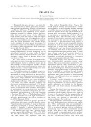
Fig. 1. A. Drawing of a hypothetical Chaetonotida Paucitubulatina. B. Drawing of a hypothetical Macrodasyida.<br />
C. Neodasys (Chaetonotida, Multitubulatina). Aao, accessory adhesive organs; an, anus; cat, caudal adhesive<br />
tubes; cog, caudal organ; cyr, cyrtocytes; eg, egg; fog, frontal organ; fu, furca; hp, hypostomion; int, intestine;<br />
lat, lateral adhesive tubes; lc, locomotor cilia; mth, mouth; pha, pharynx; phij, pharyngeo-intestinal junction;<br />
php, pharyngeal pores; pl, pleuria; sk, keeled scale; sns, scales with notched spines; sp, sperm; ss, smooth<br />
scales; sss, scales with simple spines; tbr, tactile bristle; xog, X-organ. A, B, modified from Balsamo & Todaro<br />
(2003); C, Modified from Ruppert (1988).<br />
5a (4a) Cuticle smooth; dorsal side of the trunk<br />
bare; chordoid organ absent. Common to<br />
rare; marine and brackish: interstitial. DAC-<br />
TYLOPODOLIDAE (Fig. 2). ........................6<br />
5b (4a) Cuticle forming crests and thickenings,<br />
if smooth dorsal side of the trunk bearing<br />
long rod-like structure; chordoid organ<br />
present. Rare; marine: interstitial. XENO-<br />
DASYIDAE (Fig. 3). ......................................8<br />
6a (5a) Head simple or with a pair of laterally<br />
directed tentacles; cuticle smooth; posterior<br />
end bilobed. Lateral adhesive tubes present.<br />
Often common; marine: interstitial. .............<br />
.................................... Dactylopodola (Fig. 2A)<br />
hp<br />
pl<br />
sss<br />
lc<br />
sss<br />
cyr<br />
sns<br />
sp<br />
lc<br />
ss<br />
sk<br />
xog<br />
tbr<br />
fu<br />
mth<br />
pha<br />
lat<br />
php<br />
aao<br />
phij<br />
sp<br />
int<br />
eg<br />
fog<br />
cog<br />
an<br />
cat<br />
A B C<br />
6b (5a) Head simple or with tentacles; cuticle<br />
smooth; posterior end bifurcate. Lateral<br />
adhesive tubes absent. ..................................7<br />
7a (6b) Head simple, without tentacles; cuticle<br />
smooth. Rare; marine: interstitial. ................<br />
..................................... Dendropodola (Fig. 2B)<br />
7b (6b) Head with elongate crenulated lateral<br />
lobes. Uncommon; marine: interstitial. ........<br />
...................................... Dendrodasys (Fig. 2C)<br />
8a (5b) Trunk lacking rod-like structure, but<br />
showing conspicuous indentations along the<br />
lateral margins; posterior end furcate, each<br />
branch ending with short adhesive tubes.<br />
Rare; marine: interstitial. ................................<br />
..........................................Xenodasys (Fig. 3A)<br />
Todaro & Hummon: Key to genera of the phylum <strong>Gastrotricha</strong>
A B C<br />
Fig. 2. Macrodasyida, Dactylopodolidae – Representatives of the genera Dactylopodola (A), Dendropodola (B),<br />
Dendrodasys (C). Scale bars = 100 μm. A, modified from Ruppert (1988); B, modified from Hummon et al. (1998);<br />
C modified from Hummon et al. (1993).<br />
8b (5b) Trunk showing several tentacles; lateral<br />
margins of the trunk parallel, without<br />
indentations; posterior end furcate; each<br />
branch ending with an adhesive pad. Rare;<br />
marine: interstitial. ..........................................<br />
..................................Chordodasiopsis (Fig. 3B)<br />
9a (4b) Anterior adhesive tubes (generally 4 or<br />
more per side, occasionally 2 or 3) borne on<br />
extensible fleshy base; pharyngeal pores<br />
located at base of pharynx. ........................10<br />
9b (4b) Anterior adhesive tubes borne more or<br />
less directly on ventral body surface (generally<br />
1 to 3 per side (occasionally 4 or more);<br />
pharyngeal pores located at base of pharynx<br />
or in mid-pharyngeal region. ....................16<br />
10a (9a) Head usually well delimited posteriorly<br />
by a constriction; posterior end broadly<br />
expanded, rounded, truncated, or tapered<br />
into a medial process, but not bilobed. LEPI-<br />
DODASYIDAE (part) (Fig. 4). ...................11<br />
10b (9a) Head usually not well delimited; posterior<br />
end bilobed. TURBANELLIDAE<br />
(part) (Fig. 5). ...............................................12<br />
Meiofauna Marina, Vol. 16<br />
A B<br />
Fig. 3. Macrodasyida, Xenodasyidae – Representatives<br />
of the genera Xenodasys (A), Chordodasiopsis (B). Scale<br />
bars = 200 μm. A, modified from Ruppert 1988; B, modified<br />
from Rieger et al. (1974).<br />
7
8<br />
A<br />
G<br />
B<br />
C<br />
D<br />
Fig. 4. Macrodasyida, Lepidodasyidae – Representatives of the genera Cephalodasys (A), Pleudodasys (B), Mesodasys<br />
(C), Megadasys (D), Dolichodasys (E), Lepidodasys (F), Paradasys (G). Scale bar = 200 μm. A-F, modified<br />
from Ruppert (1988); G, original (W. D. Hummon).<br />
E<br />
Todaro & Hummon: Key to genera of the phylum <strong>Gastrotricha</strong><br />
F
Meiofauna Marina, Vol. 16<br />
A<br />
E F<br />
Fig. 5. Macrodasyida, Turbanellidae – Representatives of the genera Dinodasys (A), Pseudoturbanella (B), Paraturbanella<br />
(C), Turbanella (D), Prostobuccantia (E), Desmodasys (F). Scale bars = 200 μm. A, original (W. D. Hummon);<br />
B-D, modified from Ruppert (1988); E, modified from Evans & Hummon (1991); F, modified from Clausen<br />
(2000).<br />
B<br />
D<br />
C<br />
9
10<br />
11a (10a) A pair of laterally directed accessory<br />
adhesive organs present near the pharyngeointestinal<br />
junction, each comprised of four<br />
adhesive tubes; a pair of drumstick-like<br />
organs on the dorsal side of the pharyngeal<br />
region. Rare, marine: interstitial. ..................<br />
........................................ Pleurodasys (Fig. 4B)<br />
11b (10a) Laterally directed accessory adhesive<br />
and drumstick-like organs, such as those<br />
described above, absent. Regionally common;<br />
marine and brackish: interstitial. ........<br />
..................................... Cephalodasys (Fig. 4A)<br />
12a (10b) Head bearing elongate (> 0.5 × head<br />
width) lateral tentacles. ..............................13<br />
12b (10b) Head perhaps bearing conical lobes<br />
(< 0.5 × head width), but not tentacles. ....14<br />
13a (12a) Lateral adhesive tubes numerous.<br />
Uncommon; marine: interstitial. ...................<br />
.......................................... Dinodasys (Fig. 5A)<br />
13b (12a) Lateral adhesive tubes absent, a single<br />
pair of ventral adhesive tubes inserting just<br />
behind pharyngo-intestinal junction. Rare;<br />
marine: interstitial. ..........................................<br />
................................Pseudoturbanella (Fig. 5B)<br />
14a (12b) A pair of posteriorly directed ventrolateral<br />
accessory adhesive organs present in<br />
the anterior portion of the pharyngeal region,<br />
each organ comprising 2 adhesive tubes of<br />
unequal lengths. Common; marine and<br />
brackish: interstitial. ........................................<br />
.................................. Paraturbanella (Fig. 5 C)<br />
14b (12b) Ventrolateral accessory adhesive organs<br />
such as those described above absent,<br />
or located in different region of the body. ..<br />
........................................................................15<br />
15a (14b) Accessory adhesive organs, absent.<br />
Common; marine and brackish: interstitial.<br />
..........................................Turbanella (Fig. 5D)<br />
15b (14b) Accessory adhesive organs arising near<br />
the pharyngo-intestinal junction. Rare; marine:<br />
interstitial. ......Prostobuccantia (Fig. 5E)<br />
16a (9b) Pharyngeal pores located in mid-pharyngeal<br />
region, posterior end of body tapered<br />
into a medial process. MACRODASYIDAE<br />
(Fig. 6). ..........................................................17<br />
16b (9b) Pharynpeal pores located at base of<br />
pharynx; posterior end of body not tapered<br />
into a medial process. .................................18<br />
17a (16a) Posterior process short (< 0.2 × length<br />
of head, trunk). Common; marine: interstitial.<br />
.................................Macrodasvs (Fig. 6A)<br />
17b (16a) Posterior process elongate (> 0.8 ×<br />
length of head, trunk). Regionally common;<br />
marine: interstitial and epibenthic................<br />
.............................................Urodasys (Fig. 6B)<br />
18a (16b) Cuticular structure present in the form<br />
of thickenings, scales, papillae or hooks. ....<br />
........................................................................19<br />
18b (16b) Cuticle naked, lacking armature. ....25<br />
19a (18a) Mouth opening narrow (< 0.4 × head<br />
width); cuticular armature of elongate,<br />
keeled thickenings. Uncommon; marine:<br />
interstitial. LEPIDODASYIDAE (part). .......<br />
.........................................Lepidodasys (Fig. 4F)<br />
19b (18a) Mouth opening-broad (> 0.6 × head<br />
width); cuticular armature of broadened<br />
scales, papillae or variously spined<br />
hooks. THAUMASTODERMATIDAE (part)<br />
(Fig. 7). ..........................................................20<br />
20a (l9b) Cuticular armature with broadened<br />
scales or papillae. ........................................21<br />
20b (l9b) Cuticular armature with uni- or multi-<br />
spined hooks. ...............................................22<br />
21a (20a) Cuticular scales present, but not papillae;<br />
a single row of wide spines present on<br />
either side of body; testes paired. Uncommon;<br />
marine: interstitial. ................................<br />
......................................... Diplodasys (Fig. 7A)<br />
21b (20a) Cuticular papillae present, but not<br />
scales or spines; testis present on right side<br />
only. Uncommon; marine: interstitial. .........<br />
.......................................... Platydasys (Fig. 7B)<br />
22a (20b) Cuticular amature with uni-spined<br />
hooks; testes paired. Common; marine: interstitial.<br />
.....................Acanthodasys (Fig. 7C)<br />
22b (20b) Cuticular armature with multi-spined<br />
hooks; testis present on right side only. ......<br />
........................................................................23<br />
23a (22b) Buccal palps (flashy grasping structures<br />
preceeding on either sides the mouth basket),<br />
present; hooks 3-, 4- or 5-spined (triancres,<br />
tetrancres and pentancres). Common; marine:<br />
interstitial. ...... Pseudostomella (Fig. 7D)<br />
23b (22b) Buccal palps absent; hooks 3-, 4- or<br />
5-spined (tri-, tetra- or pentancres). ........24<br />
Todaro & Hummon: Key to genera of the phylum <strong>Gastrotricha</strong>
24a (23b) Head with 2 pairs of laterally directed<br />
tentacles; hooks 4-spined. Common; marine:<br />
interstitial. .............Thaumastoderma (Fig. 7E)<br />
24b (23b) Head with 0 or 1, pair of laterally directed<br />
tentacles; hooks 3- 4- or 5-spined. Very<br />
common; marine: interstitial. ........................<br />
.............................. Tetranchyroderma (Fig. 7F)<br />
25a (18b) Anterior adhesive tubes several to<br />
many, borne in a tuft; lateral adhesive tubes<br />
absent. Rare; marine: interstitial. TUR-<br />
BANELLIDAE (part) (Fig. 5). ........................<br />
.......................................Desmodasys (Fig. 5H)<br />
25b (18b) Anterior adhesive tubes few to many,<br />
but not borne in a tuft; lateral adhesive tubes<br />
usually present or, if absent, then anterior<br />
adhesive tubes few. .....................................26<br />
26a (25b) Anterior, lateral and posterior adhesive<br />
tube groups with many tubes each (> 10 per<br />
side); mouth narrow (< 0.4 × head width) and<br />
posterior end distinctly bilobed. PLANO-<br />
DASYIDAE (Fig. 8). ....................................27<br />
26b (25b) At least one group with few to several<br />
adhesive tubes (< 6 per side); mouth narrow<br />
to broad, if narrow, then posterior end not<br />
distinctly bilobed. ........................................28<br />
27a (26a) Tail lobes form oval appendages on<br />
the posterior end; most anterior adhesive<br />
tubes arranged transversely; bursa elongate.<br />
Rare; marine: interstitial. ................................<br />
.........................................Planodasys (Fig. 8A)<br />
27b (26a) Tail lobes form furcate extensions on<br />
the posterior end; most anterior adhesive<br />
tubes arranged longitudinally; bursa ovate.<br />
Uncommon; marine: interstitial. ...................<br />
..............................................Crasiella (Fig. 8B)<br />
28a (26b) Mouth narrow (< 0.4 × head width);<br />
testes paired. LEPIDODASYIDAE (part)<br />
(Fig. 4). ..........................................................29<br />
28b (26b) Mouth broad (> 0,6 × head width) or,<br />
if narrow, leading to a large buccal cavity<br />
surrounded by an oral hood; testis present<br />
on right side only. THAUMASTODERMA-<br />
TIDAE (part). ...............................................32<br />
29a (28a) fully grown adults (both sexual apparati<br />
and mature gametes present) usually<br />
less than 1 mm in total length. ..................30<br />
29b (28a) fully grown adults up to 3.5 mm in<br />
total length, always exceeding 1 mm. ......31<br />
Meiofauna Marina, Vol. 16<br />
A<br />
B<br />
11<br />
Fig. 6. Macrodasyida, Macrodasyidae – Representatives<br />
of the genera Macrodasys (A), Urodasys (B). Scale<br />
bar = 200 μm. A, B, modified from Ruppert (1988).<br />
30a (29a) Anterior adhesive tubes in two groups<br />
of 1-4 tubes per side; lateral adhesive tubes<br />
absent or less than 6 per side. Uncommon;<br />
marine: Interstitial. .........Paradasys (Fig. 4G)<br />
30b (29a) Anterior adhesive tubes few to several;<br />
lateral adhesive tubes several to many per<br />
side. Common; marine: interstitial. ..............<br />
..........................................Mesodasys (Fig. 4C)<br />
31a (29b) Anterior adhesive 1 per side; lateral<br />
adhesive tubes inconspicuous (in form of<br />
papillae). Uncommon; marine: interstitial. .<br />
.......................................Dolichodasys (Fig. 4E)<br />
31b (29b) Anterior adhesive few to several; lateral<br />
adhesive tubes distinct, many per side.<br />
Uncommon; marine: interstitial. ...................<br />
......................................... Megadasys (Fig. 4D)
12<br />
A<br />
E F<br />
32a (28b) Mouth broad; ventral locomotor cilia<br />
not restricted to pharyngeal region; male<br />
genital pore lacking cuticular plates. Common;<br />
marine: interstitial. ................................<br />
...................................Ptychostomella (Fig. 7G)<br />
32b (28b) Mouth narrow, leading to a large buccal<br />
cavity surrounded, by an oral hood;<br />
ventral locomotor cilia restricted to pharyn-<br />
B<br />
Fig. 7. Macrodasyida, Thaumastodermatidae – Representatives of the genera Diplodasys (A), Platydasys (B), Acanthodasys<br />
(C), Pseudostomella (D), Thaumastoderma (E), Tetranchyroderma (F), Ptychostomella (G), Hemidasys (H).<br />
A-F, H, bar scale = 200 μm, G, bar scale = 50 μm. A, Modified from Luporini et al. (1973); B-F, modified from<br />
Ruppert (1988); G, modified from Hummon et al. (1993); H, modified from Claparéde (1864).<br />
C<br />
G<br />
geal region; male genital pore surrounded<br />
by cuticular plates. Very rare (possibly extinct);<br />
marine: intestitial. ................................<br />
......................................... Hemidasys (Fig. 7H)<br />
33a (3b) Body length from 300 to 400 μm; two<br />
pairs of anterior ventral adhesive tubes. Rare,<br />
interstitial. ........................Redudasys (Fig. 9B)<br />
Todaro & Hummon: Key to genera of the phylum <strong>Gastrotricha</strong><br />
D<br />
H
33b (3b) Body length up to 220 μm; one pair of<br />
anterior (possibly ventral) adhesive tubes.<br />
Rare, interstitial. ..........Marinellina (Fig. 9A)<br />
34a (1a) Locomotor cilia beneath pharyngeal<br />
region inserted as tightly-packed “hypotrichous”<br />
cirri, occurring in two longitudinal<br />
rows. Marine and brackish. XENOTRI-<br />
CHULIDAE (Fig. 10)...................................35<br />
34b (1a) Locomotor cilia beneath pharyngeal<br />
region inserted individually, occurring in<br />
longitudinal rows, loose tufts or as a uniform<br />
field, never organised in cirri. Marine, brackish<br />
and freshwater. ......................................37<br />
35a (34a) Locomotor cirri of two or more sizes,<br />
generally with 1-2 transverse rows of tiny<br />
cirri anteriorly and two isolated tufts of<br />
cirri in the mid-trunk region; pharynx bearing<br />
a bulb anteriorly. Common; marine and<br />
brackish: interstitial. ........................................<br />
.......................... Heteroxenotrichula (Fig. 10A)<br />
35b (34a) Locomotor cirri of nearly the same size;<br />
two isolated tufts of cirri in the mid-trunk<br />
region present or absent (not seen); pharynx<br />
cylindrical, without bulb. ..........................36<br />
Meiofauna Marina, Vol. 16<br />
A<br />
A<br />
B<br />
13<br />
Fig. 8. Macrodasyida, Planodasyidae – Representatives<br />
of the genera Planodasys (A), Crasiella (B). Scale<br />
bar = 200 μm. A, B, modified from Ruppert (1988).<br />
Fig. 9. Incertae sedis – Representatives of the genera Marinellina (A), Redudasys (B). Scale bars = 50 μm. A, Modified<br />
from Ruttner-Kolisko (1955); B, modified from Kisielewski (1987).<br />
B
14<br />
A<br />
36a (35b) Male apparatus absent; head well<br />
defined; dorsal scales flat; lateral mid-trunk<br />
scales peduncolated; single spine on either<br />
side of the base of the caudal furca. Common;<br />
marine: interstitial. ..........................................<br />
................................... Draculiciteria (Fig. 10B)<br />
B C<br />
Fig. 10. Chetonotida, Xenotrichulidae – Representatives of the genera Heteroxenotrichula (A), Draculiciteria (B),<br />
Xenotrichula (C). Scale bars = 50 μm. A, C, original (W. D. Hummon); B, modified from Luporini et al. (1973).<br />
A<br />
B C<br />
Fig. 11. Chetonotida: A, Dichaeturidae – Representative<br />
of the genus Dichaeura, B, C, Proichthydidae – Representatives<br />
of the genera Proichthydium (B), Proichthydioides<br />
(C). Scale bars = 50 μm. A, modified from Balsamo<br />
(1983); B, modified from Cordero (1918); C, Redrawn<br />
from Sudzuki (1971).<br />
36b (35b) Male apparatus present; pharynx<br />
without anterior bulb; head usually indistinct;<br />
lateral mid-trunk scales simple, flat, or<br />
pedunculated; if pedunculated, similar to<br />
dorsal mid-trunk scales. Common; marine<br />
and brackish: interstitial. ................................<br />
.................................... Xenotrichula (Fig. 10C)<br />
37a (34b) Furcal branches present, with or without<br />
adhesive tubes. .....................................38<br />
37b (34b) Furcal branches absent; posterior body<br />
end truncated or rounded with possible<br />
presence of two protuberances or spines. ...<br />
........................................................................54<br />
38a (37a) Posterior end bifurcate, with 4 adhesive<br />
tubes or 2 adhesive tubes and 2 spiny processes;<br />
cuticle naked, lacking armature. Rare;<br />
freshwater: interstitial. DICHAETURI-<br />
DAE. ............................. Dichaetura (Fig. 11A)<br />
38b (37a) Posterior end truncated, bilaterally<br />
projected into protuberances, or furcate, with<br />
0, 2 or 4 adhesive tubes; cuticular armature<br />
in form of scales and/or spines present or<br />
absent. ...........................................................39<br />
39a (38b) Cuticular armature absent; caudal<br />
adhesive tubes sickle-shaped; cephalic cilia<br />
not grouped into tufts. Very rare; freshwater:<br />
hyperbenthic or semiplanktonic. PROICH-<br />
THYDIIDAE (Fig. 11B,C). ..........................40<br />
Todaro & Hummon: Key to genera of the phylum <strong>Gastrotricha</strong>
39b (38b) Cuticular scales and spines generally<br />
present; if present caudal adhesive tubes<br />
mostly straight, long to very short; cephalic<br />
cilia grouped into tufts or in a band encircling<br />
a muzzle-like anterior projection of head,<br />
which bears the mouth. ..............................41<br />
40a (39a) A transverse row of short dorsal cephalic<br />
cilia; ventral cilia arranged in tufts,<br />
only present on the head and neck regions.<br />
Fresh water: hyperbenthic. ............................<br />
..................................Proichthydium (Fig. 11B)<br />
40b (39a) No dorsal cephalic cilia; ventral cilia<br />
arranged in two longitudinal bands. Fresh<br />
water: semiplanktonic. ...................................<br />
...............................Proichthydioides (Fig. 11C)<br />
41b (39b) Cephalic cilia in 1 or 2 pairs of tufts,<br />
inserting dorso-or ventro laterally on head.<br />
Common (except Arenotus and Undula);<br />
marine, brackish and freshwater: periphytic,<br />
epibenthic and interstitial CHAETONOTI-<br />
DAE (Fig. 12). .............................................42<br />
41a (39b) Cephalic cilia in a band, encircling a<br />
muzzle-like anterior projection of head,<br />
which bears the mouth; two or four posterior<br />
adhesive tubes. Uncommon to rare,<br />
marine: interstitial or infaunal. MUSELLI-<br />
FERIDAE (Fig. 15). ......................................61<br />
42a (41b) Caudal appendages with adhesive<br />
tubes. .............................................................43<br />
42b (41b) Caudal appendages without adhesive<br />
tubes. Rare; fresh water: epibenthic. ............<br />
.............................................Undula (Fig. 12A)<br />
43a (42a) Caudal furca very long (up to 1 /3 of the<br />
total length), segmented, naked or bearing<br />
very small scales or spines. Common; freshwater:<br />
epibenthic, periphytic. ........................<br />
.....................................Polymerurus (Fig. 12B)<br />
43b (42a) Caudal furca from mid length to very<br />
short, unsegmented, without scales or<br />
spines.............................................................44<br />
44a (43b) Body cuticle smooth or with numerous,<br />
non-spined scales; occasionally a few spines<br />
at the furcal base. ........................................45<br />
44b (43b) Body cuticle covered with numerous<br />
spined and/or keeled scales; short to very<br />
long spines, simple or with 1-2 lateral notches.<br />
...................................................................52<br />
Meiofauna Marina, Vol. 16<br />
15<br />
45a (44a) Body cuticle smooth. .........................46<br />
45b (44a) Body cuticle with non-spined scales.<br />
........................................................................48<br />
46a (45a) Thin, smooth cuticle which may show<br />
very tiny, longitudinal lines; rarely a few<br />
spines at the furcal base. Common; freshwater,<br />
rarely marine or brackish-water: epibenthic,<br />
periphytic, interstitial. ........................47<br />
46b (45a) Very thick, smooth, cuticle clearly<br />
distinguishable from the epidermis. Rare;<br />
freshwater: interstitial. .. Arenotus (Fig. 12C)<br />
47a (46a) Furcal base pedunculate; ventral locomotory<br />
cilia arranged in tufts. Uncommon;<br />
marine: interstitial. ..........................................<br />
.............................. Caudichthydium (Fig. 12D)<br />
47b (46a) Furcal base not pedunculate; vental<br />
locomotory cilia arranged in two longitudinal<br />
bands. Common; freshwater, rarely<br />
marine or brackish-water. ..............................<br />
....................................... Ichthydium (Fig. 12E)<br />
48a (45b) Scales small with a stalk or a keel. 49<br />
48b (45b) Scales large, flat, polygonal, rhomboidal<br />
or circular in shape. ..............................50<br />
49a (47a) Scales with a stalk. Common; freshwater,<br />
brackish-water, marine: epibenthic, periphytic,<br />
interstitial. ...........................................<br />
...................................Aspidiophorus (Fig. 12F)<br />
49b (47a) Scales with a keel. Common; freshwater,<br />
brackish-water, marine: epibenthic, periphytic,<br />
interstitial. ...........................................<br />
.......................... Heterolepidoderma (Fig. 12G)<br />
50a (48b) Numerous polygonal scales. Common;<br />
freshwater, rarely brackish-water and marine:<br />
epibenthic, periphytic, interstitial ........<br />
.................................Lepidodermella (Fig. 12H)<br />
50b (48b) Scales rhomboidal or circular in<br />
shape. ............................................................51<br />
51a (50b) Few circular scales. Rare; freshwater:<br />
periphytic. ................... Fluxiderma (Fig. 12I)<br />
52b (50b) Numerous rhomboidal scales. Rare;<br />
freshwater: periphytic. ...................................<br />
...............................Rhomballichthys (Fig. 12L)<br />
52a (44b) Dorsal scales with a double anterior<br />
edge, lacking a keel but with or without a<br />
spine; ventral, interciliary scales similar in<br />
shape to the dorsal scales; some pairs of long
16<br />
A<br />
B<br />
C D<br />
E F G H I<br />
L M N O<br />
Fig. 12. Chetonotida, Chaetonotidae – Representative of the genera Undula (A), Polymerurus (B), Arenotus (C), Caudichthydium<br />
(D), Ichthydium (E), Aspidiophorus (F), Heterolepidoderma (G), Lepidodermella (H), Fluxiderma (I), Rhomballichthys<br />
(L), Lepidochaetus (M), Halichaetonotus (N), Chaetonotus (O). Scale bars = 50 μm. A, modified from<br />
Kisielewski (1991); B, E-H, M, Modified from Balsamo (1983); C, Modified from Kisielewski (1987); D, Modified<br />
from Mock (1979); I, L, modified from Swank 1991; N, Modified from Schrom (1972); O, modified from Hummon<br />
et al. (1992).<br />
Todaro & Hummon: Key to genera of the phylum <strong>Gastrotricha</strong>
A B<br />
Fig. 13. Chetonotida, Neogosseidae – Representative of the genera Neogossea (A), Kijanebalola (B). Scale<br />
bars = 50 μm. A, modified from Balsamo (1983); B, modified from Kisielewski (1990).<br />
and very thin spines at the sides of the furcal<br />
base. Quite common; freshwater: epibenthic,<br />
periphytic. ................................................<br />
..................................Lepidochaetus (Fig. 12M)<br />
52b (44b) Dorsal scales with a single anterior<br />
edge and a keel or a keel and/or a spine;<br />
ventral, interciliary scales different in shape<br />
from the dorsal scales. Very common; marine,<br />
brackish-water and freshwater: interstitial,<br />
epibenthic, periphytic. ....................53<br />
53a (52b). Ventrolateral scales adjacent to locomotor<br />
ciliary tract with spines bearing lamellae<br />
(hydrofoil scales); dorsal scales with a<br />
keel; if present spines restricted to 1-3 scales.<br />
Common; marine and brachish-water: interstitial.<br />
....................Halichaetonotus (Fig. 12N)<br />
53a (52b). Hydrofoil scales usually absent; if<br />
present dorsal scales with spines. Very common;<br />
marine, brackish-water and freshwater:<br />
interstitial, epibenthic, periphytic. ................<br />
.....................................Chaetonotus (Fig. 12O)<br />
54a (37b) Two club-shaped, cephalic tentacles;<br />
small scales with very short spines on the<br />
trunk; truncated or rounded body end bearing<br />
several spines. Rare; freshwater: epibenthic<br />
and semipelagic. NEOGOSSEIDAE<br />
(Fig. 13). ........................................................55<br />
54b (37b) No cephalic tentacles; scales reduced<br />
or absent; very long and motile spines arranged<br />
into groups on the trunk; truncated<br />
Meiofauna Marina, Vol. 16<br />
17<br />
or rounded body end which may show<br />
bristly protuberances or spines. Rare; freshwater:<br />
epibenthic, periphytic, hyperbenthic<br />
and semipelagic. DASYDYTIDAE (Fig.<br />
14). .................................................................56<br />
55a (54a) Truncated body end showing two<br />
protuberances, each with a tuft of long<br />
spines; fine spined scales. Epibenthic and<br />
semipelagic. ...................Neogossea (Fig. 13A)<br />
55b (54a) Rounded body end with a central group<br />
of spines and no protuberances; keeled<br />
scales. Epibenthic and semipelagic. .............<br />
...................................... Kijanebalola (Fig. 13B)<br />
56a (54b) Several long spines, up to 1 /4 of the body<br />
length, scattered on the dorsal trunk region,<br />
or only two caudal spines; two longitudinal,<br />
ventral ciliary bands; pharynx with two<br />
bulbs. ...................Anacanthoderma (Fig. 14A)<br />
56b (54b) Long, lateral spines arranged into<br />
groups or longitudinal rows; tufts of ventral<br />
cilia; pharynx with one or no bulbs. ........57<br />
57a (56b) Lateral spines with or without one<br />
lateral denticle; few large and elliptic scales,<br />
if present; pharynx with no bulb. .............58<br />
57b (56b) Lateral spines with bifurcate apex and<br />
one lateral denticle, or with a sharp apex and<br />
2-3 lateral denticles; if present, numerous,<br />
small, keeled scales; pharynx with one<br />
bulb. ...............................................................59
18<br />
A<br />
58a (57a) Dorsal spines; two caudal spines per<br />
side; thick trunk and caudal spines with an<br />
evident lateral denticle; a few large dorsal<br />
scales with a lace-like surface. .....................<br />
....................................Ornamentula (Fig. 14B)<br />
58b (57a) No dorsal spines; one caudal spine per<br />
side or none; if very long, the lateral spines<br />
are strongly bent at the base gradually becoming<br />
thinner up to a hair-like apical portion;<br />
spines with or without a lateral denticle.<br />
If present, small and weakly keeled scales.<br />
........................................................................60<br />
59a (57b) Lateral spines with a sharp apex and<br />
2-3 lateral denticles; scales absent; body end<br />
extending into two bristled protuberances.<br />
.......................................Stylochaeta (Fig. 14C)<br />
C<br />
B<br />
D E<br />
Fig. 14. Chetonotida, Dasydytidae – Representative of the genera Anacanthoderma (A), Ornamentula (B), Stylochaeta<br />
(C), Dasydytes (D), Haltidytes (E), Setopus (F). Scale bars = 50 μm. A, F, modified from Balsamo (1983);<br />
B-E, modified from Kisielewski (1990).<br />
59b (57b) Lateral spines with a bifurcate apex<br />
and one lateral denticle; scales present;<br />
rounded body end. ..... Dasydytes (Fig. 14D)<br />
60a (58b) Caudal spines present or absent;<br />
straight, lateral spines of medium length;<br />
ventral saltatorial spines absent. ...................<br />
............................................. Setopus (Fig. 14F)<br />
60b (58b) Caudal spines absent; very long,<br />
strongly bent lateral spines extending up the<br />
dorsal side; ventral saltatorial spines<br />
present. ...........................Haltidytes (Fig. 14E)<br />
61a (41a) Posterior end furcated with two adhesive<br />
tubes; cuticular armature made up of<br />
keelless spinate scales. Rare, marine: interstitial<br />
or infaunal. Uncommon: interstitial or<br />
infaunal. .........................Musellifer (Fig. 15A)<br />
Todaro & Hummon: Key to genera of the phylum <strong>Gastrotricha</strong><br />
F
61b (41a) Posterior end furcate with four adhesive<br />
tube; cuticular armature made up of<br />
keel scales. Rare, marine: interstitial. ...........<br />
.......................................Diuronotus (Fig. 15B)<br />
Meiofauna Marina, Vol. 16<br />
References<br />
Balsamo, M. (1983). Gastrotrichi. Guide C.N.R. per il<br />
riconoscimento delle specie animali delle acque<br />
interne italiane, 20, Roma, 92 pp.<br />
Balsamo, M. & M. A. Todaro (2002). <strong>Gastrotricha</strong>. In:<br />
Rundle, S. D., A. L. Robertson & J. M. Schmid-Araya<br />
(eds), Freshwater meiofauna: Biology and Ecology,<br />
pp. 45-61. Backhuys Publishers, Leiden.<br />
Balsamo, M., L. Guidi, L. Pierboni, R. Marotta, M. A. Todaro<br />
& M. Ferraguti (2007). Living without mitochondria<br />
: spermatozoa and spermatogenesis in two<br />
species of Urodasys (<strong>Gastrotricha</strong>, Macrodasyida)<br />
from dysoxic sediments. Invert. Biol. 126: 1-9.<br />
Claparède, E. (1867). Miscellaneous zoologiques. III.<br />
Type d’un nouveau genere de gastrotriches. Ann.<br />
Sci. Nat. Zool. 8: 16-23.<br />
Clausen, C. (2000). <strong>Gastrotricha</strong> Macrodasyida from the<br />
Trömso region, northern Norway. Sarsia 85: 357-384.<br />
Cordero, E. H. (1918). Notes sur les Gastrotriches. Buenos<br />
Aires, Phys. Rev. Soc. Arg. Sci. Nat. 4: 241-244.<br />
Coull, B. S. (1985). Long-term variability of estuarine<br />
meiobenthos: an 11 year study. Mar. Ecol. Progress<br />
Ser. 24: 205-218.<br />
Evans,W. A. & W. D Hummon (1991). A new genus and<br />
species of <strong>Gastrotricha</strong> from the Atlantic coast of<br />
Florida, U.S.A. Trans. Am. Microsc. Soc. 110: 321-327.<br />
Hochberg, R. (1999). Spatiotemporal size-class distribution<br />
of Turbanella mustela (<strong>Gastrotricha</strong>: Macrodasyida)<br />
on a northern California beach and its effect<br />
on tidal suspension. Pacific Sci. 53: 50-60.<br />
Hochberg , R. & M. K. Litvaitis (2000). Hexamethyldisilazane<br />
for scanning electron microscopy of<br />
<strong>Gastrotricha</strong>. Biotech. Histochem. 75: 41-44.<br />
d’Hondt, J. L. (1971). <strong>Gastrotricha</strong>. Oceanogr. Mar. Biol.<br />
Annu. Rev. 9: 141-192.<br />
Hummon W. D. (1973). A working key to the genera of<br />
the <strong>Gastrotricha</strong>. Psammonalia, 22<br />
(1982). <strong>Gastrotricha</strong>, In: S. P. Parker, ed. Synopsis and<br />
Classification of Living Organisms , Vol. 1, 857-863.<br />
McGraw-Hill, New York.<br />
— (1981). Extraction by seiving: A biased procedure in<br />
studies of stream meiobenthos. Trans. Am. Microsc.<br />
Soc. 100: 278-284.<br />
— (2007) <strong>Gastrotricha</strong>. Light and Smith Manual:<br />
Intertidal Invertebrates from Central California to<br />
Oregon (ed. by J. T.Carlton), pp. 267-268. University<br />
of California Press, Berkeley.<br />
Hummon, W. D., M. Balsamo & M. A. Todaro (1992).<br />
Italian marine <strong>Gastrotricha</strong>: I. Six new and one<br />
redescribed species of Chaetonotida. Boll. Zool.<br />
59: 499-516.<br />
A B<br />
19<br />
Fig. 15. Chetonotida, Muselliferidae – Representative<br />
of the genera Musellifer (A), Diuronotus (B). Scale<br />
bar = 200 μm. A, B, modified from Ruppert (1988).<br />
Hummon, W. D., M. A., Todaro, M. Balsamo & P.<br />
Tongiorgi (1990). Effects of pollution on marine<br />
<strong>Gastrotricha</strong> in the northwestern Adriatic Sea. Mar.<br />
Pollut. Bull. 21: 241-243.<br />
Hummon,W. D., M. A. Todaro & P. Tongiorgi (1993).<br />
Italian marine <strong>Gastrotricha</strong>: II. One new genus<br />
and ten new species of Macrodasyida. Boll. Zool.<br />
60: 109-127.<br />
Hummon, W. D., M. A. Todaro, P. Tongiorgi & M. Balsamo<br />
(1998). Italian marine <strong>Gastrotricha</strong>: V. Four<br />
new and one redescribed species of Macrodasyida<br />
in the Dactylopodolidae and Thaumastodermatidae.<br />
Ital. J. Zool. 65, 109-119.<br />
Kisielewski, J. (1987). Two new interesting genera of<br />
<strong>Gastrotricha</strong> (Macrodasyida and Chaetonotida)<br />
from the Brazilian freshwater psammon. Hydrobiologia<br />
153: 23-30.<br />
— (1991). Inland-water <strong>Gastrotricha</strong> from Brazil. Ann.<br />
Zool. (Warsaw) 43 Suplement 2: 1-168.<br />
Leasi, F. & M. A. Todaro (2008). The muscular system<br />
of Musellifer delamarei (Renaud-Mornant, 1968) and<br />
other chaetonotidans with implications for the phylogeny<br />
and systematisation of the Paucitubulatina<br />
(<strong>Gastrotricha</strong>). Biol. J. Linn. Soc. DOI 10.1111/j.1095-<br />
8312.2008.00974.x.
20<br />
Lee, J. M. & C. Y. Chang (2003). Two new marine gastrotrichs<br />
of the genus Ptychostomella (Macrodasyida,<br />
Thaumastodermatidae) from South Korea. Zool.<br />
Sci. 20: 481-489.<br />
Luporini, P., G. Magagnini & P. Tongiorgi (1973). Gastrotrichi<br />
Macrodasioidei delle coste della Toscana.<br />
Pubbl. Stn. Zool. Napoli 38: 267-288.<br />
Pfannkuche, O. & H. Thiel (1988). Sampling processing.<br />
In: Higgins, R. P. & H. Thiel (eds) Introduction to<br />
the study of meiofauna, pp. 134-145. Smithsonian<br />
Institution, Press. Washington, D.C.<br />
Rieger, R. M., E. E. Ruppert, G. E. Rieger & C. Schoepfer-<br />
Sterrer (1974). On the fine structure of gastrotrichs,<br />
with description of Chordodasys antennatus sp. n.<br />
Zool. Scr. 3: 219-237.<br />
Ruppert, E. E. (1988). <strong>Gastrotricha</strong>. In: Higgins, R. P. &<br />
H. Thiel (eds) Introduction to the study of meiofauna,<br />
pp. 302-311. Smithsonian Institution, Press.<br />
Washington, D.C.<br />
— (1991). <strong>Gastrotricha</strong>. In: Harrison, F. W. & E. E. Ruppert<br />
(eds.), Microscopic Anatomy of Invertebrates,<br />
Vol. 4, Aschelminthes, pp 41-109. Wiley-Liss, New<br />
York.<br />
Ruttner-Kolisko, A. (1955). Rheomorpha neiswestnovae<br />
und Marinellina flagellata, zwei phylogenetisch interessante<br />
Wurmtypen aus dem Süsswasserpsammon.<br />
Österr. Zool. Z. 6: 55-69.<br />
Schrom, H. (1972). Nordadriatische Gastrotrichen. Helgoländer<br />
Wiss. Meeresunters. 23: 286-351.<br />
Sudzuki, M. (1971). Die das Kapillarwasser des Lueckensystems<br />
Bewohnenden Gastrotrichen Japans. I. Zool.<br />
Mag. 80: 256-257.<br />
Schwank, P. (1990). <strong>Gastrotricha</strong>. In: Brauer, A. (ed.),<br />
Süsswasserfauna von Mitteleuropas, 3/1. , pp. 1-252.<br />
G. Fischer Verlag, Stuttgart.<br />
Todaro, M.A. (1992). Contribution to the study of the<br />
Mediterranean meiofauna: <strong>Gastrotricha</strong> from the<br />
Island of Ponza, Italy. Boll. Zool. 59: 321-333.<br />
Todaro, M. A. & C. E. F Rocha (2004). Diversity and<br />
distribution of marine <strong>Gastrotricha</strong> along the northern<br />
beaches of the state of Sao Paulo (Brazil), with<br />
description of a new species of Macrodasys (Macrodasyida,<br />
Macrodasyidae). J. Nat. Hist. 38: 1605-1634.<br />
— (2005). Further data on marine gastrotrichs from the<br />
State of São Paulo and the first records from the State<br />
of Rio de Janeiro (Brazil). Meiofauna Mar. 14: 27-31.<br />
Todaro, M. A., J. W. Fleeger & W. D. Hummon (1995).<br />
Marine gastrotrichs from the sand beaches of the<br />
northern Gulf of Mexico: species list and distribution.<br />
Hydrobiologia 310: 107-117.<br />
Todaro, M. A., F, Leasi, N, Bizzarri & P. Tongiorgi<br />
(2006c). Meiofauna densities and gastrotrich community<br />
composition in a Mediterranean sea cave.<br />
Mar. Biol. 149: 1079-1091.<br />
Todaro, M. A., D. T. J. Littlewood, M. Balsamo, E.<br />
A. Herniou, S. Cassanelli, G. Manicardi, A. Wirz<br />
& P. Tongiorgi (2003). The interrelationships of<br />
the <strong>Gastrotricha</strong> using nuclear small rRNA subunit<br />
sequence data, with an interpretation based on<br />
morphology. Zool. Anz. 242: 145-156.<br />
Todaro, M. A., M. J. Telford, A. E. Lockyer & D. T. J.<br />
Littlewood (2006a). Interrelationships of the <strong>Gastrotricha</strong><br />
and their place among the Metazoa inferred<br />
from 18S rRNA genes. Zool. Scr. 35: 251-259.<br />
Todaro, M. A., L. Guidi, F. Leasi & P. Tongiorgi<br />
(2006b). Morphology of Xenodasys (<strong>Gastrotricha</strong>):<br />
the first species from the Mediterranean Sea and<br />
the establishment of Chordodasiopsis gen. nov and<br />
Xenodasyidae fam. nov. J. Mar. Biol. Ass. U. K. 86:<br />
1005-1015.<br />
Todaro & Hummon: Key to genera of the phylum <strong>Gastrotricha</strong>
<strong>MEIOFAUNA</strong> <strong>MARINA</strong><br />
Biodiversity, morphology and ecology of small benthic organisms<br />
Meiofauna Marina continues the journal Microfauna Marina.<br />
It invites papers on all aspects of permanent and temporary<br />
marine meiofauna, especialls those dealing with their taxonomy,<br />
biogeography, ecology, morphology and ultrastructure.<br />
Manuscripts on the evolution of marine meiofauna are also<br />
welcome. Publication of larger reviews or special volumes are<br />
possible, but need to be requested for. Meiofauna Marina will<br />
be published once a year. All contributions undergo a thorough<br />
pro cess of peer-review.<br />
Manuscript format: Manuscripts must be in English with<br />
metric units throughout. All parts of the manuscript must be<br />
typed, double-spaced, with margins at least 2.5 cm. Number all<br />
pages. Submit original plus 2 copies to facilitate reviewing and<br />
editing. Online-submission of manuscripts via the Meiofauna<br />
Marina homepage (www.meiofauna-marina.com) will be possible<br />
soon, but one of the editors must additionally be notifi ed<br />
by e-mail or mail.<br />
Page 1: Cover page including title of the paper; name(s) and<br />
address(es) of author(s); number of fi gures and tables. Suggest<br />
up to 5 keywords not in the title, and a short running title of no<br />
more than 50 characters. Indicate to which author correspondence<br />
and proofs should be sent; include e-mail, phone and fax<br />
numbers for this person.<br />
Page 2: Concise abstract summarizing the main fi ndings,<br />
conclusions, and their signifi cance.<br />
Page 3 and following pages: The Introduction, usually a brief<br />
account of background and goals, must be titled. Subsequent<br />
sections also bear titles, usually Material and Methods, Results,<br />
Discussion, Acknowledgements and References, but these<br />
may vary to suit the content. Subsections may be sub titled<br />
(don’t number subtitles).<br />
Figure legends, tables, and footnotes (in that order) should<br />
follow on extra pages following the References.<br />
Citations and references: Complete data for all published<br />
works and theses cited, and only those cited, must be listed<br />
in References in alphabetical order; include papers accepted<br />
for publication (Cramer, in press), but not those merely submitted<br />
or in preparation. In the text, cite works in chronological<br />
order: (Smith & Ruppert 1988, Cook et al. 1992, Ax 1998a,b).<br />
Cite unpublished data and manuscripts from one of the autors<br />
(Smith, unpublished) or other individuals (E. E. Ruppert,<br />
pers. comm.) with no entry in References. Consult BIOSIS for<br />
journal-title abbreviations.<br />
Examples of reference style:<br />
Pesch, G. G., C. Müller & C. E. Pesch (1988). Chromosomes of<br />
the marine worm Nephtys incisa (Annelida: Polychaeta).<br />
Ophelia 28: 157-167.<br />
Fish, A. B. & C. D. Cook (1992). Mussels and other edible<br />
Bivalves. Roe Publ., New York.<br />
Smith, X. Y. (1993). Hydroid development. In: Development<br />
of Marine Invertebrates, vol. 2, Jones, M. N. (ed.), pp.<br />
123-199. Doe Press, New York.<br />
Meiofauna Marina, Vol. 16<br />
INSTRUCTIONS TO CONTRIBUTORS<br />
201<br />
Illustrations and data: In designing tables, fi gures, and<br />
multiple-fi gure plates, keep in mind the fi nal page size and<br />
proportions: 140 mm wide and maximally 200 mm high. Figures<br />
may occupy one column (68 mm) or two columns (140 mm).<br />
Details of all fi gures (graphs, line drawings, halftones) must be<br />
large enough to remain clear after reduction; type should be<br />
1.5 mm high after reduction. Please submit original line drawings;<br />
they will be reduced to fi nal size by the publisher.<br />
Copies (submitted as hard copies or online) must be suffi ciently<br />
good for reviewers to judge their quality. Include a scale bar<br />
and its value in each fi gure (value may be stated in the legend);<br />
do not use magnifi cation. Authors are encouraged to submit<br />
extra, unlabelled photographs or drawings (black and white or<br />
colour) to be considered for the back cover of the journal. For<br />
fi nal publication, photographic prints must be mounted, leaving<br />
no space between multiple prints on a plate. Protect each fi gure<br />
with a tissue cover sheet, and keep all materials within the size<br />
of the manuscript sheets, for safe and easy mailing.<br />
Digital images and charts must be of high quality and professionally<br />
built. For more information visit “www.pfeil-verlag.<br />
de/div/eimag.php”. Even if photographs or line drawings are<br />
processed with graphics programs, original slides, negatives<br />
or drawings must always be submitted.<br />
Scientifi c names: For all species studied, the complete<br />
scientifi c name with taxonomic author and date (e.g., Hesionides<br />
arenaria Friedrich, 1937) should be given either at the fi rst<br />
mention in the text of the paper or in the Material and Methods,<br />
but not in the title or abstract. Thereafter, use the full binomial<br />
(Hesionides arenaria) at the fi rst mention in each section of the<br />
paper, and then abbreviate (H. arenaria, not Hesion ides unless<br />
referring to the genus). Names for higher taxa should refer to<br />
monophyletic units, not to paraphyla (use, e.g., Macrostomida<br />
or Dinophilidae but not designations such as Turbellaria or<br />
Archiannelida). International nomenclature conventions must<br />
be observed, especially the International Code of Zoological<br />
Nomenclature (IRZN). The Latin name of any taxon is treated<br />
as a singular noun, not a plural or an adjective. Strictly, a taxon<br />
should not be confused with its members (the taxon Cnidaria<br />
does not bear nematocysts, but cnidarians do). Avoid terms<br />
of Linnean classifi cation above the genus level.<br />
Submitting a diskette: To facilitate speed and accuracy of<br />
publication, authors should supply a diskette after acceptance<br />
of the manuscript. Authors should retain a computer fi le that<br />
corresponds exactly to the hard-copy manuscript. Use a single<br />
standard font, a single space between sentences, and a single<br />
tab to indent each paragraph; avoid justifying, hyphenating,<br />
etc. Specialized word-processing commands (except boldface,<br />
italics, superscript, subscript) will have to be stripped from<br />
the fi nal fi le. Use italics for species and genus names only.<br />
Complete instructions for diskettes will be sent with notifi cation<br />
of acceptance.<br />
Proofs, reprints, charges: 20 reprints are free of charge.<br />
Color plates must be paid by the authors. Additional reprints<br />
can be ordered by the authors.
202<br />
ISSN 1611-7557<br />
<strong>MEIOFAUNA</strong> <strong>MARINA</strong><br />
Biodiversity, morphology and ecology<br />
of small benthic organisms<br />
Volume 16<br />
C O N T E N T S<br />
Todaro, M. Antonio and William D. Hummon: An overview and a dichotomous<br />
key to genera of the phylum <strong>Gastrotricha</strong> .............................................................................. 3<br />
Sørensen, Martin V. and Fernando Pardos: Kinorhynch systematics and biology – an<br />
introduction to the study of kinorhynchs, inclusive identifi cation keys to the genera ... 21<br />
Fontaneto, Diego, Willem H. De Smet and Giulio Melone: Identifi cation key to the genera of<br />
marine rotifers worldwide ........................................................................................................ 75<br />
Hochberg, Rick: <strong>Gastrotricha</strong> of Bocas del Toro, Panama: A Preliminary Report ................. 101<br />
Hummon, William D.: Brackish-Water <strong>Gastrotricha</strong> of the Polish Baltic Coast ..................... 109<br />
Hummon, William D.: <strong>Gastrotricha</strong> of the North Atlantic Ocean: 1. Twenty four new and<br />
two redescribed species of Macrodasyida .............................................................................. 117<br />
Rothe, Birgen H. and Andreas Schmidt-Rhaesa: Variation in the nervous system in three<br />
species of the genus Turbanella (<strong>Gastrotricha</strong>, Macrodasyida) ............................................ 175<br />
Mitwally, Hanan M. and Ahmed A. Abada: Spatial Variability of Meiofauna and Macrofauna<br />
in a Mediterranean Protected Area, Burullus Lake, Egypt ....................................... 185