Skeletal Muscle Pathology after Spinal Cord Injury: Our 20 Year ...
Skeletal Muscle Pathology after Spinal Cord Injury: Our 20 Year ...
Skeletal Muscle Pathology after Spinal Cord Injury: Our 20 Year ...

Create successful ePaper yourself
Turn your PDF publications into a flip-book with our unique Google optimized e-Paper software.
It is evident that other co-factors as spasticity and microvascular<br />
damage, contribute to the induction of the<br />
marked morphological and enzyme histochemical<br />
changes seen in the paralyzed skeletal muscle.<br />
The present review reports the results of our <strong>20</strong> year<br />
experience on skeletal muscle morphology, on muscle<br />
histochemical and metabolic profile and on muscle microcirculation<br />
from paraplegics with SCI.<br />
<strong>Skeletal</strong> <strong>Muscle</strong> Studies<br />
<strong>Muscle</strong> fibre morphology and morphometry<br />
Morphological and morphometric studies were performed<br />
on different paralyzed muscles. Morphological<br />
studies were performed on muscle transverse sections in<br />
paraffine-embedded material with routine stains as<br />
hematoxylin and eosin and Van Gieson. Quantitative<br />
analysis of muscle fibre diameter was performed using an<br />
automatic interactive image analysis system (IBAS I-II.<br />
Kontron, Bilanalyse, Munich). In the first study we analyzed<br />
open biopsies of the rectus femoris muscle in 22<br />
paraplegic patients aged 16-66 years in subsequent stages<br />
(1-17 months) starting from the occurrence of SCI [38].<br />
Next, our morphometric studies regarded open biopsies<br />
from the gastrocnemius and soleus muscle (composed<br />
predominantly of type 2 slow fibres) of 10 paraplegics<br />
aged 16-54 years, grouped on the basis to the time<br />
elapsed from SCI (1 to 10 months) [<strong>20</strong>, 21], and biopsies<br />
from rectus femoris muscle in 10 young paraplegics aged<br />
16-28 years, divided in 2 groups, 1-5 months and 6-14<br />
months post SCI, respectively [45]. More recently, a<br />
morphometric analysis on needle biopsies of quadriceps<br />
femoris muscle was performed in 15 male paraplegics<br />
aged <strong>20</strong>-30 years, 7-14 months post SCI [41].<br />
<strong>Skeletal</strong> muscle from healthy subjects is composed by<br />
trophic fibres with multiple subsarcolemmal nuclei. The<br />
histographic analysis of the normal vastus lateralis muscle<br />
indicate a mean fibre diameter of 67.2 µm [37]. Fibres<br />
are surrounded by a thin endomysium composed by re-<br />
Paraplegia and muscle<br />
- 76 -<br />
ticular connective tissue and by 4-8 capillaries. In Table<br />
1, a summary of quantitative findings on rectus femoris<br />
and quadriceps femoris fibres and capillaries (the fibre<br />
atrophy grade, the fibre type percentage and the capillary<br />
density and percentage) in different patient groups are<br />
reported. Particularly, in a study on rectus femoris muscle<br />
in paraplegics [38], in the early times post SCI (1-2<br />
months) muscle fibre atrophy was evident with mean fibre<br />
diameter 26 µm. The fibre diameter decreased progressively<br />
<strong>after</strong> SCI and, at least in the first year <strong>after</strong> injury,<br />
was directly proportional to the age of the cord lesion.<br />
In this period denervation atrophy patterns with<br />
small groups of angulated atrophic or targetoid fibres<br />
were observed. 7-9 months post SCI the mean fibre diameter<br />
was <strong>20</strong> µm, and at 10-17 months 16.5 µm.<br />
The muscle atrophy in paraplegics is of central type<br />
and depends on the disuse and loss of upper connections<br />
of the lower motor neuron, sometimes associated to the<br />
loss of anterior horn cells and transinaptic degeneration<br />
[13, 28, 38]. The last alteration may be responsible for<br />
the denervation changes seen in early stages post SCI<br />
[42]. In the later stages of paraplegia (10-17 months<br />
post SCI) diffuse muscle atrophy with reduction of the<br />
muscle fascicle dimension is associated to fat infiltration<br />
and endomysial fibrosis. In all stages post SCI, almost<br />
all patients showed myopathic changes, as internal<br />
nuclei, fibre degeneration and cytoplasmic vacuolation<br />
due to lipid accumulation (see Figure 1).<br />
<strong>Muscle</strong> fibre ultrastructure<br />
Many different ultrastructural changes have been observed<br />
in paralyzed muscle [38, 42]. The sarcolemma of<br />
atrophic fibres frequently present irregular projections<br />
containing many mitochondria, dilated sarcoplasmic reticulum<br />
and glycogen granules. In the biopsies obtained<br />
10-17 months <strong>after</strong> SCI, numerous fibres show vesicular<br />
nuclei that sometimes migrate inside the fibres and<br />
many sarcoplasmic glycogen granules and vacuoles<br />
containing lipid osmiophilic material. These alterations<br />
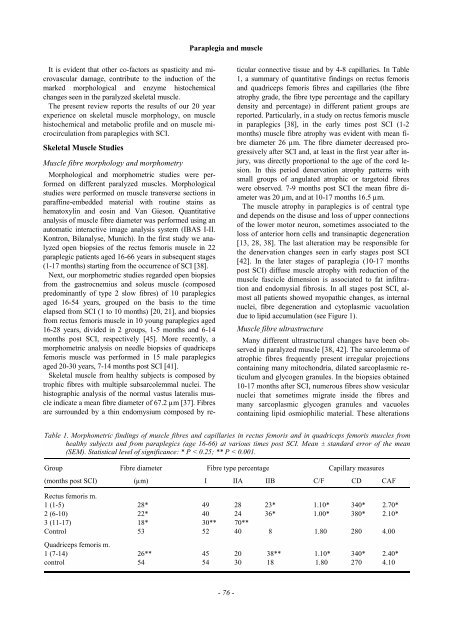
Table 1. Morphometric findings of muscle fibres and capillaries in rectus femoris and in quadriceps femoris muscles from<br />
healthy subjects and from paraplegics (age 16-66) at various times post SCI. Mean ± standard error of the mean<br />
(SEM). Statistical level of significance: * P < 0.25; ** P < 0.001.<br />
Group Fibre diameter Fibre type percentage Capillary measures<br />
(months post SCI) (µm) I IIA IIB C/F CD CAF<br />
Rectus femoris m.<br />
1 (1-5) 28* 49 28 23* 1.10* 340* 2.70*<br />
2 (6-10) 22* 40 24 36* 1.00* 380* 2.10*<br />
3 (11-17) 18* 30** 70**<br />
Control 53 52 40 8 1.80 280 4.00<br />
Quadriceps femoris m.<br />
1 (7-14) 26** 45 <strong>20</strong> 38** 1.10* 340* 2.40*<br />
control 54 54 30 18 1.80 270 4.10