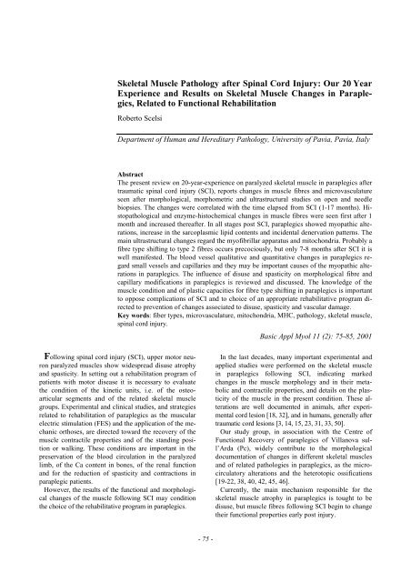
Skeletal Muscle Pathology after Spinal Cord Injury: Our 20 Year ...
Skeletal Muscle Pathology after Spinal Cord Injury: Our 20 Year ...
Skeletal Muscle Pathology after Spinal Cord Injury: Our 20 Year ...

You also want an ePaper? Increase the reach of your titles
YUMPU automatically turns print PDFs into web optimized ePapers that Google loves.
<strong>Skeletal</strong> <strong>Muscle</strong> <strong>Pathology</strong> <strong>after</strong> <strong>Spinal</strong> <strong>Cord</strong> <strong>Injury</strong>: <strong>Our</strong> <strong>20</strong> <strong>Year</strong><br />
Experience and Results on <strong>Skeletal</strong> <strong>Muscle</strong> Changes in Paraplegics,<br />
Related to Functional Rehabilitation<br />
Roberto Scelsi<br />
Department of Human and Hereditary <strong>Pathology</strong>, University of Pavia, Pavia, Italy<br />
Abstract<br />
The present review on <strong>20</strong>-year-experience on paralyzed skeletal muscle in paraplegics <strong>after</strong><br />
traumatic spinal cord injury (SCI), reports changes in muscle fibres and microvasculature<br />
seen <strong>after</strong> morphological, morphometric and ultrastructural studies on open and needle<br />
biopsies. The changes were correlated with the time elapsed from SCI (1-17 months). Histopathological<br />
and enzyme-histochemical changes in muscle fibres were seen first <strong>after</strong> 1<br />
month and increased there<strong>after</strong>. In all stages post SCI, paraplegics showed myopathic alterations,<br />
increase in the sarcoplasmic lipid contents and incidental denervation patterns. The<br />
main ultrastructural changes regard the myofibrillar apparatus and mitochondria. Probably a<br />
fibre type shifting to type 2 fibres occurs precociously, but only 7-8 months <strong>after</strong> SCI it is<br />
well manifested. The blood vessel qualitative and quantitative changes in paraplegics regard<br />
small vessels and capillaries and they may be important causes of the myopathic alterations<br />
in paraplegics. The influence of disuse and spasticity on morphological fibre and<br />
capillary modifications in paraplegics is reviewed and discussed. The knowledge of the<br />
muscle condition and of plastic capacities for fibre type shifting in paraplegics is important<br />
to oppose complications of SCI and to choice of an appropriate rehabilitative program directed<br />
to prevention of changes associated to disuse, spasticity and vascular damage.<br />
Key words: fiber types, microvasculature, mitochondria, MHC, pathology, skeletal muscle,<br />
spinal cord injury.<br />
Following spinal cord injury (SCI), upper motor neuron<br />
paralyzed muscles show widespread disuse atrophy<br />
and spasticity. In setting out a rehabilitation program of<br />
patients with motor disease it is necessary to evaluate<br />
the condition of the kinetic units, i.e. of the osteoarticular<br />
segments and of the related skeletal muscle<br />
groups. Experimental and clinical studies, and strategies<br />
related to rehabilitation of paraplegics as the muscular<br />
electric stimulation (FES) and the application of the mechanic<br />
orthoses, are directed toward the recovery of the<br />
muscle contractile properties and of the standing position<br />
or walking. These conditions are important in the<br />
preservation of the blood circulation in the paralyzed<br />
limb, of the Ca content in bones, of the renal function<br />
and for the reduction of spasticity and contractions in<br />
paraplegic patients.<br />
However, the results of the functional and morphological<br />
changes of the muscle following SCI may condition<br />
the choice of the rehabilitative program in paraplegics.<br />
- 75 -<br />
Basic Appl Myol 11 (2): 75-85, <strong>20</strong>01<br />
In the last decades, many important experimental and<br />
applied studies were performed on the skeletal muscle<br />
in paraplegics following SCI, indicating marked<br />
changes in the muscle morphology and in their metabolic<br />
and contractile properties, and details on the plasticity<br />
of the muscle in the present condition. These alterations<br />
are well documented in animals, <strong>after</strong> experimental<br />
cord lesion [18, 32], and in humans, generally <strong>after</strong><br />
traumatic cord lesions [3, 14, 15, 23, 31, 33, 50].<br />
<strong>Our</strong> study group, in association with the Centre of<br />
Functional Recovery of paraplegics of Villanova sull’Arda<br />
(Pc), widely contribute to the morphological<br />
documentation of changes in different skeletal muscles<br />
and of related pathologies in paraplegics, as the microcirculatory<br />
alterations and the heterotopic ossifications<br />
[19-22, 38, 40, 42, 45, 46].<br />
Currently, the main mechanism responsible for the<br />
skeletal muscle atrophy in paraplegics is tought to be<br />
disuse, but muscle fibres following SCI begin to change<br />
their functional properties early post injury.
It is evident that other co-factors as spasticity and microvascular<br />
damage, contribute to the induction of the<br />
marked morphological and enzyme histochemical<br />
changes seen in the paralyzed skeletal muscle.<br />
The present review reports the results of our <strong>20</strong> year<br />
experience on skeletal muscle morphology, on muscle<br />
histochemical and metabolic profile and on muscle microcirculation<br />
from paraplegics with SCI.<br />
<strong>Skeletal</strong> <strong>Muscle</strong> Studies<br />
<strong>Muscle</strong> fibre morphology and morphometry<br />
Morphological and morphometric studies were performed<br />
on different paralyzed muscles. Morphological<br />
studies were performed on muscle transverse sections in<br />
paraffine-embedded material with routine stains as<br />
hematoxylin and eosin and Van Gieson. Quantitative<br />
analysis of muscle fibre diameter was performed using an<br />
automatic interactive image analysis system (IBAS I-II.<br />
Kontron, Bilanalyse, Munich). In the first study we analyzed<br />
open biopsies of the rectus femoris muscle in 22<br />
paraplegic patients aged 16-66 years in subsequent stages<br />
(1-17 months) starting from the occurrence of SCI [38].<br />
Next, our morphometric studies regarded open biopsies<br />
from the gastrocnemius and soleus muscle (composed<br />
predominantly of type 2 slow fibres) of 10 paraplegics<br />
aged 16-54 years, grouped on the basis to the time<br />
elapsed from SCI (1 to 10 months) [<strong>20</strong>, 21], and biopsies<br />
from rectus femoris muscle in 10 young paraplegics aged<br />
16-28 years, divided in 2 groups, 1-5 months and 6-14<br />
months post SCI, respectively [45]. More recently, a<br />
morphometric analysis on needle biopsies of quadriceps<br />
femoris muscle was performed in 15 male paraplegics<br />
aged <strong>20</strong>-30 years, 7-14 months post SCI [41].<br />
<strong>Skeletal</strong> muscle from healthy subjects is composed by<br />
trophic fibres with multiple subsarcolemmal nuclei. The<br />
histographic analysis of the normal vastus lateralis muscle<br />
indicate a mean fibre diameter of 67.2 µm [37]. Fibres<br />
are surrounded by a thin endomysium composed by re-<br />
Paraplegia and muscle<br />
- 76 -<br />
ticular connective tissue and by 4-8 capillaries. In Table<br />
1, a summary of quantitative findings on rectus femoris<br />
and quadriceps femoris fibres and capillaries (the fibre<br />
atrophy grade, the fibre type percentage and the capillary<br />
density and percentage) in different patient groups are<br />
reported. Particularly, in a study on rectus femoris muscle<br />
in paraplegics [38], in the early times post SCI (1-2<br />
months) muscle fibre atrophy was evident with mean fibre<br />
diameter 26 µm. The fibre diameter decreased progressively<br />
<strong>after</strong> SCI and, at least in the first year <strong>after</strong> injury,<br />
was directly proportional to the age of the cord lesion.<br />
In this period denervation atrophy patterns with<br />
small groups of angulated atrophic or targetoid fibres<br />
were observed. 7-9 months post SCI the mean fibre diameter<br />
was <strong>20</strong> µm, and at 10-17 months 16.5 µm.<br />
The muscle atrophy in paraplegics is of central type<br />
and depends on the disuse and loss of upper connections<br />
of the lower motor neuron, sometimes associated to the<br />
loss of anterior horn cells and transinaptic degeneration<br />
[13, 28, 38]. The last alteration may be responsible for<br />
the denervation changes seen in early stages post SCI<br />
[42]. In the later stages of paraplegia (10-17 months<br />
post SCI) diffuse muscle atrophy with reduction of the<br />
muscle fascicle dimension is associated to fat infiltration<br />
and endomysial fibrosis. In all stages post SCI, almost<br />
all patients showed myopathic changes, as internal<br />
nuclei, fibre degeneration and cytoplasmic vacuolation<br />
due to lipid accumulation (see Figure 1).<br />
<strong>Muscle</strong> fibre ultrastructure<br />
Many different ultrastructural changes have been observed<br />
in paralyzed muscle [38, 42]. The sarcolemma of<br />
atrophic fibres frequently present irregular projections<br />
containing many mitochondria, dilated sarcoplasmic reticulum<br />
and glycogen granules. In the biopsies obtained<br />
10-17 months <strong>after</strong> SCI, numerous fibres show vesicular<br />
nuclei that sometimes migrate inside the fibres and<br />
many sarcoplasmic glycogen granules and vacuoles<br />
containing lipid osmiophilic material. These alterations<br />
Table 1. Morphometric findings of muscle fibres and capillaries in rectus femoris and in quadriceps femoris muscles from<br />
healthy subjects and from paraplegics (age 16-66) at various times post SCI. Mean ± standard error of the mean<br />
(SEM). Statistical level of significance: * P < 0.25; ** P < 0.001.<br />
Group Fibre diameter Fibre type percentage Capillary measures<br />
(months post SCI) (µm) I IIA IIB C/F CD CAF<br />
Rectus femoris m.<br />
1 (1-5) 28* 49 28 23* 1.10* 340* 2.70*<br />
2 (6-10) 22* 40 24 36* 1.00* 380* 2.10*<br />
3 (11-17) 18* 30** 70**<br />
Control 53 52 40 8 1.80 280 4.00<br />
Quadriceps femoris m.<br />
1 (7-14) 26** 45 <strong>20</strong> 38** 1.10* 340* 2.40*<br />
control 54 54 30 18 1.80 270 4.10
Figure 1. <strong>Spinal</strong> cord injury.<br />
are limited to the degenerating fibres and are considered<br />
myopathic changes in the immobilized and hypoxiemic<br />
muscle. The main ultrastructural alterations in paraplegics<br />
regard the myofibrillar apparatus, mitochondria and<br />
the sarcoplasmic lipid content of the muscle fibres.<br />
In Table 2, morphometric results of the analysis of<br />
mitochondria (mean mitochondrial size and % of mitochondria<br />
per fibre area) and of lipid droplet percentage<br />
per fibre area are also reported.<br />
Myofibrillar apparatus<br />
Remarkable alterations in the myofibrillar apparatus<br />
have been frequently observed in the medium and old<br />
stage of the lesion, with loss and disruption of the myofilaments,<br />
abnormalities of the Z line, including smearing<br />
and streaming, and frequent formation of rods.<br />
These alterations are generally considered as consequence<br />
of muscle fibre degeneration.<br />
Mitochondria<br />
The mitochondria are generally small in size and<br />
sometimes show swelling and intracristal inclusions.<br />
The mitochondrial size and percentage significantly<br />
decrease in paraplegics in comparison with normal<br />
muscle [38]. Similar changes were observed in muscle<br />
disuse in old aged subjects as result of low utilisation of<br />
the lipid energy sources [30, 37]. In the immobilized<br />
and atrophic muscle, the morphological mitochondrial<br />
alterations are accompanied by impairment of the mitochondrial<br />
oxidative enzyme activities [51]. Whereas<br />
some authors described diffuse morphological damage<br />
of mitochondria in disuse atrophy of skeletal muscle<br />
Paraplegia and muscle<br />
DISUSE SPASTICITY<br />
MUSCLE FIBRE ATROPHY<br />
CAPILLARY DECREASE<br />
MYOPATHIC CHANGES<br />
- 77 -<br />
FIBRE TYPE TRANSFORMATION<br />
Satellite cells, myotubes FAST TYPE 2 FIBRE PREDOMINANCE<br />
[51], in our studies these changes are confined to rare<br />
interspersed fibres.<br />
Lipid content<br />
Interspersed round vacuoles either empty or filled with<br />
osmiophylic lipid material, are seen in normal skeletal<br />
muscle fibres. Lipids are normally utilized by mitochondria<br />
to release energy for metabolism and muscle contraction.<br />
They increase in the muscle with aging and are<br />
expression of disuse and of abnormal mitochondrial<br />
function [26, 37]. In paraplegics, the lipid droplets were<br />
widely distributed in the cytoplasm, mainly in atrophic<br />
fibres, and their percentage per fibre area significantly<br />
increased (Table 3) [38]. This phenomenon may be related<br />
to decrease in mitochondrial percentage and to the<br />
less efficient utilisation of the lipid energy sources following<br />
muscle inactivity [26, 38].<br />
Previous reports on skeletal muscle fibre morphology<br />
and composition have been performed on patients at different<br />
times post SCI, using standard qualitative enzymehistochemical<br />
methods. All authors reported increase<br />
in the relative proportion of the fast type II fibres<br />
in longstanding paralyzed muscles, if compared to normal<br />
healthy control subjects. In some patients, this fibre<br />
type transformation appears to be important, with rarefaction<br />
of slow type I fibre in the biopsies [14, 15, 23,<br />
31, 33, 50]. Morphological and morphometric analysis<br />
of paralyzed muscle post SCI indicates a progressive<br />
muscle fibre atrophy, with important structural alterations.<br />
A 50% decrease in the fibre diameter was present<br />
in the early stages of paraplegia. The disuse may be the<br />
Table 2. Morphometric results of mitochondrial and lipid droplet content in quadriceps femoris muscle from young paraplegics<br />
7-12 months post SCI and control healthy subjects (mean age: 25). Mean ± standard error of the mean (SEM).<br />
Statistical level of significance: * P < 0.25; ** P < 0.001.<br />
Mean mitochondrial size % mitochondria % lipid droplets<br />
size (µm) per fibre area per fibre area<br />
Controls 0.14 ± 0.09 2.53 ± 0.3 0.48 ± 0.<strong>20</strong><br />
Paraplegics 0.008 ± 00.4** 1.70 ± 0.5* 3.<strong>20</strong> ± 1.00**
main cause of muscle atrophy in paraplegics. In the recent<br />
cord lesion, the degree of muscle atrophy was evident<br />
and denervation changes were also seen. In the<br />
later stages of paraplegia the muscle atrophy degree increased<br />
and myopathic changes with focal necrosis and<br />
more extensive fibre degeneration in about 4% of fibres<br />
were observed. The degenerative changes in paralyzed<br />
muscle may be due to the vascular changes reported<br />
below, and the denervation patterns might reflect the<br />
presence of some coincidental alterations of the peripheral<br />
nerve or trans-synaptic degeneration in the<br />
lower motor neuron [13, 28, 38]. In advanced stages<br />
post SCI, muscle atrophy and increase in the interstitial<br />
endomysial connective tissues and perifascicular fatty<br />
infiltration were evident. The significance of the ultrastructural<br />
changes, particularly of the decrease in the<br />
number of muscle mitochondria and of the increase in<br />
cytoplasmic lipid vacuoles, are related to muscle inactivity<br />
and degeneration.<br />
<strong>Muscle</strong> fibre type composition<br />
Normal quadriceps femoris muscle is composed of two<br />
major types of fibre characterized by enzymehistochemical<br />
methods (myofibrillar ATPase pH 9.6 and 4.6): type 1<br />
and type 2 fibres, and numerous sub-types. In our studies<br />
the normal type 1 fibre diameter measures 52.2 µm and<br />
the type 2 fibre diameter 48.2 µm [<strong>20</strong>]. Type 1 fibres are<br />
often of smaller diameter, contain many mitochondria<br />
and do not stain histochemically for myosin ATP-ase activity.<br />
Type 2 fibres are often broad and possess few mitochondria<br />
but they stain intensely for myosin ATP-ase<br />
activity [8]. In Table 1 and 3, morphometric findings on<br />
the fibre diameter and on the fibre type percentage in<br />
rectus femoris, quadriceps femoris, soleus and gastrocnemius<br />
muscle biopsies from paraplegic patients at times<br />
ranging over 1-17 months post SCI are reported. In significantly<br />
early time periods post injury (1-4 months post<br />
SCI) evident preferential atrophy of type 2 fibres was observed,<br />
but no changes in the relative percentage of type 1<br />
and 2 fibres were remarked.<br />
Paraplegia and muscle<br />
Table 3. Mean muscle fibre type diameter in soleus and gastrocnemius muscles of paraplegics (age 16-54) and healthy control<br />
subjects.<br />
Group Soleus muscle Gastrocnemius muscle<br />
(months post SCI)<br />
Fibre type Fibre type<br />
I IIA IIB I IIA IIB<br />
1 (1-2) 30 24 26 28 22 24<br />
2 (3-4) 28 22 24 28 24 24<br />
3 (5-6) 28 24 22 26 24 22<br />
4 (7-8) 26 24 24 24 22 <strong>20</strong><br />
5 (9-10) 16 21 <strong>20</strong> 14 <strong>20</strong> 18<br />
Control 50 48 44 48 46 46<br />
- 78 -<br />
Biopsies performed 4-9 months post SCI showed atrophy<br />
of both fibre types with a reduction in the relative<br />
percentage of type 1 fibres. Following long term SCI<br />
(10-17 months post SCI), upper motor neuron paralysed<br />
muscles lose the normal type 1 and 2 type mosaic pattern<br />
and become predominantly composed of type 2 fibres.<br />
Interesting results have been obtained from the<br />
study of the paretic soleus muscle, that normally is predominantly<br />
composed by slow type 1 fibres. A significant<br />
shift of type 1 fibres to type 2B was observed in the<br />
7-10 months post SCI patient group [<strong>20</strong>, 21, 38, 42].<br />
The above described results support the presence of<br />
progressive changes in paralyzed muscles probably occurring<br />
early <strong>after</strong> cord injury, but most evident 4 months<br />
post SCI. The main interesting change in the contractile<br />
properties of paralyzed muscle <strong>after</strong> SCI is the fibre type<br />
transformation phoenomenon, with type 1 fibre change to<br />
type 2, representing down-regulation of the slow MHC<br />
isoform and upper-regulation of the fast isoform in those<br />
fibres. The shift to type II fibres was more evident in<br />
quadriceps and rectus femoris, and in soleus muscle,<br />
while in the gastrocnemius muscle the fibre type conversion<br />
was less remarkable. Moreover, the plastic modifications<br />
of the fibre types was more evident in young<br />
paraplegics than in older subjects. This result is probably<br />
related to modifications in muscle morphology and in<br />
their contractile properties described in the normal sedentary<br />
aging man [30, 37]. The fast type II predominance<br />
in longstanding paraplegia may explain the problem of<br />
muscle fatigability encountered during rehabilitation exercise<br />
using FES. Studies on experimental spinal cord<br />
transaction showed changes in the rat and cat skeletal<br />
muscle, with almost complete type I to type II fibre transformation<br />
[18, 52]. These changes are different from<br />
those seen in immobilisation in which large increase in<br />
the ratio for fast resistant and slow units are seen [24]. It<br />
may be suggested that in long term paraplegia the loss of<br />
the upper motor neuron control and the spasticity may<br />
induce phoenomena of fibre type transformation. The described<br />
muscular changes in paraplegics are reversible
<strong>after</strong> FES and electrically induced training, with partial<br />
recovery of the muscle atrophy and with modification in<br />
the fibre type transformation [25].<br />
Myosin heavy chain isoform profile<br />
Studies on myosin heavy chain (MHC) content in<br />
muscle fibres from parapegic patients are very scarce.<br />
Paraplegia and muscle<br />
Figure 2. A. Fascicular atrophy with increase of perimysial fatty tissue in the rectus femoris muscle. 15 months post<br />
SCI. Giemsa. X 60. B. Pattern of denervation in groups of angulated atrophic fibres in the rectus femoris muscle.<br />
1 month post SCI. DPNH diaphorase. X 250. C. Numerous lipid vacuoles in the muscle fibre cytoplasm. 6<br />
months post SCI. H & E. X 250 D. Normal type I and type II (dark) fibre distribution in the quadriceps femoris<br />
muscle. 18 days post SCI. Myosin ATPase pH 9.4. X 150. E. Some areas of type 2 fibre predominance in the<br />
rectus femoris muscle. 9 months post SCI. Myosin ATPase pH 9.4. X 150. F. Diffuse type II fibre predominance<br />
in the soleus muscle. 17 months <strong>after</strong> SCI. Myosin ATPase pH 9.4. X 150.<br />
- 79 -<br />
The adult normal skeletal muscle fibres express only<br />
one MHC isoform. Type 1 fibres have the slow MHC<br />
isoform, type 2 fibres have the fast MHC isoform and<br />
type C fibres co-express both MHC isoforms. In our<br />
studies, the expression in MHC isoform content of the<br />
whole biopsy was analyzed with an electrophoretic separation<br />
technique [6] on medial gastrocnemius and soleus<br />
muscles in paraplegics 1-10 months post SCI.
The results indicated in the early period post SCI (1-6<br />
months) a predominant type 2 muscle fibre atrophy,<br />
without changes in the relative percentage of fibre types<br />
and MHC content. Eight months post injury, we remarked<br />
atrophy of both fibre types with increase in the<br />
relative percentage of type 2 fibres and fast MHC content.<br />
However, the shift of the type 1 to type 2 fibres was<br />
evident 7-8 months post SCI [42]. Fibre type transformation<br />
of type 1 to type 2 fibres goes through a transitional<br />
phase where they co-express slow and fast myosin isoforms.<br />
Data presented by Burnham et al [3], by means of<br />
immunofluorescence determination of single fibre MCH<br />
isoform, suggest that the onset of fibre shifting to type 2<br />
fibres in paraplegics may to be earlier than previously described,<br />
probably occurring about 1 month post SCI. This<br />
study was performed on vastus lateralis muscle biopsies<br />
from 12 paraplegics with SCI over a period of 1-219<br />
months <strong>after</strong> SCI. Thus, following long term SCI induced<br />
paralysis, fibres in the quadriceps femoris and vastus lateralis<br />
muscle take on a new state profile defined by a shift<br />
in the MHC expression to the fast isoform.<br />
Paraplegia and muscle<br />
Figure 3. A. Normal capillary distribution in a paraffin embedded transverse section of muscle fibres from an healthy subject.<br />
Rectus femoris muscle. Immunohistochemical reaction for CD 36. X 60. B. Reduction in the number of capillaries<br />
and marked dilatation of the lumen of small blood vessels in gastrocnemius muscle. 12 months <strong>after</strong> SCI. Immunohistochemical<br />
reaction for CD36. X 60.<br />
- 80 -<br />
FES and electrically induced training determine in the<br />
paralyzed muscle expressing a majority of MHC isoform<br />
II B fibres, a fibre type transformation towards the<br />
more fatigue resistant MHC isoform II A [25].<br />
Interesting results on paralyzed muscle derive from<br />
the characterization of fibre type profile with enzymehistochemistry<br />
and from the study of the MHC isoforms<br />
over a wide range of post SCI periods. The findings<br />
support the concept of progressive stages of change in<br />
muscle from paraplegics, suggesting that the earliest alterations<br />
post SCI are confined to the morphological<br />
muscle aspects (fibre atrophy, focal fibre degeneration<br />
with changes in the ultrastructural profile and capillary<br />
dilatation). The onset of the fibre type shifting to type 2<br />
fibre probably occurs 1 month <strong>after</strong> SCI, but only 7-8<br />
months <strong>after</strong> injury the shift of the type 1 to type 2 fibres<br />
is well manifested. This fibre type conversion is<br />
related to muscle plasticity, defined as a functional fibre<br />
remodeling <strong>after</strong> different normal and pathological conditions,<br />
inducing important changes of the subcellular
structure, of the enzyme content, of the metabolic and<br />
biochemical characteristics, and of the contractile properties<br />
of muscle fibres [10, 34]. The spasticity causes<br />
pathological changes in refex function, such as loss of<br />
reciprocal inhibition, cocontraction of agonist and antagonist<br />
muscles and the loss of activity in the inhibitory<br />
pathways. These alterations, together with disuse and<br />
other co-factors, are undoubtedly an appropriate opportunity<br />
for the development of the plastic changes in the hypertonic<br />
paralyzed muscle [4].<br />
<strong>Muscle</strong> microcirculation studies<br />
Blood vessels<br />
The first studies on microvasculature from human<br />
skeletal muscle concerned normal healthy adults, athletes<br />
and normal subjects trained in endurance or strength [1,<br />
2, 7]. They demonstrated a close relationship among the<br />
muscle fibre diameter, fibre type and capillary number.<br />
The greatest number of capillaries surrounds the type 1<br />
slow fibres. Literature data on normal muscle microcirculation<br />
concern qualitative and quantitative analysis of<br />
capillaries. In our studies on muscle microcirculation in<br />
paraplegics, microscopic qualitative studies were performed<br />
using histological and ultrastructural methods.<br />
More recently these studies utilized the immunohistochemical<br />
characterization of capillaries by mean of antibodies<br />
as endotheline and CD 36.<br />
The quantitative evaluation of capillaries was performed<br />
with an automatic Interactive Image Analysis<br />
Sistem- IBAS I,II (Kontron-Bildanalyse Munich, Germany)<br />
on histological sections, the number of capillaries<br />
per fibre (C/F), capillaries surrounding a single fibre<br />
(CAF), and the capillary density per square millimeter<br />
(CD) were determined (Table 1). The clinical and morphological<br />
evidences of vascular alterations in paraplegics,<br />
as vasomotor disturbances and decrease in the venous<br />
distensibility and capacity [16] and structural alterations<br />
in the muscle blood vessels [45] may be important<br />
causes of the degenerative myopathic changes in paralyzed<br />
muscle fibres. These alterations are similar to those<br />
seen in patients with chronic venous insufficiency, who<br />
develop microangiopathy of cutaneous blood and lymphatic<br />
capillaries [11]. In recent SCI (1-4 months post<br />
injury), when paralysis with profound hypotonic flaccidity<br />
is evident, marked capillary dilatation and interstitial<br />
vasogenic oedema were seen. In later stages of paraplegia<br />
(10-17 months post SCI), when spasticity becomes evident,<br />
paraplegics showed thickening of the arteriolar wall<br />
with reduplication of the basal lamina of capillaries and<br />
Paraplegia and muscle<br />
- 81 -<br />
of capillary wall associated to reduction in the capillary<br />
number [<strong>20</strong>, 38, 42, 45]. The above mentioned microvascular<br />
changes are also present in the skin of paralyzed<br />
muscle and may be expression of a microangiopathy in<br />
paraplegics <strong>after</strong> SCI [11, <strong>20</strong>, 42]. These alteration are<br />
important and similar changes, as the reduplication and<br />
thickening of the capillary basal lamina, have been described<br />
in diabetes mellitus, in dystrophia myotonica and<br />
in other different microvascular diseases [36, 48]. Human<br />
and experimental studies on skeletal muscle inactivity<br />
which causes muscle atrophy, showed reduction of capillaries<br />
as well as the long standing denervation atrophy<br />
that cause reduction and damage of mitochondria and of<br />
capillaries [5, 35, 51].<br />
On the basis of these observations, the capillary<br />
qualitative and qualitative changes in paraplegics may<br />
be due to interaction of different factors: disuse fibre<br />
atrophy, reduction in muscle volume, incidental overimposed<br />
denervation, loss of type 1 fibres that are normally<br />
surrounded by a great number of capillaries, and<br />
shift to the type 2 muscle fibres. Moreover they could<br />
be interpreted as a negative phoenomenon inducing the<br />
ischemic degenerative myopathic changes described in<br />
muscle fibres in long term paraplegia [42].<br />
The results of a study on young paraplegics <strong>after</strong> use<br />
of gait orthoses (HGO,ORLAU Parawalker), the period<br />
<strong>after</strong> supply ranging from 5-6 months [41, 43] support<br />
the latter hypothesis.<br />
In Table 4, overall capillary and fibre morphometry in<br />
rectus femoris muscle from untrained and Parawalker<br />
trained paraplegics were reported. After orthotic walking<br />
training there was a discrete preservation of the<br />
muscle fibre diameter and increase of the muscle capillary<br />
supply in comparison with untrained paraplegics.<br />
These results are in agreement with those described in<br />
experimental spinal cord lesions in animals in which<br />
favourable exercise-induced changes in biochemical and<br />
contractile properties of muscle fibres and in muscle<br />
capillarity were observed [9, 17].<br />
Lymphatics<br />
In paraplegics, immobilization, prolonged bed rest and<br />
loss of muscle contraction are main causes of venous stasis<br />
in the lower extremities. The venous stasis is frequently<br />
associated to microlymphatic changes of the skin<br />
of the paralyzed leg and it is an important cause of thromboembolic<br />
disease in paraplegics. In the patients, the increase<br />
of the deep venous resistance and the venous stasis<br />
determinate microangiopathic alterations in the skin, in-<br />
Table 4. Overall capillary and fibre morphometry in rectus femoris muscle from healthy control subjects, untrained paraplegics<br />
and Parawalker trained paraplegics. Mean ± standard error of the mean (SEM). P < 0.0001.<br />
Untrained paraplegics 26.0 ± 4.2* 47.0 ± 4.0 1.10 ± 0.12 340 ± 8 2.70 ± 0.1*<br />
Parawalker users 33.0 ± 5.4* 50.0 ± 5.6 1.46 ± 0.6* 300 ± 8 3.60 ± 0.4*<br />
Normal adults 53.3 ± 7.4* 52.0 ± 4.2 1.80 ± 0.1* 280 ± 18 4.00 ± 0.2
Paraplegia and muscle<br />
Figure 4. A. Disorganized myofibrils with smearing and streaming of the Z line. Rectus femoris muscle. 7 months post<br />
SCI. Electron micrograph. X 7,000. B. Extensive myofibrillar degeneration and vacuolation with presence of lysosomal<br />
bodies in the cytoplasm of two adjacent muscle fibres. Rectus femoris muscle. 17 months post SCI.<br />
Electron micrograph. X 6,000. C. Two adjacent muscle fibres with subsarcolemmal lysosome accumulation and<br />
two small vessels with thickening of the basal lamina. 4 months post SCI. Soleus muscle. Electron micrograph. X<br />
6,000. D. Thickened intramuscular capillary with reduplication of the basal lamina. Soleus muscle. 10 months<br />
post SCI. Electron micrograph. X 10,000.<br />
- 82 -
volving blood and lymphatic vessels and eventually leading<br />
to skin dystrophic changes and oedema (phlegmasia<br />
alba dolens). The morphological identification of lymphatic<br />
vessel in the skeletal muscle is difficult. However,<br />
in a study, we analyzed lymphatic vessels in the skin of<br />
the lower extremities from young male paraplegic patients<br />
with and without thromboembolic disease (TED) [44, 46].<br />
In paraplegics with TED of deep veins, skin lymphatic<br />
vessels of paretic legs showed dilated lumen and distended<br />
wall. The endothelial cells were attenuated and<br />
numerous channels among endothelial cells were present.<br />
Perivascular collagen and elastic fibres were dissociated<br />
by oedema and by the presence of granular material. In<br />
paraplegics without TED similar but more rare microlymphatic<br />
changes are present.<br />
These morphological changes demonstrated a lymphatic<br />
microangiopathy in paraplegics, with lymph stasis<br />
and an increased transcapillary diffusion of the<br />
lymph material into perivascular dermic tissues, clinically<br />
resulting in oedema and reduced removal of tissue<br />
catabolites. These leg terminal lymphatic changes and<br />
the blood cutaneous microangiopathy probably determinates<br />
the extent of the trophic disturbances and the ulcer<br />
formation in paraplegics. The microangiopathy is the<br />
basis for reduction of PO2 and of for destruction of the<br />
lymphatic capillary network, seen respectively <strong>after</strong><br />
transcutaneous PO2 measurements and by fluorescent<br />
microlymphography in long term paraplegia and in patients<br />
with chronic venous insufficiency [11, 46].<br />
Conclusions and Perspectives<br />
From a rehabilitation perspective it is necessary to<br />
evaluate the conditions of the motor unit and the muscle<br />
fibre plastic potentialities and reversibility in paraplegics<br />
<strong>after</strong> SCI. The results from the present studies<br />
demonstrated changes in the fibre morphology and in<br />
their contractile properties, and in the muscle capillarity<br />
of paralyzed muscle fibres <strong>after</strong> SCI. There is a number<br />
of rehabilitative programs that are able to modify muscle<br />
atrophy and to prevent some clinical complications, by<br />
means of physical training, FES, aerobic exercise trainers<br />
and bio-mechanic orthoses. In numerous reports, prevention<br />
of muscle disuse and improvement in the fibre<br />
oxidative capacities <strong>after</strong> muscle electric stimulation in<br />
paraplegics were demonstrated [12, 49]. In adult paraplegics,<br />
FES induce changes of morphological, biochemical<br />
and histochemical profile, and of contractile<br />
properties of muscle fibres and improve the muscle<br />
capillarity [31], as well as the use of bio-mechanic orthoses<br />
[27, 41, 43] and of aerobic exercise trainers [29].<br />
The muscle fibre atrophy and the fibre type transformation<br />
process in paraplegics was partially normalized <strong>after</strong><br />
electrical induced cycle training [25]. These supports<br />
for paraplegic locomotion require high energy cost and<br />
may be utilized in patients without cardiovascular and<br />
respiratory diseases [27]. The knowledge of the muscle<br />
Paraplegia and muscle<br />
- 83 -<br />
condition and of plastic capacities for fiber type shifting is<br />
not only important in attenuating the adverse muscle impact<br />
and complications of SCI, but it is also crucial in the<br />
choice of an appropriate rehabilitative program directed to<br />
preventing the changes associated to disuse and to retrain<br />
contractile properties associated to spasticity and microvascular<br />
damage. In fact, it is well known that the therapeutic<br />
exercise setting out to isotonic concentric or eccentric<br />
contracture determines selective recruitment of fast or<br />
slow fibres which prevents selective fibre type atrophies in<br />
paretic muscle. Finally, in an attempt to restore function<br />
and regain motor control, many laboratories are now<br />
focusing a rehabilitation program based on engineering<br />
devices that substitute a motor controlled function (SCI<br />
transcutaneous FES walking). It is easily comprehensible<br />
that these recovery techniques can be utilized only<br />
in well-preserved muscle <strong>after</strong> SCI. Since it is evident<br />
that muscle atrophy and transformation in their fundamental<br />
properties occur precociously post SCI, rehabilitative<br />
interventions need to be instituted as early as<br />
safetly possible.<br />
Acknowledgements<br />
We gratefully acknowledge for they cooperation: Dr<br />
Sergio Lotta, head of the Rehabilitation Center for paraplegics<br />
G Verdi of Villanova sull’Arda (Pc), Prof Carla<br />
Marchetti and Prof Paola Poggi, professors of the Histology<br />
and Human Anatomy of the University of Pavia,<br />
and Prof Ugo Carraro, head of the CNR Unit for <strong>Muscle</strong><br />
Biology and Physiopathology of the Institute of General<br />
<strong>Pathology</strong> of the University of Padova (Italy).<br />
Address correspondence to:<br />
Prof. R. Scelsi, Istituto di Anatomia e Istologia Patologica,<br />
via Forlanini 14, 27100 Pavia, Italy, phone +39<br />
0382 528476, fax +39 0382 525866, Email apat@unipv.it.<br />
References<br />
[1] Anderson P: Capillary density in skeletal muscle of<br />
man. Acta Physiol Scand 1975; 95: <strong>20</strong>3-<strong>20</strong>5.<br />
[2] Brodal P, Ingjer F, Hermansen L: Capillary supply<br />
of skeletal muscle in trained and endurance-trained<br />
men. Am J Physiol 1977; 232: 705-712.<br />
[3] Burke D: Spasticity as an adaptation to pyramidal<br />
tract injury. In Waxman SC (ed): Functional recovery<br />
in neurological diseases. Advances in Neurology.<br />
New York, Raven Press, 1988; 47: 401-423.<br />
[4] Burnham R, Martin T, Stein R, Bell G, MacLean<br />
I, Steadward R: <strong>Skeletal</strong> muscle fibre type transformation<br />
following spinal cord injury. <strong>Spinal</strong><br />
<strong>Cord</strong> 1997; 35: 86-91.<br />
[5] Carpenter S, Karpati G: Necrosis of capillaries in<br />
denervation atrophy of human skeletal muscle.<br />
<strong>Muscle</strong> Nerve 1982; 5: 250-254.<br />
[6] Carraro U, Catani C: A sensitive SDS-PAGE<br />
method separating myosin heavy chain isoforms of<br />
rat skeletal muscle reveals the heterogeneous nature
of the embryonic myosin. Biochem Biophys Res<br />
Commun 1983; 116: 793-802.<br />
[7] Carry MR, Ringel SP, Starcevich JM: Distribution<br />
of capillaries in normal and diseased human skeletal<br />
muscle. <strong>Muscle</strong> Nerve 1986; 9: 250-254.<br />
[8] Dubowitz V: <strong>Muscle</strong> biopsy. A practical approach.<br />
2nd Edn. Baillire. London. 1985.<br />
[9] Edgerton VR, Smith LA, Elred E, Mendell LM:<br />
<strong>Muscle</strong> and motor unit properties of exercised and<br />
non-exercised chronic spinal cats. In Pette D (ed):<br />
Symposium on plasticity of muscle. New York, De<br />
Gruyter, 1980, pp 355-372.<br />
[10] Eisenberg BR, Salmons S: The reorganisation of subcellular<br />
structure in muscle undergoing fast to slow type<br />
transformation. Cell Tissue Res 1981; 2<strong>20</strong>: 449-471.<br />
[11] Franzeck UK, Haselbach P, Speiser D, Bollinger A:<br />
Microangiopathy of cutaneous blood and lymphatic<br />
capillaries in chronic venous insufficiency. Yale J<br />
Biol Med 1993; 66: 37-46.<br />
[12] Gibson JNA, Smith K, Rennie MJ: Prevention of<br />
disuse muscle atrophy by means of electrical<br />
stimulation. Lancet 1988; Oct: 767-770.<br />
[13] Goldkamp O: Electromyographic and nerve conduction<br />
studies in 116 patients with hemiplegia.<br />
Arch Phys Med 1987; 48: 59-63.<br />
[14] Greeve JMD: Functional electric stimulation<br />
(FES): muscle histochemical analysis. Paraplegia<br />
1993; 31: 764-770.<br />
[15] Grimby G, Broberg C, Krotkiewsky M: <strong>Muscle</strong> fibre<br />
composition in patients with traumatic spinal<br />
cord lesion. Scand J Rehab Med 1976; 8: 37-42.<br />
[16] Hopman MTE, Nommensen E, et al: Properties of the<br />
venous vascular system in the lower extremities of individuals<br />
with paraplegia. Paraplegia 1994; 32: 810-816.<br />
[17] Johnson DJ, Smith LA, Elred E, Edgerton VR: Exercise-induced<br />
changes of biochemical, histochemical<br />
and contractile properties of muscle in cordotomized<br />
kittens. Exp Neurol 1982; 76: 414-427.<br />
[18] Leiber R, Johansson CAB, Wahlsing HL, Hargens<br />
AR, Feringa ER: Long-term effects of spinal cord<br />
transection on fast and slow rat skeletal muscle.<br />
Exp Neurol 1986; 91: 423-434.<br />
[19] Lotta S, Lommi G, Scelsi R, Poggi P, Marchetti C,<br />
Foresti G: Composizione delle fibre muscolari striate<br />
nei postumi di lesioni complete del motoneurone<br />
centrale. Studio morfologico in funzione riabilitativa.<br />
In Perfetti C, Ambrosino N (eds): La<br />
programmazione della esperienza post lesionale.<br />
Atti 12° congresso Soc. It. Medicina Fisica e Riabilitativa.<br />
Il Ciocco, 1981, pp 353-371.<br />
[<strong>20</strong>] Lotta S, Scelsi L, Scelsi R: Microvascular changes in<br />
the lower extremities of paraplegics with heterotopic<br />
ossifications. <strong>Spinal</strong> <strong>Cord</strong> <strong>20</strong>01; 39: 595-598.<br />
[21] Lotta S, Scelsi R, Alfonsi E, Saitta A, Nicolotti D,<br />
Epifani P,Carraro U: Morphometric and neurophysiological<br />
analysis of skeletal muscle in para-<br />
Paraplegia and muscle<br />
- 84 -<br />
plegic patients with traumatic cord lesion. Paraplegia<br />
1991; 29: 247-252.<br />
[22] Lotta S, Scelsi R: <strong>Skeletal</strong> muscle atrophy <strong>after</strong> spinal<br />
cord lesion. Temporal changes in the paretic muscle<br />
morphology. In Capodaglio P, Narici MV (eds): <strong>Muscle</strong><br />
atrophy. Disuse and disease. Advances in Occupational<br />
Medicine and Rehabilitation. 1998; 4: 83-87.<br />
[23] Martin TP, Stein RB, Hoeppner PH, Reid DC: Influence<br />
of electrical stimulation on the morphologic<br />
and metabolic properties of paralyzed muscle. J<br />
Appl Physiol 1992; 72: 1401-1406.<br />
[24] Mayer RF, Burke RE, Toop J, Hodgson JA: The effect<br />
of spinal cord transection on motor units in cat medial<br />
gastrocnemius muscle. <strong>Muscle</strong> Nerve 1984; 7: 21-23.<br />
[25] Mohr T, Andersen J, Biering-Sorensen F, Galbo H,<br />
Kjaer M: Long term adaptation to electrically induced<br />
cycle training in severe spinal cord injured<br />
individuals. <strong>Spinal</strong> <strong>Cord</strong> 1997; 35: 1-16.<br />
[26] Morgan-Hughes JA: The mitochondrial myopathies.<br />
In Engel AG, Ranker BSL (eds): Myology. Mac<br />
Graw Hill Book Co, 1986, pp 1706-1743.<br />
[27] Nene AV, Orth D, Patrick JH: Energy cost of paraplegic<br />
locomotion with the ORLAU parawalker.<br />
Paraplegia 1989; 27: 5-18.<br />
[28] Ochs S: System of material transport in nerve fibres<br />
related to nerve function and trophic control. Ann<br />
NY Acad Sci 1974; 228: <strong>20</strong>2-210.<br />
[29] Petrofsky J, Chandler P, Almeida J, Briggs R:<br />
Aerobic trainer with physiological monitoring for<br />
exercise in paraplegic and quadriplegic patients. J<br />
Clin Engineering 1985; 4: 307-316.<br />
[30] Poggi P, Marchetti C, Scelsi R: Automatic morphometric<br />
analysis of skeletal muscle fibres in the<br />
aging man. Anatomical Record 1987; 217: 30-34.<br />
[31] Rochester L, Barron MJ: Influence of electrical<br />
stimulation of the tibialis anterior muscle in paraplegic<br />
patients. Morphological and histochemical<br />
properties. Paraplegia 1995; 33: 514-522.<br />
[32] Round JM, Barr FMD, Moffat B, Jones DA: Fibre<br />
areas and histochemical fibre types in the quadriceps<br />
muscle of paraplegic subjects. J Neurol Sci<br />
1993; 116: <strong>20</strong>7-211.<br />
[33] Roy RR, Baldwin KM, Edgerton VR: The plasticity<br />
of skeletal muscle: effects on neuromuscular activity.<br />
In Hollosky J (ed): Exercise and Sport Review. Baltimore,<br />
Williams and Wilkins, 1991; 19: 269-312.<br />
[34] Salmons S, Henriksson J: The adaptive response of<br />
skeletal muscle to increased use. <strong>Muscle</strong> Nerve<br />
1981; 4: 94-105.<br />
[35] Sargeant AJ, Davies CTM, Edwards RHT, Maunder<br />
C, Young A: Functional and structural changes<br />
<strong>after</strong> disuse of human muscle. Clin Sci Mol Med<br />
1977; 52: 337-342.<br />
[36] Scelsi R, Lotta S, Lommi G, Poggi P, Marchetti C:<br />
Hemiplegic atrophy. Morphological findings in the
anterior tibial muscle of patients with cerebral vascular<br />
accidents. Acta Neuropathol (Berl) 1984; 62: 324-331.<br />
[37] Scelsi R, Lotta S, Poggi P, Bocchi R, Scelsi L: Microlymphatic<br />
and thromboembolic disease in acute<br />
spinal cord injury. A morphological study on skin<br />
biopsies. European J Lymphology 1994; 4: 115-119.<br />
[38] Scelsi R, Lotta S, Scelsi L: <strong>Skeletal</strong> muscle morphology<br />
during restoration of gait with orthoses in<br />
thoracic paraplegia. In: 9 th International Conference<br />
on Mechanics in Medicine Biology. Ljubljana, June<br />
1996, Abstracts N 34.<br />
[39] Scelsi R, Lotta S: Morphological properties of<br />
skeletal muscle in spastic paraplegia. Basic Appl<br />
Myol 1991; 1: 317-326.<br />
[40] Scelsi R, Marchetti C, Poggi P, Lotta S, Lommi G:<br />
<strong>Muscle</strong> fiber type morphology and distribution in<br />
paraplegic patients with traumatic cord lesion. Acta<br />
Neuropathol (Berl) 1982; 57: 243-248.<br />
[41] Scelsi R, Marchetti C, Poggi P: Histochemical and<br />
ultrastructural aspects of M Vastus Lateralis in sedentary<br />
old people (age 65-89 years). Acta Neuropath<br />
(Berl) 1980; 51: 99-105.<br />
[42] Scelsi R, Poggi P, Nappi G, Sandrini G: Peripheral<br />
microcirculatory lesions in myotonic dystrophy. A<br />
light and electron microscopic study. Acta Neurol<br />
1978; 33: 137-148.<br />
[43] Scelsi R, Poggi P, Padovani R, Lotta S, Saitta A:<br />
<strong>Skeletal</strong> muscle changes following myelotomy in<br />
paraplegic patients. Paraplegia 1986; 24: 250-259.<br />
[44] Scelsi R, Scelsi L, Bocchi R, Lotta S: Morphological<br />
changes in the skin microlymphatics in recent<br />
injured paraplegic patients with ilio-femoral thrombosis.<br />
Paraplegia 1995; 33: 472-475.<br />
Paraplegia and muscle<br />
- 85 -<br />
[45] Scelsi R, Scelsi L, Lotta S: Microvascular changes<br />
in skeletal muscle from paraplegic patients with<br />
spinal cord lesion. In Carraro U, Salmons S (eds):<br />
Basic and Applied Myology. Perspectives for the<br />
90s. Padova, Unipress, 1995, pp 163-170.<br />
[46] Scelsi R, Scelsi L, Lotta S: <strong>Skeletal</strong> muscle morphology<br />
in paraplegic patients <strong>after</strong> Parawalker orthotic<br />
walking. In: Activity-induced muscle injury<br />
and repair: International Symposium on Basic and<br />
Applied Myology. Abano Terme, May 30, 1991.<br />
[47] Scelsi R, Scelsi L, Lotta S: <strong>Skeletal</strong> muscle morphology<br />
in paraplegics <strong>after</strong> Parawalker orthotic<br />
training. In: Proceedings of the 4 th Vienna International<br />
Workshop on Functional Electrostimulation.<br />
Vienna, September 24-27, 1992, pp 75-78.<br />
[48] Siperstein MD, Raskin PR, Burn H: Electron microscopic<br />
quantification of diabetic microangiopathy.<br />
Diabetes 1973; 22: 514-524.<br />
[49] Stein RB: Optimal stimulation of paralysed muscle<br />
<strong>after</strong> human spinal cord injury. J Appl Physiol<br />
1992; 72: 1393-1400.<br />
[50] Stilwill EW, Sahgal V: Histochemical and morphologic<br />
changes in skeletal muscle following cervical<br />
cord injury: a study of upper and lower motor neuron<br />
lesions. Arch Phys Med Rehabil 1977; 58: <strong>20</strong>1-<strong>20</strong>6.<br />
[51] Weheman HJ, Max SR: Mitochondrial ultrastructure<br />
in disuse atrophy of skeletal muscle. Federation<br />
Proc 1972; 31: 253.<br />
[52] West SP, Roland RR, Reggie Edgerton V: Fibre type<br />
and fibre size in cat ankle, knee and hip extensor and<br />
flexors following low thoracic spinal cord transection<br />
in an early age. Exp Neurol 1986; 91: 174-182.