

Staphylococcal Pyelonephritis and Cystitis in a California Sea - OERS
Staphylococcal Pyelonephritis and Cystitis in a California Sea - OERS
Staphylococcal Pyelonephritis and Cystitis in a California Sea - OERS

You also want an ePaper? Increase the reach of your titles
YUMPU automatically turns print PDFs into web optimized ePapers that Google loves.
Journal of Mar<strong>in</strong>e Animals <strong>and</strong> Their Ecology<br />
Copyright © 2008 Oceanographic Environmental Research Society<br />
<strong>Staphylococcal</strong> <strong>Pyelonephritis</strong> <strong>and</strong> <strong>Cystitis</strong> <strong>in</strong> a <strong>California</strong> <strong>Sea</strong> Lion<br />
(Zalophus <strong>California</strong>nus)<br />
Vanessa Fravel, Richard H. Evans<br />
Pacific Mar<strong>in</strong>e Mammal Center, 20612 Laguna Canyon Road, Laguna Beach, <strong>California</strong> USA<br />
Abstract<br />
An adult, female <strong>California</strong> sea lion (Zalophus californianus)<br />
was str<strong>and</strong>ed <strong>in</strong> Laguna Beach, <strong>California</strong> (33 o 32’43.96”N x<br />
117 o 47’51.10”W) on January 13, 2007. Cl<strong>in</strong>ical signs <strong>in</strong>cluded<br />
marked lethargy, head-bobb<strong>in</strong>g <strong>and</strong> ‘wet-dog shak<strong>in</strong>g’.<br />
A presumptive diagnosis of Domoic Acid <strong>in</strong>toxication was<br />
made <strong>and</strong> supportive therapy <strong>in</strong>clud<strong>in</strong>g anti-seizure treatment<br />
was <strong>in</strong>stituted. About thirty hours later, seizure activity had<br />
abated but the animal cont<strong>in</strong>ued to deteriorate to a semicomatose<br />
state <strong>and</strong> euthanasia was elected. Postmortem exam<strong>in</strong>ation<br />
revealed severe, bilateral pyelonephitis <strong>and</strong> cystitis<br />
from which a pure culture of Staphylococcus aureus was isolated.<br />
With the exception of Leptospira sp <strong>in</strong>terstitial nephritis,<br />
bacterial ur<strong>in</strong>ary tract <strong>in</strong>fections are very rare <strong>in</strong> mar<strong>in</strong>e mammals.<br />
To the author’s knowledge this is the first report of<br />
Staphylococcus aureus pyleonephritis <strong>and</strong> cystitis <strong>in</strong> the <strong>California</strong><br />
sea lion. [JMATE. 2008;1(1):12-14]<br />
Keywords: <strong>California</strong> sea lion, bacterial nephritis, Staphylococcus<br />
aureus<br />
——————————————————————————————————<br />
Introduction<br />
An adult, female <strong>California</strong> sea lion (Zalophus californianus)<br />
str<strong>and</strong>ed on the beach at Shaw’s Cove, Laguna Beach, <strong>California</strong><br />
(33 o 32’43.96”N x 117 o 47’51.10”W) on the afternoon of<br />
January 13, 2007. A rescue team from the Pacific Mar<strong>in</strong>e Mammal<br />
Center (PMMC) was dispatched to the scene <strong>and</strong> noted the<br />
sea lion to be very lethargic, reluctant to move <strong>and</strong> exhibit<strong>in</strong>g<br />
periodic head-bobb<strong>in</strong>g <strong>and</strong> generalized ‘wet-dog shak<strong>in</strong>g’. Domoic<br />
acid (algae tox<strong>in</strong>) <strong>in</strong>toxication was suspected <strong>and</strong> the<br />
animal was impounded <strong>and</strong> transported to PMMC.<br />
Physical exam<strong>in</strong>ation revealed moderate, generalized body<br />
wast<strong>in</strong>g <strong>and</strong> <strong>in</strong>termittent low-grade tonic-clonic seizure activity<br />
with head-bobb<strong>in</strong>g. Heart rate was 86 bpm, respirations ~30/<br />
m<strong>in</strong>ute <strong>and</strong> temperature 38˚C (100.4˚F) . Rehydration was<br />
<strong>in</strong>stituted by subcutaneous adm<strong>in</strong>istration of lactated R<strong>in</strong>gers.<br />
Diazepam (5 mg/ml Benzodiazep<strong>in</strong>e) was given <strong>in</strong>tramuscularly<br />
(dosage = 5 mg) to control seizures. Attempts at blood collection<br />
via the caudal gluteal <strong>and</strong> jugular ve<strong>in</strong>s were unsuccessful.<br />
Twenty four hours later, the sea lion’s condition had stabilized<br />
with no obvious seizure activity. However, the follow<strong>in</strong>g morn<strong>in</strong>g<br />
she had deteriorated to a semi-comatose state with very<br />
sluggish to non-existent reflexes <strong>and</strong> moderate bradycardia<br />
<strong>and</strong> dyspnea. Because of a poor prognosis, euthanasia <strong>and</strong><br />
postmortem exam<strong>in</strong>ation was elected. Follow<strong>in</strong>g euthanasia,<br />
blood samples were collected for pick-up <strong>and</strong> analysis (CBC<br />
<strong>and</strong> serum chemistry) by a local, commercial veter<strong>in</strong>ary laboratory.<br />
Unfortunately, the samples were accidentally destroyed <strong>in</strong><br />
-route to the laboratory.<br />
Received December 29, 2007; Accepted March 5, 2008.<br />
Correspondance: Pacific Mar<strong>in</strong>e Mammal Center, 20612 Laguna Canyon Road,<br />
Laguna Beach, <strong>California</strong>, 92651, revans@pacificmmc.org, 949-494-3050<br />
Vol 1, No1, 2008<br />
Pr<strong>in</strong>ted <strong>in</strong> Canada<br />
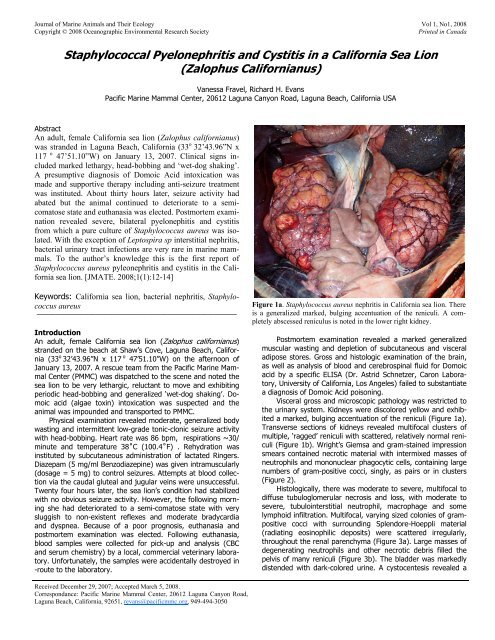
Figure 1a. Staphylococcus aureus nephritis <strong>in</strong> <strong>California</strong> sea lion. There<br />
is a generalized marked, bulg<strong>in</strong>g accentuation of the reniculi. A completely<br />
abscessed reniculus is noted <strong>in</strong> the lower right kidney.<br />
Postmortem exam<strong>in</strong>ation revealed a marked generalized<br />
muscular wast<strong>in</strong>g <strong>and</strong> depletion of subcutaneous <strong>and</strong> visceral<br />
adipose stores. Gross <strong>and</strong> histologic exam<strong>in</strong>ation of the bra<strong>in</strong>,<br />
as well as analysis of blood <strong>and</strong> cerebrosp<strong>in</strong>al fluid for Domoic<br />
acid by a specific ELISA (Dr. Astrid Schnetzer, Caron Laboratory,<br />
University of <strong>California</strong>, Los Angeles) failed to substantiate<br />
a diagnosis of Domoic Acid poison<strong>in</strong>g.<br />
Visceral gross <strong>and</strong> microscopic pathology was restricted to<br />
the ur<strong>in</strong>ary system. Kidneys were discolored yellow <strong>and</strong> exhibited<br />
a marked, bulg<strong>in</strong>g accentuation of the reniculi (Figure 1a).<br />
Transverse sections of kidneys revealed multifocal clusters of<br />
multiple, ‘ragged’ reniculi with scattered, relatively normal reniculi<br />
(Figure 1b). Wright’s Giemsa <strong>and</strong> gram-sta<strong>in</strong>ed impression<br />
smears conta<strong>in</strong>ed necrotic material with <strong>in</strong>termixed masses of<br />
neutrophils <strong>and</strong> mononuclear phagocytic cells, conta<strong>in</strong><strong>in</strong>g large<br />
numbers of gram-positive cocci, s<strong>in</strong>gly, as pairs or <strong>in</strong> clusters<br />
(Figure 2).<br />
Histologically, there was moderate to severe, multifocal to<br />
diffuse tubuloglomerular necrosis <strong>and</strong> loss, with moderate to<br />
severe, tubulo<strong>in</strong>terstitial neutrophil, macrophage <strong>and</strong> some<br />
lymphoid <strong>in</strong>filtration. Multifocal, vary<strong>in</strong>g sized colonies of grampositive<br />
cocci with surround<strong>in</strong>g Splendore-Hoeppli material<br />
(radiat<strong>in</strong>g eos<strong>in</strong>ophilic deposits) were scattered irregularly,<br />
throughout the renal parenchyma (Figure 3a). Large masses of<br />
degenerat<strong>in</strong>g neutrophils <strong>and</strong> other necrotic debris filled the<br />
pelvis of many reniculi (Figure 3b). The bladder was markedly<br />
distended with dark-colored ur<strong>in</strong>e. A cystocentesis revealed a
Figure 1b. Transverse sections of formal<strong>in</strong> fixed right kidney. Multiple<br />
foci of contiguous, ‘ragged’, <strong>in</strong>flamed renucli are apparent, however<br />
several adjacent reniculi are relatively unaffected (medullary pyramid<br />
lower right, top section). The large vacuolated portion <strong>in</strong> the bottom<br />
section represents pelvic dilation secondary to corticomedullary loss<br />
from necrotiz<strong>in</strong>g <strong>in</strong>flammation.<br />
ur<strong>in</strong>e specific gravity 1.2, pH 7.5, moderate to marked levels of<br />
blood, ketones, glucose, prote<strong>in</strong>, urobil<strong>in</strong>ogen <strong>and</strong> bilirub<strong>in</strong>.<br />
The ur<strong>in</strong>e sediment conta<strong>in</strong>ed large numbers of neutrophils,<br />
many macrophages conta<strong>in</strong><strong>in</strong>g gram-positive cocci <strong>and</strong> some<br />
plasma cells <strong>and</strong> lymphocytes. The bladder wall conta<strong>in</strong>ed<br />
moderate to severe, multifocal areas of denud<strong>in</strong>g necrotic<br />
urothelium with moderate to marked, submucosal <strong>in</strong>filtration<br />
by moderate to marked numbers of neutrophils <strong>and</strong> macrophages<br />
with occasional scattered colonies of gram-positive<br />
cocci.<br />
Swabs of asceptically obta<strong>in</strong>ed, clotted blood <strong>and</strong> the deep<br />
parenchyma of the right kidney were taken <strong>and</strong> submitted to a<br />
local veter<strong>in</strong>ary laboratory (IDEXX) for bacterial cultur<strong>in</strong>g. No<br />
bacteria were cultured from the clotted blood but a pure culture<br />
of oxacill<strong>in</strong>-sensitive Staphylococcus sp. was obta<strong>in</strong>ed from<br />
the kidney. Culture samples were submitted to the University<br />
of <strong>California</strong> at Davis, College of Veter<strong>in</strong>ary Medic<strong>in</strong>e, Cl<strong>in</strong>ical<br />
Laboratory for speciation. The organism was found to be a<br />
coagulase-positive Staphylococcus, phenotypically identified as<br />
Staphylococcus aureus.<br />
Staphylococcus aureus is a gram-positive, coagulasepositive<br />
bacterium commonly found as a benign, commensal<br />
on the sk<strong>in</strong> <strong>and</strong> mucous membranes of the respiratory, gastro<strong>in</strong>test<strong>in</strong>al<br />
<strong>and</strong> urogenital tracts of wild <strong>and</strong> domestic mammals<br />
<strong>and</strong> birds. However, when these membrane barriers are compromised<br />
by physical trauma <strong>and</strong>/or immunopathies, Staphylococci<br />
may ga<strong>in</strong> entry <strong>in</strong>to the tissues, result<strong>in</strong>g <strong>in</strong> bacteremia<br />
<strong>and</strong> dissem<strong>in</strong>ated <strong>in</strong>flammatory disease. A classic example <strong>in</strong><br />
<strong>Staphylococcal</strong> Nephritis <strong>in</strong> <strong>California</strong> <strong>Sea</strong> Lion<br />
humans <strong>and</strong> other animal species is lower ur<strong>in</strong>ary tract trauma<br />
result<strong>in</strong>g <strong>in</strong> ascend<strong>in</strong>g disease through the bladder <strong>and</strong> <strong>in</strong>to the<br />
kidneys (5). In this case, the fact that <strong>Staphylococcal</strong> bacteremia<br />
was not detected from an aseptically collected blood sample<br />
gives credence to an ascend<strong>in</strong>g disease etiology, i.e. the<br />
bacteria probably ga<strong>in</strong>ed entrance through the vulvar area, up<br />
the urethra, <strong>in</strong>to the bladder <strong>and</strong> f<strong>in</strong>ally the kidney.<br />
The normal kidneys of a <strong>California</strong> sea lion are multilobed<br />
or reniculate, be<strong>in</strong>g composed of hundreds of lobes or<br />
‘reniculi’ (LL, Dim., “Little Kidneys”), which are further divided<br />
<strong>in</strong>to the st<strong>and</strong>ard morphologic subunits of metanephric kidneys,<br />
i.e. a dist<strong>in</strong>ct layer of cortical tissue completely surround<strong>in</strong>g<br />
a medullary pyramid with<strong>in</strong> a s<strong>in</strong>gle calyx (1,6,7). Unlike the<br />
Phoca <strong>in</strong> which each reniculus is dist<strong>in</strong>ctly separated by an<br />
external capsule, <strong>in</strong> Zalophus, only a very th<strong>in</strong> fibrous capsule<br />
is noted surround<strong>in</strong>g the reniculus which allows <strong>in</strong>flammatory<br />
disease to readily spread between reniculi as depicted <strong>in</strong> this<br />
case (Figure 1b) (1).<br />
Howard’s 1983 study on the pathobiology of mar<strong>in</strong>e mammal<br />
diseases noted kidney <strong>in</strong>fections to be “uncommon” <strong>in</strong><br />
mar<strong>in</strong>e mammals, but referenced a case of Leptospiral nephritis<br />
<strong>in</strong> a northern fur seal (3). A review of the literature s<strong>in</strong>ce<br />
this time, clearly shows Leptospira are now a common etiology<br />
for chronic <strong>in</strong>terstitial nephritis <strong>in</strong> the <strong>California</strong> sea lion (2,11).<br />
However, only a few cases of Staphylococcus-associated renal<br />
disease have been reported <strong>in</strong> mar<strong>in</strong>e mammals. In a case<br />
published <strong>in</strong> 1973 <strong>in</strong>volv<strong>in</strong>g attempts to rehabilitate a newborn<br />
Harbour seal str<strong>and</strong>ed <strong>in</strong> Alaska, pustular dermatitis was noted<br />
at 12 days of age, followed by death at 21 days with the postmortem<br />
isolation of a coagulase-positive, Staphylococcus<br />
aureus from the lung, liver <strong>and</strong> kidneys (10). In 1974, Ketterer<br />
<strong>and</strong> Rosenfeld reported a Staphylococcus aureus subcutaneous<br />
abscess <strong>in</strong> a dolph<strong>in</strong> (Trusiops truncatus), result<strong>in</strong>g <strong>in</strong> fatal<br />
septicemia <strong>and</strong> septic embolic nephritis (4). In 2001 <strong>and</strong> 2002,<br />
Staphylococcus aureus septicemia <strong>and</strong> severe, supparative<br />
pyleonephritis was found follow<strong>in</strong>g postmortem exam<strong>in</strong>ations <strong>in</strong><br />
Harbour porpoises (Phocoena phocoena) str<strong>and</strong>ed <strong>in</strong> the German<br />
North <strong>and</strong> Baltic <strong>Sea</strong>s (8,9). To the authors’ knowledge,<br />
this case appears to be the first report of ur<strong>in</strong>ary tract <strong>in</strong>fection<br />
attributed to Staphylococcus aureus <strong>in</strong> the <strong>California</strong> sea lion.<br />
In conclusion, there are several important po<strong>in</strong>ts to made<br />
<strong>and</strong> lessons to be learned from this case: 1) Generalized body<br />
Figure 2. Cut-surface touch impression of right kidney. Note mononuclear,<br />
phagocytic cells conta<strong>in</strong><strong>in</strong>g proliferat<strong>in</strong>g bacterial cocci arranged<br />
<strong>in</strong> couples or vary<strong>in</strong>g sized clusters. Wrights-Giemsa sta<strong>in</strong>, 40x<br />
12
Figure 3. A, Obliteration of normal kidney architecture by marked, diffuse<br />
tubular necrosis <strong>and</strong> <strong>in</strong>flammation. Inset upper left: Gram-positive<br />
cocci bacterial colony with<strong>in</strong> <strong>in</strong>terstitium with a surround<strong>in</strong>g layer of<br />
Splendore-Hoeppli material. H & E 4x. Figure B, Renicular pelvis with<br />
marked <strong>in</strong>filtration of neutrophils, active macrophages <strong>and</strong> plasma cells,<br />
transitional urothelial hypertrophy <strong>and</strong> hyperplasia <strong>and</strong> <strong>in</strong>terstitial mixed<br />
<strong>in</strong>flammatory cell <strong>in</strong>filtration. H & E 40x<br />
wast<strong>in</strong>g, lethargy <strong>and</strong> neurologic signs are not etiologic specific<br />
f<strong>in</strong>d<strong>in</strong>gs, but rather they are found <strong>in</strong> a myriad of subacute to<br />
chronic organ-system based diseases, 2) It is necessary to conduct<br />
gross postmortem exam<strong>in</strong>ations on all mortalities occurr<strong>in</strong>g<br />
<strong>in</strong> a rehabilitation center. In this case, had such an exam<strong>in</strong>ation<br />
not been done, an erroneous cl<strong>in</strong>ical diagnosis of Domoic<br />
Acid <strong>in</strong>toxication might have been made, 3) Cytological<br />
exam<strong>in</strong>ation of tissues is an easily performed, “low tech” procedure<br />
that has significant diagnostic utility. In this case, sta<strong>in</strong><strong>in</strong>g<br />
A<br />
B<br />
<strong>Staphylococcal</strong> Nephritis <strong>in</strong> <strong>California</strong> <strong>Sea</strong> Lion<br />
of impression smears of grossly abnormal kidneys with a<br />
Wright-Giemsa <strong>and</strong> a Gram’s sta<strong>in</strong>, easily revealed a severe,<br />
gram-positive coccoid bacterial associated, <strong>in</strong>flammatory disease<br />
process.<br />
References<br />
1. Green RF. Observations on the anatomy of some Cetaceans<br />
<strong>and</strong> P<strong>in</strong>nipeds, Kidney. In: Mammals of the <strong>Sea</strong>, edited<br />
by Ridgway SH: Charles C. Thomas, 1972.<br />
2. Gull<strong>and</strong> FMD, Koski M, Lowenst<strong>in</strong>e LJ, Colagross A, Morgan<br />
L, Spraker T.<br />
3. Leptospirosis <strong>in</strong> <strong>California</strong> <strong>Sea</strong> Lions Str<strong>and</strong>ed along the<br />
Central <strong>California</strong> Coast, 1981-1994, J Wildl Dis 32(4): 572<br />
-580, 1996.<br />
4. Howard EB, Britt JO, Matsumoto GK, Itahara R, Nagano<br />
CN. Bacterial Diseases. In: Pathobiology of Mar<strong>in</strong>e Mammal<br />
Diseases, Volume 1, edited by Howard EB: CRC Press,<br />
1983.<br />
5. Ketterer, PJ <strong>and</strong> Rosenfeld LE. Septic embolic nephritis <strong>in</strong> a<br />
dolph<strong>in</strong> caused by Staphylococcus aureus, Australian Veter<strong>in</strong>ary<br />
Journal 50: 123, 1974.<br />
6. Kloos WE <strong>and</strong> Bannerman TL. Staphylococcus <strong>and</strong> Micrococcus.<br />
In: Manual of Cl<strong>in</strong>ical Microbiology, 7 th Edition,<br />
edited by Murray PR, et al.: American Society of Microbiology<br />
Press, 1999.<br />
7. Ortiz R. Osmoregulation <strong>in</strong> Mar<strong>in</strong>e Mammals, J Exp Biol<br />
204:1831-1844, 2001<br />
8. Rommel SA, Lowenst<strong>in</strong>e LJ. Anatomy <strong>and</strong> Physiology of<br />
Mar<strong>in</strong>e Mammals, Gross <strong>and</strong> Microscopic Anatomy. In: CRC<br />
H<strong>and</strong>book of Mar<strong>in</strong>e Mammal Medic<strong>in</strong>e, edited by Dierauf,<br />
LA <strong>and</strong> Gull<strong>and</strong>, FMD: CRC Press, 2001<br />
9. Siebert U, Wünschmann A, Weiss R, Frank H, Benke H,<br />
Frese K. Post-mortem F<strong>in</strong>d<strong>in</strong>gs <strong>in</strong> Harbour Porpoises<br />
(Phocoena phocoena) from the German North <strong>and</strong> Baltic<br />
<strong>Sea</strong>s. Journal of Comparative Pathology 124: 102-114,<br />
2001.<br />
10. Siebert U, Müller G, Desportes G, Weiss R, Hansen K,<br />
Baumgärtner W. Pyogranulomatous myocarditis due to<br />
Staphylococcus aureus septicaemia <strong>in</strong> two harbour porpoises<br />
(Phocoena phocoena). The Veter<strong>in</strong>ary Record 150:<br />
273-277, 2002.<br />
11. Van Pelt RW <strong>and</strong> Dieterich RA. <strong>Staphylococcal</strong> Infection<br />
<strong>and</strong> Toxoplasmosis <strong>in</strong> a young Harbor <strong>Sea</strong>l, J Wildl Dis 9:<br />
258-261, 1973<br />
12. Vedkos N, Smith AW, Schonewald J, Migaki G, Hubbard<br />
RC. Leptospirosis Epizootic among <strong>California</strong> <strong>Sea</strong> Lions.<br />
Science 172: 1250-1251, 1971.<br />
JMATE<br />
13