STED Microscopy: Different Approaches and Applications
STED Microscopy: Different Approaches and Applications
STED Microscopy: Different Approaches and Applications

Create successful ePaper yourself
Turn your PDF publications into a flip-book with our unique Google optimized e-Paper software.
Introduction<br />
Until recently, it was widely accepted that lensbased<br />
(far-field) optical microscopes cannot visualize<br />
details much finer than about half the wavelength of<br />
light. Therefore, to image fine details as of densely<br />
packed synaptic vesicles or the synaptic cleft,<br />
electron microscopy was always the technique of<br />
choice. In the last two decades, however, a family of<br />
light microscopy techniques has emerged that breaks<br />
through the diffraction limit which was established<br />
by Abbe (1873).<br />
<strong>STED</strong> was the first concrete <strong>and</strong> feasible concept<br />
showing that, in fluorescence microscopy, the<br />
diffraction barrier can be broken. <strong>STED</strong> st<strong>and</strong>s for<br />
Stimulated Emission Depletion <strong>Microscopy</strong> <strong>and</strong> was<br />
introduced in 1994 (Hell <strong>and</strong> Wichmann, 1994). It<br />
is now viewed as part of a family of concepts that<br />
utilize reversible saturable optical (fluorescence)<br />
transitions, which was named RESOLFT for short<br />
(Hell et al., 2003, 2009). All these concepts have<br />
in common that the fluorescent marker is switched<br />
between two distinct, interchangeable states A <strong>and</strong><br />
B, whereby one of these states is fluorescent <strong>and</strong> at<br />
least one of the transitions between A <strong>and</strong> B can be<br />
driven by light.<br />
Recently, RESOLFT microscopy was complemented<br />
by powerful methods that are based on the sequential<br />
stochastic switching of individual photoactivatable<br />
fluorophores in wide-field illumination (Betzig et<br />
al., 2006; Hess et al., 2006, 2009; Rust et al., 2006).<br />
The photochromic molecules are switched to a<br />
conformational state, leading to m >> 1 consecutive<br />
photon emissions <strong>and</strong> allowing the calculation of the<br />
position of individual fluorophores.<br />
The following sections of this chapter explain the basic<br />
idea of <strong>STED</strong>, which led to a modification of Abbe’s<br />
equation describing the diffraction<br />
limit. It then focuses on the <strong>STED</strong><br />
setup <strong>and</strong> different approaches<br />
to simplify the equipment for a<br />
<strong>STED</strong> microscopy setup, e.g., by<br />
using simple continuous wave<br />
lasers or a supercontinuum light<br />
source. Finally, it discusses an<br />
application of <strong>STED</strong> microscopy<br />
to neuroscience.<br />
Principles of<br />
<strong>STED</strong> <strong>Microscopy</strong><br />
In a spot-scanning (nonconfocal)<br />
fluorescence microscope, the<br />
resolution in the focal plane can<br />
© 2009 Willig<br />
STeD <strong>Microscopy</strong>: <strong>Different</strong> <strong>Approaches</strong> <strong>and</strong> <strong>Applications</strong><br />
be described by the spatial extent of the fluorescing<br />
area, which in turn is equal to the excitation area<br />
h exc(r). Because of diffraction, h exc(r) is a blurred spot<br />
with a full width at half-maximum (FWHM) of<br />
�r = �/2n sin� (Born <strong>and</strong> Wolf, 2002), with �, n, <strong>and</strong><br />
� denoting the wavelength of the focused light, the<br />
refractive index, <strong>and</strong> the semi-aperture angle of the<br />
objective lens, respectively.<br />
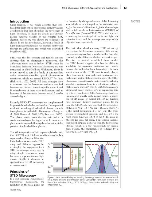
The basic idea behind scanning <strong>STED</strong> microscopy<br />
is to confine the fluorescence emission of fluorescent<br />
markers to a region that is much smaller than that<br />
covered by the diffraction-limited excitation spot.<br />
Therefore, a second, red-shifted beam (called<br />
the <strong>STED</strong> beam) is applied that has the ability to<br />
annihilate the molecular excitation <strong>and</strong> thereby<br />
prevent the molecules from fluorescing. The focal<br />
spatial extent of the <strong>STED</strong> beam is usually shaped<br />
like a doughnut in order to de-excite molecules only<br />
in the outer region of the excitation spot. The <strong>STED</strong><br />
photons act primarily on the excited state S 1, inducing<br />
stimulated emission down to a vibrational sublevel<br />
of the ground state S 0 vib (Fig. 1, left). Subpicosecond<br />
vibrational decay empties S 0 vib , so repumping into<br />
S 1 is largely ineffective. <strong>STED</strong> microscopy has been<br />
implemented mostly with pulsed beams, whereby<br />
the <strong>STED</strong> pulses of typical 0.1–1.0 ns duration<br />
have followed (shorter) excitation pulses. By the<br />
time the <strong>STED</strong> pulse has vanished, the population<br />
of the S 1 is N(h <strong>STED</strong>) = N0 exp(–�h <strong>STED</strong>), where N 0<br />
is the initial population, � ≈ 10 –16 cm 2 the crosssection<br />
for stimulated emission, <strong>and</strong> h <strong>STED</strong>(r) is the<br />
point-spread function (PSF) of the <strong>STED</strong> pulse in<br />
photons per area per pulse. This formula assumes<br />
that the <strong>STED</strong> pulse is shorter than the fluorescence<br />
lifetime, which is a few nanoseconds for organic<br />
dyes. Hence, the fluorescence is reduced by a<br />
factor �(h <strong>STED</strong>) = exp(–�h <strong>STED</strong>).<br />
Figure 1. Left, Jablonski diagram showing the energy states of a fluorescent molecule.<br />
Right, Breaking the diffraction barrier using <strong>STED</strong>. Normalized intensity profiles<br />
of the excitation PSF h exc , <strong>STED</strong> PSF h <strong>STED</strong> , De-excitation probability (1-�) <strong>and</strong><br />
effective PSF h eff .<br />
13<br />
NoTeS

![[Authors]. [Abstract Title]. - Society for Neuroscience](https://img.yumpu.com/8550710/1/190x245/authors-abstract-title-society-for-neuroscience.jpg?quality=85)