List of Abbreviations and Acronyms - Walter Reed Army Institute of ...
List of Abbreviations and Acronyms - Walter Reed Army Institute of ...
List of Abbreviations and Acronyms - Walter Reed Army Institute of ...

You also want an ePaper? Increase the reach of your titles
YUMPU automatically turns print PDFs into web optimized ePapers that Google loves.
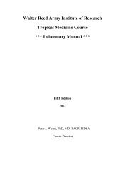
<strong>Walter</strong> <strong>Reed</strong> <strong>Army</strong> <strong>Institute</strong> <strong>of</strong> Research<br />
Tropical Medicine Course<br />
*** Laboratory Manual ***<br />
Third Edition<br />
2010<br />
Edited by<br />
COL Peter J. Weina PhD, MD, FACP, FIDSA<br />
Course Director<br />
MAJ Luis J. Martinez, MD, FACP<br />
Deputy Course Director
DEPARTMENT OF THE ARMY<br />
Lieutenant General Eric B. Schoomaker<br />
THE SURGEON GENERAL<br />
UNITED STATES ARMY MEDICAL RESEARCH AND<br />
MATERIEL COMMAND<br />
Major General James K. Gilman<br />
COMMANDER<br />
WALTER REED ARMY INSTITUTE OF RESEARCH<br />
Colonel Kent E. Kester, MC<br />
DIRECTOR AND COMMANDANT<br />
Colonel Peter J. Weina, MC<br />
Director, Division <strong>of</strong> Viral Diseases<br />
COURSE DIRECTOR<br />
Major Luis J. Martinez, MC<br />
Division <strong>of</strong> Viral Diseases<br />
DEPUTY COURSE DIRECTOR<br />
Ms. Natalie E. Slepski, BS, MPH<br />
Division <strong>of</strong> Viral Diseases<br />
SCIENTIFIC EDUCATION SPECIALIST<br />
3
Preface<br />
The <strong>Walter</strong> <strong>Reed</strong> <strong>Army</strong> <strong>Institute</strong> <strong>of</strong> Research (WRAIR)<br />
was established in 1893 as the <strong>Army</strong> Medical School by War<br />
Department General Orders No. 51, dated 24 Jan 1893. The<br />
Tropical Medicine course began in that school in July 1941<br />
while BG Russell Callendar was Comm<strong>and</strong>ant. At that time,<br />
the course ran for 30 days <strong>and</strong> consisted <strong>of</strong> didactic <strong>and</strong><br />
laboratory sessions similar to today’s course. Very much like<br />
the majority <strong>of</strong> the 52 years that this course was <strong>of</strong>fered at<br />
WRAIR, that first course presented continuing education to<br />
approximately 30 <strong>of</strong>ficers.<br />
Over the next fifty years, the course changed names<br />
<strong>and</strong> length but remained dedicated to teaching continuing<br />
tropical medicine education to military <strong>of</strong>ficers. In 1954, the<br />
<strong>Institute</strong> began the “Advanced Military Preventive Medicine<br />
Course” which carried on the tropical medicine education<br />
tradition begun in 1941. This course was eventually supplanted<br />
by the “Global Medicine Course” in December <strong>of</strong> 1966. During<br />
the next four <strong>and</strong> a half years, the Global Medicine course was<br />
<strong>of</strong>fered on 8 separate occasions. This 12 week course was<br />
divided into 4 weeks <strong>of</strong> “Epidemiology <strong>and</strong> Applied<br />
Biostatistics”, 3 weeks <strong>of</strong> “Ecology <strong>and</strong> Disease”, <strong>and</strong> 5 weeks<br />
<strong>of</strong> “Tropical Medicine”. In February <strong>of</strong> 1972, the Global<br />
Medicine course was split into a 5 week course called “Military<br />
Medical Ecology” <strong>and</strong> a 6 week course called the “Tropical<br />
Medicine Course”. The first Tropical Medicine Course was<br />
<strong>of</strong>fered in July <strong>and</strong> August <strong>of</strong> 1972 <strong>and</strong> was attended by 11<br />
medical <strong>of</strong>ficers <strong>and</strong> 4 clinical clerks. The course endured until<br />
1993 <strong>and</strong> was the only surviving remnant <strong>of</strong> the original <strong>Army</strong><br />
Medical School educational <strong>of</strong>ferings.<br />
In 1991, the <strong>Institute</strong> celebrated its 50 year tradition <strong>of</strong><br />
tropical medicine education. In memory <strong>of</strong> his significant<br />
contributions to tropical medicine education, the <strong>Institute</strong><br />
established “The Colonel George W. Hunter III Certificate”.<br />
This award was to be presented yearly to no more than two<br />
course lecturers who embody excellence <strong>and</strong> longevity as<br />
senior lecturers in the course. The first two recipients <strong>of</strong> the<br />
award were Dr. Jay P. Sanford (former University President<br />
5
<strong>and</strong> Dean <strong>of</strong> the Medical School at the Uniformed Services<br />
University <strong>of</strong> the Health Sciences) <strong>and</strong> Dr. Theodore E.<br />
Woodward (Emeritus Pr<strong>of</strong>essor <strong>of</strong> Medicine at the University<br />
Of Maryl<strong>and</strong> School <strong>of</strong> Medicine). A special presentation <strong>of</strong><br />
this award was made to Colonel Richard N. Miller, former<br />
Tropical Medicine Course Director, for his significant<br />
contributions to this course <strong>and</strong> its organization over the<br />
previous 12 years. The 50 year celebration also was<br />
particularly honored by the commencement address given by<br />
Dr. Theodore E. Woodward who attended the first course in<br />
1941.<br />
Due to operational needs <strong>of</strong> the Special Operations Comm<strong>and</strong><br />
<strong>and</strong> the newly formed Africa Comm<strong>and</strong>, in 2010 it was decided<br />
to resurrect the former 6 week course at WRAIR <strong>and</strong> convert it<br />
to a targeted short course that would provide a broader<br />
spectrum <strong>of</strong> individuals with the knowledge they need to<br />
combat international infectious disease threats. Operational<br />
dem<strong>and</strong>s upon the U.S. military facing wars on multiple fronts<br />
in areas affected with tropical disease identified a vital need for<br />
an intensely focused short course to familiarize medical<br />
personnel at all educational levels in tropical medicine.<br />
I hope that this edition <strong>of</strong> the manual will continue to<br />
contribute to the strength <strong>of</strong> a course that has served both<br />
military <strong>and</strong> civilian physicians for the last 70 years. As we<br />
find “tropical medicine” more <strong>and</strong> more becoming “world<br />
medicine”, this course, <strong>and</strong> all it has to <strong>of</strong>fer, will remain a<br />
necessity for physicians <strong>and</strong> other medical personnel for years<br />
to come.<br />
This laboratory manual is written for use in the<br />
Tropical Medicine Course. It represents years <strong>of</strong> accumulated<br />
knowledge from those who have been associated with the<br />
course over the last 70 years. I would be negligent if I did not<br />
at least recognize those who made significant contributions to<br />
the present manuscript. For the new (3 rd Edition) laboratory<br />
manual, contributors include Lieutenant Colonel Thomas J.<br />
Steinbach, Dr. Edgar Rowton, Dr. Harold Harlan, Lieutenant<br />
Colonel Jason H. Richardson, <strong>and</strong> Mr. Juan Mendez. Without<br />
Ms. Natalie Slepski’s hard work, this edition would never have<br />
6
come about, <strong>and</strong> to her we are all grateful. Among those who<br />
have contributed historically to past editions include Lieutenant<br />
Colonel Peter V. Perkins, Lieutenant Colonel Phillip G.<br />
Lawyer, Lieutenant Colonel Wilbur Milhous, Dr. Jacob L.<br />
Frenkel, <strong>and</strong> Dr. Claudia F. Golenda. This is by no means an<br />
exhaustive list <strong>of</strong> contributors, <strong>and</strong>, to those excluded by a<br />
lapse <strong>of</strong> memory, I apologize.<br />
Peter J. Weina, PhD, MD, FACP, FIDSA<br />
Colonel, Medical Corps, USA<br />
7
8<br />
Table <strong>of</strong> Contents<br />
Page<br />
<strong>List</strong> <strong>of</strong> <strong>Abbreviations</strong> <strong>and</strong> <strong>Acronyms</strong> ......................... 10<br />
Leishmaniasis ............................................................... 12<br />
Transport/Shipping <strong>of</strong> Parasites & Tissue ......... 15<br />
Slide Preparation <strong>and</strong> Staining ........................... 19<br />
Slide Reading ..................................................... 21<br />
rK39 Serum Test Strip Procedure ...................... 22<br />
Package Literature ................................. 23<br />
Appendix I: Diagrams ................................................. 25<br />
Appendix II: Photos .....................................................31<br />
Entomology ................................................................... 36<br />
Mosquito-borne Diseases ................................... 36<br />
S<strong>and</strong> Fly-borne Diseases .................................... 46<br />
Tick-borne Diseases ........................................... 48<br />
Vector Surveillance <strong>and</strong> Suppression ................ 48<br />
VECTOR MANAGEMENT (CONTROL) ................... 50<br />
POINT SOURCE THREATS .................................... 52<br />
USEFUL WEBSITES .............................................. 60<br />
REFERENCES ....................................................... 62<br />
Pathology ...................................................................... 67
Preservation <strong>of</strong> a Stool Specimen ...................... 69<br />
Macroscopic Examination <strong>of</strong> Stool Specimen .. 70<br />
Microscopic Examination <strong>of</strong> Stool Specimen ... 70<br />
Capillary Puncture for Blood Parasites ............. 71<br />
Preparation <strong>of</strong> Thick <strong>and</strong> Thin Smears .............. 73<br />
Medical Teleconsultation ............................................ 75<br />
9
<strong>List</strong> <strong>of</strong> <strong>Abbreviations</strong> <strong>and</strong> <strong>Acronyms</strong>:<br />
ALTHA – Armed Forces Health Longitudinal Technology<br />
Application (Formerly CHC2)<br />
CAP – College <strong>of</strong> American Pathologists<br />
CDC – Centers for Disease Control <strong>and</strong> Prevention<br />
CFR – Code <strong>of</strong> Federal Regulations<br />
CHCS – Composite Health Care System<br />
CHIKV – Chikungunya<br />
CL – Cutaneous Leishmaniasis<br />
CLIA – Clinical Laboratory Improvement Amendments<br />
CONUS – Continental United States<br />
DENV – Dengue Virus<br />
DHF – Dengue Hemorrhagic Fever<br />
DSS – Dengue Shock Syndrome<br />
DOD – Department <strong>of</strong> Defense<br />
IATA – International Air Transport Association<br />
ICAO – International Civil Aviation Organization<br />
JE – Japanese encephalitis<br />
LDL – Leishmania Diagnostic Laboratory<br />
LF – Lymphatic Filariasis<br />
MCL – Mucocutaneous Leishmaniasis<br />
PBSS – Phosphate Buffered Saline Solution<br />
OCONUS – Outside the Continental United States<br />
POC – Point <strong>of</strong> Contact<br />
SOP – St<strong>and</strong>ard Operating Procedure<br />
USACHPPM – United States <strong>Army</strong> Center for Health<br />
Promotion <strong>and</strong> Preventive Medicine<br />
VL – Visceral Leishmaniasis<br />
10
WHO – World Health Organization<br />
WRAIR – <strong>Walter</strong> <strong>Reed</strong> <strong>Army</strong> <strong>Institute</strong> <strong>of</strong> Research<br />
WRAMC – <strong>Walter</strong> <strong>Reed</strong> <strong>Army</strong> Medical Center<br />
11
12<br />
Leishmaniasis<br />
Leishmaniasis is a disease caused by an intracellular<br />
protozoa parasite, <strong>and</strong> it affects as many as 12 million people<br />
worldwide, with 1.5-2 million new cases each year. The global<br />
incidence <strong>of</strong> leishmaniasis has increased in recent years<br />
because <strong>of</strong> increased international leisure- <strong>and</strong> military-related<br />
travel, human alteration <strong>of</strong> vector habitats, <strong>and</strong> concomitant<br />
factors that increase susceptibility, such as HIV infection <strong>and</strong><br />
malnutrition.<br />
The recent conflicts in Iraq <strong>and</strong> Afghanistan have led<br />
to approximately 2000 laboratory-confirmed cases (<strong>and</strong> at least<br />
double the number <strong>of</strong> unconfirmed cases) <strong>of</strong> cutaneous<br />
leishmaniasis <strong>and</strong> 5 laboratory-confirmed cases <strong>of</strong> visceral<br />
leishmaniasis in American soldiers alone from 2003-2008. In<br />
Colombia, the military fighting the Fuerzas Armadas<br />
Revolucionarias de Colombia (FARC) has seen more than<br />
30,000 cases <strong>of</strong> leishmaniasis in the last 3 years. Of course, a<br />
significantly larger burden <strong>of</strong> diseases is borne by the local<br />
populations <strong>of</strong> these countries where Leishmania species are<br />
endemic. In these populations, leishmaniasis contributes<br />
greatly to morbidity <strong>and</strong> mortality.<br />
Infection is transmitted by the bite <strong>of</strong> a s<strong>and</strong>fly, which<br />
is usually one half to one third the size <strong>of</strong> a mosquito. The<br />
clinical spectrum <strong>of</strong> leishmaniasis ranges from a self-resolving<br />
cutaneous ulcer to a mutilating mucocutaneous disease <strong>and</strong>,<br />
depending on the species <strong>of</strong> Leishmania involved, even a lethal<br />
systemic illness. Infection with different Leishmania species<br />
can lead to a remarkably broad range <strong>of</strong> disease states.<br />
The clinical spectrum can range from insignificant<br />
pustules to fatal systemic disease. General underst<strong>and</strong>ing <strong>of</strong><br />
this clinical spectrum, although once believed to be quite<br />
predictable, continues to evolve as new diagnostic techniques<br />
contribute to the elucidation <strong>of</strong> the variety <strong>of</strong> clinical<br />
manifestations <strong>of</strong> an infection with even a single species <strong>of</strong><br />
Leishmania. The particular species associated with certain<br />
disease states originally was determined based only on clinical
manifestations <strong>and</strong> location found. In current practice,<br />
molecular techniques have shown a very different parasite-todisease<br />
association than was ever appreciated previously.<br />
Diagnosis is <strong>of</strong>ten difficult because <strong>of</strong> the small size <strong>of</strong><br />
the protozoa sequestered within macrophages <strong>of</strong> the skin, bone<br />
marrow, <strong>and</strong> reticuloendothelial system. Therapy has long been<br />
a challenge in the more severe forms <strong>of</strong> the disease <strong>and</strong> is made<br />
more difficult by the emergence <strong>of</strong> drug resistance. No<br />
effective vaccine for leishmaniasis is available.<br />
Localized cutaneous leishmaniasis usually manifests as<br />
a nonspecific ulcer that can mimic many other infectious <strong>and</strong><br />
noninfectious skin conditions. The vast majority <strong>of</strong> cases<br />
patients spontaneously with scarring <strong>and</strong> never come to the<br />
attention <strong>of</strong> clinicians. Even in US troops stationed in Iraq, it is<br />
currently felt, by many most closely associated with the disease<br />
<strong>and</strong> familiar with the epidemiology in the military, that less<br />
than 25% <strong>of</strong> all disease ever concerns afflicted soldiers enough<br />
to seek medical attention.<br />
In both the localized cutaneous <strong>and</strong> mucocutaneous<br />
forms <strong>of</strong> leishmaniasis, cell-mediated immunity to the parasite<br />
is vigorous <strong>and</strong> organism density in the skin <strong>and</strong>/or mucosa is<br />
low, especially in long-st<strong>and</strong>ing disease (although very early in<br />
the disease large numbers <strong>of</strong> the parasites are frequently<br />
found). Therefore, growing organisms in culture can be<br />
difficult, as can finding them in pathological specimens.<br />
Malnourished individuals are at greater risk <strong>of</strong> acquiring<br />
leishmaniasis <strong>and</strong> respond less well to treatment than those<br />
with adequate nutrition.<br />
The general consensus is that less than 5% <strong>of</strong><br />
individuals infected by Leishmania brasiliensis, <strong>and</strong> a smaller<br />
percentage <strong>of</strong> individuals infected by Leishmania panamensis<br />
<strong>and</strong> Leishmania guyanensis, develop mucosal metastases<br />
several months to years after the apparent resolution <strong>of</strong><br />
cutaneous disease. However, no rigorous studies prove this<br />
commonly accepted rate. Without treatment though, even in<br />
this small number, destruction <strong>of</strong> the oral <strong>and</strong> nasopharyngeal<br />
mucosa can be quite devastating <strong>and</strong> relentless.<br />
13
Symptoms <strong>of</strong> visceral leishmaniasis can be confused<br />
with many other infectious diseases; however, in endemic<br />
areas, the typical patient has wasting <strong>and</strong> presents with massive<br />
splenomegaly, pancytopenia, hypergammaglobulinemia, <strong>and</strong><br />
intermittent fevers (although they are less acutely ill than<br />
patients with malaria).<br />
A typical lesion <strong>of</strong> localized cutaneous leishmaniasis<br />
begins as an inflammatory papule, which later progresses to an<br />
ulcer. This may be associated with sporotrichotic lymphatic<br />
spread. In the vast majority <strong>of</strong> cases, the ulcers heal<br />
spontaneously with scarring.<br />
14
Transport or Shipping <strong>of</strong> Parasites <strong>and</strong> Tissue<br />
1. Procedures<br />
A. General Regulations to follow<br />
1) Note that there is a restrictive DOD Directive on the<br />
shipment <strong>of</strong> DOD hazardous materials (guidance for<br />
this directive is 49 CFR 173).<br />
2) Only <strong>of</strong>fer for transportation shipments <strong>of</strong> hazardous<br />
materials, including biomedical materials after approval<br />
by a DOD certifying <strong>of</strong>ficial.<br />
3) The DOD certifying <strong>of</strong>ficial must be approved by the<br />
comm<strong>and</strong>er in writing after receiving appropriate<br />
training (i.e. Transport <strong>of</strong> Biomedical Materials Course<br />
from USACHPPM).<br />
4) Ensure any packages are certified by the DOD<br />
certifying <strong>of</strong>ficial before shipment. DO NOT package or<br />
ship any biomedical material without certification.<br />
B. First, determine whether the sample to be shipped is<br />
considered an infectious substance or a diagnostic<br />
specimen:<br />
1) Diagnostic specimen: Unknown whether contents are<br />
infectious to humans, or shipment for identification.<br />
a) Make shipment in a plain box, removing any<br />
biohazard labels.<br />
b) Do not use an airbill for CONUS diagnostic<br />
shipments. See local logistician to enter shipment<br />
information into shipment computer.<br />
c) For OCONUS diagnostic shipments, use the<br />
following instructions.<br />
(1) Mark To: <strong>and</strong> From: with appropriate addresses<br />
on the side <strong>of</strong> box.<br />
(2) Place both diagnostic <strong>and</strong> directional arrow<br />
stickers on the side <strong>of</strong> box.<br />
(3) Ensure to over pack box with absorbent<br />
material.<br />
15
16<br />
(4) Ship only a MAXIMUM <strong>of</strong> 50mls <strong>of</strong> media per<br />
package.<br />
(5) Place FED EX sleeve onto top <strong>of</strong> box that<br />
contains airbill <strong>and</strong> CDC permit.<br />
(6) Follow these instructions for airbill:<br />
-Fill out To: Receiver’s information.<br />
-Fill out From: Lab Director information.<br />
-Ensure account information is correct.<br />
-Check FED EX Priority overnight (4A).<br />
-Check ”Yes” for dangerous goods (6).<br />
-Weigh box <strong>and</strong> note on airbill.<br />
-Make copy <strong>of</strong> airbill to track pack.<br />
(7) Sign airbill at release signature block. Follow<br />
common procedures for completion.<br />
2) Infectious substance: Known to contain material<br />
infectious to humans <strong>and</strong> concentrated.<br />
a) Make shipment with a 4G/Class 6.2 Isocontainer<br />
from Casing Corp.<br />
b) Ensure proper address labels are affixed to box.<br />
(1) From: Physician <strong>and</strong> facility information<br />
(receiving).<br />
(2) To: Lab Director information<br />
c) Enter the Name <strong>of</strong> responsible party (at top <strong>and</strong><br />
bottom <strong>of</strong> box) such as “COL Peter Weina (301) 319-<br />
9956 / 9497”.<br />
d) Ensure proper labels attached to side <strong>of</strong> box:<br />
(1) Infectious substance (6) sticker<br />
(2) UN2814 Infectious substance [fill out “Liquid<br />
Leishmania (amount in mls)”]<br />
(3) Etiologic agents / biomedical material (6) sticker
e) Ship only a MAXIMUM <strong>of</strong> 50mls <strong>of</strong> media per<br />
package.<br />
f) Place FED EX sleeve on top <strong>of</strong> box that contains<br />
airbill, CDC permit, <strong>and</strong> Shipment <strong>of</strong> dangerous<br />
goods form<br />
g) Follow these instructions for airbill:<br />
(1) Fill out To: Receiver’s information<br />
(2) Fill out From: Lab Director’s information.<br />
(3) Ensure account information is correct.<br />
(4) Check FED EX Priority overnight (4A).<br />
(5) Check “Yes” for dangerous goods (6).<br />
(6) Weigh box <strong>and</strong> note on airbill.<br />
(7) Make copy <strong>of</strong> airbill to track package.<br />
h) Sign airbill at release signature block.<br />
C. Instructions for declaration <strong>of</strong> dangerous goods.<br />
1) Write in “Shipper”: Lab Director’s information.<br />
2) Write in “Consignee”: Receiver’s information.<br />
3) Blot out Cargo Aircraft only <strong>and</strong> radioactive boxes.<br />
4) Type everything under nature <strong>and</strong> quantity <strong>of</strong><br />
dangerous goods in capital letters.<br />
a) Under proper shipping name: “INFECTIOUS<br />
SUBSTANCE AFFECTING HUMANS (LIQUID)<br />
LEISHMANIA”<br />
b) Class or division: “6.2”<br />
c) UN or ID No.: “UN2814”<br />
d) Under quantity <strong>and</strong> type <strong>of</strong> packaging: 4G, # <strong>of</strong><br />
flasks contained, amount <strong>of</strong> total media in mls.<br />
e) Packing instructions: “602”<br />
f) Under additional h<strong>and</strong>ling information: “FOLLOW<br />
DIRECTIONS ON CONTAINER PRIOR<br />
ARRANGEMENTS AS REQUIRED BY THE IATA<br />
17
18<br />
DANGEROUS GOODS REGULATION 1.3.3.1<br />
HAVE BEEN MADE IN ACCORDANCE WITH<br />
ICAO AND IATA REGULATIONS”<br />
g) Under emergency Telephone # place local contact,<br />
such as: 301-319-9956 / 9497, pager 301 369-5413,<br />
fax 301-319-7360<br />
h) Fill out signature block with sender’s information.<br />
i) Sign signature block.<br />
D. Common Procedures<br />
1) Wrap in brown paper <strong>and</strong> tape up final package.<br />
Attach the final FED EX sleeve to the top <strong>of</strong> box.<br />
2) Ensure the following:<br />
a) Sign release signature.<br />
b) Make copy <strong>of</strong> airbill to track package.<br />
c) Give top copy <strong>of</strong> airbill goes to comptroller for<br />
accountability.<br />
d) Bring box to shipping section.<br />
e) Place box in FED EX shipping area.<br />
f) Log in / fill out shipment notice form (POC “name<br />
<strong>and</strong> telephone”).
Slide Preparation <strong>and</strong> Staining<br />
1. Materials <strong>and</strong> Equipment<br />
A. Slide preparation<br />
1) Slides<br />
2) Cover slips<br />
3) Methanol<br />
4) Plastic disposable pipettes<br />
5) Cytospin<br />
6) Timer<br />
7) Cytospin holder<br />
8) Pencil, marker<br />
9) PBSS<br />
10) Tissue<br />
11) Disposable alcohol wipes<br />
12) Small Avery labels<br />
B. Staining<br />
1) Methanol<br />
2) Giemsa / Diff-Quick Solutions 1 & 2<br />
3) Forceps<br />
4) Copel<strong>and</strong> jars<br />
5) Water (Analytical type I reagent grade water)<br />
6) Water (Sigma® sterile water)<br />
7) Microscope<br />
8) Paper towels<br />
9) Permount<br />
10) Immersion oil<br />
19
20<br />
11) Slide box<br />
12) Filters (0.22μm)<br />
2. Procedures<br />
A. Slide Preparation<br />
1) Direct Slide Preparation<br />
a) Clean slide with alcohol wipe. With forceps grasp<br />
the tissue <strong>and</strong> make impression on the slide by either<br />
making a dabbing or circular motion on the slides with<br />
the tissue (let it dry).<br />
b) Place a drop <strong>of</strong> mixture on the slide <strong>and</strong> smear the<br />
slide, if a tissue/PBSS mixture. Air dry slide before<br />
use.<br />
2) Using Cytospin<br />
a) Put 2 - 3 drops <strong>of</strong> ground tissue on each slide <strong>and</strong><br />
place in separate cytospin holder. Ensure an equal<br />
amount is placed in on each slide.<br />
b) Spin for 3 min @ 800 rpm. Repeat if necessary.<br />
c) Remove slide from holder <strong>and</strong> let dry for staining.<br />
B. Staining<br />
1) Using Diff-Quik<br />
a) Fix slide with methanol. Methanol should cover the<br />
entire slide area. Let slide air dry under hood.<br />
b) Filter stain prior to use.<br />
c) Get 4 Copel<strong>and</strong> jars ready for staining by filling the<br />
jars 3/4 full with the following:<br />
(1) Jar 1: Diff-Quick I<br />
(2) Jar 2: type I water<br />
(3) Jar 3: Diff-Quik II<br />
(4) Jar 4: type I water.<br />
d) Grasp the slide <strong>and</strong> submerge it into Diff-Quik<br />
solution I for 45 seconds using forceps.
e) Remove slide <strong>and</strong> wash gently in jar 2 type I water.<br />
f) Submerge slowly in type I water 3 times.<br />
g) Damp slide edge on paper towel.<br />
h) Submerge slide into Diff-Quik solution II for 45<br />
seconds.<br />
i) Remove slide <strong>and</strong> wash gently in jar 4, type I water.<br />
j) Submerge slowly in type I water 3 times.<br />
k) Let slide air dry.<br />
2) Giemsa Stain<br />
a) Fix slide in methanol <strong>and</strong> let air dry.<br />
b) Prepare a 30% concentration <strong>of</strong> Giemsa in Sigma®<br />
water using 50 ml conical tube. Filter 30% Giemsa<br />
solution prior to use.<br />
c) Pour 30% Giemsa solution on slides <strong>and</strong> leave slide<br />
flooded with solution for 30 minutes<br />
d) Wash slide thoroughly with type I water.<br />
e) Let slide air dry under hood.<br />
f) Read.<br />
Slide Reading<br />
1. Materials <strong>and</strong> Equipment<br />
A. Immersion oil<br />
B. Cover slip<br />
C. Smeared <strong>and</strong> stained slide<br />
D. Microscope<br />
E. Permount with 10% xylem<br />
F. Control positive slide<br />
2. Procedures<br />
A. Place slide on microscope after airdrying under hood.<br />
21
22<br />
B. Use an oil immersion lens in order to view the slides<br />
C. Complete a comprehensive review <strong>of</strong> the slide in order to<br />
visualize any amastigotes (Leishmania parasites which lack<br />
a tale) on the slide See Appendix 2, Figure 1 for an image <strong>of</strong><br />
amastigotes on the Giemsa stained slide.<br />
D. Use a positive control slide if necessary to confirm<br />
visualization <strong>of</strong> amastigotes.<br />
E. Write a report with the technician’s impressions on the<br />
slide quality <strong>and</strong> slide result. Put this with the patient file.<br />
F. Label the slides <strong>and</strong> slide box with the local control<br />
number, date, patient name, <strong>and</strong> the technician’s initials.<br />
G. Bring the slides to the Lab Director for confirmation.<br />
rK39 Serum Test Strip<br />
1. Procedures<br />
A. Follow product insert from INBIOS for rK39 strip test<br />
for visceral leishmaniasis.<br />
B. Record results in notes section <strong>of</strong> LDL Accession<br />
spreadsheet <strong>and</strong> notify Lab Director <strong>of</strong> results.<br />
C. Follow these procedures for archiving patient serum<br />
1) Fully aliquot positive serums into as many 1.8ml<br />
cryotubes are needed <strong>and</strong> labeled by local control<br />
number.<br />
2) Aliquot negative serums once <strong>and</strong> properly label by<br />
local control number.<br />
3) Store all serum aliquots in chronological order.
APPENDIX I<br />
Figure 1: Relationships <strong>of</strong> the various protozoans<br />
phylogenetically<br />
25
Figure 2: Lifecycles <strong>of</strong> the various Trypomastigotes<br />
26
Figure 3: Various body forms for the Trypomastigotes<br />
(from <strong>Army</strong> / Air Force manual AFM 160-48/TM 8-227-2,<br />
1August 1974)<br />
27
Figure 4: Various Parasitic Trematode Eggs (from <strong>Army</strong> /<br />
Air Force manual AFM 160-48/TM 8-227-2, 1August 1974)<br />
28
Figure 5: Various Tapeworms found in humans (from<br />
<strong>Army</strong> / Air Force manual AFM 160-48/TM 8-227-2,<br />
1August 1974)<br />
29
Figure 6: Various Parasitic Nematide Eggs (from <strong>Army</strong> /<br />
Air Force manual AFM 160-48/TM 8-227-2, 1August 1974)<br />
30
APPENDIX II:<br />
Figure 1: Amastigotes on the Giemsa stained slide<br />
Figure 2: S<strong>and</strong>fly size comparison (S<strong>and</strong>fly on left,<br />
Mosquito on right)<br />
31
Figure 3: Cutaneous Leishmaniasis punch biopsy<br />
Figure 4: Where to make dermal punch for Leishmaniasis<br />
32
Figure 5: Cutaneous Leishmaniasis lesion<br />
Figure 6: Cutaneous Leishmaniasis over a tattoo<br />
33
Figure 7: Fecal specimen container<br />
Figure 9: Stool preservation kit<br />
34<br />
Figure 11: Hemoccult test<br />
Figure 8: Stool specimen<br />
preservation<br />
Figure 10: Hemoccult test
Figure 12: Materials that may be mistaken for parasites:<br />
Fat Globule (left) <strong>and</strong> Plant Cell (right).<br />
Figure 13: Protozoa species<br />
Figure 14: Example <strong>of</strong> thick smear (left) <strong>and</strong> thin smear (right)<br />
35
36<br />
Entomology<br />
Mosquito-borne diseases. Mosquitoes are the most important<br />
group <strong>of</strong> vectors <strong>of</strong> human diseases. Throughout recorded<br />
history they have transmitted hundreds <strong>of</strong> different pathogens<br />
<strong>of</strong> humans, including: protozoans, viruses, <strong>and</strong> parasites that<br />
kill millions <strong>of</strong> people a year even today <strong>and</strong> debilitate<br />
hundreds <strong>of</strong> millions more. An example <strong>of</strong> some major<br />
pathogen groups is detailed briefly below. Websites that<br />
contain details about mosquito-borne human diseases, their<br />
vectors, distributions <strong>and</strong> prevention include:<br />
1. Malaria<br />
Malaria transmission is endemic in many tropical regions<br />
around the world, <strong>and</strong> transmission may occur nearly year<br />
round in many tropical countries, except at higher elevations<br />
(>500 m). The World Health Organization (WHO) estimated<br />
in 2009 that there are >300 Million new cases <strong>of</strong> malaria each<br />
year <strong>and</strong> that >1.5 Million <strong>of</strong> those are fatal each year with<br />
most <strong>of</strong> those cases <strong>and</strong> fatalities occurring in sub-Saharan<br />
Africa, India, <strong>and</strong> Southeast Asia. Vector mosquito activity<br />
<strong>and</strong> related intensity <strong>of</strong> transmission increase <strong>and</strong> decrease<br />
based on rainfall timing <strong>and</strong> patterns <strong>and</strong> also vary with the<br />
particular vector species, but are usually most intense during or<br />
shortly after peaks <strong>of</strong> rainfall in warm months. Strains <strong>of</strong><br />
Plasmodium falciparium <strong>and</strong> P. vivax resistant to<br />
chemoprophylactic drugs have been reported to be present in<br />
some countries.<br />
Adult females <strong>of</strong> at least 60 species <strong>of</strong> Anopheles,<br />
worldwide, have been proven capable <strong>of</strong> spreading the four<br />
species Plasmodium that cause human malarias. Many <strong>of</strong> these<br />
are species complexes <strong>and</strong> are still not well studied. Insecticide<br />
resistance is not regularly monitored in many countries, nor<br />
well reported. Historically, many countries have reported<br />
resistance or tolerance by several species <strong>of</strong> vector mosquitoes<br />
to commonly used insecticides.
Anyone going into a country or region where malaria is<br />
known or reported to be present should seek preventive <strong>and</strong><br />
prophylactic advice (anti-malarial materials <strong>and</strong> dose guidance)<br />
from their higher medical authorities. They may also wish to<br />
do a search beforeh<strong>and</strong> on websites <strong>of</strong> the WHO<br />
(www.who.int), the U.S. CDC (www.cdc.gov), or similar<br />
travelers’ health sites, for their latest reported status <strong>and</strong><br />
currently suggested chemoprophylactic drugs (<strong>and</strong> respective<br />
doses). Malaria may be a significant threat to either short or<br />
long term military operations in every place where it is<br />
endemic or has been recently reported to be actively<br />
transmitted. Appropriate personal protective techniques should<br />
be used routinely to help prevent infection by any vector-borne<br />
disease(s) wherever you may be in the world.<br />
World distribution <strong>of</strong> Malaria - 2008<br />
37
Malaria Disease Cycle(s)<br />
38
Comparison <strong>of</strong> characteristics <strong>of</strong> three major mosquito genera.<br />
39
40<br />
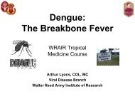
2. Dengue<br />
Dengue virus (DENV) is widespread <strong>and</strong> essentially<br />
endemic throughout many tropical areas <strong>of</strong> the world. Periodic<br />
outbreaks <strong>of</strong> all four strains (serotypes DENV-1, -2, -3, & -4)<br />
occur <strong>and</strong> tend to go through poorly defined cycles <strong>of</strong><br />
transmission at roughly 10-15 year intervals. Both dengue<br />
hemorrhagic fever (DHF) <strong>and</strong> dengue shock syndrome<br />
(DSS) have been reported currently or very recently from a<br />
number <strong>of</strong> tropical countries. Populations <strong>of</strong> the primary<br />
vector, Aedes aegypti, have greatly increased due partly to<br />
rapid <strong>and</strong> uncontrolled urbanization in much <strong>of</strong> Southeast Asia.<br />
Aedes albopictus is an important vector in peri-urban <strong>and</strong> rural<br />
areas <strong>of</strong> some regions <strong>and</strong> a cold-hardy strain <strong>of</strong> this species<br />
has spread from Northern Asia to the central U.S. <strong>and</strong> Brazil,<br />
<strong>and</strong> it was recently reported as established in northern Europe<br />
(i.e., the Netherl<strong>and</strong>s). Dengue is a debilitating disease that<br />
would be a significant threat to military forces <strong>and</strong> there is no<br />
effective preventive vaccine or curative treatment, only<br />
supportive care (for current specifics see Heinemann 2009, <strong>and</strong><br />
search the U.S. CDC <strong>and</strong> the WHO websites).<br />
Distribution <strong>of</strong> Dengue:
Dengue Disease Cycle (an example <strong>of</strong> an arbovirus):<br />
41
Some Vectors <strong>of</strong> Dengue: Comparison <strong>of</strong> Adult Female<br />
Aedes aegypti vs. Aedes albopictus.<br />
42
3. Chikungunya virus (CHIKV)<br />
Chikungunya (CHIKV) virus is endemic to Africa <strong>and</strong> Asia<br />
<strong>and</strong> is transmitted by the same mosquitoes as DENV, Ae.<br />
aegypti <strong>and</strong> Ae. albopictus. CHIKV causes an illness with<br />
symptoms similar to dengue fever. CHIKV manifests itself<br />
with an acute febrile phase <strong>of</strong> the illness lasting only two to<br />
five days, followed by a prolonged arthralgic disease that<br />
affects the joints <strong>of</strong> the extremities. The pain associated with<br />
CHIKV infection <strong>of</strong> the joints persists for weeks or months, or<br />
in some cases years.<br />
4. Japanese encephalitis (JE)<br />
Japanese encephalitis (JE) virus has caused widespread<br />
epidemics in Japan <strong>and</strong> the Republic <strong>of</strong> Korea <strong>and</strong> is endemic<br />
in Southeast Asia. The virus is maintained in nature by<br />
mosquitoes <strong>and</strong> non-human vertebrates, <strong>and</strong> man becomes<br />
accidentally involved. In temperate countries like Japan, the<br />
disease occurs in the warm weather; in the tropics it could<br />
occur during any season, although the risk is higher during <strong>and</strong><br />
immediately after the rainy season when the mosquito<br />
population increases. Culex tritaeniorhynchus, a rice field<br />
breeding mosquito is the principal vector <strong>and</strong> feeds mainly on<br />
large animals <strong>and</strong> birds. Elsewhere in the area <strong>of</strong> distribution,<br />
Cx. gelidus (predominantly a pig biter) <strong>and</strong> Cx. vishnui group<br />
mosquitoes are also involved. Japanese encephalitis is<br />
predominantly a rural disease <strong>and</strong> in most <strong>of</strong> southeastern Asia,<br />
associated with rice cultivation <strong>and</strong> mosquitoes which breed in<br />
rice fields. Transmission is by bite. A licensed vaccine is<br />
available.<br />
43
Distribution <strong>of</strong> reported FEV cases from<br />
Tiroumourougane1 et al 2002.<br />
44<br />
5. Filariasis<br />
Bancr<strong>of</strong>tian filariasis, caused by Wuchereria bancr<strong>of</strong>ti, <strong>and</strong><br />
Brugian filariasis, caused by Brugia malayi, are now lumped<br />
by the WHO as “Lymphatic Filariasis” (LF). According to<br />
the WHO (2007), LF is currently endemic <strong>and</strong> widespread, <strong>and</strong><br />
poses serious public health problems in Southeast Asia, South<br />
Asia, <strong>and</strong> sub-Saharan Africa. Mapping <strong>of</strong> the populations at<br />
risk <strong>and</strong> related programs <strong>of</strong> mass drug administration (MDA),<br />
using oral dosing with diethylcarbamazine citrate (DEC), are in<br />
progress in a number <strong>of</strong> countries, <strong>and</strong> seem to be reducing the<br />
levels <strong>of</strong> LF morbidity therein. The main vectors <strong>of</strong><br />
Bancr<strong>of</strong>tian filariasis, Culex pallens <strong>and</strong> Cx. quinquefasciatus,<br />
have become more abundant as breeding sites have been<br />
exp<strong>and</strong>ed due to increased urbanization <strong>and</strong> poor sanitation.<br />
Nocturnally periodic forms <strong>of</strong> B. malayi <strong>and</strong> W. bancr<strong>of</strong>ti are<br />
also currently endemic in parts <strong>of</strong> Southeast Asia <strong>and</strong> some
southwest Pacific isl<strong>and</strong>s. At least 16 different species <strong>of</strong><br />
tropical area mosquitoes are reported to be effective vectors <strong>of</strong><br />
filariases (LF), including several species <strong>of</strong> Anopheles.<br />
Distribution <strong>of</strong> Filariasis:<br />
Filariasis disease cycle (Bancr<strong>of</strong>tian):<br />
45
Typical transmission cycles for Wuchereria bancr<strong>of</strong>ti <strong>and</strong><br />
Brugia malayi:<br />
S<strong>and</strong> Fly-borne Diseases. One <strong>of</strong> the issues with s<strong>and</strong> fly<br />
borne diseases is that there are no vaccines or prophylactics<br />
available for them. For this reason vector control <strong>and</strong> personal<br />
protective measures are the only defenses against contracting<br />
these diseases. S<strong>and</strong> flies are much smaller than most<br />
mosquitoes. Like mosquitoes only females take a blood meal<br />
<strong>and</strong> are capable <strong>of</strong> disease transmission. Unlike mosquitoes<br />
they do not have an aquatic phase to their life cycle. They are<br />
found in temperate <strong>and</strong> warmer environments, ranging from<br />
forests to deserts. The larvae feed on decaying organic matter<br />
from decaying vegetation to feces. Adult s<strong>and</strong> flies can be<br />
distinguished from most other blood-sucking arthropods<br />
including their related mosquitoes in that they hold their wings<br />
in a “V” shape rather than laying them flat against their<br />
46
abdomens. They also tend to “hop” after lighting on surfaces.<br />
When examining light trap collections their small size, hunched<br />
back, long legs, <strong>and</strong> the fine hairs (rather than scales) along<br />
with their wings held away from their bodies helps to separate<br />
them from other insects.<br />
1. Leishmaniasis<br />
Leishmaniasis is an infection <strong>of</strong> animals <strong>and</strong> humans<br />
caused by protozoa in the genus Leishmania. At least 23<br />
Leishmania species cause leishmaniasis. There are three forms<br />
<strong>of</strong> the disease: cutaneous (CL), mucocutaneous (MCL) <strong>and</strong><br />
visceral (VL). CL appears as a non-healing ulcer lasting<br />
months to years if untreated. MCL patients develop ulcerative<br />
or granulomatous (granular) lesions <strong>of</strong> the nasal, oral, <strong>and</strong><br />
pharyngeal linings, which generally occur after or concurrent<br />
with CL lesions. VL, the most severe form <strong>of</strong> leishmaniasis<br />
with 95% mortality in untreated cases, is a chronic disease<br />
involving the liver <strong>and</strong> spleen.<br />
Transmission occurs through the bite <strong>of</strong> infective female<br />
phlebotomine s<strong>and</strong> flies in the genera Phlebotomus (Old<br />
World) <strong>and</strong> Lutzomyia (New World). Generally, s<strong>and</strong> flies feed<br />
at dusk <strong>and</strong> during the evening; however, some species are<br />
opportunistic <strong>and</strong> will feed during the day if disturbed. The<br />
uninfected s<strong>and</strong> fly generally acquires the infection by feeding<br />
on a reservoir host. Reservoirs for leishmaniasis, which vary<br />
depending on location, include domestic dogs, rodents<br />
(including rats, hyraxes <strong>and</strong> gerbils), sloths, marsupials, <strong>and</strong> in<br />
some endemic areas, humans. It is reported that 90 species <strong>of</strong><br />
Lutzomyia <strong>and</strong> at least 39 species <strong>of</strong> Phlebotomus feed on<br />
humans.<br />
Protect Yourself from Leishmaniasis. Limit outdoor<br />
activity at dusk <strong>and</strong> during the evening when possible as this is<br />
when the s<strong>and</strong> fly is most active. If possible building should<br />
have window screens or other barriers to keep s<strong>and</strong> flies from<br />
entering. S<strong>and</strong> flies bite in <strong>and</strong> outdoors; although generally<br />
nocturnal, they may feed during the day in buildings. Avoid the<br />
bites <strong>of</strong> s<strong>and</strong> flies by using protective clothing <strong>and</strong> insect<br />
repellents (See Personal Protective Measures in the Vector<br />
Management Section).<br />
47
48<br />
2. S<strong>and</strong> Fly Fever<br />
S<strong>and</strong> Fly Fever is a Phlebovirus, (Bunyaviridae) with two<br />
major serotypes, Sicilian <strong>and</strong> Naples. It occurs in the<br />
Mediterranean, Mid-East <strong>and</strong> North Africa. A major vector is<br />
Phlebotomus papatasi; however, several other species <strong>of</strong><br />
Phlebotomus also serve as vectors.<br />
Control <strong>and</strong> Personal Protective Measures would be those<br />
used for other s<strong>and</strong> fly borne diseases such as leishmaniasis.<br />
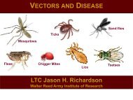
Tick-borne Diseases. Tick-borne illnesses are caused by<br />
infection with a variety <strong>of</strong> pathogens, including rickettsia <strong>and</strong><br />
other types <strong>of</strong> bacteria, viruses, <strong>and</strong> protozoa. Because ticks<br />
can harbor more than one disease-causing agent, patients can<br />
be infected with more than one pathogen at the same time,<br />
compounding the difficulty in diagnosis <strong>and</strong> treatment. The<br />
geographic range <strong>of</strong> many tick-borne diseases is not well<br />
known <strong>and</strong> symptoms are <strong>of</strong>ten similar to other bacterial or<br />
viral diseases. Prevention <strong>of</strong> tick bites should be stressed using<br />
methods described below.<br />
Vector Surveillance <strong>and</strong> Suppression. Light traps can be<br />
used to collect night-biting mosquitoes, phlebotomine s<strong>and</strong><br />
flies, <strong>and</strong> Culicoides. Not all species are attracted to light <strong>and</strong><br />
adding carbon dioxide (CO2) to light traps (figure 1) increases<br />
the number <strong>of</strong> species <strong>and</strong> total numbers <strong>of</strong> females collected.<br />
Traps baited with animals or humans or placed around their<br />
habitations can be useful for determining risk in these areas <strong>and</strong><br />
may also increase catch. (the use <strong>of</strong> humans as attractants may<br />
be subject to the requirements <strong>of</strong> formal human-use protocols).<br />
Adults can be collected from indoor <strong>and</strong> outdoor resting sites<br />
using a mechanical aspirator <strong>and</strong> flashlight. For mosquitoes
systematic larval sampling with a long-h<strong>and</strong>led white dipper<br />
provides information on species composition <strong>and</strong> population<br />
dynamics <strong>of</strong> species breeding locally that can be used to plan<br />
control measures. Larva sampling are generally not effective<br />
for s<strong>and</strong> flies.<br />
Typical light trap used in the surveillance <strong>of</strong> night biting<br />
insects. These traps come in different formats but consist<br />
basically <strong>of</strong> a light <strong>and</strong> a fan which drives the insects into a<br />
collection jar or net when they get close to the light. Carbon<br />
dioxide can be added to the trap in the form dry ice or<br />
compressed gas cylinder.<br />
Figure 12. Mosquito light trap.<br />
49
Vector Management (Control)<br />
50<br />
1. Personal protective measures<br />
Personal protective measures are the first line <strong>of</strong> defense<br />
against arthropod-borne disease <strong>and</strong>, in some cases, may be the<br />
only protection for deployed military personnel. Proper<br />
wearing <strong>of</strong> the uniform <strong>and</strong> appropriate use <strong>of</strong> repellents can<br />
provide high levels <strong>of</strong> protection against blood-sucking<br />
arthropods. The uniform fabric provides a significant<br />
mechanical barrier to mosquitoes <strong>and</strong> other blood-sucking<br />
insects. Therefore, the uniform should be worn to cover as<br />
much skin as possible if weather <strong>and</strong> physical activity permit.<br />
Some newly developed repellents provide military<br />
personnel unprecedented levels <strong>of</strong> protection. An aerosol<br />
formulation <strong>of</strong> permethrin (NSN 6840-01-278-1336) can be<br />
applied to a uniform according to label directions, but not to<br />
the skin. This will impart both repellent <strong>and</strong> insecticidal<br />
properties to the uniform material that will be retained through<br />
numerous washings. An extended formulation lotion <strong>of</strong> N, Ndiethyl-m-toluamide<br />
(DEET) (NSN 6840-01-284-3982) has<br />
been developed to replace the older 2 oz. bottles <strong>of</strong> 75% deet in<br />
alcohol. This lotion contains 33% active ingredient. It is less<br />
irritating to the skin, has less odor <strong>and</strong> is generally more<br />
acceptable to users.<br />
A properly worn Uniform (e.g., ACU) impregnated with<br />
permethrin, combined with use <strong>of</strong> extended duration DEET on<br />
exposed skin, has been demonstrated to provide nearly 100%<br />
protection against a variety <strong>of</strong> blood-sucking arthropods. This<br />
dual strategy is termed “the DoD Arthropod Repellent<br />
System.” In addition, permethrin may be applied to bednets,<br />
tents, <strong>and</strong> other field items as appropriate. Complete details<br />
regarding these <strong>and</strong> other personal protective measures are<br />
provided in TG 36, Personal Protective Techniques Against<br />
Insects <strong>and</strong> Other Arthropods <strong>of</strong> Military Significance (2009).<br />
2. Physical Controls<br />
Soldiers <strong>of</strong>ten do not have the option to avoid potential<br />
disease vector or their typical habitats, but physical controls
may sometimes be possible. Covering structural openings<br />
(e.g., doors <strong>and</strong> windows) with screen fine enough to exclude<br />
particular vectors or biting pests may be very practical,<br />
especially in permanent or semi-permanent camps. Sealing<br />
cracks <strong>and</strong> joints in even temporary structures <strong>and</strong> vehicles<br />
whenever possible or covering such openings with netting (like<br />
a bed net, or part <strong>of</strong> an old one) might keep out a lot <strong>of</strong> filth<br />
flies that can spread food-borne diseases), as well as scorpions<br />
<strong>and</strong> similar crawling pests that can pose a health threat. The<br />
use <strong>of</strong> fly swatters or similar devices against individual<br />
invaders falls within this same category <strong>of</strong> control. Use a bed<br />
net while sleeping; NSN 7210-00-266-9736 (Netting), NSN<br />
7210-00-267-5641 (Poles). Because s<strong>and</strong> flies are small<br />
enough to pass through the mesh <strong>of</strong> the st<strong>and</strong>ard bed net,<br />
permethrin (aerosol spray) should be applied to netting. There<br />
is enough permethrin in one spray can to treat one uniform <strong>and</strong><br />
a bed net.<br />
3. Source Reduction<br />
Eliminating st<strong>and</strong>ing water or treating it with non-toxic or<br />
minimally toxic (to humans) larvicides like Bti or an insect<br />
growth regulator (an IGR) within a 100-ft. radius <strong>of</strong> any point<br />
can greatly reduce the local breeding <strong>and</strong> short-range attraction<br />
<strong>of</strong> a number <strong>of</strong> mosquitoes, Phlebotomine s<strong>and</strong> flies, <strong>and</strong><br />
certain other vectors <strong>and</strong> pests. Surface drainage, filling pot<br />
holes, <strong>and</strong> similar actions can also help reduce the numbers <strong>of</strong> a<br />
given vector or pest species that any particular habitat can<br />
support. Removing trash, garbage <strong>and</strong> rubbish frequently<br />
could reduce the habitat for a number <strong>of</strong> arthropod <strong>and</strong> some<br />
vertebrate pests so many fewer can live near your site.<br />
4. Chemical Controls<br />
Chemical control <strong>of</strong> vectors or pests is not usually a<br />
practical option for soldiers, but the use <strong>of</strong> residual insecticides<br />
to treat the interior <strong>of</strong> semi-permanent or permanent buildings<br />
<strong>and</strong> other selected surfaces can be a very important strategy in<br />
some situations. Specific chemicals (e.g., repellents) applied to<br />
bed nets, screening, or movable structural elements (like<br />
temporary walls or partitions) can greatly reduce or eliminate<br />
vector or pest populations in many situations (especially<br />
51
crawling pests <strong>and</strong> even certain mosquitoes or other biting<br />
arthropods). When properly planned <strong>and</strong> performed, wide area<br />
treatments with ULV or fogging devices can knock down a<br />
large portion <strong>of</strong> currently active population <strong>of</strong> infected vectors,<br />
<strong>and</strong> thus, possibly prevent or greatly reduce local area<br />
outbreaks <strong>of</strong> some diseases (e.g., some relatively deadly<br />
encephalitides). Such treatments are beyond the capabilities <strong>of</strong><br />
most soldiers or tactical units.<br />
52<br />
5. Preventing tick, chigger <strong>and</strong> flea bites<br />
When operating in areas infested with ticks, chiggers or fleas,<br />
the pants should be bloused into the boots to prevent access to<br />
the skin by ticks <strong>and</strong> other crawling arthropods such as<br />
chiggers. Check yourself frequently when walking through<br />
tick-infested areas. Upon returning from infested areas, remove<br />
all clothing <strong>and</strong> examine yourself for ticks <strong>and</strong> chiggers.<br />
Infected ticks may require several hours <strong>of</strong> feeding before<br />
pathogens are transmitted. Therefore, personnel who operate in<br />
tick-infested areas should check themselves frequently for ticks<br />
<strong>and</strong> remove them as soon as possible. If ticks become attached,<br />
the simplest <strong>and</strong> best method <strong>of</strong> removal is by a slow, steady<br />
pull with a pair <strong>of</strong> tweezers or forceps. Do not squeeze the<br />
body but grasp the tick where the mouthparts enter the skin <strong>and</strong><br />
pull firmly until the tick is extracted. Be careful not to break <strong>of</strong>f<br />
the mouthparts <strong>and</strong> leave them in the skin. Wipe the bite area<br />
with an antiseptic. If h<strong>and</strong>s have touched the tick during<br />
removal, wash them thoroughly with soap <strong>and</strong> water or an<br />
antiseptic, since tick secretions may contain pathogens.<br />
Point Source Threats<br />
There are many different potential point source threats that can<br />
impact any given deployment. Following are some examples <strong>of</strong><br />
more common arthropod point source threats <strong>and</strong> a brief<br />
description <strong>of</strong> their potential impact to military personnel.<br />
1. Spiders<br />
Spiders, in general, are harmless to people. However, a few<br />
species are capable <strong>of</strong> causing serious damage or death in<br />
victims.
A. Black Widow Spiders<br />
Black widow spiders, Latrodectus spp. are among the<br />
most dangerous spiders in the world. They are normally<br />
timid, medium-sized spiders (
54<br />
perhaps the most recognized member <strong>of</strong> this group. The<br />
fiddle-shaped mark on the cephalothorax, long legs <strong>and</strong><br />
sleek, brown coloration are characteristic <strong>of</strong> this group.<br />
- The brown recluse bite is not particularly painful <strong>and</strong><br />
may not be felt at all. Multiple bites in a single attack are<br />
not uncommon.<br />
- The venom is necrotic, <strong>and</strong> destroys the tissues <strong>of</strong> the<br />
victim. However, not all bites cause necrosis <strong>and</strong> the<br />
extent <strong>of</strong> necrosis is highly variable among victims<br />
ranging from a small “pimple” to severe “craters” that<br />
may take months to heal.<br />
C. Tarantulas<br />
Tarantulas are widely feared, but they are not considered<br />
to be dangerous. Their bites are similar to a bee or wasp<br />
sting. Their fangs are quite large approaching the size <strong>of</strong><br />
a pair <strong>of</strong> large needles <strong>and</strong> bites are quite painful.<br />
2. Scorpions<br />
Scorpions have painful stings <strong>and</strong> several species can be<br />
deadly to humans. Generally, small species with slender<br />
claws are the most dangerous, whereas larger species with<br />
big claws tend to be less venomous -- however, untrained<br />
personnel should never attempt to gauge the danger<br />
potential <strong>of</strong> scorpions based on size alone.<br />
- Most areas <strong>of</strong> the world have one or more species <strong>of</strong><br />
particularly dangerous scorpions.<br />
- In Chihuahuan <strong>and</strong> Sonoran deserts <strong>of</strong> North America <strong>and</strong><br />
Mexico, there are only a few dangerous species <strong>of</strong><br />
scorpion. Stings from these species typically do not swell<br />
or redden. Those <strong>of</strong> most other scorpions in North America<br />
do one or both.<br />
3. Honeybee<br />
Honeybee stings kill more people annually around the<br />
world than poisonous snakes. Bee stings are painful, but,<br />
for most people, that is the only effect. Others however,
can have an allergic reaction to the sting <strong>and</strong> can die from<br />
anaphylactic shock. A single bee sting can result in death.<br />
Deaths caused by honeybee stings are due to anaphylactic<br />
shock. An initial sting sensitizes the body’s immune<br />
system, a subsequent sting, which may occur years later,<br />
causes shock <strong>and</strong> sometimes death.<br />
- Africanized honey bees are becoming more widely<br />
distributed in the United States since crossing the border<br />
from Mexico in the early 1990s. These bees are more<br />
aggressive, attack in larger numbers <strong>and</strong> pursue further, but<br />
their venom has no more toxicity than “tamer” races <strong>of</strong><br />
honey bees.<br />
- If you are “investigated” by a honey bee, do not<br />
antagonize it. This could cause the bee to sting <strong>and</strong> release<br />
an “alarm pheromone” which signals other bees to attack.<br />
- If attacked, literally “run for your life”-- get indoors, in a<br />
vehicle, tent, any kind <strong>of</strong> shelter, if possible, <strong>and</strong> if not, run<br />
through brush in a zig-zag pattern to disorient the bees.<br />
- Honey bee stingers are barbed <strong>and</strong> they, along with the<br />
attached venom gl<strong>and</strong>, will remain imbedded in the skin<br />
because they are pulled from the bees abdomen following<br />
the attack. As long as the stinger remains inserted in the<br />
skin, the venom gl<strong>and</strong> will continue to pump venom into<br />
the host until the supply is exhausted. To prevent this from<br />
occurring, the stinger(s) should be removed as quickly as<br />
possible using a straight, sharp edge such as a fingernail,<br />
credit card, knife blade or similar tools. Never attempt to<br />
remove a stinger with the fingertips as this may actually<br />
force more venom into the victim.<br />
4. Fire Ants<br />
Fire ants in the southern United States <strong>and</strong> southward<br />
through South America can be a severe problem because <strong>of</strong><br />
their unusually large numbers <strong>and</strong> potent venom. - Their<br />
venom is necrotic, like that <strong>of</strong> the brown recluse spider, but<br />
not as potent. Characteristically, a blister forms, the liquid<br />
55
56<br />
within solidifies <strong>and</strong> when the blister goes away there is a<br />
small pit that may persist several weeks. Normally one<br />
does not get just one fire ant sting, <strong>and</strong> the stings are <strong>of</strong>ten<br />
quite numerous, because they swarm their victim <strong>and</strong><br />
release a chemical signaling others to sting, <strong>of</strong>ten<br />
overwhelming their victim as a result.<br />
5. Wasps <strong>and</strong> Hornets<br />
Wasps <strong>and</strong> hornets are present in virtually all areas <strong>of</strong> the<br />
world except the poles. They resemble each other in<br />
appearance, <strong>and</strong> in having painful stings. Unlike<br />
honeybees, wasps <strong>and</strong> hornets have straight stingers <strong>and</strong><br />
they can sting multiple times. Most stings caused by wasps<br />
<strong>and</strong> hornets only cause pain <strong>and</strong> are more a nuisance than<br />
anything else, <strong>and</strong> are rarely fatal.<br />
6. Centipedes<br />
Centipedes normally are harmless, but larger species are<br />
capable <strong>of</strong> inflicting painful itching “bites.” The “bite is<br />
actually produced by the first pair <strong>of</strong> legs which are<br />
modified into claws capable <strong>of</strong> injecting venom. Centipede<br />
“bites” have a characteristic appearance, a series <strong>of</strong> paired<br />
puncture wounds caused by the centipede “chewing” with<br />
its poison claws to inject poison.<br />
7. Chiggers<br />
Chiggers are small mites, which insert their mouthparts<br />
into pores <strong>and</strong> inject their saliva. This affects the<br />
surrounding cells <strong>and</strong> causes intense <strong>and</strong> long-lasting<br />
itching. They will characteristically crawl up the body until<br />
they reach an area constricted by clothing (sock top,<br />
underwear b<strong>and</strong>, etc.) <strong>and</strong> feed in that area. Throughout<br />
Asia, chigger mites are capable <strong>of</strong> transmitting scrub<br />
typhus.<br />
8. Scabies<br />
Scabies mites also cause severe <strong>and</strong> very long-term itching.<br />
The mites burrow beneath the skin surface <strong>and</strong> live out<br />
their life cycles there. Scabies is spread by dermal contact,<br />
<strong>and</strong> they can be found on most parts <strong>of</strong> the body but most
commonly on h<strong>and</strong>s, feet, groin, folds <strong>of</strong> buttocks <strong>and</strong><br />
under breasts. The rash caused by scabies is very easy to<br />
detect. Scabies may lead to secondary infection if scratched<br />
with dirty fingernails (all bites can he infected this way).<br />
9. Biting Bugs<br />
Biting bugs, including wheel bugs, giant water bugs,<br />
backswimmers, bed bugs, (Cimex lectularius) can inflict<br />
very painful bites with their piercing-sucking mouthparts;<br />
some can inject non-lethal toxic venom. The pain stops<br />
spontaneously in one to four hours. Although such bites are<br />
not life-threatening, severe psychological distress may<br />
result.<br />
10. Blister Beetles<br />
Blister beetles are not a severe pest in terms <strong>of</strong> pain <strong>and</strong><br />
suffering, but they can inflict fairly serious lesions. -<br />
Blister beetles are s<strong>of</strong>t-bodied beetles 1/4 to 1 inch long, in<br />
various colors <strong>and</strong> color patterns, with a well-defined<br />
“neck” <strong>and</strong> “shoulders.” - When crushed against skin,<br />
they release a substance that causes a large, painless blister<br />
or vesicle. The lesion requires careful management <strong>and</strong><br />
there is danger <strong>of</strong> secondary infection. If a blister ruptures<br />
additional blistering may occur where the fluid touches the<br />
skin. Thus, scratching can lead to extensive damage in<br />
some individuals.<br />
11. Urticating insects<br />
Urticating insects, primarily caterpillars that have hollow<br />
hairs filled with poison can be encountered around the<br />
world. When contacted by humans, hairs embed, break <strong>of</strong>f,<br />
<strong>and</strong> release venom. Venom causes local <strong>and</strong> sometimes<br />
systemic reaction. This produces an urticarial rash. An<br />
example is the puss caterpillar. - The lesion from puss<br />
caterpillar contact <strong>of</strong>ten looks like an outline <strong>of</strong> the<br />
caterpillar. Stings are extremely painful, <strong>and</strong> the victim<br />
may be sick for 2 or 3 days. Some require overnight<br />
hospitalization for supportive care. - First aid is to remove<br />
hairs with adhesive tape -- stick on the area, then pull <strong>of</strong>f.<br />
Then cleanse the wound with alcohol. Secondary exposure<br />
57
58<br />
<strong>of</strong> patient care providers does occur <strong>and</strong> medical staff<br />
should wear protective gloves <strong>and</strong> take care not to come<br />
into contact with the hairs.<br />
12. Head <strong>and</strong> Body Lice<br />
Head <strong>and</strong> Body Lice have seldom been a problem to troops<br />
since World War II, however, the possibility <strong>of</strong> epidemic<br />
typhus is ever-present, especially in refugee situations like<br />
those faced during many recent humanitarian assistance<br />
deployments. The crab or pubic louse does not vector<br />
disease but causes severe itching where it feeds. Proper<br />
personnel hygiene <strong>and</strong> permethrin treated clothing serve to<br />
prevent louse infestation among U.S. military forces.<br />
13. Fleas<br />
Fleas can be a problem in areas where dogs <strong>and</strong> cats roam<br />
free in urban areas, <strong>and</strong> in “wild” areas where wild rodents<br />
<strong>and</strong> their fleas can be contacted. Fleas normally are not a<br />
severe nuisance, but can be when they are present in large<br />
numbers. Flea bites feel like a strong pin prick, producing<br />
reddening, swelling, itching. Site encampments should be<br />
located in areas distant from rodent burrows <strong>and</strong> their<br />
associated fleas. Tunga fleas (chigoes) found in tropical<br />
areas differ from other fleas in that they burrow into the<br />
skin, <strong>of</strong>ten under toenails. Chigoes should be removed in a<br />
sterile manner to prevent secondary infection, which can<br />
lead to autoamputation <strong>of</strong> the digit under extreme<br />
conditions like those encountered in contingency<br />
conditions.<br />
14. Biting Flies<br />
Biting Flies rank among the most annoying insect pests <strong>and</strong><br />
can be a severe distraction for military members in an<br />
operational environment. For example, horseflies <strong>and</strong><br />
deerflies can cause severe biting trauma. They tear a<br />
wound into the flesh with their mouthparts <strong>and</strong> then lap up<br />
the blood -- the bite is very painful. In some areas these<br />
flies occur in large numbers, thus making outdoor activities<br />
difficult. Stable Flies (or dog flies) are very persistent,<br />
painful biters <strong>and</strong> they occur throughout most <strong>of</strong> the
Americas. Black flies have painful, irritating bites, <strong>and</strong><br />
because they are fairly strong flyers, they can become a<br />
nuisance some distance from breeding sites. S<strong>and</strong> flies,<br />
although small in size, have very irritating bites. Biting<br />
midges (no-see-ums, s<strong>and</strong> gnats) arguably are the most<br />
irritating <strong>of</strong> all the small flies that feed on people. They<br />
occur periodically, at certain times <strong>of</strong> the day, but can drive<br />
people inside at those times. Some people become very<br />
allergic to no-see-um bites <strong>and</strong> experience reactions that<br />
resemble the lesions produced from contact with blister<br />
beetle. Mosquitoes can cause serious annoyance problems<br />
in addition to spreading diseases, <strong>and</strong> their bites can<br />
produce itchy wheals that can become secondarily infected.<br />
Tsetse flies can inflict painful bites in addition to being<br />
vectors <strong>of</strong> African sleeping sickness, <strong>and</strong> they can pose a<br />
significant threat to force health during deployments to<br />
central Africa.<br />
15. Allergens<br />
Allergens originating from arthropods can cause a<br />
multitude <strong>of</strong> problems. The tussock moth is a hairy<br />
caterpillar whose hairs are not venomous but when molting<br />
in large numbers, hairs are shed <strong>and</strong> may be ingested,<br />
inhaled, or rubbed into skin, causing severe allergic “hay<br />
fever” symptoms.<br />
59
Useful Websites:<br />
Some websites that contain details about vector-borne human<br />
diseases, their vectors, distributions <strong>and</strong> prevention include:<br />
The Acarological Society <strong>of</strong> Amer. (ASA) web site<br />
info/ACARI (mites & Ticks)<br />
www.Acariweb.com/ASA/<br />
AFPMB website: www.afpmb.org (some suggested specific<br />
references)<br />
http://www.afpmb.org/pubs/Field_Guide/field_guide.htm<br />
http://www.afpmb.org/pubs/Living_Hazards/living_hazards.ht<br />
m<br />
http://www.afpmb.org/dveps/dveps.htm (then select the ones <strong>of</strong><br />
interest)<br />
http://www.afpmb.org/coweb/guidance_targets/ppms/TG36/T<br />
G36.pdf<br />
American Arachnological Society at:<br />
www.americanarachnology.org<br />
The CIA Factbook:<br />
http://www.cia.gov/library/publications/the-worldfactbook/geos/uv.html<br />
U.S. Centers for Disease Control <strong>and</strong> Prevention (CDC):<br />
www.cdc.gov (search by topic)<br />
The U.S. CDC, Travelers’ Health, “searchable” on-line<br />
reference (Yellow Book, 2008) is at:<br />
http://wwwn.cdc.gov/travel/contentYellowBookAbout.aspx<br />
60
Link to NCMI website (MEDIC, copy/info):<br />
https://www.intelink.gov/ncmi/medic_downloadable.php<br />
Link for the New Reptile Database:<br />
http://www.reptiliaweb.org<br />
Pan Amer. J. Publ. Hlth in e-format 4 free & open access at:<br />
http://journal.paho.org & http://www.scielo.org<br />
The Scorpion Files at: http://www.ub.ntnu.no/scorpion-files/<br />
Toxinology website, Adelaide, Australia at:<br />
www.toxinology.com<br />
<strong>Walter</strong> <strong>Reed</strong> Biosystematics Unit at: www.wrbu.org<br />
The World Health Organization (WHO):<br />
www.who.int (then search by topic)<br />
WHO Pesticide Evaluation Scheme (WHOPES):<br />
http://www.who.int/whopes<br />
WHO searchable Snake/Antivenin Database (still under<br />
development):<br />
http://apps.who.int/bloodproducts/snakeantivenoms/database/<br />
61
References <strong>and</strong> information sources:<br />
Some important published references about vector-borne<br />
diseases, their vectors, distributions <strong>and</strong> prevention include:<br />
Military Publications:<br />
1966. Poisonous snakes <strong>of</strong> the world, a manual for use by U.S.<br />
amphibious forces. NAVMED P-5099, BUMED, Department<br />
<strong>of</strong> the Navy, U.S. Govt. Print. Off., Washington, DC. 212 pp.<br />
1991. Technical Guide (TG) 138. Guide to commensal rodent<br />
control. U.S. <strong>Army</strong> Environmental Hygiene Agency<br />
(USAEHA), Aberdeen Proving Ground (APG), MD. 91 pp.<br />
This item (in color) can be reached at: www.afpmb.org - select<br />
“Publications etc.”; then select “Literature Retrieval System”<br />
(LRS) = the next to last line on the sub-page; then select<br />
“Advanced Search”; then enter the LRS Accession No.<br />
“181762”.<br />
1991. Venomous snakes <strong>of</strong> the Middle East. AFMIC, Fort<br />
Detrick, MD. DST-1810S-469-91, 168 pp.<br />
1995. USACHPPM TG 196. Guide to poisonous <strong>and</strong> toxic<br />
plants. U.S. <strong>Army</strong> Center for Health Promotion <strong>and</strong> Preventive<br />
Medicine (USACHPPM), APG, MD. 77 pp.<br />
Regional Disease Vector Ecology Pr<strong>of</strong>iles (DVEP)s. AFPMB.<br />
http://www.afpmb.org/pubs/dveps/dveps.htm<br />
2009. TG 36. Personal protective techniques against insects<br />
<strong>and</strong> other arthropods <strong>of</strong> military significance. AFPMB. 61 pp.<br />
http://www.afpmb.org/coweb/guidance_targets/ppms/TG36/T<br />
G36.pdf<br />
62
2006. Field Guide to venomous <strong>and</strong> medically important<br />
invertebrates affecting military operations: Identification,<br />
biology, symptoms, treatment. Version 2.0, 31 July 2006.<br />
Bowles, D., <strong>and</strong> J. Swaby (eds). Go to:<br />
http://www.afpmb.org/pubs/Field_Guide/field_guide.htm.<br />
2006. USAF School <strong>of</strong> Aerospace Medicine. 2006. “United<br />
States Air Force Guide to Operational Surveillance <strong>of</strong><br />
Medically Important Vectors <strong>and</strong> Pests “Operational<br />
Entomology”, Version 2.1, 15 Aug. 2006. 116 pp. (available<br />
via the AFPMB website at:<br />
http://www.afpmb.org/coweb/guidance_targets/vector_<strong>and</strong>_pes<br />
tcontrol/Operation_Surveillance_Guide.pdf)<br />
2008. TG 24. Contingency pest management guide. AFPMB,<br />
36 pp. (May).<br />
Non-Military Publications:<br />
Baker, E., T. Evans, D. Gould, W. Hull, <strong>and</strong> H. Keegan. 1956.<br />
A Manual <strong>of</strong> Parasitic Mites <strong>of</strong> Medical or Economic<br />
Importance. NPCA, Dunn Loring, VA, [reprinted – 1967, by<br />
Henry Tripp, Woodhaven, NY]. 170 pp.<br />
Balashov, Yu. S. 1972. Bloodsucking ticks (Ixodoidea) -<br />
vectors <strong>of</strong> diseases <strong>of</strong> man <strong>and</strong> animals. Bull. Entomol. Soc.<br />
Amer. 8: 161 - 376.<br />
Foote, R., <strong>and</strong> D. Cook. 1959. Mosquitoes <strong>of</strong> medical<br />
importance. USDA H<strong>and</strong>book No. 152, USDA, ARS,<br />
Washington, DC. 158 pp.<br />
Goddard, J. 2007. 5th ed. Physician’s guide to arthropods <strong>of</strong><br />
medical importance. CRC Press, Boca Raton, FL. 457 p. (plus<br />
a complete digital version on an enclosed CD)<br />
63
Halstead, B. 1988. 2nd ed. Poisonous <strong>and</strong> Venomous Marine<br />
Animals <strong>of</strong> the World. Darwin Press, Inc., Princeton, NJ. 288<br />
pp.<br />
Heymann, D. (ed.) 2009. 19th ed. Control <strong>of</strong> Communicable<br />
Diseases Manual. Amer. Public Health Assn., Washington, DC.<br />
736 pp.<br />
Horsfall, W. 1955. Mosquitoes. Their bionomics <strong>and</strong> relation to<br />
disease. Ronald Press, New York, NY. 723 pp. [1972.<br />
Facsimile Edition, by Hafner Publ., New York, NY.]<br />
James, W. 1989. Know your poisonous plants, Poisonous<br />
plants found in field <strong>and</strong> garden. Naturegraph Publishers, Inc.,<br />
Happy Camp, CA. 99 pp.<br />
Keegan, H. 1980. Scorpions <strong>of</strong> Medical Importance. Univ.<br />
Press <strong>of</strong> Mississippi, Jackson, MS. 140 pp.<br />
Kingsbury, J. 1972. Deadly harvest, A guide to common<br />
poisonous plants. Holt, Reinhart, & Winston, New York, NY.<br />
128 pp.<br />
Lawyer, P.G. <strong>and</strong> P.V. Perkins. 2000. Leishmaniasis <strong>and</strong><br />
trypanosomiasis. Chapter 8 In: Medical Entomology. B.<br />
Eldridge & J. Edman (eds.). Kluwer Academic Publ., Norwell,<br />
MA.<br />
Lewis, W. 1977. Medical Botany: Plants affecting man’s<br />
health. John Wiley & Sons, New York, NY. 515 pp.<br />
64
Mullen, G., <strong>and</strong> L. Durden (eds.). 2009. 2ed. Medical <strong>and</strong><br />
Veterinary Entomology. Elsevier, Burlington, MA. 637 pp.<br />
Munstermann, L. 2005. Phlebotomine s<strong>and</strong> flies, the<br />
Psychodidae. Chap. 12, pp. 141-151, In: Biology <strong>of</strong> Disease<br />
Vectors. 2 ed. (W. Marquardt, ed.). Elsevier Academic Press,<br />
Burlington, MA. 785 pp.<br />
O’Shea, M. 2005. Venomous Snakes <strong>of</strong> the World. New<br />
Holl<strong>and</strong> Publishers, Ltd., London, UK. 160 pp.<br />
Peters, W. 1992. A Color Atlas <strong>of</strong> Arthropods in Clinical<br />
Medicine. Wolfe Publishing, Ltd., London, UK. 304 pp.<br />
Russell, F. 1983. Snake Venom Poisoning. Scholium Internat’l,<br />
Inc., Great Neck, NY. 562 pp.<br />
Service, M. 1993. 2nd ed. Mosquito ecology field sampling<br />
methods. Chapman & Hall, New York, NY. 988 pp.<br />
Sprawls, S., <strong>and</strong> B. Branch. 1995. The Dangerous Snakes <strong>of</strong><br />
Africa. Ralph Curtis Publ., Inc., Sanibel Isl<strong>and</strong>, FL. 192 pp.<br />
Tiroumourougane1, SV., P. Raghava, S. Srinivasan. 2002.<br />
Japanese viral encephalitis.<br />
Postgrad Med J 2002;78:205-215.<br />
Warrell, D., <strong>and</strong> H. Gilles. (eds.). 2002. 4th ed. Essential<br />
Malariology. Arnold Publ., New York, NY. 348 pp.<br />
World Health Organization (WHO). 2006. International<br />
meeting on preventing <strong>and</strong> controlling plague: The old<br />
65
calamity still has a future. Weekly Epidem. Rec., 81(28): 278-<br />
284. WHO, Geneva, Switzerl<strong>and</strong>. http://www.who.int/wer” (14<br />
July 2006).<br />
World Health Organization. 2008. Scientific Working Group<br />
Report on Dengue. 1-5 October 2006. WHO, TDR/SWG/08,<br />
Geneva, Switzerl<strong>and</strong>. 116 pp.; go to: www.who.int/tdr .<br />
World Health Organization. 2008. Global programme to<br />
eliminate lymphatic filariasis. WHO Weekly epidemiological<br />
record, Nos. 37/38, 2008,83: 333-348 (12 Sept. 2008).<br />
66
Pathology<br />
With the frequency <strong>of</strong> world travel, immigration <strong>and</strong><br />
refugee populations today, parasitic infections present a<br />
growing problem in many areas <strong>of</strong> the world. A primary<br />
concern <strong>of</strong> health care facilities is the control <strong>of</strong> parasitic<br />
diseases. A successful program for the treatment, control, or<br />
eradication <strong>of</strong> these parasitic infections cannot be realized<br />
unless the diagnostic forms <strong>of</strong> these organisms are rapidly <strong>and</strong><br />
accurately recovered <strong>and</strong> identified in the medical laboratory.<br />
a. Proper collection <strong>and</strong> h<strong>and</strong>ling <strong>of</strong> specimens – essential<br />
to ensure parasites will be recovered.<br />
b. Old, inadequate or poorly preserved specimens – <strong>of</strong><br />
limited value <strong>and</strong> may lead to inaccurate results.<br />
c. Some medications may interfere with examination<br />
– Some examples are antacids, antidiarrheal<br />
compounds <strong>and</strong> some antibiotics<br />
d. All specimens should be labeled according to local<br />
SOP<br />
– Minimum information needed for labeling would<br />
be last name, first name, <strong>and</strong> full social security<br />
number.<br />
67
68<br />
e. Fecal specimens are collected in clean, wide mouthed<br />
containers that have a tight-fitting seal. (See Appendix<br />
II, Figure 7 for example)<br />
1) The container does not have to be sterile<br />
2) Collect sample directly into container to avoid<br />
contamination<br />
3) Some contaminants such as urine <strong>and</strong> water may<br />
destroy organisms or may contain free living<br />
organisms.<br />
NOTE: There are several ways to collect the specimen while<br />
avoiding contamination. For example, the patient may initially<br />
pass the specimen into a bedpan, plastic trash bag, or<br />
newspaper <strong>and</strong> then transfer a representative portion to the<br />
specimen container.<br />
f. Several stool examinations are necessary before<br />
parasitic infection can be ruled out.<br />
g. Timely processing <strong>of</strong> samples are very important<br />
1) Specimens can be refrigerated at 3-5 degrees C for no<br />
more than 24 hours<br />
2) If specimen cannot be examined within the<br />
recommended time frame, it should be preserved in a<br />
suitable preservative<br />
NOTE: Stool specimens should NEVER be frozen or placed in<br />
an incubator
Stool Specimen Preservation<br />
NOTE: Most laboratories use two vial systems<br />
(PVA/Formalin) available from commercial manufacturers<br />
(See Appendix II, Figures 8 <strong>and</strong> 9 for examples)<br />
a. Polyvinyl Alcohol (PVA)<br />
1) Excellent preservative for the morphologic features<br />
<strong>of</strong> intestinal protozoa<br />
b. Formalin preserved specimens<br />
1) 10% aqueous formalin – best for preparation <strong>of</strong><br />
most organisms<br />
2) 5% formalin-saline solution – adequately preserves<br />
protozoan cysts, helminth eggs <strong>and</strong> larvae<br />
NOTE: If the zinc sulfate procedure is to be performed<br />
on specimens preserved in formalin, it will be<br />
necessary to adjust the specific gravity <strong>of</strong> the zinc<br />
sulfate solution (1.18 to 1.20<br />
c. Merthiolate-Iodine Formalin (MIF) Preparation<br />
1) A combination preservative <strong>and</strong> stain for fecal<br />
specimens<br />
2) Especially useful in field surveys<br />
3) Recovery <strong>of</strong> intestinal protozoa, helminth eggs <strong>and</strong><br />
larvae from temporary wet mount preparations<br />
NOTE: Always utilize local SOP or package inserts<br />
for instructions on how to use any specimen<br />
preservative.<br />
69
Macroscopic examination <strong>of</strong> stool specimen<br />
70<br />
a. Consistency <strong>of</strong> Stool Specimen – hard, s<strong>of</strong>t or liquid<br />
b. Color – Normal, black, red, gray or chalky<br />
c. Gross blood – Blood present in large amounts<br />
d. Mucus – visible white patches on the stool specimen<br />
(should be reported if in excess)<br />
e. Occult blood – Blood present in trace amounts<br />
1) Additional testing need for the detection <strong>of</strong><br />
occult blood<br />
a) Guaiac test<br />
b) Hemoccult test (See Appendix II,<br />
Figures 10 <strong>and</strong> 11 for examples)<br />
NOTE: The fecal specimen should be observed for the<br />
presence <strong>of</strong> adult helminthes or segments <strong>of</strong> adult<br />
tapeworms, which may occasionally be found on the<br />
surface <strong>of</strong> the sample.<br />
Microscopic Examination<br />
a. Some material may be mistaken for parasites. These<br />
objects are <strong>of</strong>ten referred to as artifacts. (See Appendix<br />
II, Figure 12 for examples)<br />
1) Examples are animal or plant hair, starch or pollen<br />
granules<br />
b. Most widely used procedures for examination <strong>of</strong> fresh<br />
<strong>and</strong> preserved fecal specimens<br />
1) Direct wet smears<br />
2) Flotation or sedimentation concentration<br />
procedures
3) Preparation <strong>of</strong> permanent-stained smears<br />
c. Direct Wet Mounts<br />
Procedure<br />
1) Purpose – recommended for the detection <strong>of</strong> motile<br />
trophozoites from fresh fecal specimens<br />
2) This method is the simplest method because<br />
limited materials are needed<br />
– Add on drop <strong>of</strong> saline to a clean microscope slide<br />
– Obtain specimen on the tip <strong>of</strong> an applicator stick<br />
– Mix the specimen <strong>and</strong> saline into a uniform suspension<br />
– Place cover slip over the preparation<br />
– Density – should be thing enough so that fine print can be<br />
read through it<br />
NOTE: Intestinal parasites are readily found in direct smears<br />
when present in large quantities. However, in most cases,<br />
concentration is required for the detection <strong>of</strong> these parasites.<br />
When properly performed, concentration techniques are more<br />
reliable by insuring a higher recovery for protozoa <strong>and</strong><br />
helminthes. (See Appendix II, Figure 13 for example <strong>of</strong><br />
Protozoa spp)<br />
Capillary Puncture for the Recovery <strong>of</strong> Blood Parasites<br />
NOTE: Please refer to figure A-1 on page 74<br />
a. A minor surgical procedure, therefore, aseptic technique<br />
is a must <strong>and</strong> all equipment must be sterile<br />
b. Preferred method <strong>of</strong> collection – when only a small<br />
amount <strong>of</strong> blood is required<br />
71
72<br />
c. Simplest method <strong>of</strong> obtaining blood when making slides<br />
for the study <strong>of</strong> Plasmodium spp <strong>and</strong> other blood<br />
parasites<br />
1) Procedure<br />
a) Sites<br />
(1) Finger – preferably ring finger<br />
(2) Ear – lobe <strong>of</strong> the ear (has higher concentration <strong>of</strong><br />
white blood cells)<br />
(3) Heel – used on newborn infants<br />
b) Finger Puncture<br />
(1) Warm puncture site to assure good circulation <strong>of</strong><br />
blood<br />
(2) Cleanse the site to be punctured with alcohol<br />
soaked gauze to remove dirt <strong>and</strong> skin debris<br />
(3) Let alcohol air dry<br />
(4) Hold patient’s finger between the thumb <strong>and</strong> index<br />
finger while puncturing <strong>and</strong> collecting blood<br />
(5) Do not touch puncture site<br />
(6) Puncture the skin by using a quick, firm stroke <strong>of</strong><br />
the lancet<br />
(7) Collect blood for smears or tests being performed<br />
(8) Apply pressure to the wound using a sterile gauze<br />
pad or apply a b<strong>and</strong>age<br />
c) Precautions<br />
(1) Wipe away the first drop, then place collecting<br />
utensils into the blood; do not touch the skin<br />
(2) Avoid squeezing finger near the puncture site (it<br />
may shut <strong>of</strong>f the blood supply <strong>and</strong> introduce tissue<br />
fluid)
(3) Have patient look away during finger stick to<br />
prevent a reflex action <strong>of</strong> pulling the arm<br />
(4) Dispose <strong>of</strong> the blood lancets immediately in a<br />
proper receptacle (sharps container)<br />
Preparation <strong>of</strong> Thick <strong>and</strong> Thin Smears<br />
NOTE: Please refer to Figure A-2 on page 74<br />
A. Purpose – preparation <strong>of</strong> both a thick <strong>and</strong> thin blood<br />
smear is the preferred method for laboratory analysis<br />
<strong>of</strong> malaria parasites<br />
1) Thick smear – provides increased sensitivity <strong>and</strong> is<br />
useful for screening purposes<br />
2) Thin smear – makes identification easier due to the<br />
characteristics <strong>of</strong> the intact red blood cells that<br />
have been infected with Plasmodium spp.<br />
3) Density <strong>of</strong> thick smear – should be able to read<br />
fine print through it<br />
4) Blood smears from venipuncture<br />
a. If possible, do not use anticoagulants –<br />
they interfere with staining reactions<br />
NOTE: See Appendix II, Figure 14 for example <strong>of</strong> thick <strong>and</strong><br />
thin smears<br />
73
Medical Teleconsultation<br />
Get a second opinion tele-consult from medical specialists by<br />
submitting a non-secure email message with relevant<br />
attachments (JPEG, ECG, ect.):<br />
Teleconsultation Format:<br />
- Used by deployed healthcare pr<strong>of</strong>essionals<br />
* Physician, PA, Nurse, SF/DMT, Medic<br />
- No Patient identifying information<br />
- No classified information<br />
- Patient demographics:<br />
* Age, Branch <strong>of</strong> Service, Location<br />
- Duration <strong>of</strong> Problem<br />
- Symptoms Now<br />
* Getting better? Worse? Staying same?<br />
- Previous treatment & outcome/effectiveness<br />
- Test & results<br />
- Your Diagnosis/Differential Diagnosis<br />
- Limitations to care for patient<br />
* Medications? Procedures? Evacuation?<br />
- Attachments<br />
* Digital images, scanned EKG<br />
* Lab findings, Radiographs<br />
- Include Physician in email<br />
- Consultation answered 7 days/week<br />
75
Consultations with established contact groups:<br />
76<br />
Burn Trauma: burntrauma.consult@us.army.mil<br />
Cardiology: cards.consult@us.army.mil<br />
Dermatology: derm.consult@us.army.mil<br />
Infectious Diseases: id.consult@us.army.mil<br />
Nephrology: nephrology.consult@us.army.mil<br />
Ophthalmology: eye.consult@us.army.mil<br />
Pediatrics: picu.consult@us.army.mil<br />
Pediatric Intensive Care: picu.consult@us.army.mil<br />
Preventative Medicine/Occupational Medicine:<br />
pmom.consult@us.army.mil<br />
Rheumatology: rheum.consult@us.army.mil<br />
Toxicology: toxicology.consult@us.army.mil<br />
Internal Medicine: im.consult@us.army.mil<br />
Neurology: neuron.consult@us.army.mil
Orthopedics <strong>and</strong> Podiatry: ortho.consult@us.army.mil<br />
Urology: urology.consult@us.army.mil<br />
Other specialties as requested include: Allergy, Flight<br />
Medicine, Endocrinology, EENT, Gastroenterology, General<br />
Surgery, Hematology, Legal, Neurosurgery, Nutrition, OB-<br />
GYN, Oncology, Oral Pathology, Pharmacy, Plastic Surgery,<br />
Pulmonary Diseases, Psychiatry, Radiology, Speech Pathology,<br />
<strong>and</strong> Vascular Surgery<br />
For consultations for all other specialties email:<br />
Mr. (LTC-Retired) Charles Lappan:<br />
chuck.lappan@us.army.mil<br />
OTSG Telemedicine Teleconsultation<br />
Project Manager<br />
POC:<br />
COL Ron Poropatich, MD<br />
Senior Clinical Advisor<br />
Telemedicine & Advanced Technology Research Center (TATRC)<br />
USAMRMC Liason to the Department <strong>of</strong> Homel<strong>and</strong> Security<br />
U.S.<strong>Army</strong> Medical Research <strong>and</strong> Materiel Comm<strong>and</strong> (MRMC)<br />
ron.potopatich@amedd.army.mil<br />
77
78<br />
NOTES
NOTES<br />
79
80<br />
NOTES