ICTIOSIS - AEDV
ICTIOSIS - AEDV
ICTIOSIS - AEDV

You also want an ePaper? Increase the reach of your titles
YUMPU automatically turns print PDFs into web optimized ePapers that Google loves.
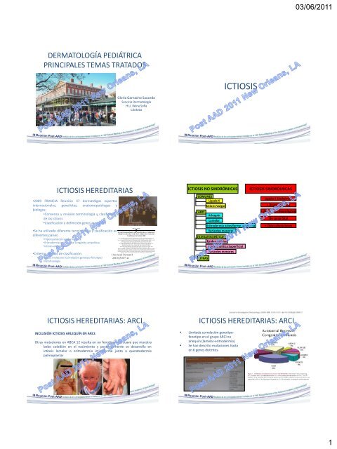
DERMATOLOGÍA PEDIÁTRICA<br />
PRINCIPALES TEMAS TRATADOS<br />
<strong>ICTIOSIS</strong> HEREDITARIAS<br />
•2009 FRANCIA Reunión 37 dermatólgos expertos<br />
internacionales, genetistas, anatomopatólogos y<br />
biólogos:<br />
•Consenso y revisión terminología y clasificación<br />
de las ictiosis<br />
•Clasificación y definición genes asociados<br />
•Se ha utilizado diferente terminología y clasificación en<br />
diferentes países<br />
•Hiperqueratosis epidermolítica<br />
•Eritrodermia ictiosiforme congénita ampollosa<br />
•Ictiosis ampollosa<br />
•Criterios nuevos de clasificación:<br />
•Causa molecular (Correlación genotipo-fenotipo)<br />
•Patofisiología<br />
Gloria Garnacho Saucedo<br />
Servicio Dermatología<br />
H.U. Reina Sofía<br />
Córdoba<br />
<strong>ICTIOSIS</strong> HEREDITARIAS: ARCI<br />
INCLUSIÓN <strong>ICTIOSIS</strong> ARLEQUÍN EN ARCI:<br />
Otras mutaciones en ABCA 12 resulta en un fenotipo más suave que muestra<br />
bebe colodión en el nacimiento y posteriormente se desarrolla en<br />
ictiosis lamelar o eritrodermia ictiosiforme junto a queratodermia<br />
palmoplantar<br />
<strong>ICTIOSIS</strong><br />
<strong>ICTIOSIS</strong> NO SINDRÓMICAS <strong>ICTIOSIS</strong> SINDRÓMICAS<br />
COMUNES<br />
Ligada X<br />
ARCI<br />
QUERATINOPÁTICAS<br />
Epidermolítica<br />
OTRAS<br />
Ictiosis Vulgar<br />
Arlequín<br />
Lamelar<br />
Eritrodermia ictiosiforme congénita<br />
Variantes menores<br />
Epidermolítica Superficial<br />
Variantes menores<br />
Ligada X Sindrómica<br />
+ Anomalías Pelo<br />
+ Anomalías Neurológicas<br />
De curso fatal<br />
+ Otras alteraciones<br />
<strong>ICTIOSIS</strong> HEREDITARIAS: ARCI<br />
Limitada correlación genotipofenotipo<br />
en el grupo ARCI no<br />
arlequín (lamelar-eritrodermia)<br />
Se han descrito mutaciones hasta<br />
en 6 genes distintos.<br />
03/06/2011<br />
1
<strong>ICTIOSIS</strong> HEREDITARIAS: ARCI<br />
Bebe colodion afectación levemoderada<br />
Ectropion-eclavion sólo 1/3<br />
Descamación generalizada: ALOX12B<br />
suave, ligera y blanca marrón suave.<br />
ALOX3 marrón, moderada y adherente;<br />
con eritema leve (12B) o ausente (3)<br />
Hiperlinealidad palmar con (ALOX12B) o<br />
sin (ALOX3) queratodermia<br />
<strong>ICTIOSIS</strong> HEREDITARIAS: ARCI<br />
<strong>ICTIOSIS</strong> EN TRAJE DE BAÑO.<br />
BJD 2010; 162: 448-451. Eur J Dermatol 2005; 15: 433-6<br />
•Variante minor de ictiosis lamelar (forma localizada).<br />
•Bebe colodión al nacimiento<br />
•Posteriormente (primeros meses vida) desarrollo de escamas grandes que<br />
afectan únicamente áreas corporales de alta temperatura, generalmente bajo<br />
la ropa (34ºC)<br />
•Localización: axilas, cuero cabelludo, tronco. Respeta extremidades y parte de<br />
la cara.<br />
•Genética:<br />
• Acúmulo de mutaciones espontáneas en dominios catalíticos de la<br />
enzima TGM1<br />
• La enzima es temperatura sensible y pierde actividad a altas<br />
temperaturas<br />
Variantes menores<br />
Variantes menores<br />
Variantes mayores <strong>ICTIOSIS</strong> HEREDITARIAS: QUERATINOPÁTICAS<br />
ERITRODERMIA ICTIOSIFORME RETICULAR CONGÉNITA.<br />
<strong>ICTIOSIS</strong> EN CONFETTI.<br />
•Alteraciones y mutaciones en KRT10 (17q) que llevan a<br />
una alteración de la keratina 10 (“tailless k10”)<br />
•Keratina 10 reducida en la epidermis<br />
•“Mosaicismo Reversible”: alta frecuencia de remisiones<br />
espontáneas clonales por mutaciones a lo normal<br />
•“Recombinación mitótica”: Pérdida de heterocigosidad<br />
en manchas revertidas (presumiblemente en los clones<br />
de células madres). Pérdida de alelos mutados<br />
•Alto número de manchas revertidas indica: Selección<br />
fuertemente positiva y/o alto grado de recombinación<br />
mitótica<br />
03/06/2011<br />
2
<strong>ICTIOSIS</strong> HEREDITARIAS: QUERATINOPÁTICAS<br />
ERITRODERMIA ICTIOSIFORME RETICULAR CONGÉNITA.<br />
•Herencia AD?? (casos aislados)<br />
<strong>ICTIOSIS</strong> EN CONFETTI.<br />
•Inicio en momento del nacimiento como una<br />
eritrodermia ictiosiforme congénita con amplias<br />
áreas formando un patrón reticular que predomina<br />
en extremidades<br />
•Durante la infancia y pubertad característico<br />
patrón parcheado con áreas que se van resolviendo<br />
•Escamas de color amarillo marrón finas con<br />
eritema pronunciado y afectación palmoplantar<br />
KLICK: KERATOSIS LINEALES-<strong>ICTIOSIS</strong><br />
CONGÉNITA-QUERATODERMIA<br />
•En el momento del nacimiento ictiosis<br />
generalizada con eritema variable<br />
•Más tarde, eritema leve o ausente con fina o<br />
focal descamación (codos y rodillas).<br />
•Placas hiperqueratósicas articulares<br />
•Progresivamente hiperqueratosis linear en<br />
“rayas” sobre todo en pliegues cutáneos<br />
•Queratodermia palmoplantar difusa,<br />
constricciones digitales escleróticas y<br />
contracturas de las flexuras<br />
•Histopatología: Hipergranulosis, granulos de<br />
queratohialina anormalmente grandes,<br />
abundantes y redondos.<br />
KLICK: KERATOSIS LINEALES-<strong>ICTIOSIS</strong><br />
CONGÉNITA-QUERATODERMIA<br />
KLICK. KERATOSIS LINEALES-<strong>ICTIOSIS</strong> CONGÉNITA-KERATODERMIA<br />
JAAD 2010; 63: 607-641<br />
•Herencia autosómica recesiva<br />
•Gen alterado POMP gen 13q<br />
•Proteina alterada: Proteinas de maduración de proteosomas (complejos<br />
proteicos degradadores proteinas) (Chaperonas)<br />
•Mecanismo: Las proteasomas controlan la vida media de varias proteínas<br />
reguladoras que son degradas constantemente, tales como aquellas que están<br />
involucradas en el ciclo celular. Su fallo conducen al crecimiento celular<br />
descontrolado y desdiferenciación<br />
03/06/2011<br />
3
•AR<br />
SINDROME MEDNIK:<br />
RETRASO MENTAL-ENTEROPATÍA-SORDERA-<br />
NEUROPATÍA-<strong>ICTIOSIS</strong>-QUERATODERMIA<br />
•Gen implicado AP1S1<br />
•Proteina: unidad menor del complejo adaptador de proteina AP1 crucial para<br />
la organización y el transporte intracelular de proteinas<br />
•Mecanismo: imposibilidad para el ensamblaje de vesículas, salida proteinas y<br />
tráfico vesicular<br />
SINDROME MEDNIK:<br />
RETRASO MENTAL-ENTEROPATÍA-SORDERA-<br />
NEUROPATÍA-<strong>ICTIOSIS</strong>-QUERATODERMIA<br />
PLoS Genetics 2008; 4: e1000296.<br />
•Diagnosticado en familias de la región de Kamouraska en quebec con<br />
antecesores comunes<br />
•Piel: Eritroqueratodermia variabilis-like, hiperqueratosis congénita y parches<br />
eritematosos de tamaños y formas variables así como duración. Inicialmente<br />
denominado EKV3<br />
•Otros hallazgos: sordera neurosensorial, neuropatía periférica, retraso<br />
psicomotor severo.<br />
•Claves diagnósticas:<br />
• Diarrea crónica congénita<br />
• Niveles muy elevados de ácidos grasos de cadena larga<br />
03/06/2011<br />
4
CEDNIK:<br />
DISGENESIA CEREBRAL-NEUROPATÍA-<strong>ICTIOSIS</strong>-<br />
QUERATODERMIA PALMOPLANTAR<br />
Am J Hum Genet. 2005; 77 (2): 242-51.<br />
•Debut 5-12 meses tras nacimiento.<br />
•Curso: Progresiva, desenlace fatal<br />
•Piel: Ictiosis lamelar severa y queratodermia palmoplantar<br />
•Neurológico: Microcefalia progresiva, hipotonía truncal, ausencia de reflejos<br />
tendones profundos, retraso psicomotor severo que comienza en el primer<br />
año de vida<br />
•Ojos: nistagmo, atrofia macular<br />
•Sordera neurosensorial<br />
•Dismorfismo facial<br />
•Cerebro RM: alteración en la migración neuronal. Displasia cortical,<br />
paquigiria, agenesia cuerpo calloso<br />
ARC:<br />
ARTROGRIPOSIS-DISFUNCIÓN RENAL-COLESTASIS<br />
Turk J Pediatr 2005; 47: 67-70.<br />
•Inicio tras el nacimiento<br />
•Piel: Desarrollo progresivo de escamas lamelares en el tronco, cuero<br />
cabelludo y extremidades. Dismorfismo.<br />
•Hueso y músculos: Artrogriposis múltiple congénita, costillas estrechas,<br />
osteopenia, atrofia muscular neurogénica, contracturas<br />
•Riñones: Síndrome de Fanconi renal (anormalidades función plaquetaria).<br />
Disfunción renal tubular: aminoaciduria, glycosuria, acidosis, diabetes<br />
nefrogénica insípida, deshidratación severa, FTT.<br />
•Colestasis: colestasis neonatal, ictericia, hipoplasia biliar intrahepática<br />
•Otros: ausencia cuerpo calloso, diarrea recurrente, infecciones, sordera<br />
ARC:<br />
ARTROGRIPOSIS-DISFUNCIÓN RENAL-COLESTASIS<br />
•Herencia AR, muy raro, consanguinidad en las familias de Pakistan y otras<br />
etnias<br />
•Gen: VPS33B<br />
•Proteina: VPS33B está involucrada principalmente en al regulación de la<br />
exocitosis y en la fusión de membrana SNARE dependiente<br />
•Mecanismo: Imposibilidad de las proteinas intracelulares para transporte<br />
desde Golgi a las vacuolas<br />
•La inactivación del VP33B lleva a la secreción anomal y atrapamiento de<br />
granulos lamelares, alteraciones de la permeabilidad de la barrera cutánea y<br />
sistema urinario y biliar<br />
CEDNIK:<br />
DISGENESIA CEREBRAL-NEUROPATÍA-<strong>ICTIOSIS</strong>-<br />
QUERATODERMIA PALMOPLANTAR<br />
•Herencia AR<br />
•Gen SNAP 29<br />
•Proteina: SNARE proteina en las estructuras de la membrana intracelular,<br />
involucrada en la fusión de vesículas, regulación endo/exocitosis, vesículas<br />
que cargan en el aparato de Golgi, interactúa en la sinapsis<br />
•Mecanismo: imposibilidad global del SNARE para mediar el transporte y la<br />
fusión de vesículas<br />
•Piel: Maduración anormal de los granulos lamelares, retención de los mismos<br />
en estrato córneo, secreción reducida de lípidos epidérmicos y proteasa,<br />
alteración del mecanismo de barrera y reducción de la descamación<br />
•Hallazgos ultraestructurales ME<br />
ARC:<br />
ARTROGRIPOSIS-DISFUNCIÓN RENAL-COLESTASIS<br />
•Curso: progresivo, normalmente fatal en un año (neumonía y otras<br />
infecciones)<br />
•Laboratorio: clave diagnóstica: plaquetas grandes, pálidas y agranulares en<br />
sangre periférica. Hiperbilirrubinemia directa, AP elevada, GGT<br />
•Microscopia electrónica: Granulos lamelares retenidos en estrato córneo,<br />
imposibilidad de secreción de los contenidos (lípidos, proteasas).<br />
03/06/2011<br />
5
<strong>ICTIOSIS</strong> PREMATURIDAD<br />
•Comienzo intrauterino: Polihidramnios, líquido<br />
amniótico opaco por aumento células epidérmicas<br />
(cielo estrellado), nacimiento prematuro<br />
•En el nacimiento: Asfixia severa (aspiración) y<br />
síndrome de distrés respiratorio, eosinofilia<br />
transitoria, hiperqueratosis masiva en cuero<br />
cabelludo y otras localizaciones (piel gruesa, caseosa,<br />
roja y edematosa con posterior descamación)<br />
•AUTORRESOLUTIVO<br />
•Posteriormente: marcada mejoría en unos meses,<br />
más tarde piel seca o moderada hiperqueratosis sin<br />
descamación. Atopia y eosinofilia son posibles<br />
DERMATITIS ATÓPICA (DA)<br />
ALTERACIÓN<br />
INMUNITARIA<br />
GENÉTICA<br />
DISFUNCIÓN<br />
BARRERA<br />
EPIDÉRMICA<br />
FACTORES<br />
AMBIENTALES<br />
<strong>ICTIOSIS</strong> PREMATURIDAD<br />
•Clave diagnóstica en ME: Agregados en estrato<br />
córneo y granuloso patognomónicos<br />
•Herencia AR Relativamente frecuente en<br />
Scandianavia y Europa<br />
•Gen: SLC 27A4 9q34<br />
•Proteina FATP4 proteina transportadora de<br />
ácidos grasos<br />
•Mecanismo: Imposibilidad transporte ácidos<br />
grasos, acumulación de masas de lípidos en la<br />
epidermis superior y alteraciones de la barrera<br />
epidérmica<br />
DERMATITIS ATÓPICA<br />
DERMATITIS ATÓPICA<br />
FACTORES<br />
AMBIENTALES<br />
• Aumento contínuo de la prevalencia 15-30% que varía según zonas<br />
geográficas<br />
• Influencia nivel socioeconómico: directamente proporcional a la educación<br />
familiar<br />
• Gradiente urbano-rural: mayor prevalencia en ciudades (tráfico y polución)<br />
• Influencia unidad familiar: divorcio, estrés, adopción…<br />
• Dureza del agua<br />
• Infecciones (teoría higienista)<br />
03/06/2011<br />
6
• Identificación factores genéticos y ambientales<br />
• Cohorte 762 niños recién nacidos con alto riesgo de desarrollar atopia<br />
(mínimo uno de los padres atópico)<br />
Abstract<br />
• Determinan genotipos que aumentan el riesgo de DA: CD14C-159T y<br />
IL4Rx175V<br />
• Exposición a perros disminuye riesgo de DA<br />
• El papel protector de la exposición a los perros es más evidente en los<br />
examination.<br />
portadores mutaciones anteriores<br />
• Predisposición en función mutaciones de la filagrina<br />
• Filagrina (1q21) localizada dentro del “Complejo de diferenciación<br />
epidérmica”: múltiples genes involucrados en la diferenciación final de la<br />
epidermis y la formación del estrato córneo (3q21.11, 16q, 17q25, 20p12,<br />
3p26.13)<br />
• Poligénica >40 mutaciones descritas<br />
• Genotipos de filagrina determinan fenotipos de dermatitis atópica<br />
• Análisis variantes mutaciones filagrina 116 niños chinos 2-5 años DA y 212<br />
controles sanos<br />
• Genotipo P478S aumenta riesgo DA (p100KU/L<br />
FACTORES<br />
AMBIENTALES<br />
Filaggrin Gene Defects Are Independent Risk Factors for<br />
Atopic Asthma in a Polish Population: A Study in ECAP<br />
Cohort<br />
Joanna Ponińska 1 , Bolesław Samoliński 2 , Aneta Tomaszewska 2 , Filip Raciborski 2 , Piotr Samel-Kowalik 2 ,<br />
Artur Walkiewicz 2 , Agnieszka Lipiec 2 , Barbara Piekarska 2 , Jarosław Komorowski 2 , Edyta Krzych-Fałta 2 ,<br />
Andrzej Namysłowski 2 , Jacek Borowicz 2 , Graz_yna Kostrzewa 1 , Sławomir Majewski 3 , Rafał Płoski 1 *<br />
1 Department of Medical Genetics, Medical University of Warsaw, Warsaw, Poland, 2 Department of Prevention of Environmental Hazards and Allergology, Medical<br />
University of Warsaw, Warsaw, Poland, 3 Department of Dermatology and Venereology, Medical University of Warsaw, Warsaw, Poland<br />
Background: FLGnull variants of which 2282del4 and R501X are the most frequent in Caucasians are established risk factors<br />
for atopic dermatitis (AD) with an effect probably mediated through impairment of epidermal barrier. Among subjects with<br />
AD FLGdefects are also consistently associated with asthma and allergic rhinitis (AR) but it is less clear to what extent these<br />
associations are also present independently from skin disease. The aim of the present study was to evaluate the role of<br />
2282del4 and R501X in predisposing to these allergic phenotypes in a Polish population.<br />
Methodology: 2282del4 and R501X were typed among 3,802 participants of the Epidemiology of Allergic Diseases in<br />
Poland (ECAP) survey, a cross-sectional population-based study using ECRHS II and ISAAC questionnaires, and ambulatory<br />
Principal Findings: The FLG null variants were associated with AD (OR= 2.01, CI: 1.20–3.36, P= 0.007), allergic rhinitis (in<br />
particular persistent form, OR= 1.69, CI:1.12–2.54, P= 0.011), and asthma (in particular atopic asthma, OR= 2.22, CI:1.24–3.96,<br />
P= 0.006). Association with atopic asthma (but not persistent allergic rhinitis) was also present in the absence of AD,<br />
(OR= 2.02, CI: 1.07–3.81, P= 0.027) as well as in the absence of AD and history of broadly defined inflammatory skin disease<br />
(OR= 2.30, CI: 1.07–4.93, P= 0.03). Association to atopic asthma would have not been found if diagnosis was made by<br />
questionnaire only (OR= 1.15, CI: 0.58–2.32, P= 0.8). We did not observe an association between FLG variants and allergic<br />
sensitizations (P= 0.8) or total IgE. (P= 0.6).<br />
Conclusions/Significance: In a Polish population FLG 2282del4 and R501X carriage increases risk for development of AD<br />
and atopic asthma (also in the absence of AD or history thereof). This suggests that interventions aimed at restoring<br />
epidermal barrier may have a general role in asthma prophylaxis/treatment.<br />
Citation: Ponińska J, Samoliński B, Tomaszewska A, Raciborski F, Samel-Kowalik P, et al. (2011) Filaggrin Gene Defects Are Independent Risk Factors for Atopic<br />
Asthma in a Polish Population: A Study in ECAP Cohort. PLoSONE 6(2): e16933. doi:10.1371/journal.pone.0016933<br />
Editor: Jacques Zimmer, Centre de Recherche Public de la Santé (CRP-Santé), Luxembourg<br />
Received October 14, 2010; Accepted January 3, 2011; Published February 18, 2011<br />
Copyright: ß 2011 Ponińska et al. This is an open-access article distributed under the terms of the Creative Commons Attribution License, which permits<br />
unrestricted use, distribution, and reproduction in any medium, provided the original author and source are credited.<br />
Funding: The work was financed by grants from Polish Ministry of Science and Higher Education, Ministry of Health grant nr 6P052005C/06572, and Warsaw<br />
Medical University grant nr 1WY/NK1W/2009. The funders had no role in study design, data collection and analysis, decision to publish, or preparation of the<br />
manuscript.<br />
Competing Interests: The authors have declared that no competing interests exist.<br />
* E-mail: rploski@wp.pl<br />
Introduction<br />
and R501X with originally reported carrier rates in general<br />
population of , 2 and 6%, respectively [2]. Both variants result in<br />
Filaggrin gene(FLG) isstrongly expressed in the granular cellsof<br />
the epidermis leading to production of a large precursor protein<br />
profilaggrin. In the process of differentiation profilaggrin is<br />
a complete loss of processed filaggrin due to premature<br />
termination codons within the first FLG repeat. Whereas several<br />
new FLG variants have been reported they are substantially less<br />
proteolytically cleaved into functional filaggrin peptides which bind prevalent and qualitatively different with some residual function<br />
and collapsethekeratin cytoskeleton and subsequently aredegraded [4].<br />
into hydrophilic amino acids forming the natural moisturizing Among subjects with AD FLG defects are associated with other<br />
factor. The N-terminal domain of profilaggrin is likely to have an allergic disease such as asthma and allergic rhinitis (AR) however,<br />
additional function asit specifically localizesto thenucleus. All these<br />
processes areGENÉTICA critical for creation of epidermal barrier with<br />
it is less clear to what extent these associations are present<br />
independently from skin disease[5]. Two meta-analysesconcluded<br />
appropriate mechanical and biochemical properties [1].<br />
that there was no association between FLG null variants and<br />
FLG null variants are strong risk factor for AD [2,3]. In asthma among subjects without AD although the ORs from<br />
Caucasians two such variants are particularly common: 2282del4 pooled estimates suggested a trend in the direction of association<br />
PLoS ONE | www.plosone.org 1 February 2011 | Volume 6 | Issue 2 | e16933<br />
GENÉTICA<br />
• Nuevos genes implicados en la barrera cutánea: claudin-1, Loricrina,<br />
Corneodesmosin, TSLP…<br />
• Tight junction bajo estrato córneo regulando permeabilidad paracelular<br />
• Proteina Tight junction claudin-1 codificada gen CLDN1<br />
• Comparan expresión claudin-1 en epitelio de enfermos DA y no atópicos<br />
• Reducción expresión claudin-1 y 23 sólo en pacientes con DA<br />
• La expresión de claudin-1 se correlaciona inversamente con los<br />
biomarcadores de la respuesta inmunitaria TH2<br />
• Pacientes con ictiosis Peeling Skin Desease con fenotipo de atopia<br />
GENÉTICA<br />
• Mutaciones en el gen de la profilagrina (1q21) presente 10% europeos y<br />
norteamericanos<br />
• Aumenta el riesgo de desarrollo de sensibilización (marcha atópica), DA y<br />
rinitis alérgica y asma<br />
• Fuerte relación entre mutaciones filagrina y DA: mayor severidad y<br />
persistencia de la enfermedad<br />
• Restauración función barrera epidérmica en etapas precoces de la vida<br />
previene el desarrollo y la progresión de la enfermedad alérgica:<br />
SCREENING DEFECTOS FILAGRINA EN CORDON O SANGRE FETAL O EN<br />
FROTIS BUCAL EN NIÑOS MAYORES (117 EUROS)<br />
Filaggrin Gene Defects Are Independent Risk Factors for<br />
Atopic Asthma in a Polish Population: A Study in ECAP<br />
Cohort<br />
Joanna Ponińska 1 , Bolesław Samoliński 2 , Aneta Tomaszewska 2 , Filip Raciborski 2 , Piotr Samel-Kowalik 2 ,<br />
Artur Walkiewicz 2 , Agnieszka Lipiec 2 , Barbara Piekarska 2 , Jarosław Komorowski 2 , Edyta Krzych-Fałta 2 ,<br />
Andrzej Namysłowski 2 , Jacek Borowicz 2 , Graz_yna Kostrzewa 1 , Sławomir Majewski 3 , Rafał Płoski 1 *<br />
1 Department of Medical Genetics, Medical University of Warsaw, Warsaw, Poland, 2 Department of Prevention of Environmental Hazards and Allergology, Medical<br />
University of Warsaw, Warsaw, Poland, 3 Department of Dermatology and Venereology, Medical University of Warsaw, Warsaw, Poland<br />
Abstract<br />
Background: FLGnull variants of which 2282del4 and R501X are the most frequent in Caucasians are established risk factors<br />
for atopic dermatitis (AD) with an effect probably mediated through impairment of epidermal barrier. Among subjects with<br />
GENÉTICA<br />
AD FLGdefects are also consistently associated with asthma and allergic rhinitis (AR) but it is less clear to what extent these<br />
• associations Mutaciones are also present independently filagrina from skin aumentan disease. The aim ofriesgo the present study dewas asma to evaluatecon the role of independencia de la<br />
2282del4 and R501X in predisposing to these allergic phenotypes in a Polish population.<br />
enfermedad cutánea<br />
Methodology: 2282del4 and R501X were typed among 3,802 participants of the Epidemiology of Allergic Diseases in<br />
Poland (ECAP) survey, a cross-sectional population-based study using ECRHS II and ISAAC questionnaires, and ambulatory<br />
• examination. Población polaca. Mutaciones FLG 2282del4 y R501X<br />
• Principal Rol Findings: alteraciones The FLG null variants werebarrera associated with AD epidérmica (OR= 2.01, CI: 1.20–3.36, P= en 0.007), el allergic rhinitis desarrollo, (in profilaxis y<br />
particular persistent form, OR= 1.69, CI:1.12–2.54, P= 0.011), and asthma (in particular atopic asthma, OR= 2.22, CI:1.24–3.96,<br />
P= 0.006). Association with atopic asthma (but not persistent allergic rhinitis) was also present in the absence of AD,<br />
tratamiento del asma<br />
(OR= 2.02, CI: 1.07–3.81, P= 0.027) as well as in the absence of AD and history of broadly defined inflammatory skin disease<br />
(OR= 2.30, CI: 1.07–4.93, P= 0.03). Association to atopic asthma would have not been found if diagnosis was made by<br />
questionnaire only (OR= 1.15, CI: 0.58–2.32, P= 0.8). We did not observe an association between FLG variants and allergic<br />
sensitizations (P= 0.8) or total IgE. (P= 0.6).<br />
Conclusions/Significance: In a Polish population FLG 2282del4 and R501X carriage increases risk for development of AD<br />
and atopic asthma (also in the absence of AD or history thereof). This suggests that interventions aimed at restoring<br />
epidermal barrier may have a general role in asthma prophylaxis/treatment.<br />
Citation: Ponińska J, Samoliński B, Tomaszewska A, Raciborski F, Samel-Kowalik P, et al. (2011) Filaggrin Gene Defects Are Independent Risk Factors for Atopic<br />
Asthma in a Polish Population: A Study in ECAP Cohort. PLoS ONE 6(2): e16933. doi:10.1371/journal.pone.0016933<br />
Editor: Jacques Zimmer, Centre de Recherche Public de la Santé (CRP-Santé), Luxembourg<br />
Received October 14, 2010; Accepted January 3, 2011; Published February 18, 2011<br />
Copyright: ß 2011 Ponińska et al. This is an open-access article distributed under the terms of the Creative Commons Attribution License, which permits<br />
unrestricted use, distribution, and reproduction in any medium, provided the original author and source are credited.<br />
Funding: The work was financed by grants from Polish Ministry of Science and Higher Education, Ministry of Health grant nr 6P052005C/06572, and Warsaw<br />
Medical University grant nr 1WY/NK1W/2009. The funders had no role in study design, data collection and analysis, decision to publish, or preparation of the<br />
manuscript.<br />
Competing Interests: The authors have declared that no competing interests exist.<br />
* E-mail: rploski@wp.pl<br />
Introduction<br />
Filaggrin gene (FLG) isstrongly expressed in the granular cellsof<br />
the epidermis leading to production of a large precursor protein<br />
profilaggrin. In the process of differentiation profilaggrin is<br />
proteolytically cleaved into functional filaggrin peptides which bind<br />
and collapsethekeratin cytoskeleton and subsequently aredegraded<br />
into hydrophilic amino acids forming the natural moisturizing<br />
factor. The N-terminal domain of profilaggrin is likely to have an<br />
additional function asit specifically localizesto thenucleus. All these<br />
processes are critical for creation of epidermal barrier with<br />
appropriate mechanical and biochemical properties [1].<br />
FLG null variants are strong risk factor for AD [2,3]. In<br />
Caucasians two such variants are particularly common: 2282del4<br />
and R501X with originally reported carrier rates in general<br />
population of , 2 and 6%, respectively [2]. Both variants result in<br />
a complete loss of processed filaggrin due to premature<br />
termination codons within the first FLG repeat. Whereas several<br />
new FLG variants have been reported they are substantially less<br />
prevalent and qualitatively different with some residual function<br />
[4].<br />
Among subjects with AD FLG defects are associated with other<br />
allergic disease such as asthma and allergic rhinitis (AR) however,<br />
it is less clear to what extent these associations are present<br />
independently from skin disease [5]. Two meta-analyses concluded<br />
that there was no association between FLG null variants and<br />
asthma among subjects without AD although the ORs from<br />
pooled estimates suggested a trend in the direction of association<br />
DERMATITIS ATÓPICA<br />
PLoS ONE | www.plosone.org 1 February 2011 | Volume 6 | Issue 2 | e16933<br />
• Aumento de IgE relacionado con SCORAD<br />
• Muchos prick test positivos<br />
• Asocia asma, conjuntivitis…<br />
• Se transfiere en trasplante de médula osea<br />
• Citokinas alérgicas disminuyen la expresión de filagrina<br />
ALTERACIÓN<br />
INMUNOLÓGICA<br />
• Presencia en infiltrado inflamatorio de células NKT que producen altos<br />
niveles de IL4 e IFN gamma<br />
• Mutaciones TLR2 (toll-like receptor 2) aumentan riesgo de DA<br />
03/06/2011<br />
7
DERMATITIS ATÓPICA Y CALIDAD DE VIDA<br />
• Gran afectación calidad de vida pacientes y sus<br />
familias (66,4% afectación moderada) Pediatric<br />
Dermatology 2010; 27 (6): 618-623)<br />
• DA con alteración sueño riesgo de desarrollar<br />
problemas emocionales y de conducta a los 10<br />
años (hiperactividad/inatención)<br />
• Círculo vicioso estrés psicológico y síntomas DA<br />
(prurito). Acta Derm Venereol 2010; 90:582-588<br />
DERMATITIS ATÓPICA:<br />
¿Justificación dietas restrictivas?<br />
PRO: Alergólogos CONTRA: Dermatólogos<br />
• Los alimentos como<br />
desencadenante y exacerbación<br />
brotes<br />
• FALSOS POSITIVOS: 1/3 de los<br />
tests de alimentos son positivos<br />
en la urticaria aguda, urticaria<br />
de contacto, angioedema y rash<br />
morbiliforme transitorio<br />
• Sensibilización: MARCHA<br />
ATÓPICA. Los antígenos<br />
estimulan mucho más por la piel<br />
que por las mucosas<br />
• No justificado Prick-tests<br />
• Quitar la comida no mejora.<br />
DERMATITIS ATÓPICA:<br />
¿Influencia tipo de dieta?<br />
DERMATITIS ATÓPICA SEVERIDAD<br />
• SCORAD Y EASI<br />
• PO-SCORAD Y SA-EASI<br />
© 2009 S. Karger AG, Basel<br />
1018–8665/09/2183–0246$26.00/0<br />
Fax +41 61 306 12 34<br />
E-Mail karger@karger.ch Accessible online at:<br />
www.karger.com<br />
www.karger.com/drm<br />
Clinical and Laboratory Studies<br />
Dermatology 2009;218:246–251<br />
DOI: 10.1159/000193997<br />
Patient-Oriented SCORAD: A Self-<br />
Assessment Score in Atopic Dermatitis<br />
A Preliminary Feasibility Study<br />
M. Vourc’h-Jourdain a S. Barbarot a A. Taieb b T. Diepgen c M. Ambonati d<br />
V. Durosier d V. Sibaud d J.F. Stalder a<br />
a Clinique Dermatologique, CHU Hôtel-Dieu, Nantes , et b Service de Dermatologie, CHU Bordeaux, Bordeaux , France;<br />
c Department of Social Medicine, Occupational and Environmental Dermatology, University of Heidelberg,<br />
Heidelberg , Germany; d Pierre Fabre Laboratoire, Fondation dermatite atopique, Boulogne, France<br />
Key Words<br />
Atopic dermatitis Self-assessment score Scoring Atopic<br />
Dermatitis index, patient-oriented<br />
Received: August 15, 2008<br />
Accepted: October 22, 2008<br />
Published online: January 16, 2009<br />
the PO-SCORAD. Conclusion: This study shows that self-as-<br />
sessment is feasible in AD, and that there is a correlation between<br />
the physician and the patient scores. This study was<br />
the first step in validating the PO-SCORAD.<br />
Copyright © 2009 S. Karger AG, Basel<br />
Abstract<br />
Background: The SCORing Atopic Dermatitis (SCORAD) index<br />
is used worldwide to assess the severity of atopic ecze- Introduction<br />
ma. Patient involvement in the treatment process is of major<br />
current interest. There are very few validated patient self- Atopic dermatitis (AD) affects between 5 and 20% of<br />
assessment tools for atopic dermatitis (AD). Objective: To children under 11 years old [1, 2] and has significant im-<br />
develop a self-assessment score for AD patients, the patientplications in terms of patient quality of life and health<br />
oriented SCORAD (PO-SCORAD) based on the SCORAD in- economically [3–5] . Although treatments aim to resolve<br />
dex, and to assess its acceptability in a pilot study. Methods: patients’ symptoms, more attention is being paid (in both<br />
A multicenter working group decided on the initial form of daily practice and clinical trials) to assessment based on<br />
the PO-SCORAD. A prospective, single-center pilot study objective clinical signs [6, 7] . The principal tools used in<br />
was then carried out to assess its acceptability and validity. AD are: SCORAD (severity SCORing of Atopic Derma-<br />
A SCORAD and a PO-SCORAD were applied at baseline and titis index [8] ), EASI score (Eczema Area and Severity<br />
after 18 days; the acceptability of the tool was assessed by Index [9] ) and Six-Area Six-Sign Atopic Dermatitis sever-<br />
questionnaire, its validity by comparing SCORAD and PO- ity score [10] which all combine several clinical criteria<br />
SCORAD on both visits. Results: The study involved 15 chil- with a varying, but always lower, weight for subjective<br />
dren and 18 adults. 80% of the respondents found the ques- symptoms.<br />
tions clear and the form easy to fill in; 96% spent less than 10 Patient involvement in the treatment process is of<br />
min on it. A correlation was found between the SCORAD and topical interest and is widely recommended by public<br />
M. Vourc’h-Jourdain<br />
Clinique Dermatologique, CHU Hôtel-Dieu<br />
Place Alexis-Ricordeau<br />
FR–44000 Nantes (France)<br />
Tel. +33 240 083 116, Fax +33 240 083 117, E-Mail mvourch@yahoo.fr<br />
DERMATITIS ATÓPICA:<br />
¿Justificación dietas restrictivas?<br />
DERMATITIS ATÓPICA:<br />
¿Papel simbióticos?<br />
• Niños DA
DERMATITIS ATÓPICA:<br />
¿Papel hipoclorito y antibióticos?<br />
• Porcentaje CA-MARSA no tan alto como se pensaba<br />
• Pediatrics 2009;123;e808-e814<br />
Grupos a favor y en contra de la utilización lejía como prevención<br />
DOI: 10.1542/peds.2008-2217<br />
• Utilización agentes<br />
sobreinfección<br />
antibacterianos sólo si signos clínicos de<br />
Paller<br />
Treatment of<br />
Jennifer T. Huang, Melissa Abrams, Brook Tlougan, Alfred Rademaker and Amy S.<br />
The online version of this article, along with updated information and services, is<br />
located on the World Wide Web at:<br />
http://www.pediatrics.org/cgi/content/full/123/5/e808<br />
Staphylococcus aureus Colonization in Atopic Dermatitis Decreases<br />
Disease Severity<br />
PEDIATRICS is the official journal of the American Academy of Pediatrics. A monthly<br />
publication, it has been published continuously since 1948. PEDIATRICS is owned, published,<br />
and trademarked by the American Academy of Pediatrics, 141 Northwest Point Boulevard, Elk<br />
Grove Village, Illinois, 60007. Copyright © 2009 by the American Academy of Pediatrics. All<br />
rights reserved. Print ISSN: 0031-4005. Online ISSN: 1098-4275.<br />
Downloaded from www.pediatrics.org by on March 19, 2011<br />
DERMATITIS ATÓPICA: Miscelánea<br />
• Aceite de girasol destilado (repleto ácidos grasos esenciales) en<br />
enfermedades inflamatorias<br />
• Aumenta síntesis lipídica epidérmica y reduce inflamación<br />
• Estimula PPAR: diferenciación queratinocitos, mejora función barrera<br />
epidérmica, potencia metabolismo lipídico<br />
• Ahorrador corticoides<br />
• Mejora calidad vida<br />
• Reduce infecciones nosocomiales en recién nacidos pretérmino (41%<br />
menos) en comparación con vaselina<br />
• Reduce mortalidad infantil 26% (p=0,042) comparado con otros<br />
emolientes<br />
• Al 2% en emulsión: 90% AGE (oleico y linoleico), 5% fitosterol y 1% Vit E<br />
DERMATITIS ATÓPICA:<br />
Tratamiento tópico<br />
DERMATITIS ATÓPICA:<br />
¿Papel hipoclorito y antibióticos?<br />
• Porcentaje CA-MARSA no tan alto como se pensaba<br />
• Grupos a favor y en contra de la utilización lejía como prevención<br />
• Utilización agentes antibacterianos sólo si signos clínicos de<br />
sobreinfección<br />
DERMATITIS ATÓPICA<br />
Miscelánea ttos tópicos<br />
DERMATITIS ATÓPICA:<br />
Tratamiento tópico<br />
03/06/2011<br />
9
DERMATITIS ATÓPICA:<br />
Tratamiento tópico<br />
• Desarrollo inmunidad celular, humoral y<br />
respuesta inmune a vacunas en grupo<br />
pimecrolimus vs corticoides (774 pacientes)<br />
• Poblaciones LB y LC descienden similar en 2<br />
grupos sin diferencias significativas<br />
• Incremento IG similar ambos grupos sobre<br />
todo E<br />
• Respuesta a vacunas similar en ambos<br />
grupos<br />
DERMATITIS ATÓPICA<br />
Tratamiento sistémico<br />
• CICLOSPORINA:<br />
• Rescate 3-5-7 mg/k/dia 2-4-6 semanas (nifedipino si HTA o<br />
hiperplasia gingival)<br />
• Estabilizador 2,5 mg/Kg/dia >6 meses. Aumenta población<br />
Linfocitos T reguladores<br />
• No más de un año<br />
• PREDNISONA:<br />
• Crisis<br />
• Enlazar ttos<br />
• Ensayo clínico Novartis PIMECROLIMUS 1%<br />
crema DA leve-moderada lactates 3-12 meses vs<br />
corticoides potencia baja-moderada<br />
• Estudio multicéntrico, randomizado, abierto,<br />
grupos paralelos<br />
• Seguridad corto y largo plazo 5 años<br />
• Infecciones más comunes: nasofaringitis, fiebre,<br />
bronquitis, otitis media y catarro VA<br />
• NO DIFERENCIAS ENTRE 2 GRUPOS<br />
• Mayor sobreinfección eccema en pimecrolimus<br />
DERMATITIS ATÓPICA<br />
Consideraciones tto sistémico<br />
CANDIDATOS:<br />
1.Dermatitis atópica moderada-severa con recidivas frecuentes y mala calidad<br />
de vida<br />
2.Brotes muy intensos entre los tratamientos<br />
A TENER EN CUENTA:<br />
1.Todas las indicaciones están fuera de ficha técnica<br />
2.Consideraciones con las vacunas<br />
3.Consentimiento padres<br />
DERMATITIS ATÓPICA<br />
Tratamiento sistémico<br />
• Seguridad a largo plazo en la utilización de corticoides sistémicos y<br />
ciclosporina oral en el tratamiento de la dermatitis atópica<br />
• Densitometría 60 niños (5-16 años) DA moderada-severa<br />
tratamiento corticoides sistémicos y ciclosporina 5 años previos<br />
• No diferencias densitometría con población general sana<br />
03/06/2011<br />
10
DERMATITIS ATÓPICA<br />
Tratamiento sistémico<br />
• FOTOTERAPIA:<br />
• UVB BE: elección. Eficacia similar dosis medias-altas UVA1<br />
• PUVA: en casos refractarios. Superioridad en velocidad de<br />
respuesta y en el tiempo de remisión tras el tratamiento (12 vs<br />
4 semanas)<br />
• Nuevo sistema fototerapia “Full-spectrum light phototherapy”:<br />
utilización previa en enfermedades neuropsiquiátricas, similar<br />
helioterapia, no aporta mayores ventajas que UVB<br />
DERMATITIS ATÓPICA:<br />
Tratamiento sistémico casos recalcitrantes<br />
• IFN Gamma (?)<br />
• Inmunoglobulinas (niños)<br />
• Rituximab<br />
• Alefacept<br />
• Omalizumab<br />
• Ustekinumab<br />
• Etanercept<br />
• Adalimumab<br />
DERMATITIS ATÓPICA:<br />
Control PRURITO INCOHERCIBLE<br />
• ANTAGONISTAS RECEPTORES OPIÁCEOS: NALTREXONE 25-50<br />
mg/24h 2-3 semanas<br />
DERMATITIS ATÓPICA<br />
Tratamiento sistémico<br />
• AZATIOPRINA:<br />
• Eficacia y seguridad TPMT<br />
• Tiempo medio respuesta muy largo (2-4 meses)<br />
• MOFETIL MICOFENOLATO:<br />
• Eficacia y seguridad a largo plazo (ideal mantenimiento)<br />
• Myfortic®<br />
• METOTREXATO:<br />
• Eficacia reducciones SCORAD hasta 70%<br />
• Dosis medias<br />
• Más efectivo en casos que no asocian otros síntomas de atopia<br />
ni niveles elevados de IgE (DA intrínseca)<br />
• ROSIGLITAZONA:<br />
• Ligando PPAR (disminuidos en ambas fases DA)<br />
• Retirado en EEUU por riesgo cardiovascular<br />
DERMATITIS ATÓPICA:<br />
Control PRURITO INCOHERCIBLE<br />
• Participación neuropéptidos: Sustancia P y Factor neurotrófico<br />
derivado del cerebro<br />
• Aumento leucotrienos a través activación cascada ácido<br />
araquidónio<br />
• AGONISTAS PARCIALES: SEDIEL®(TANDOSPIRONE CITRATO) 30<br />
mg/24h 4 semanas. Mejoría SCORAD, ansiedad, depresión, estrés e<br />
insomnio<br />
HEMANGIOMAS<br />
MALFORMACIONES<br />
VASCULARES<br />
03/06/2011<br />
11
SÍNDROME PHACE DOI:<br />
Consensus Statement on Diagnostic Criteria for PHACE Syndrome<br />
Denise Metry, Geoffrey Heyer, Christopher Hess, Maria Garzon, Anita Haggstrom,<br />
Peter Frommelt, Denise Adams, Dawn Siegel, Karla Hall, Julie Powell, Ilona Frieden<br />
and Beth Drolet<br />
Pediatrics 2009;124;1447-1456; originally published online Oct 26, 2009;<br />
10.1542/peds.2009-0082<br />
The online version of this article, along with updated information and services, is<br />
located on the World Wide Web at:<br />
http://www.pediatrics.org/cgi/content/full/124/5/1447<br />
PEDIATRICS is the official journal of the American Academy of Pediatrics. A monthly<br />
publication, it has been published continuously since 1948. PEDIATRICS is owned, published,<br />
and trademarked by the American Academy of Pediatrics, 141 Northwest Point Boulevard, Elk<br />
Grove Village, Illinois, 60007. Copyright © 2009 by the American Academy of Pediatrics. All<br />
rights reserved. Print ISSN: 0031-4005. Online ISSN: 1098-4275.<br />
Downloaded from www.pediatrics.org by on February 11, 2011<br />
DERMATOLOGÍA PEDIÁTRICA<br />
•Determinar prevalencia de manifestaciones extracutáneas PHACE en niños<br />
con hemangiomas faciales grandes<br />
• Alteraciones vasculares 91%<br />
• Anomalías cardiacas 67%<br />
•33/108 (31%) niños con hemangioma facial >22 cm2 tenían PHACE<br />
• SEG. 1 Frontotemporal 28/33<br />
• SEG. 2 Maxilar 1/33<br />
• SEG. 3 Mandibular 24/33<br />
• 2 o más SEG. 15/33<br />
•En niños con hemangiomas faciales grandes 1/3 asociaban manifestaciones<br />
extracutáneas PHACE (arteriales intracraneales y cervicales)<br />
SINDROME LUMBAR<br />
•Anomalías intraespinales en 21 de los 41<br />
pacientes estudiados (RNM) con<br />
hemangiomas >2,5 cm en región media<br />
lumbosacra (51,2%)<br />
•Mayor riesgo si asocia ulceración u otras<br />
alteraciones cutáneas adicionales<br />
marcadores de disrafismo<br />
•Asintomáticos en el momento del estudio<br />
•Si sólo hemangiomas esperar RNM 4-6<br />
meses<br />
SÍNDROME PHACE<br />
Consensus Statement on Diagnostic Criteria for PHACE Syndrome<br />
Denise Metry, Geoffrey Heyer, Christopher Hess, Maria Garzon, Anita Haggstrom,<br />
Peter Frommelt, Denise Adams, Dawn Siegel, Karla Hall, Julie Powell, Ilona Frieden<br />
and Beth Drolet<br />
Pediatrics 2009;124;1447-1456; originally published online Oct 26, 2009;<br />
DOI: 10.1542/peds.2009-0082<br />
•Podrían ser demasiado restrictivos:<br />
located on the World Wide Web at:<br />
• Hemangiomas http://www.pediatrics.org/cgi/content/full/124/5/1447<br />
a penas perceptibles<br />
• Hemangiomas de cualquier localización<br />
(hemangiomas del brazo, cuello y torso de<br />
hallazgos similares)<br />
•Necesarios para la investigación<br />
•En última instancia puede que necesiten revisión<br />
•TAC cerebral estructural y angiografía intracraneal y<br />
cervical en niños con riesgo de PHACE.<br />
The online version of this article, along with updated information and services, is<br />
PEDIATRICS is the official journal of the American Academy of Pediatrics. A monthly<br />
publication, it has been published continuously since 1948. PEDIATRICS is owned, published,<br />
and trademarked by the American Academy of Pediatrics, 141 Northwest Point Boulevard, Elk<br />
Grove Village, Illinois, 60007. Copyright © 2009 by the American Academy of Pediatrics. All<br />
rights reserved. Print ISSN: 0031-4005. Online ISSN: 1098-4275.<br />
Downloaded from www.pediatrics.org by on February 11, 2011<br />
SÍNDROME PHACE<br />
•Anomalías arteriales cerebrales y cervicales son las alteraciones no cutáneas<br />
más frecuentemente asociadas al PHACE (84%)<br />
•Se desconoce localización y tipo<br />
•Intentan establecer fenotipo de arteriopatías asociadas y relación con<br />
hemangiomas y alteraciones cerebrales<br />
•Angiografías 70 niños: 57% >1 forma de arteriopatía<br />
•5 tipo lesiones:<br />
1. Disgenesia 56%<br />
2. Curso (trayecto) u origen anómalo 47%<br />
3. Estrechamiento 39%<br />
4. No visualización 20%<br />
5. Conexiones anómalas 20%<br />
•Hemangiomas ipsilaterales a la alteración arterial (+/- cerebral)<br />
•Segmentos frontotemporal S1 y mandibular S3 97%<br />
•En todos los casos de alteraciones de la fosa posterior presentaban o<br />
conexiones anómalas persistentes embrionarias carotido-basilar o<br />
alteraciones de la arteria cerebral interna<br />
HEMANGIOMAS ABORTIVOS<br />
• Hemangiomas con componente proliferativo
HEMANGIOMAS: Tratamiento tópico y ulceración<br />
Dra. M. Garzón. Columbia<br />
HEMANGIOMAS CON FASE DE<br />
CRECIMIENTO PROLONGADA<br />
• 23 pacientes con hemangiomas con fase de crecimiento prolongada >9<br />
meses de edad (17 meses)<br />
• Importante componente profundo<br />
• Morfología segmentaria o indeterminada<br />
• 39% afectación glándula parotídea<br />
• 20 de 23 tratamiento corticoideo prolongado<br />
RAS0PATIAS<br />
•CM-AVM nuevo desorden hereditario<br />
caractarizado por múltiples<br />
malformaciones vasculares atípicas<br />
multifocales y lesiones de alto flujo<br />
(cutáneas, subcutáneas, musculares,<br />
oseas y cerebrales)<br />
•CM-AVM asociado a mutaciones RASA 1<br />
(AD)<br />
•Algunos Parkes-Weber<br />
•Relación genotipo-fenotipo no está<br />
clara<br />
•Fenotipo se está aumentando<br />
•Infrecuente pero no rara<br />
RAS0PATIAS<br />
•Describen 42 mutaciones RASA 1 y su<br />
fenotipo en 44 familias<br />
•Todos MV<br />
•1/3 asociaban lesiones alto flujo:<br />
•Algunos tumore neurales (reminiscencias<br />
neurofibromatosis)<br />
03/06/2011<br />
13
RAS0PATIAS<br />
•NUEVA ASOCIACIÓN CON LESIONES ARTERIOVENOSAS EN MÉDULA ESPINAL<br />
•Screening con RNM en pacientes con múltiples malformaciones vasculares<br />
multifocales y síntomas neurológicos<br />
MALFORMACIONES CEREBRALES CAVERNOSAS<br />
•Clínica neurológica: cefalea, hemorragia intracraneal,<br />
déficit neurológicos, calcificaciones intracraneales,<br />
convulsiones…<br />
•Clínica cutánea:<br />
•Lo más característico: malformaciones capilares y<br />
venosas hiperqueratósicas (angioqueratoma like)<br />
•Otras menos específicas<br />
•Otras alteraciones:<br />
•Ojos: Malformaciones vasculares retinianas<br />
•Malformaciones vasculares hepáticas<br />
•Malformaciones vasculares musculares y de los<br />
tejidos blandos<br />
•La RNM es la mejor técnica de imagen para detectarla<br />
•Debut 16-33 años<br />
MALFORMACIONES CEREBRALES CAVERNOSAS<br />
• Anomalías vasculares prevalentes caracterizadas por<br />
vasos sinuosos intracraneales de pared delgada<br />
• Heterogenicidad genetica. Mutaciones 3 tipos genes:<br />
1. CCM1-KRIT 1<br />
2. CCM2-Malcavernin<br />
3. CCM3-PDCD 10<br />
• Autosómico dominante de penetrancia incompleta<br />
• Múltiples lesiones en casos familiares<br />
• Lesiones aisladas en casos esporádicos<br />
• Se asocia con elementos del citoesqueleto,<br />
componentes de señales de transducción y uniones<br />
celulares<br />
• En México hay unos 30.000 individuos afectos de<br />
400 familias efecto conquistador español<br />
MACROCEFALIA-MALFORMACIÓN CAPILAR<br />
•Síndrome infrecuente-raro<br />
• Reticular MVOporto<br />
• Nevus simple facial central<br />
prominente<br />
•Otros frecuentes hallazgos: piel suave<br />
pastosa, macrosomia, sindactilia y<br />
sobrecrecimiento y asimetría de<br />
extremidades, anomalías craneofaciales<br />
•Publicados criterios diagnósticos<br />
propuestos<br />
MACROCEFALIA-MALFORMACIÓN CAPILAR<br />
•Alteraciones neurológicas: retraso madurativo, hipotonía, enfermedad<br />
neurológica progresiva especialmente ventriculomegalia y herniación<br />
amígdala<br />
•Se recomienda RNM 2 veces al año hasta la edad de 2 años y posteriormente<br />
anualmente<br />
•El alcance de la enfermedad aún está definiéndose<br />
03/06/2011<br />
14
HEMANGIOMAS<br />
INDICACIONES TRATAMIENTO<br />
Pediatrics 2006, 118: 882-887.<br />
SITUACIONES QUE MOTIVAN TRATAMIENTO<br />
• Desfigurantes<br />
• Ulceración<br />
• Compromiso visual<br />
• Obstrucción vía aérea<br />
• Insuficiencia cardiaca<br />
• Obstrucción auditiva<br />
CONSIDERACIONES TRATAMIENTO TÓPICO<br />
• Tamaño (área a tratar)<br />
• Localización<br />
• Morfología<br />
HEMANGIOMAS:<br />
Tratamiento tópico<br />
3. Timolol tópico:<br />
• 0,5% gel o solución 2/24h 3<br />
meses. Rápido (2 sem). Muy<br />
bien en lesiones muy<br />
superficiales.<br />
• Acorta fase proliferativa. Mejor<br />
y más rápida involución<br />
HEMANGIOMAS ULCERADOS<br />
• Complicación más frecuente (16%-25% estudios prospectivos 10%<br />
lesiones)<br />
• Ocurre sobre los 4 meses<br />
• FACTORES DE RIESGO:<br />
1. Segmentarios<br />
2. Gran tamaño<br />
3. Mixtos<br />
4. Localización anatómica: Región anogenital (50%), labios y cuello.<br />
• Complicaciones: dolor, anemia, sangrado, infecciones<br />
• Causa desconocida<br />
• MOMENTO ULCERACIÓN:<br />
1. Lesión precursora<br />
2. Fase proliferativa<br />
3. Ulceración tardía (4-6 m fin fase proliferativa. Mayoría)<br />
4. Durante tratamiento<br />
HEMANGIOMAS:<br />
Tratamiento tópico<br />
1. Corticoides tópicos:<br />
• Perioculares y lesiones faciales<br />
delgadas<br />
• Mecanismo: inhibe VEGF-A y<br />
disminución proliferación de las<br />
stem cells<br />
• 35% buena respuesta 8sem<br />
• E2º: glaucoma y catarata<br />
posterior subcapsular<br />
• 1-2 semanas máximo<br />
2. Imiquimod:<br />
• 3-7/sema 16 sem<br />
• Actuación comp.superficial<br />
• Inhibe VEGF (IFNα)<br />
HEMANGIOMAS:<br />
Tratamiento sistémico<br />
MECANISMOS ACCIÓN PROPANOLOL:<br />
1. VASOCONSTRICCIÓN (1-3 dias. precoz)<br />
2. INHIBICIÓN-ANGIOGÉNESIS (intermedia en<br />
crecimiento: VEFG, BFFG, metaloproteasas)<br />
3. INDUCCIÓN-APOPTOSIS<br />
(etapa tardía. Endotelio vascular)<br />
SEGURIDAD:<br />
• Usos previos a 8mg/kg/24h sin grandes<br />
efectos adversos cardiovasculares<br />
• Precaución: hipotensión, bradicardia,<br />
hipoglucemia<br />
HEMANGIOMAS ULCERADOS<br />
• ALARMA: BLANQUEAMIENTO O COLOR GRIS EN
HEMANGIOMAS ULCERADOS<br />
• TRATAMIENTO: NINGÚN TRATAMIENTO HA DEMOSTRADO SER EFECTIVO<br />
EN EL CONTROL DE LA PROGRESIÓN DE LA ULCERACIÓN. NO EXISTE<br />
GOLD STANDAR<br />
• OPCIONES:<br />
1. Cuidado habitual heridas con metronidazol gel<br />
2. Láser vascular<br />
3. PDGF Beclapernin REGRANEX®<br />
4. Corticoides orales<br />
5. Propanolol<br />
6. Cirugía<br />
03/06/2011<br />
16