pBR322 DNA/BsuRI (HaeIII) Marker - Biotools
pBR322 DNA/BsuRI (HaeIII) Marker - Biotools
pBR322 DNA/BsuRI (HaeIII) Marker - Biotools

You also want an ePaper? Increase the reach of your titles
YUMPU automatically turns print PDFs into web optimized ePapers that Google loves.
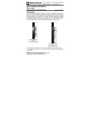
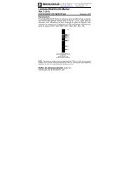
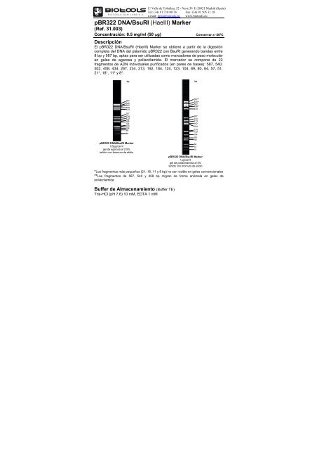
C/ Valle de Tobalina, 52 - Nave 39 E-28021 Madrid (Spain)Tel: (34) 91 710 00 74 Fax: (34) 91 505 31 18e-mail: info@biotools.eu www.biotools.eu<strong>pBR322</strong> <strong>DNA</strong>/<strong>BsuRI</strong> (<strong>HaeIII</strong>) <strong>Marker</strong>(Ref. 31.003)Concentración: 0.5 mg/ml (50 µg)Conservar a -20ºCDescripciónEl <strong>pBR322</strong> <strong>DNA</strong>/<strong>BsuRI</strong> (<strong>HaeIII</strong>) <strong>Marker</strong> se obtiene a partir de la digestióncompleta del <strong>DNA</strong> del plásmido <strong>pBR322</strong> con <strong>BsuRI</strong> generando bandas entre8 bp y 587 bp, aptas para ser utilizadas como marcadores de peso molecularen geles de agarosa y poliacrilamida. El marcador se compone de 22fragmentos de ADN individuales purificados (en pares de bases): 587, 540,502, 458, 434, 267, 234, 213, 192, 184, 124, 123, 104, 89, 80, 64, 57, 51,21*, 18*, 11* y 8*.bpbp5875405024584342672342131921841241231048980<strong>pBR322</strong> <strong>DNA</strong>/<strong>BsuRI</strong> <strong>Marker</strong>0.5µg/carrilgel de agarosa al 2.5%teñido con bromuro de etidio<strong>pBR322</strong> <strong>DNA</strong>/<strong>BsuRI</strong> <strong>Marker</strong>1µg/carrilgel de poliacrilamida al 5%teñido con bromuro de etidio*Los fragmentos más pequeños (21, 18, 11 y 8 bp) no son visible en geles convencionales**Los fragmentos de 587, 540 y 458 bp migran de forma anómala en geles depoliacrilamidaBuffer de Almacenamiento (Buffer TE)Tris-HCl (pH 7.6) 10 mM, EDTA 1 mM587**540**502458**4342672342131921841241231048980645751
AlmacenamientoAlmacenar a -20ºC. Para uso frecuente y a fin de evitar ciclos decongelación/ descongelación se recomienda realizar alícuotas, o bienalmacenar a 4ºC en presencia de buffer de carga (estable durante 4 meses).Protocolo de uso1- Preparar la siguiente mezcla (para carriles de 5 mm de longitud + ):Geles de Agarosa Geles de Poliacrilamida• <strong>pBR322</strong> <strong>DNA</strong> <strong>Marker</strong> (0.5-1 µg) 1 µl 2 µl• Buffer de carga 6X 1 µl 0.5 µl• Agua destilada 4 µl 0.5 µlVolumen final 6 µl 3 µl2- Mezclar suavemente3- No calentar4- Cargar la totalidad de la mezcla en el carril del gel5- Visualizar el ADN por tinción con bromuro de etidio o con SYBR® Green I+La mezcla debe ser escalada en función del ancho del carril. Utilizaraproximadamente 0.1-0.2 µg de ADN/mm de longitud del carril.Si bien el <strong>pBR322</strong> <strong>DNA</strong>/<strong>BsuRI</strong> <strong>Marker</strong> no está diseñado para la cuantificaciónprecisa de ADN, puede utilizarse para una cuantificación aproximada (verTabla 1). Para cuantificar se recomienda ajustar la masa de ADN en lamuestra a la banda del marcador de tamaño más próximo.Tabla 1. Porcentaje y masa de fragmentos individuales para 0.5 µg de <strong>pBR322</strong> <strong>DNA</strong>/<strong>BsuRI</strong> <strong>Marker</strong>masaFragmento Tamaño %(ng/0.5µg)1 587 13.5 67.32 540 12.4 61.93 502 11.5 57.64 458 10.5 52.55 434 10.0 49.86 267 6.1 30.67 234 5.4 26.88 213 4.9 24.49 192 4.4 22.010 184 4.2 21.111 124 2.8 14.212 123 2.8 14.113 104 2.4 11.914 89 2.0 10.215 80 1.8 9.2Aviso a usuariosUso exclusivo en investigación y aplicaciones in vitro. Ed. 03 – Junio 10