Colonización por Candida albicans ... - Medicinaoral.com
Colonización por Candida albicans ... - Medicinaoral.com
Colonización por Candida albicans ... - Medicinaoral.com

You also want an ePaper? Increase the reach of your titles
YUMPU automatically turns print PDFs into web optimized ePapers that Google loves.
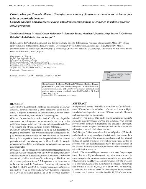
Medicina y Patología Oral / Oral Medicine and Pathology <strong>Colonización</strong> en prótesis dentales / Colonization in dental prosthesis<br />
<strong>Colonización</strong> <strong>por</strong> <strong>Candida</strong> <strong>albicans</strong>, Staphylococcus aureus y Streptococcus mutans en pacientes <strong>por</strong>tadores<br />
de prótesis dentales<br />
<strong>Candida</strong> <strong>albicans</strong>, Staphylococcus aureus and Streptococcus mutans colonization in patients wearing<br />
dental prosthesis<br />
Tania Baena Monroy (1) , Víctor Moreno Maldonado (2) , Fernando Franco Martínez (1) , Beatriz Aldape Barrios (1) , Guillermo<br />
Quindós (3) , Luis Octavio Sánchez Vargas (1,3)<br />
(1) Laboratorio de Patología Experimental, área de Microbiología, División de Estudios de Postgrado e Investigación, México DF, México<br />
(2) Departamento de Prostodoncia Total, Facultad de Odontología Universidad Nacional Autónoma de México, México DF, México<br />
(3) Departamento de Inmunología, Microbiología y Parasitología, Facultad de Medicina y Odontología, Universidad del País Vasco-Euskal<br />
Herriko Unibertsitatea, Bilbao, España.<br />
Correspondencia / Address:<br />
Dr. Luis Octavio Sánchez Vargas<br />
Laboratorio de Genética Molecular<br />
División de Estudios de Postgrado e Investigación<br />
Facultad de Odontología, Universidad Nacional Autónoma de México<br />
Ciudad Universitaria, México DF, México.<br />
Teléfono: (0152 55) 56 225565, Fax: (0152 55) 55 50 34 97<br />
E-mail: cdlosv@yahoo.<strong>com</strong>.mx-<br />
Recibido / Received: 7-03-2004 Aceptado / Accepted: 28-11-2004<br />
RESUMEN<br />
Antecedentes: La estomatitis protética está asociada a <strong>Candida</strong><br />
<strong>albicans</strong>, diversas bacterias y otros cofactores, <strong>com</strong>o un pH<br />
ácido, la ingesta aumentada de carbohidratos, diversas enfermedades<br />
sistémicas y tratamientos farmacológicos.<br />
Objetivo: Determinar la prevalencia de C. <strong>albicans</strong>, Staphylococcus<br />
aureus y Streptococcus mutans en la mucosa y en las<br />
prótesis de los pacientes con y sin estomatitis protética atrófica<br />
y su relación con otros cofactores clínicos potenciales.<br />
Diseño del estudio: Se recolectó la saliva de 105 pacientes (62<br />
mujeres y 43 hombres) con prótesis dental para la medida del pH<br />
y se tomaron muestras orales con torunda estéril de la mucosa<br />
bucal y de la superficie interna de las prótesis dentales para su<br />
posterior estudio microbiológico. La identificación de los microorganismos<br />
aislados se realizó <strong>por</strong> métodos microbiológicos<br />
convencionales.<br />
Resultados: Las enfermedades sistémicas más frecuentes fueron<br />
diabetes mellitus e hipertensión y se observó una ingesta alta de<br />
carbohidratos en un número im<strong>por</strong>tante de pacientes. Se observó<br />
estomatitis protética atrófica en 50 pacientes y el pH salival medio<br />
en estos pacientes fue de 5,2. La presencia en las muestras<br />
de la mucosa bucal de C. <strong>albicans</strong>, S. aureus y S. mutans fue<br />
de 51,4 %, 52,4 % y de 67,6 %, respectivamente. C. <strong>albicans</strong> se<br />
aisló del 66,7% de las prótesis. S. aureus y S. mutans se aislaron<br />
del 49,5 % de las dichas prótesis. En un 86 % de los pacientes<br />
Baena-Monroy T, Moreno-Maldonado V, Franco-Martínez F, Aldape-Barrios<br />
B, Quindós G, Sánchez-Vargas LO. <strong>Candida</strong> <strong>albicans</strong>,<br />
Staphylococcus aureus and Streptococcus mutans colonization in<br />
patients wearing dental prosthesis. Med Oral Patol Oral Cir Bucal<br />
2005;10:E27-E39.<br />
© Medicina Oral S. L. C.I.F. B 96689336 - ISSN 1698-4447<br />
27<br />
Indexed in:<br />
-Index Medicus / MEDLINE / PubMed<br />
-EMBASE, Excerpta Medica<br />
-Indice Médico Español<br />
-IBECS<br />
ABSTRACT<br />
Background: Denture stomatitis is associated to <strong>Candida</strong> <strong>albicans</strong>,<br />
different bacteria and other co-factors such as an acid pH,<br />
a carbohydrate ingestion increase, different systemic illnesses<br />
and pharmacological treatments.<br />
Objective: The aim of this study was to determine <strong>Candida</strong><br />
<strong>albicans</strong>, Staphylococcus aureus and Streptococcus mutans<br />
prevalence in the mucous membrane and prosthesis of patients<br />
with and without atrophic denture stomatitis and its relationship<br />
with other potential clinical co-factors.<br />
Study Design: Saliva was collected from 105 patients (62 female<br />
and 43 male) wearing dental prosthesis in order to measure their<br />
pH. Oral samples of the mucous membrane and the internal<br />
surface of dental prosthesis were taken with sterile cotton to<br />
proceed with the microbiological study. The identification of<br />
the isolated microorganisms was performed using conventional<br />
microbiological methods.<br />
Results: Diabetes and Hypertension were the most frequent systemic<br />
illnesses. High carbohydrate ingestion was observed in<br />
numerous patients. Atrophic denture stomatitis was re<strong>por</strong>ted in<br />
50 patients and the pH average in saliva was of 5,2. The presence<br />
of C <strong>albicans</strong>, S. aureus and S. mutans in the mucous membrane<br />
and prosthesis was of 51,4%, 52,4% and 67,6%, respectively.<br />
C. <strong>albicans</strong> was isolated in 66,7% from the prosthesis, whereas<br />
S. aureus and S. mutans were isolated in 49,5% of those same
Med Oral Patol Oral Cir Bucal 2005;10:E27-E39. <strong>Colonización</strong> en prótesis dentales / Colonization in dental prosthesis<br />
con estomatitis protética atrófica se aisló C. <strong>albicans</strong> y en un<br />
<strong>por</strong>centaje parecido (84 % de los pacientes) se aisló S. aureus.<br />
El aislamiento de S. mutans fue menos frecuente, observándose<br />
en el 16% de las muestras orales de estos pacientes.<br />
Conclusiones: C. <strong>albicans</strong>, S. aureus y S. mutans colonizan con<br />
frecuencia la mucosa bucal de pacientes con prótesis dental. Este<br />
estado de <strong>por</strong>tador es más frecuente en pacientes con estomatitis<br />
protética atrófica, aunque en estas personas, la colonización de<br />
las prótesis es menor que la de la mucosa bucal.<br />
Palabras clave: <strong>Candida</strong> <strong>albicans</strong>, Staphylococcus aureus,<br />
Streptococcus mutans, prótesis dentales, estomatitis protética.<br />
INTRODUCCION<br />
La estomatitis protética es un proceso inflamatorio de la mucosa<br />
bucal. Sus principales formas clínicas son la atrófica (con<br />
presencia de lesiones eritematosas) y la hiperplásica (1). Se<br />
observa con mayor frecuencia en mujeres, principalmente con<br />
localización maxilar, en la superficie del paladar en contacto con<br />
la prótesis dental. Esta lesión tiene una etiología multifactorial<br />
y ha sido asociada con la presencia de <strong>Candida</strong> <strong>albicans</strong> y otros<br />
microorganismos bucales (1-3). El desarrollo de la estomatitis<br />
protética se ve influido, además de <strong>por</strong> la presencia de la<br />
prótesis y de <strong>Candida</strong> y otros microorganismos, <strong>por</strong> diversos<br />
factores locales y sistémicos, <strong>com</strong>o un pH salival ácido, el alto<br />
consumo de carbohidratos, tratamiento antibiótico antibacteriano<br />
prolongado, terapia hormonal, así <strong>com</strong>o <strong>por</strong> enfermedades<br />
sistémicas, <strong>com</strong>o diabetes mellitus o hipertensión arterial que<br />
tienen repercusión directa en las condiciones ambientales de la<br />
cavidad bucal (4-6).<br />
C. <strong>albicans</strong> y Staphylococcus aureus se asocian en las lesiones<br />
de muchos pacientes con queilitis angular, entidad clínica donde<br />
C. <strong>albicans</strong> juega un im<strong>por</strong>tante papel etiológico (1,5-7).<br />
C. <strong>albicans</strong> y S. aureus son microorganismos con una elevada<br />
capacidad de adhesión a la mucosa bucal (8-9). Diversos estudios<br />
(10-13) han demostrado que existe una asociación de<br />
C. <strong>albicans</strong>, y otras especies de <strong>Candida</strong>, con otras bacterias<br />
bucales <strong>com</strong>o Streptococcus sanguis, Streptococcus salivarius,<br />
Streptococcus mutans, Fusobacterium nucleatum y Actinomyces<br />
viscosus y que éstas bacterias promueven la adherencia de<br />
las levaduras al epitelio oral y a las superficies acrílicas. Esta<br />
adherencia está favorecida in vitro <strong>por</strong> la incubación simultanea<br />
de <strong>Candida</strong> con S. mutans, S. sanguis, S. salivarius u otras bacterias.<br />
C. <strong>albicans</strong> y S. aureus pueden producir lesiones mixtas<br />
en la mucosa oral, <strong>com</strong>o la queilitis angular. Sin embargo, los<br />
factores que facilitan esta co-infección no han sido identificados.<br />
Las alteraciones salivales son im<strong>por</strong>tantes factores asociados<br />
con el desarrollo de estomatitis y otras alteraciones orales. C.<br />
<strong>albicans</strong> es la especie de <strong>Candida</strong> con mayor capacidad de adhesión<br />
a las células epiteliales bucales y vaginales, hecho que<br />
está relacionado con su mayor presencia <strong>com</strong>o colonizador o patógeno<br />
estas mucosas humanas puesto que la adhesión a células<br />
y superficies es el primer paso para la posterior colonización e<br />
invasión de los tejidos (13). La adhesión de <strong>Candida</strong> a la mucosa<br />
bucal está favorecida <strong>por</strong> diferentes factores del hongo (<strong>com</strong>o<br />
la transición blastoconidio a hifa y la producción de diferentes<br />
28<br />
prosthesis. C. <strong>albicans</strong> was isolated in 86% of the patients with<br />
atrophic denture stomatitis and S. aureus was isolated in a similar<br />
percentage (84% of patients). The isolation of S. mutans was<br />
less frequent, and it was observed in 16% of the oral samples<br />
of these patients.<br />
Conclusions: C. <strong>albicans</strong>, S. aureus and S. mutans frequently<br />
colonize the oral mucous of patients wearing dental prosthesis.<br />
This illness-bearing condition is more frequent in patients with<br />
denture stomatitis, even though dental prosthesis colonization<br />
is lower than in the oral mucous.<br />
Key words: <strong>Candida</strong> <strong>albicans</strong>, Staphylococcus aureus, Streptococcus<br />
mutans, dental prosthesis, denture stomatitis.<br />
INTRODUCTION<br />
Denture stomatitis is an inflammatory process of the oral mucous.<br />
Atrophic (with erythematous lesions) and hyperplasic<br />
lesions are the main forms of denture stomatitis.(1) It is more<br />
frequently found in women, essentially in the maxillary, most<br />
of the times in the palate surface in contact with a dental prosthesis.<br />
There are many factors involved in the etiology of this<br />
lesion and it has been associated with the presence of <strong>Candida</strong><br />
<strong>albicans</strong> and other oral microorganisms.(1-3) Denture stomatitis<br />
development is influenced by the presence of a dental prosthesis,<br />
<strong>Candida</strong> and other microorganisms, but also by other local<br />
and systemic factors such as an acid salivary pH, a high carbohydrates<br />
ingestion, a long-term antibiotic therapy, hormonal<br />
therapy as well as systemic illnesses such as diabetes mellitus<br />
or arterial hypertension which have a direct repercussion in the<br />
environment of the oral cavity.(4-6)<br />
C. <strong>albicans</strong> and Staphylococcus aureus are associated in lesions<br />
of several patients with queilitis angular, where C. <strong>albicans</strong><br />
plays an im<strong>por</strong>tant etiological role (1,5-7) C. <strong>albicans</strong> and<br />
Staphylococcus aureus are microorganisms with an elevated<br />
adhesion capacity to the oral mucous. (8-9). Several studies<br />
(10-13) have demonstrated an association between C. <strong>albicans</strong><br />
or other species of <strong>Candida</strong>, and several oral bacteria such as<br />
Streptococcus sanguis, Streptococcus salivarius, Streptococcus<br />
mutans, Fusobacterium nucleatum and Actinomyces viscosus.<br />
This suggests that these bacteria promote the adhesion of yeasts<br />
in the oral epithelium and acrylic surfaces. This adherence is<br />
enhanced in vitro when <strong>Candida</strong> is incubated simultaneously<br />
with S. mutans, S. sanguis, S. salivarius or some other bacteria.<br />
C. <strong>albicans</strong> and S. aureus produce lesions in oral mucous, such<br />
as the angular queilitis. However, the factors that facilitate this<br />
co-infection have not yet been identified. The development of<br />
stomatitis includes im<strong>por</strong>tant associated factors such as changes<br />
in saliva, as well as other oral alterations.<br />
In <strong>com</strong>parison with other species of <strong>Candida</strong>, C. <strong>albicans</strong> has the<br />
highest adhesion capacity to oral and vaginal epithelial cells, fact<br />
related to its extend presence as colonizer or pathogenic agent of<br />
human mucous because the adhesion to the cells and surfaces is<br />
the first step to the subsequent colonization and tissues invasion<br />
(13) The adhesion of <strong>Candida</strong> to oral mucous is enhanced by<br />
different factors related to fungi (such as the transition from<br />
blastoconia to hypha, the production of several extra cellular
Medicina y Patología Oral / Oral Medicine and Pathology <strong>Colonización</strong> en prótesis dentales / Colonization in dental prosthesis<br />
enzimas extracelulares <strong>com</strong>o proteinasas y fosfolipasas) y condiciones<br />
del microambiente bucal (temperatura, pH, concentración<br />
de carbohidratos, etc.). Los materiales biomédicos pueden ser<br />
colonizados <strong>por</strong> microorganismos que forman una biopelícula<br />
adherente sobre la superficie de éstos. Las biopelículas están<br />
organizadas en <strong>com</strong>unidades microbianas estructuradas dentro<br />
de una matriz de material extracelular. Estos microorganismos<br />
son diferentes en fenotipo de las células libres en suspensión<br />
(planctónicas). La mayoría de las biopelículas bacterianas están<br />
producidas <strong>por</strong> S. aureus y Staphylococcus epidermidis (14-17).<br />
C. <strong>albicans</strong> y <strong>Candida</strong> parapsilosis son las más frecuentes de las<br />
especies fúngicas formadoras de biopelículas aunque la especie<br />
<strong>Candida</strong> dubliniensis, con un hábitat preferentemente oral, es<br />
una eficaz productora de biopelículas sobre superficies acrílicas<br />
(9). Las proteínas salivales, (principalmente las mucinas),<br />
que recubren la mucosa bucal y las superficies inertes de las<br />
prótesis dentales pueden actuar <strong>com</strong>o receptores específicos de<br />
C. <strong>albicans</strong> y otros microorganismos (2,5).<br />
El objetivo de éste estudio ha sido determinar la prevalencia de<br />
C. <strong>albicans</strong>, S. aureus y S. mutans <strong>com</strong>o colonizadores de la<br />
mucosa y las prótesis de los pacientes con estomatitis protética<br />
atrófica, la asociación entre estos microorganismos y su relación<br />
con otros cofactores clínicos potenciales.<br />
MATERIAL Y METODOS<br />
Pacientes, material y métodos<br />
Pacientes. Se estudiaron 105 pacientes <strong>por</strong>tadores de prótesis<br />
dentales (43 varones y 62 mujeres con una edad media de 67<br />
años) que se atendieron en la clínica de Prostodoncia Total de la<br />
Facultad de Odontología de la Universidad Nacional Autónoma<br />
de México, México DF. Se estudiaron pacientes desdentados<br />
totales <strong>por</strong>tadores de prótesis, con o sin presencia de estomatitis<br />
protésica, que no hubieran ingerido alimentos o fumado en las<br />
3 h previas a la toma de las muestras, sin haber realizado ningún<br />
procedimiento de higiene bucal y que no hubieran estado<br />
sometidos a terapia antimicrobiana en un periodo previo de 6<br />
meses antes del estudio.<br />
Muestras microbiológicas. Previo consentimiento informado,<br />
revisión clínica y realización de un cuestionario anamnésico, a<br />
cada paciente se le tomó una muestra de 2 ml de saliva no estimulada<br />
o la secretada durante 5 min. De cada paciente también<br />
se obtuvo una muestra con hisopo estéril frotando la superficie<br />
interna de la prótesis una vez retirada ésta de la cavidad bucal y<br />
una segunda muestra, frotando con un hisopo estéril la mucosa<br />
bucal con lesiones de estomatitis protética atrófica, la mucosa<br />
del paladar, la zona retromolar o cualquier otra zona mucosa en<br />
contacto con la prótesis dental. Las muestras se colocaron inmediatamente<br />
en medio de trans<strong>por</strong>te de Stuart (BBL, EE.UU.) para<br />
su posterior procesamiento.<br />
Métodos. Se midió el pH salival inmediatamente después de haberse<br />
obtenido la muestra mediante un potenciómetro (Conductronic<br />
pH120, EE.UU.). Para el aislamiento e identificación de C.<br />
<strong>albicans</strong>, las muestras se sembraron <strong>por</strong> agotamiento en placas de<br />
agar cromógeno CandiSelect (Bio-Rad, Francia), que es un medio<br />
específico para el crecimiento de levaduras que contiene una base<br />
nutritiva con glucosa, un sustrato cromógeno y antibióticos que<br />
29<br />
enzymes such as proteinases and phospholipases) and the oral<br />
microenvironment conditions (temperature, pH, carbohydrates<br />
concentration, etc.)<br />
Biomedical materials can be colonized by microorganisms<br />
producing a biofilm. Biofilms are organized in microbial <strong>com</strong>munities<br />
structured within an extra cellular material womb. The<br />
phenotype of these microorganisms is different from those of<br />
the free cells in suspension (planktonic cells)<br />
In most cases, bacterial biofilms are produced by S. aureus and<br />
Staphylococcus epidermidis. (14-17) C. <strong>albicans</strong> and <strong>Candida</strong><br />
parapsilosis are the most frequent fungi species found in these<br />
biofilms, however, <strong>Candida</strong> dubliniesis species, predominant in<br />
the oral habitat, is an effective producer of biofilms on acrylic<br />
surfaces.(9) Salivary proteins (mucines mainly), that cover the<br />
oral mucous and the inert surfaces of dental prosthesis act as specific<br />
receptors of C. <strong>albicans</strong> and other microorganisms.(2,5)<br />
The aim of this study was to find out the prevalence of C. <strong>albicans</strong>,<br />
S. aureus and S. mutans as colonizers of the oral mucous<br />
and dental prosthesis in patients wearing dental prosthesis, the<br />
association between these microorganisms and its relationship<br />
with other clinical potential co-factors.<br />
METHODOLOGY AND MATERIALS<br />
Patients, material and methodology<br />
Patients. 105 patients wearing dental prosthesis were studied<br />
(43 male and 62 female with an average age of 67 years) All<br />
the patients were treated at the Prosthodontic Clinics, School of<br />
Dentistry, Universidad Nacional Autónoma de México, Mexico,<br />
D.F. The sample was taken from edentulous patients wearing<br />
dental prosthesis, with denture stomatitis or without it. The patients<br />
had to observe a fasting period of at least 3 hours before<br />
taking the samples, without previous oral hygiene procedures<br />
and without being under any antimicrobial treatment in a period<br />
of at least 6 months before the study.<br />
Microbiologic samples. The patients who had agreed to participate<br />
in the study were subjected to a clinical check-up and<br />
filled up a questionnaire as anamnesis. A sample of 2ml of non<br />
stimulated saliva was taken from each patient for 5 minutes. For<br />
each patient, a sample of the internal surface of the prosthesis<br />
was also taken using a sterile cotton bud immediately after the<br />
prosthesis was removed from the oral cavity. A second sample<br />
was obtained from the oral mucous with atrophic denture stomatitis<br />
lesions, the palate mucous, the rear zone of the molars or<br />
any other zone in contact with the dental prosthesis using a sterile<br />
cotton bud. The samples were immediately placed in a Stuart<br />
trans<strong>por</strong>t media (BBL, EE.UU) for its subsequent processing.<br />
Methodology. The salivary pH was tested immediately after the<br />
sample was taken using a potentiometer (Conductronic pH120,<br />
EE.UU.). For the isolation and identification of C. <strong>albicans</strong>,<br />
the samples were sown using the exhaustion technique in plates<br />
of chromogenic agar media, CandiSelect (Bio-Rad, France),<br />
which is a specific culture media for the growth of yeasts. It<br />
contains a nutritious base with glucose, chromogenic substrate<br />
and antibiotics in order to prevent bacteria development. Blue<br />
colonies in this media were identified as C. <strong>albicans</strong>. For the<br />
isolation and identification of S. aureus, the samples were sown
Med Oral Patol Oral Cir Bucal 2005;10:E27-E39. <strong>Colonización</strong> en prótesis dentales / Colonization in dental prosthesis<br />
impiden el desarrollo de bacterias. En este medio las colonias<br />
azules fueron identificadas <strong>com</strong>o C. <strong>albicans</strong>. Para el aislamiento<br />
e identificación de S. aureus, las muestras se sembraron en placas<br />
de agar de Chapman o agar sal-manitol (Difco, EE.UU.), que es<br />
un medio altamente selectivo para el aislamiento de estafilococos<br />
patógenos, contiene un 7,5% de cloruro de sodio y manitol con<br />
rojo fenol <strong>com</strong>o indicador ácido-base. S. aureus, se identificó en<br />
éste medio <strong>por</strong> producir colonias con un halo de color dorado<br />
(producción de ácido <strong>com</strong>o resultado de la fermentación del manitol),<br />
adicionalmente se realizaron las pruebas de la coagulasa<br />
y la catalasa. Para el aislamiento e identificación de S. mutans,<br />
las muestras se sembraron en placas de agar Mitis- Salivarius<br />
(Difco) con 0,2U.ml -1 de bacitracina (Merck, EE.UU.), 15 g.l -1 de<br />
sacarosa y 1 ml.l -1 de solución Bacto-Chapman (telurito potásico<br />
al 1%) (BBL). En este medio, las colonias características de S.<br />
mutans aparecieron elevadas, convexas, onduladas, opacas, de<br />
color azul oscuro, con márgenes irregulares, superficie granular,<br />
más o menos adheridas. Todas las placas fueron incubadas a<br />
37 ºC y se evaluaron con lecturas del crecimiento microbiano a<br />
las 24 y 48 h. Los aislamientos de microorganismos se identificaron<br />
y confirmaron en cultivos puros <strong>com</strong>plementariamente<br />
con la tinción de Gram y con la observación de las características<br />
microscópicas (18,19).<br />
Se realizó una estadística descriptiva general para todas las variables<br />
con el programa SPSS 10.0 para Windows. Se identificó la<br />
prevalencia de C. <strong>albicans</strong>, S. aureus y S. mutans, y la asociación<br />
entre las prevalencias mediante la prueba no paramétrica de Chi 2 ,<br />
además se identificó la asociación existente entre: sexo, ingesta de<br />
carbohidratos y presencia de estomatitis, con la presencia de C.<br />
<strong>albicans</strong>, S. aureus y S mutans. Las diferencias entre los valores<br />
de pH salival se calcularon mediante un ANOVA.<br />
RESULTADOS<br />
Las características clínicas generales de los pacientes se presentan<br />
en la tabla 1. Se observó la presencia de estomatitis<br />
protética atrófica en 50 pacientes (47,6 %), con un predominio<br />
de la afectación de la zona del paladar blando (en 31,4 % de<br />
los pacientes).<br />
- C. <strong>albicans</strong>. Cincuenta y cuatro pacientes (51,4 %) presentaron<br />
una colonización de la mucosa bucal <strong>por</strong> C. <strong>albicans</strong>. Cuarenta y<br />
tres pacientes presentaron estomatitis protética atrófica asociada<br />
con la presencia en la mucosa bucal de C. <strong>albicans</strong> (Chi 2 = 48,1,<br />
p < 0,001), se encontró la presencia de C. <strong>albicans</strong> en 70 prótesis<br />
(66,7 % de los pacientes) Trece prótesis de pacientes con<br />
estomatitis protética estaban colonizadas <strong>por</strong> C. <strong>albicans</strong><br />
- S. aureus. Se aisló de la mucosa de 55 pacientes (52,4 %).<br />
En 42 pacientes con estomatitis se aisló S. aureus (Chi 2 = 48,1,<br />
p < 0,001) de la mucosa. El estudio de las muestras tomadas de<br />
las prótesis reveló la presencia de S. aureus en 52 pacientes (el<br />
49,5 % de los pacientes). Dieciocho prótesis de pacientes con<br />
estomatitis protética fueron colonizadas <strong>por</strong> S. aureus.<br />
- S. mutans. Se aisló de la mucosa de 71 pacientes (67,6 %).<br />
Únicamente ocho pacientes con estomatitis protética estaban<br />
colonizados <strong>por</strong> S. mutans en la mucosa bucal. El estudio de las<br />
muestras tomadas de las prótesis reveló también la presencia de<br />
S. mutans en 52 pacientes (el 49,5 % de los pacientes). En<br />
30<br />
in plates containing Chapman agar or Mannitol Salt Agar (Difco,<br />
EE.UU.), which is a selective media for pathogenic Staphylococcus<br />
isolation. It contains 7,5% sodium chloride and mannitol<br />
with red phenol as an acid-base marker. S. aureus was identified<br />
in this media by the production of golden color zones around<br />
the colonies. Additionally, catalase and coagulase tests were<br />
performed. The samples were sown in Mitis Salivarius Agar<br />
(Difco) plates for S. mutans identification. This media contains<br />
0.2 U.ml -1 of Bacitracin (Merck, EE.UU.), 15g.L -1 of sucrose<br />
and 1ml.L -1 of Bacto-Chapman tellurite solution (potassium<br />
tellurite 1%) (BBL). In this media, the characteristic colonies<br />
of S. mutans appeared elevated, convex, wavy, and opaque, in a<br />
dark blue color, with irregular borders, granular surface and not<br />
so adhered. All the plates were incubated at 37°C and evaluated<br />
with microbial growth readings at 24 and 48 h. Microorganisms<br />
isolation were identified and confirmed in pure cultivation.<br />
In order to <strong>com</strong>plement the identification, gram stain test and<br />
microscope observation were performed. (18,19)<br />
All the variables were analyzed through a general statistical descriptive<br />
analysis using SPSS program version 10.0 for Windows.<br />
C. <strong>albicans</strong>, S. aureus and S. mutans prevalence, as well as its<br />
association were identified using the Chi 2 non parametrical test,<br />
whereas sex association, carbohydrates ingestion and denture<br />
stomatitis were identified with C. <strong>albicans</strong>, S. aureus and S,<br />
mutans presence. ANOVA was used to calculate the differences<br />
between salivary pH values.<br />
RESULTS<br />
Table 1 shows general clinical patients characteristics. In 50<br />
patients (47,6%) denture stomatitis was observed, and in 31,4%<br />
of these cases, soft palate was the affected zone.<br />
- C. <strong>albicans</strong>. Fifty four patients (51,4%) showed oral mucous<br />
colonization by C. <strong>albicans</strong>. Forty three showed atrophic denture<br />
stomatitis associated with C. <strong>albicans</strong> presence in oral mucous.<br />
(Chi 2 =48,1, p
Medicina y Patología Oral / Oral Medicine and Pathology <strong>Colonización</strong> en prótesis dentales / Colonization in dental prosthesis<br />
Tabla 1. Características clínico-microbiológicas de los pacientes.<br />
Pacientes<br />
Características Total (n = 105) Con estomatitis protética (n = 50)<br />
N % N %<br />
Género (sexo)<br />
Masculino/ Femenino 43 / 62 41 / 59 21 / 29 42 / 58<br />
Edad (media) 67 68<br />
Padecimientos sistémicos<br />
Diabetes mellitus 23 21,9 15 30<br />
Hipertensión arterial 18 17,1 12 24<br />
Tratamiento farmacológico<br />
Hipoglucemiantes 21 20 15 30<br />
Antihipertensivos 21 20 12 24<br />
Duración del tratamiento farmacológico<br />
1-5 años 29 27,6 15 30<br />
6-10 años 11 10,5 9 18<br />
11-15 años 2 1,9 2 4<br />
16-20 años 7 6,7 4 8<br />
+ 20 años 3 2,9 2 4<br />
Ingesta de carbohidratos<br />
Alta 75 71,5 38 86<br />
Baja 30 28,5 12 24<br />
pH salival (media) 5,6 5,2<br />
Estomatitis protética (localización)<br />
Paladar blando 33 66<br />
Paladar duro 5 10<br />
Paladar duro y blando 8 16<br />
Reborde residual 4 8<br />
<strong>Colonización</strong> de mucosa bucal<br />
<strong>Candida</strong> <strong>albicans</strong> 54 51,4 43 86<br />
Staphylococcus aureus 55 52,4 42 84<br />
Streptococcus mutans 71 67,6 8 16<br />
<strong>Colonización</strong> de la prótesis<br />
<strong>Candida</strong> <strong>albicans</strong> 70 66,7 13 26<br />
Staphylococcus aureus 52 49,5 18 36<br />
Streptococcus mutans 52 49,5 20 40<br />
31
Med Oral Patol Oral Cir Bucal 2005;10:E27-E39. <strong>Colonización</strong> en prótesis dentales / Colonization in dental prosthesis<br />
Table 1. Clinical and microbiological characteristics (all patients)<br />
Patients<br />
Characteristics Total (n=105) With denture stomatitis (n=50)<br />
N % N %<br />
Gender (sex)<br />
Male/Female 43/62 41/59 21/29 42/58<br />
Age (average) 67 68<br />
Systemic Illnesses<br />
Diabetes mellitus 23 21,9 15 30<br />
Arterial Hypertension 18 17,1 12 24<br />
Pharmacological Treatment<br />
Hypoglycemics 21 20 15 30<br />
Anti-hypertensive 21 20 12 24<br />
Pharmacological Treatment Period<br />
1-5 years 29 27,6 15 30<br />
6-10 years 11 10,5 9 18<br />
11-15 years 2 1,9 2 4<br />
16-20 years 7 6,7 4 8<br />
+ 20 years 3 2,9 2 4<br />
Carbohydrates ingestion<br />
High 75 71,5 38 86<br />
Low 30 28,5 12 24<br />
pH saliva (average) 5,6 5,2<br />
Prosthetic stomatitis location<br />
Soft Palate 33 66<br />
Hard Palate 5 10<br />
Hard and Soft Palate 8 16<br />
Residual Edging 4 8<br />
Mucous Colonization<br />
<strong>Candida</strong> <strong>albicans</strong> 54 51,4 43 86<br />
Staphylococcus aureus 55 52,4 42 84<br />
Streptococcus mutans 71 67,6 8 16<br />
Prosthesis Colonization<br />
<strong>Candida</strong> <strong>albicans</strong> 70 66,7 13 26<br />
Staphylococcus aureus 52 49,5 18 36<br />
Streptococcus mutans 52 49,5 20 40<br />
32
Medicina y Patología Oral / Oral Medicine and Pathology <strong>Colonización</strong> en prótesis dentales / Colonization in dental prosthesis<br />
Tabla 2. <strong>Colonización</strong> <strong>por</strong> C. <strong>albicans</strong>, S. aureus y S. mutans en mucosa bucal y prótesis dental en el total de los pacientes.<br />
Presencia de microorganismos en<br />
mucosa<br />
C. <strong>albicans</strong><br />
Presencia de microorganismos en<br />
prótesis dental<br />
C. <strong>albicans</strong><br />
* Chi 2 significativa (p ≤ 0,001)<br />
33<br />
S. aureus S. mutans<br />
si no si no<br />
si 45* 8 6 47<br />
no 10 42 27 25<br />
si no si no<br />
si 19* 17 19* 17<br />
no 34 35 34 35<br />
Table 2. Colonization by C. <strong>albicans</strong>, S. aureus and S. mutans in oral mucous and dental prosthesis (total of patients).<br />
Presence of microorganisms in mucous S. aureus S. mutans<br />
C. <strong>albicans</strong><br />
Presence of microorganism in dental<br />
prosthesis<br />
C. <strong>albicans</strong><br />
* Significative Association p≤0,001<br />
yes no yes no<br />
Yes 45* 8 6 47<br />
No 10 42 27 25<br />
yes no yes no<br />
Yes 19* 17 19* 17<br />
No 34 35 34 35
Med Oral Patol Oral Cir Bucal 2005;10:E27-E39. <strong>Colonización</strong> en prótesis dentales / Colonization in dental prosthesis<br />
Tabla 3. <strong>Colonización</strong> <strong>por</strong> C. <strong>albicans</strong>, S. aureus y S. mutans en mucosa bucal y prótesis dental de los pacientes con estomatitis protética.<br />
Presencia de microorganismos en la<br />
mucosa bucal<br />
C. <strong>albicans</strong><br />
Presencia de microorganismos en<br />
prótesis dental<br />
C. <strong>albicans</strong><br />
* Chi 2 significativa (p ≤ 0,001)<br />
34<br />
S. aureus S. mutans<br />
si no si no<br />
si 39* 4 6 37<br />
no 3 4 2 5<br />
si no si no<br />
si 6 7 7 6<br />
no 12 25 13 24<br />
Table 3. Colonization by C.°<strong>albicans</strong>, S.°aureus and S.°mutans in oral mucous and dental prosthesis in patients with denture stomatitis.<br />
Presence of microorganisms in the oral<br />
mucous<br />
C. <strong>albicans</strong><br />
Presence of microorganisms in dental<br />
prosthesis<br />
C. <strong>albicans</strong><br />
* Significative Association p≤0,001<br />
S. aureus S. mutans<br />
yes no yes no<br />
Yes 39* 4 6 37<br />
No 3 4 2 5<br />
yes no yes no<br />
Yes 6 7 7 6<br />
No 12 25 13 24
Medicina y Patología Oral / Oral Medicine and Pathology <strong>Colonización</strong> en prótesis dentales / Colonization in dental prosthesis<br />
pacientes con estomatitis 20 prótesis fueron colonizadas <strong>por</strong><br />
S. mutans.<br />
Los valores de pH salival más ácidos (5,2) se observaron en los<br />
pacientes con estomatitis protética y en aquellos pacientes que<br />
referían una alta ingesta de carbohidratos (5,4), en <strong>com</strong>paración<br />
con el resto de los pacientes estudiados (ANOVA, p < 0,001).<br />
En 45 pacientes se observó una asociación de C. <strong>albicans</strong> y<br />
S. aureus en la mucosa bucal (Chi 2 = 45,3, p < 0,001) y en 19<br />
pacientes la asociación entre ambos microorganismos se observaba<br />
en las muestras de sus prótesis dentales. La asociación entre<br />
C. <strong>albicans</strong> y S. mutans se encontró en seis pacientes en mucosa<br />
bucal y en 19 en sus prótesis dentales. (Chi 2 = 20, p < 0,001)<br />
(Tabla 2). En 39 de los 50 pacientes con estomatitis protética<br />
se encontró una asociación entre C. <strong>albicans</strong> y S. aureus en<br />
mucosa bucal (Chi 2 = 10,25 p ≤ 0,001). La asociación entre<br />
estos microorganismos con S. mutans, tanto en la mucosa bucal<br />
<strong>com</strong>o en la prótesis era mucho menor (Tabla 3).<br />
Al relacionar la presencia de estomatitis protética y la colonización<br />
microbiana en mucosa y prótesis con otros cofactores<br />
clínicos, es de resaltar que se encontró que existe una preferencia<br />
<strong>por</strong> los pacientes del género femenino en la colonización de C.<br />
<strong>albicans</strong> (28 mujeres), S. aureus (31 mujeres) y S. mutans (23<br />
mujeres) se observó que de los padecimientos sistémicos se<br />
presentó un mayor <strong>por</strong>centaje de pacientes con diabetes mellitus<br />
(23 pacientes 21,9%), seguido de hipertensión arterial (18<br />
pacientes 17,1%), estos y otros factores que se mostraron con<br />
una baja frecuencia así <strong>com</strong>o la alta ingesta de carbohidratos no<br />
mostraron correlación estadística con la presencia de estomatitis<br />
protética o la colonización microbiana. Pero si se observo que en<br />
los pacientes con estomatitis protética los valores de pH salival<br />
fueron más ácidos cuando su mucosa bucal estaba colonizada<br />
<strong>por</strong> C. <strong>albicans</strong> y S. aureus (ANOVA, p < 0,001) (Figura 1).<br />
DISCUSION<br />
La estomatitis protética atrófica es una lesión oral muy frecuente<br />
en pacientes <strong>por</strong>tadores de prótesis dentales. En el presente estudio<br />
se observaba que el 58 % de los pacientes con estomatitis<br />
protética atrófica eran mujeres y la edad media de estos pacientes<br />
fue de 69 años, lo que confirma que afecta predominantemente<br />
a mujeres y personas de edad avanzada (1,7,20,21). Es probable<br />
que las mujeres se vean más afectadas debido a los cambios<br />
hormonales que presentan asociados a la menopausia, que<br />
influyen sobre la <strong>com</strong>posición de la microbiota oral. La edad<br />
avanzada se asocia con el desarrollo de enfermedades sistémicas,<br />
modificaciones en los hábitos alimenticios e higiénicos de<br />
la cavidad bucal, y de la <strong>com</strong>posición salival que junto con el uso<br />
de prótesis dentales facilitan los cambios de la microbiota oral<br />
y la aparición de lesiones en la mucosa oral (5,6,20,21). En este<br />
sentido nosotros observamos que de 23 pacientes con diabetes,<br />
12 desarrollaron estomatitis protética y de los 18 pacientes con<br />
hipertensión arterial, todos presentaron estomatitis. Es también<br />
probable que la estomatitis protética atrófica esté asociada con<br />
los tratamientos farmacológicos que alteran las condiciones de la<br />
saliva y, a su vez, la <strong>com</strong>posición de la microbiota oral y podrían<br />
ser cofactores a considerar en su implicación con la aparición<br />
de estomatitis protética y otras lesiones orales (4). En el estudio<br />
35<br />
C. <strong>albicans</strong> and S. mutans was found in the oral mucous of six<br />
patients and in the dental prosthesis of 19 patients. (Chi 2 =20,<br />
p
Med Oral Patol Oral Cir Bucal 2005;10:E27-E39. <strong>Colonización</strong> en prótesis dentales / Colonization in dental prosthesis<br />
Salivary pH<br />
pH salival<br />
Fig. 1. pH salival de pacientes con estomatitis y su relación con la colonización <strong>por</strong> C. <strong>albicans</strong>, S. aureus y S. mutans.<br />
5.8<br />
5.7<br />
5.6<br />
5.5<br />
5.4<br />
5.3<br />
5.2<br />
5,8<br />
5,7<br />
5,6<br />
5,5<br />
5,4<br />
5,3<br />
5,2<br />
5.4<br />
5,4<br />
5.6<br />
5,6<br />
5.4<br />
5,4<br />
5.5<br />
5,5<br />
5.8<br />
5,8<br />
Fig. 1. Salivary pH of patients with stomatitis and its relationship with the colonization by C. <strong>albicans</strong>, S. aureus and S. mutans<br />
presente, el 30 % de los pacientes estaban sometidos a terapias<br />
hipoglucemiantes y el 26 % a terapias antihipertensivas.<br />
Las condiciones bajas del pH salival favorecen la colonización<br />
<strong>por</strong> C. <strong>albicans</strong>. Se ha observado que las condiciones bajas de<br />
pH salival favorecen la adherencia de C. <strong>albicans</strong> tanto a los<br />
materiales acrílicos <strong>com</strong>o a la mucosa oral (22-24).<br />
En el estudio presente, se observaba que el pH salival era más<br />
ácido en pacientes con estomatitis colonizados <strong>por</strong> C. <strong>albicans</strong>,<br />
S. aureus y S. mutans, lo que apoya lo referido <strong>por</strong> otros autores<br />
(4,5) que un pH salival ácido podría favorecer el desarrollo de<br />
36<br />
5.6<br />
C. <strong>albicans</strong> S. aureus S. mutans<br />
Species Isolated<br />
5,6<br />
C. <strong>albicans</strong> S. aureus S. mutans<br />
Especie aislada<br />
Mucosa<br />
Prótesis<br />
Mucous<br />
Prosthesis<br />
explanations about the influence of an acid pH in C. <strong>albicans</strong><br />
colonization, the increase in adhesion capacity of the fungi to<br />
the oral epithelial cells and prosthetic materials and the acidic<br />
and acidophilic nature of the species of <strong>Candida</strong>. (5,9,23-25),<br />
besides, it has been demonstrated that the secretory IgA, which<br />
is an effective mechanism against C. <strong>albicans</strong> and other oral<br />
pathogenic agents, is more effective when the salivary pH is<br />
between 5,9 and 7,5, as in normal saliva. Its effectiveness is<br />
lesser or it can even be abolished in microenvironments with<br />
an acid or alkaline pH(22-25). This acid ambient is found in the
Medicina y Patología Oral / Oral Medicine and Pathology <strong>Colonización</strong> en prótesis dentales / Colonization in dental prosthesis<br />
estomatitis protética. Este pH salival ácido también puede deberse<br />
a que una dieta alta en carbohidratos favorece el metabolismo<br />
fermentativo de C. <strong>albicans</strong> y S. mutans, lo que acidifica aún<br />
más la saliva, promueve el desarrollo de estos microorganismos<br />
y fomenta su adhesión. Dos explicaciones posibles sobre<br />
la influencia del pH ácido en el aumento de la colonización <strong>por</strong><br />
C. <strong>albicans</strong> son el aumento en la capacidad de adhesión de este<br />
hongo a las células epiteliales bucales y a los materiales protéticos<br />
y la naturaleza acidúrica y acidófila de las diferentes especies<br />
de <strong>Candida</strong> (5,9,23-25), adicionalmente se ha demostrado que la<br />
IgA secretoria que es un mecanismo eficaz frente a C. <strong>albicans</strong><br />
y otros patógenos bucales es más eficaz cuando el pH bucal está<br />
entre 5,9 y 7,5, <strong>com</strong>o en la saliva normal. Su eficacia es mucho<br />
menor e, incluso, puede estar abolida en microambientes con un<br />
pH más ácido o más alcalino (22-25). Este ambiente más ácido<br />
se encuentra en el dorso de la lengua, en la mucosa debajo de<br />
la prótesis dental y en las lesiones candidiásicas (5), factores a<br />
los que nosotros asociamos mayor acidez salival.<br />
En el estudio presente, hemos observado la presencia conjunta<br />
de C. <strong>albicans</strong> y S. aureus en la mucosa bucal de 45 pacientes y<br />
en cultivos de las prótesis de 19 pacientes. Nikawa y Saramanarayake<br />
(5,26) han demostrado que los carbohidratos de la dieta<br />
favorecen la formación de biopelículas de C. <strong>albicans</strong> sobre<br />
materiales acrílicos. Kagermeier y Callaway (3) indican que la<br />
biopelícula formada en las prótesis dentales de pacientes con<br />
estomatitis es predominantemente bacteriana. Sin embargo, en el<br />
estudio presente, hemos observado un aislamiento más frecuente<br />
de <strong>Candida</strong> de la superficie de las prótesis dentales (C. <strong>albicans</strong><br />
se aisló de 70 pacientes, y S. aureus y S. mutans se aislaron de<br />
52). Por el contrario, el aislamiento en cultivos de muestras de<br />
la mucosa bucal fue predominantemente bacteriano (S. mutans<br />
en 71 pacientes, S. aureus en 55 pacientes y C. <strong>albicans</strong> en 54).<br />
La prevalencia de colonización <strong>por</strong> <strong>Candida</strong> en mucosa oral y<br />
en superficie protética fue mayor en el estudio presente que la<br />
observada <strong>por</strong> otros autores(2,5,25-28). Dar-Odeh y Shehabi<br />
(27) observaban una colonización del 28 % de los pacientes<br />
<strong>por</strong> diferentes especies de <strong>Candida</strong> aunque consideraban que<br />
el 72 % de las estomatitis protéticas estaban causadas <strong>por</strong> C. <strong>albicans</strong>.<br />
Barbeau y colaboradores (28) observaron en pacientes<br />
con estomatitis protética que la extensión e intensidad de la<br />
inflamación de las lesiones, el tabaquismo o el mantenimiento<br />
de la prótesis dental durante el sueño se relacionaban con una<br />
mayor colonización y adhesión <strong>por</strong> C. <strong>albicans</strong>. La presencia de<br />
S. aureus en la mucosa oral y las prótesis dentales en la mitad de<br />
los pacientes estudiados debe considerarse <strong>com</strong>o representativo<br />
de una alta prevalencia de <strong>por</strong>tadores de este microorganismo<br />
en personas con prótesis. Esta elevada colonización corrobora<br />
con lo observado <strong>por</strong> otros autores (4,15) que sugieren<br />
que S. aureus es un colonizador habitual de la cavidad oral y<br />
que esta colonización puede ser un reservorio potencialmente<br />
peligroso para el paciente <strong>com</strong>o foco de diseminación o otras<br />
localizaciones cor<strong>por</strong>ales y <strong>com</strong>o fuente de transmisión a otras<br />
personas, alimentos y objetos. Debemos considerar que tanto en<br />
la cavidad oral, las superficies acrílicas de las prótesis dentales<br />
y otros materiales biomédicos se forman biopelículas, las cuales<br />
han sido descritas en su mayoría <strong>com</strong>o monomicrobianas; sin<br />
embargo, también se han descrito biopelículas polimicrobianas<br />
37<br />
dorsum of tongue, in the mucous under the dental prosthesis as<br />
well as in candidosis lesions (5), factors that we associate to a<br />
higher acidity in saliva.<br />
In this study it was observed a jointly presence of C. <strong>albicans</strong><br />
and S. aureus both in oral mucous of 45 patients as well as in<br />
the prosthesis of 19 patients. Nikawa and Samaranayake (5,26)<br />
have demonstrated that carbohydrates in the diet help the biofilm<br />
formation of C. <strong>albicans</strong> on acrylic materials. Kagermeier and<br />
Callaway (3) showed that the biofilm formed in dental prosthesis<br />
of patients with stomatitis is predominately bacterial. How ever,<br />
C. <strong>albicans</strong> was isolated more frequently from the prosthetic<br />
surface in this study (C. <strong>albicans</strong> was isolated from 70 patients<br />
and S. aureus and S. mutans were isolated from 52) , contrary<br />
to the isolation in cultures of the oral mucous samples which<br />
was mainly bacterial (S. mutans in 71 patients, S. aureus in 55<br />
patients and C. <strong>albicans</strong> in 54). The prevailed colonization of<br />
<strong>Candida</strong> in the oral mucous and the prosthetic surface was bigger<br />
in this study than the observed by other authors. (2,5,25-28).<br />
Dar-Odeh and Shehabi (27) observed a colonization of 28% by<br />
different species of <strong>Candida</strong>, how ever, they considered that the<br />
72% of denture stomatitis were caused by C. <strong>albicans</strong>. Barbeau<br />
and cols (28) observed that the extension and intensity of<br />
inflammation in denture stomatitis lesions, smoking, or the use<br />
of dental prosthesis during the night were related to the colonization<br />
and adhesion of C. <strong>albicans</strong>. The presence of S. aureus<br />
on the oral mucous and dental prosthesis in half of the studied<br />
patients must be considered as a representative high prevalence<br />
of this microorganism in persons wearing dental prosthesis. This<br />
elevated colonization corroborates what it has been observed<br />
by other authors (4,15) who suggest that S. aureus is a normal<br />
colonizer of the oral cavity and that this colonization can be a<br />
potentially dangerous provision for the patient as a dissemination<br />
source to other cor<strong>por</strong>al sites and as a transmission source to<br />
other people, food or objects.<br />
It has to be considered that the acrylic surfaces of dental prosthesis<br />
and other biomedical materials show biofilms. These<br />
biofilms have been described in the main as monomicrobial,<br />
however, polimicrobial biofilms have also been described and<br />
the association of bacteria and fungus (Staphylococcus-<strong>Candida</strong><br />
mainly) has been related to the vocal prosthesis of silicone and<br />
the acrylic dental prosthesis(14-16), situation that confirms our<br />
results. We agree that the type of material of the prosthesis and<br />
itʼs accessories are affected in different grades by the development<br />
of the microbial biofilm (adhesion and penetration of<br />
microorganisms in the prosthetic materials) and this colonization<br />
is a powerful source of infectious problems and deterioration<br />
in the functionality of the prosthesis (14-16,29-30)<br />
The treatment of denture stomatitis is principally based in<br />
disinfection, adjustment or change of dental prosthesis, use<br />
of mouth rinses and in most of the cases the administration of<br />
anti-fungi. In Mexico, nystatine is used in most of the cases,<br />
how ever it has not been considered that the microbial biofilms<br />
have more resistance to the hostʼs defenses and less sensibility<br />
to the anti-microbial agents.(14-16,30) In the case of biofilms<br />
formed by S. epidermidis and C. <strong>albicans</strong>, there are im<strong>por</strong>tant<br />
interactions between these microorganisms which lead to the<br />
less sensibility of these mixed biofilms with a diminution of
Med Oral Patol Oral Cir Bucal 2005;10:E27-E39. <strong>Colonización</strong> en prótesis dentales / Colonization in dental prosthesis<br />
y la asociación de bacterias y hongos (Staphylococcus-<strong>Candida</strong><br />
principalmente) han sido asociadas con prótesis vocales de silicona<br />
y con prótesis dentales acrílicas(14-16), lo que confirma<br />
nuestros resultados. Coincidimos en que el tipo de materiales que<br />
<strong>com</strong>ponen la prótesis y sus accesorios se ve afectado en diferente<br />
grado <strong>por</strong> la formación de la biopelícula microbiana (adhesión<br />
y penetración de los microorganismos en los materiales protéticos)<br />
y que esta colonización puede ser una fuente potencial<br />
de problemas infecciosos y de deterioro en la funcionalidad de<br />
la prótesis(14-16,29-30).<br />
El tratamiento de la estomatitis protética se basa primordialmente<br />
en la desinfección, ajuste o cambio de las prótesis dentales,<br />
uso de colutorios y en la mayoría de los casos la administración<br />
de antifúngicos, en México se utiliza primordialmente la nistatina,<br />
sin embargo no se ha considerado que las biopelículas<br />
microbianas presentan una mayor resistencia a las defensas<br />
del hospedador y una menor sensibilidad a los agentes antimicrobianos<br />
(14-16,30). En el caso concreto de las biopelículas<br />
formadas entre S. epidermidis y C. <strong>albicans</strong> hay interacciones<br />
im<strong>por</strong>tantes entre ambos microorganismos que conducen a una<br />
menor sensibilidad de éstas biopelículas mixtas con una disminución<br />
tanto de la acción bactericida de la van<strong>com</strong>icina <strong>com</strong>o<br />
de la acción antifúngica del fluconazol (25). Esta resistencia<br />
antimicrobiana puede estar relacionada con una incapacidad<br />
del fármaco de penetrar en la matriz de la biopelícula, con el<br />
diferente ritmo de crecimiento celular de los microorganismos<br />
que <strong>com</strong>ponen la <strong>com</strong>unidad microbiana o con mecanismos<br />
específicos de expresión de genes de resistencia antimicrobiana<br />
(30). La nistatina y otros polienos de aplicación tópica poseen<br />
una acción inhibitoria sobre la adhesión de C. <strong>albicans</strong> y C.<br />
glabrata a células epiteliales bucales. Esta acción se observa<br />
incluso con concentraciones subinhibitorias de nistatina que han<br />
persistido después de un tratamiento antifúngico tópico de lesiones<br />
orales candidiásicas. Sin embargo, el efecto es mayor cuando<br />
el tratamiento se realiza en las primeras fases del desarrollo de la<br />
adhesión. El efecto añadido antimicrobiano sobre el crecimiento<br />
y viabilidad de los cocos Gram positivos (<strong>com</strong>o Staphylococcus<br />
spp. o Streptococcus spp.) puede ser útil para <strong>com</strong>batir las<br />
colonizaciones microbianas y biopelículas mixtas.<br />
CONCLUSIONES<br />
Nuestros resultados indican la asociación de bacterias y hongos<br />
(Staphylococcus-<strong>Candida</strong>, principalmente) en la colonización de<br />
la mucosa oral y las prótesis dentales de pacientes con estomatitis<br />
protética, esto aunado a la etiología multifactorial y cofactores<br />
asociados de la estomatitis protética hace que su manejo terapéutico<br />
resulte muy <strong>com</strong>plicado. Los estudios que se están<br />
realizando para definir el papel de <strong>Candida</strong> y otros microorganismos<br />
en la patogenia de este y otros procesos, en la formación<br />
de biopelículas pueden a<strong>por</strong>tar datos de gran interés para mejorar<br />
el tratamiento de está enfermedad, debemos considerar que las<br />
asociaciones microbianas encontradas indican la formación de<br />
biopelículas mixtas y que estas presentan diferentes patrones de<br />
sensibilidad a antisépticos utilizados en los colutorios bucales<br />
y a fármacos antifúngicos o antimicrobianos. El tratamiento<br />
farmacológico para la estomatitis protética deberá considerarse<br />
38<br />
the bactericide action of van<strong>com</strong>icine as well as the anti-fungi<br />
action of fluconazol (25).<br />
This anti-microbial resistance can be related to the medicament<br />
incapacity of penetration on the biofilm, with a different<br />
rhythm of cellular growth of the microorganisms that <strong>com</strong>pose<br />
the microbial <strong>com</strong>munity or with specific mechanisms for the<br />
genes expression of anti-microbial resistance (30). Nystatine<br />
and other polyenes of local application possess an inhibitory<br />
action in the adherence of C. <strong>albicans</strong> and C. glabrata to oral<br />
epithelial cells. This action is observed even with sub inhibitory<br />
concentrations of nystatine which have persisted after the local<br />
anti-fungi treatment of candidosic oral lesions. How ever, the<br />
effect is bigger when the treatment is applied in the first stage<br />
of adherence development. The antimicrobial effect added to<br />
the growth and viability of Gram positive coccus (such as Staphylococcus<br />
spp. or Streptococcus spp.) is useful in the battle<br />
against the microbial colonization and mixed biofilms.<br />
CONCLUSIONS<br />
Our results have demonstrated the association between bacteria<br />
and fungi (Staphylococcus-<strong>Candida</strong> principally) in the colonization<br />
of oral mucous and dental prosthesis in patients with denture<br />
stomatitis. Due to the multi-factor etiology and associated cofactors,<br />
therapeutic management of denture stomatitis be<strong>com</strong>es<br />
<strong>com</strong>plicated. Nowadays, several studies about pathogenesis have<br />
been developed to determine the role of <strong>Candida</strong> and other microorganisms<br />
in biofilms formation. These studies can give us<br />
im<strong>por</strong>tant information in order to improve the treatment of this<br />
illness. We must consider that microbial associations found in<br />
this study, show the formation of mixed biofilms and that these<br />
biofilms present different sensibility patterns in contact with<br />
antiseptics used as mouth rinses and anti-fungis or antibiotics.<br />
Pharmacological treatment of denture stomatitis must be considered<br />
for the fungus as well as the bacteria and it is im<strong>por</strong>tant<br />
to continue the study of these microbial associations.<br />
Luis Octavio Sanchez Vargas has been financed by the Programa de Becas<br />
MAE Curse 2002-2003 of the Agencia Española de Cooperación Internacional.<br />
Guillermo Quindós has been financed by the proyects 1/UPV 00093.327-E-<br />
14645/2002 of the Universidad del País Vasco- Euskal Herriko Unibertsitatea<br />
and PI030662/2003 of the Fondo de Investigaciones Sanitarias del Ministerio<br />
de Sanidad.
Medicina y Patología Oral / Oral Medicine and Pathology <strong>Colonización</strong> en prótesis dentales / Colonization in dental prosthesis<br />
tanto para hongos <strong>com</strong>o para bacterias y seguir estudiando estas<br />
asociaciones microbianas.<br />
Agradecimientos<br />
Luis Octavio Sánchez Vargas ha sido financiado <strong>por</strong> el Programa de Becas<br />
MAE Curso 2002-2003 de la Agencia Española de Cooperación Internacional.<br />
Guillermo Quindós ha sido financiado <strong>por</strong> los proyectos 1/UPV 00093.327-E-<br />
14645/2002 de la Universidad del País Vasco-Euskal Herriko Unibertsitatea<br />
y PI030662/2003 del Fondo de Investigaciones Sanitarias del Ministerio de<br />
Sanidad.<br />
BIBLIOGRAFÍA<br />
1. Balerdi I, Aguirre JM, Zamacona JM, Ajuria B, Pontón J, Quindós G. Analyse<br />
clinique et microbiologique de la stomatite par prothese. Actualités Odonto<br />
Stomatologiques 1994; 186: 173-183.<br />
2. Nikawa H, Taizo H. Denture plaque-past and recent concerns. J Dent 1998;<br />
26: 299-304.<br />
3. Kagermeier-Callaway A, Willershausen B. In vitro colonisation of acrylic<br />
resin denture base materials by Streptococcus oralis and Actimomyces viscosus.<br />
Int Dent J 2000; 50:79-85.<br />
4. Sánchez-Vargas LO, Pérez-Ríos P, Romo-García J, Corona-Izquierdo FP,<br />
Hidalgo-Loperena H, Franco-Martínez F. Determinación de pH salival y cultivo<br />
en pacientes con candidiasis bucal. Rev Iberoam Micol 2002; 19; 155-160.<br />
5. Samaranayake LP, MacFarlane TW (Eds.). Oral candidiasis. London: Butterworths<br />
& Co. Ltd.; 1990.<br />
6. Budtz-Jorgensen E. Non-insulin dependent diabetes mellitus as a risk factor<br />
for denture stomatitis. J Oral Pathol 1996; 25:411-415.<br />
7. Aguirre JM, Verdugo F, Zamacona JM, Quindós G, Pontón J. Cytological<br />
changes in oral mucosa in denture stomatitis. Gerodontology 1996; 13: 63-<br />
67.<br />
8. Smith AJ, Brewer A, Kirkpatrick P, Jackson MS, Young J, Watson S, Thakker<br />
B. Staphylococcal species in the oral cavity from patients in a regional burns<br />
unit. J Hosp Infect 2003; 55: 184-189.<br />
9. Vidotto V, Mantoan B, Pugliese A, Pontón J, Quindós G, Aoki S, Ito-Kuwa S.<br />
Adherence of <strong>Candida</strong> <strong>albicans</strong> and <strong>Candida</strong> dubliniensis to buccal and vaginal<br />
cells. Rev Iberoam Micol 2003; 20: 52-54.<br />
10. Jenkinson H, Harish C. Coaggregation of Streptococcus sanguis and other<br />
Streptococci with <strong>Candida</strong> <strong>albicans</strong>. Infec Immun 1990; 58:1429-1436.<br />
11. Nikawa H, Egusa H. Alteration of the coadherence of <strong>Candida</strong> <strong>albicans</strong> with<br />
oral bacteria by dietary sugars. Oral Microbiol Immunol 2001; 16: 279-284.<br />
12. Jabra-Rizk MA, Falkler WA Jr, Merz WG, Kelley JI, Baqui AAMA, Meiller<br />
TF. Coaggregation of <strong>Candida</strong> dubliniensis with Fusobacterium nucleatum. J<br />
Clin Microbiol 1999;37:1464-1468.<br />
13. Jabra-Rizk MA, Falkler WA Jr., Merz WG, Baqui AAMA, Kelley JI, Meiller<br />
TF. Cell surface hydrophobicityassociated adherence of <strong>Candida</strong> dubliniensis to<br />
human buccal epithelial cells. Rev Iberoam Micol 2001; 18: 17-22.<br />
14. Donlan RM, Costerton JW. Biofilms: survival mechanisms of clinically<br />
relevant microorganisms. Clin Microbiol Rev 2002; 15: 167-193.<br />
15. Douglas LJ. Medical im<strong>por</strong>tance of biofilms in <strong>Candida</strong> infections. Rev<br />
Iberoam Micol 2002; 19: 139-143.<br />
16. Chandra J, Kuhn DM, Mukherjee PB, Hoyer LL, McCormick T, Ghannoum<br />
MA. Biofilm formation by the fungal pathogen <strong>Candida</strong> <strong>albicans</strong>: Development,<br />
architecture, and drug resistance. J Bacteriol 2001; 183: 5385-5394.<br />
17. Ramage G, Vande Walle K, Wickes BL, López-Ribot JL. Characteristics<br />
of biofilm formation by <strong>Candida</strong> <strong>albicans</strong>. Rev Iberoam Micol 2001; 18:<br />
163-170.<br />
18. Quindós G, Pontón J. Candidiasis de la cavidad oral: Etiología, patogenia<br />
y diagnóstico de laboratorio. Med Oral 1996; 1: 85-95.<br />
19. Murray PR, Baron EJ, Pfaller MA, Tenover FC, Yolken RH (Eds.). Manual<br />
of Clinical Microbiology. 7th Ed. Washington D.C.: American Society for<br />
Microbiology; 1999.<br />
20. Erkki O. Factors predisposing to oral yeast infections. Acta Odontol Scand<br />
1990; 48:71-74.<br />
21. Vitkov L, Lugstein A. Glycaemic disorders in denture stomatitis. J Oral<br />
Pathol Med 1999; 28: 406-409.<br />
22. San Millán R, Elguezabal N, Regúlez P, Moragues MD, Quindós G, Pontón<br />
J. Effect of salivary secretory IgA and monoclonal antibodies on the adhesion<br />
of <strong>Candida</strong> <strong>albicans</strong> to plastic. Microbiology 2000; 146: 2105-2112.<br />
39<br />
23. Bikandi J, Moragues MD, Quindós G, Polonelli L, Pontón J. Influence of<br />
environmental pH on the reactivity of <strong>Candida</strong> <strong>albicans</strong> with salivary IgA. J<br />
Dent Res 2000; 79: 1439-1442.<br />
24. Pontón J, Bikandi J, Moragues MD, Arilla MC, Elósegui R, Quindós G, Fisicari<br />
P, Conti S, Polonelli L. Hypha formation in <strong>Candida</strong> <strong>albicans</strong> decreases the<br />
reactivity of the fungus with salivary sIgA. J Dent Res 1996;75: 1979-1985.<br />
25. Adam B, Baille GS, Douglas J. Mixed species biofilms of <strong>Candida</strong> <strong>albicans</strong><br />
and Staphylococcus epidermidis. J Med Microbiol 2002; 51: 344-349.<br />
26. Nikawa H, Samaranayake L. Effects of dietary sugars and, saliva and<br />
serum on <strong>Candida</strong> biofilm formation on acrylic surfaces. Mycopathologia<br />
1997; 139: 87-91.<br />
27. Dar-Odeh NS, Shehabi AA. Oral candidosis in patients with removable<br />
dentures. Mycoses 2003; 46: 187-191.<br />
28. Barbeau J, Seguin J, Goulet JP, de Koninck L, Avon SL, Lalonde B, Rompre<br />
P, Deslauriers N. Reassessing the presence of <strong>Candida</strong> <strong>albicans</strong> in dentureinduced<br />
stomatitis. Oral Surg Oral Med Oral Pathol Oral Radiol Endod 2003;<br />
95: 51-59.<br />
29. Nikawa H, Jim C, Makihira S, Egusa H, Hamada T, Kumagai H. Biofilm<br />
formation of <strong>Candida</strong> <strong>albicans</strong> on the surfaces of deteriorated soft denture<br />
lining materials caused by denture cleansers in vitro. J Oral Rehabil 2003; 30:<br />
243-250.<br />
30. Jabra-Rizk MA, Flakler WA, Meiller TF. Fungal biofilms and drug resistance.<br />
Emerg Infect Dis 2004; 10: 14-19