H/S Katheter-Set mit Integriertem Sondenkanal - CooperSurgical
H/S Katheter-Set mit Integriertem Sondenkanal - CooperSurgical
H/S Katheter-Set mit Integriertem Sondenkanal - CooperSurgical

Create successful ePaper yourself
Turn your PDF publications into a flip-book with our unique Google optimized e-Paper software.
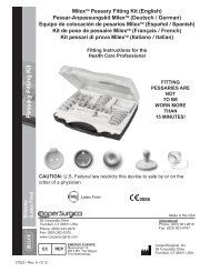
61-3005 / 61-3007<br />
H/S Catheter <strong>Set</strong> with Integrated Stylet (English)<br />
H/S <strong>Katheter</strong>-<strong>Set</strong> <strong>mit</strong> <strong>Integriertem</strong> <strong>Sondenkanal</strong> (Deutsch / German)<br />
Juego de catéter H/S con estilete integrado (Español / Spanish)<br />
Kit de cathéter H/S avec stylet intégré (Français / French)<br />
<strong>Set</strong> con catetere H/S con mandrino incorporato (Italiano / Italian)<br />
Directions for Use<br />
Gebrauchsanleitung<br />
Instrucciones de uso<br />
Mode d’emploi<br />
Istruzioni per l’uso<br />
103-300-LS • Rev. B • 3/11
61-3005 and 61-3007 • H/S Catheter <strong>Set</strong> with Integrated Stylet<br />
for Hysterosonography and Hysterosalpingography<br />
Directions for Use (English)<br />
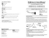
DEVICE DESCRIPTION<br />
The H/S Catheter <strong>Set</strong> consists of a latex-free balloon catheter,<br />
insertion sheath and a 1.5 cc (ml) syringe (5F catheter) or<br />
3 cc (ml) syringe (7F catheter). (See Figure 1.) The catheter<br />
can be used with aqueous based contrast media.<br />
Syringe Syringe Stopcock<br />
Clamp<br />
Blue Blue Luer Luer Hub<br />
Trapezoidal Hub Hub<br />
Insertion Sheath Sheath<br />
Balloon<br />
CAUTION: U.S. Federal law restricts this device to sale by or<br />
on the order of a physician.<br />
INTENDED USE/INDICATIONS<br />
For administering contrast media during Hysterosalpingography<br />
or Hysterosonography procedures to detect uterine<br />
pathology such as polyps, fibroids, adhesions or endometrial<br />
thickening, or patency of fallopian tubes.<br />
CONTRAINDICATIONS<br />
Suspected infection, suspected pregnancy, profuse bleeding or<br />
sexually trans<strong>mit</strong>ted disease.<br />
PRECAUTIONS<br />
Do not exceed the recommended balloon inflation volume of<br />
1.5 cc (ml) (5F catheter) or 3 cc (ml) (7F catheter) or the balloon<br />
may burst.<br />
The use of OIL-BASED contrast media such as ethyl esters<br />
may interact with the balloon of the catheter, causing rupture.<br />
The use of oil-based contrast media is not recommended.<br />
ADVERSE EVENTS<br />
Some patients may have a hypersensitivity to contrast media.<br />
DIRECTIONS FOR USE<br />
Catheter Preparation<br />
Figure 1<br />
1. Grasp the translucent insertion sheath connected to the<br />
trapezoidal hub.<br />
2. Remove and discard the crimped yellow protective cover,<br />
exposing the balloon catheter tip.<br />
3. Test the balloon integrity by inflating with air, saline or water<br />
using the syringe in the set. If performing Hysterosonography<br />
remove as much air as possible from the balloon. Deflate<br />
completely by pulling back on the syringe plunger and closing<br />
the stopcock.<br />
4. Attach a contrast media-filled syringe (not supplied) to the<br />
blue luer hub and fill the catheter with contrast media to<br />
expel air. Advance the insertion sheath so that the distal<br />
end of the catheter protrudes slightly from the distal end of<br />
the sheath. (See Figure 2.)<br />
Catheter Placement<br />
5. Visualize the external cervical os with the aid of a speculum<br />
and advance the sheath and catheter so that the tip of the<br />
catheter enters the cervical canal.<br />
6. The catheter may be shaped/curved to facilitate insertion.<br />
7. Hold the sheath stationary and advance the catheter into<br />
the cervical canal and into the uterine cavity.<br />
8. Open the stopcock and slowly inflate the balloon with up<br />
to 1.5 cc (ml) (5F catheter) or 3 cc (ml) (7F catheter) of air,<br />
saline or water. Turn the stopcock off allowing the balloon<br />
to remain inflated.<br />
9. Withdraw the catheter until it occludes the internal os of<br />
the cervix.<br />
Injection of Contrast Media<br />
10. Inject the contrast media, close the clamp and complete<br />
the study in a routine manner.<br />
11. Occasionally, when access to the uterine cavity is difficult<br />
the balloon must be inflated within the endo-cervical canal.<br />
Catheter Removal<br />
12. Open the stopcock and deflate the balloon by pulling back<br />
on the syringe plunger. Withdraw the catheter.<br />
13. Dispose of in accordance with all applicable Federal, State<br />
and local Medical/Hazardous waste practices.<br />
EXPLANATION OF SYMBOLS<br />
0086<br />
Reorder Number<br />
Batch Code<br />
Use By<br />
Irradiation Sterilization<br />
Latex Free<br />
Do Not Reuse<br />
ATTENTION: See instructions for use.<br />
Product conforms to the Medical Device<br />
Directive 93/42/EEC<br />
Authorized Representative<br />
in the European Community.<br />
Figure 2<br />
95 Corporate Drive • Trumbull, CT 06611 USA<br />
Phone: (800) 243-2974 • Fax: (800) 262-0105<br />
103-300-LS • Rev. B • 3/11<br />
International<br />
Phone: (203) 601-9818<br />
Fax: (203) 601-4747<br />
© 2011 <strong>CooperSurgical</strong>, Inc.<br />
Made in the USA<br />
Leisegang Feinmechanik GmbH<br />
Leibnizstraße 32<br />
D-10625, Berlin GERMANY
61-3005 und 61-3007 • H/S <strong>Katheter</strong>-<strong>Set</strong> <strong>mit</strong> <strong>Integriertem</strong> <strong>Sondenkanal</strong><br />
für Hysterosonographie und Hysterosalpingographie<br />
Gebrauchsanleitung (Deutsch / German)<br />
GERÄTEBESCHREIBUNG<br />
Das H/S <strong>Katheter</strong>-<strong>Set</strong> besteht aus einem latexfreien<br />
Ballonkatheter, einer Einführungskanüle sowie einer 1,5 ml (cc)<br />
-Spritze (5 F-<strong>Katheter</strong>) oder 3,0 ml (cc)-Spritze (7 F-<strong>Katheter</strong>).<br />
(Siehe Abbildung 1.) Für den <strong>Katheter</strong> können Kontrast<strong>mit</strong>tel auf<br />
Wasserbasis verwendet werden.<br />
Spritze Syringe Sperrhahn<br />
Stopcock<br />
Klemme Clamp<br />
Blauer Luer-Anschluss<br />
Blue Hub<br />
95 Corporate • Drive Trumbull, CT 06611 USA<br />
Tel: +1 (800) 243-2974 • Fax: +1 (800) 262-0105<br />
103-300-LS • Rev. B • 3/11<br />
Trapezstecker<br />
Trapezoidal Hub<br />
Einführungskanüle<br />
Insertion Sheath<br />
Abbildung 1<br />
VORSICHT: Laut US-Bundesgesetz ist der Verkauf dieses<br />
Produkts nur an Ärzte oder im Auftrag von Ärzten erlaubt.<br />
Balloon<br />
Ballon<br />
EINSATZBEREICH/INDIKATIONEN<br />
Zur Einführung eines Kontrast<strong>mit</strong>tels bei Hysterosalpingographieoder<br />
Hysterosonographiebehandlungen zur Untersuchung der<br />
Gebärmutter nach Polypen, Fibroiden, Verwachsungen oder<br />
Endometriumkarzinomen oder der Tubendurchlässigkeit.<br />
KONTRAINDIKATIONEN<br />
Verdacht auf Infektion, mögliche Schwangerschaft, starke<br />
Blutungen oder Geschlechtskrankheiten.<br />
VORSICHTSMAßNAHMEN<br />
Das empfohlene Ballonvolumen von 1,5 ml (cc) (5F-<strong>Katheter</strong>)<br />
oder 3,0 ml (cc) (7F-<strong>Katheter</strong>) darf nicht überschritten werden.<br />
Andernfalls besteht die Gefahr, dass der Ballon platzt.<br />
Die Verwendung von Kontrast<strong>mit</strong>tel auf ÖLBASIS wie z. B.<br />
Ethylester kann zu einer Wechselwirkung <strong>mit</strong> dem Ballon des<br />
<strong>Katheter</strong>s führen und einen Riss verursachen. Die Verwendung<br />
von Kontrast<strong>mit</strong>tel auf Ölbasis wird nicht empfohlen.<br />
NEBENWIRKUNGEN<br />
Einige Patienten reagieren auf das Kontrast<strong>mit</strong>tel unter<br />
Umständen <strong>mit</strong> einer Überempfindlichkeit.<br />
BEDIENUNGSANLEITUNG<br />
Vorbereitung des <strong>Katheter</strong>s<br />
1. Nehmen Sie die durchsichtige Einführungskanüle, die <strong>mit</strong><br />
dem Trapezstecker verbunden ist.<br />
2. Entfernen und entsorgen Sie die gewellte gelbe Schutzhülle,<br />
so dass die Spitze des Ballonkatheters freiliegt.<br />
3. Überprüfen Sie die Unversehrtheit des Ballons, indem<br />
Sie ihn <strong>mit</strong>hilfe der dem <strong>Set</strong> beiliegenden Spritze <strong>mit</strong> Luft,<br />
einer Kochsalzlösung oder Wasser füllen. Wird eine<br />
Hysterosonographie durchgeführt, entfernen Sie soviel Luft<br />
wie möglich aus dem Ballon. Entleeren Sie ihn vollständig,<br />
indem Sie den Kolben der Spritze zurückziehen und den<br />
Sperrhahn schließen.<br />
4. Befestigen Sie eine <strong>mit</strong> Kontrast<strong>mit</strong>tel gefüllte Spritze (nicht<br />
im Lieferumfang enthalten) am blauen Luer-Anschluss und<br />
füllen Sie den <strong>Katheter</strong> <strong>mit</strong> Kontrast<strong>mit</strong>tel, da<strong>mit</strong> die Luft<br />
vollständig entweicht. Ziehen Sie die Einführungskanüle so<br />
weit vor, bis das Distalende des <strong>Katheter</strong>s leicht über das<br />
Distalende der Kanüle hinausragt. (Siehe Abbildung 2.)<br />
International<br />
Tel: +1 (203) 601-9818<br />
Fax: +1 (203) 601-4747<br />
Einführung des <strong>Katheter</strong>s<br />
5. Machen Sie sich <strong>mit</strong>hilfe eines Spekulums ein Bild von der<br />
Öffnung des äußeren Gebärmutterhalses und schieben Sie<br />
die Kanüle und den <strong>Katheter</strong> so weit vor, bis die Spitze des<br />
<strong>Katheter</strong>s in den Zervikalkanal eindringt.<br />
6. Der <strong>Katheter</strong> kann zur leichteren Einführung geformt/gebogen<br />
werden.<br />
7. Halten Sie die Kanüle fest und führen Sie den <strong>Katheter</strong> in den<br />
Zervikalkanal und in die Gebärmutterhöhle ein.<br />
8. Öffnen Sie den Sperrhahn und füllen Sie den Ballon langsam<br />
<strong>mit</strong> bis zu 1,5 ml (cc) (5 F-<strong>Katheter</strong>) oder 3,0 ml (cc)<br />
(7 F-<strong>Katheter</strong>) Luft, Kochsalzlösung oder Wasser. Schließen<br />
Sie den Sperrhahn, so dass der Balloninhalt nicht wieder<br />
entweichen kann.<br />
9. Ziehen den <strong>Katheter</strong> zurück, bis die interne Öffnung der<br />
Zervix abgeschlossen ist.<br />
Injektion des Kontrast<strong>mit</strong>tels<br />
10. Injizieren Sie das Kontrast<strong>mit</strong>tel und schließen Sie die Klemme.<br />
Führen Sie dann die Untersuchung wie gewohnt durch.<br />
11. Sollte der Zugang zur Gebärmutterhöhle schwierig sein, muss<br />
der Ballon innerhalb des Endozervikalkanals aufgefüllt<br />
werden.<br />
Entfernung des <strong>Katheter</strong>s<br />
12. Öffnen Sie den Sperrhahn und entleeren Sie den Ballon,<br />
indem Sie den Spritzenkolben zurückziehen. Ziehen Sie den<br />
<strong>Katheter</strong> heraus.<br />
13. Entsorgung gemäß den geltenden Gesetzen zur Entsorgung<br />
medizinischer Abfälle.<br />
SYMBOLE<br />
0086<br />
Nachbestellnummer<br />
Chargencode<br />
Verfallsdatum<br />
Strahlungssterilisation<br />
Latexfrei<br />
Nicht wiederverwenden<br />
ACHTUNG: Siehe Gebrauchsanleitung.<br />
Abbildung 2<br />
Produkt entspricht der Medizinprodukterichtlinie<br />
93/42/EEG<br />
Autorisierte Vertretung in der Europäischen<br />
Gemeinschaft.<br />
© 2011 <strong>CooperSurgical</strong>, Inc.<br />
Hergestellt in USA<br />
Leisegang Feinmechanik GmbH<br />
Leibnizstraße 32<br />
D-10625, Berlin DEUTSCHLAND
61-3005 y 61-3007 • Juego de catéter H/S con estilete integrado<br />
para histeroecografía e histerosalpingografía<br />
Instrucciones de uso (Español / Spanish)<br />
DESCRIPCIÓN DEL DISPOSITIVO<br />
El juego de catéter H/S consta de un catéter con balón sin<br />
látex, una vaina de inserción y una jeringa de 1,5 ml (cc)<br />
(catéter 5 F) o de 3,0 ml (cc) (catéter 7 F). (Véase la figura 1.)<br />
El catéter puede utilizarse con medios de contraste acuosos.<br />
Jeringa Syringe<br />
Llave Stopcockde paso<br />
Pinza Clamp<br />
Conector Blue luer Luer azul Hub<br />
Conector Trapezoidal Hub trapezoidal<br />
Vaina Insertion de Sheath inserción<br />
Figura 1<br />
Balloon<br />
Balón<br />
PRECAUCIÓN: las leyes federales de EE UU li<strong>mit</strong>an la<br />
venta de este dispositivo a médicos o por prescripción médica.<br />
USO PREVISTO/INDICACIONES<br />
Para la administración de medios de contraste durante<br />
procedimientos de histerosalpingografía o histeroecografía<br />
para detectar alteraciones uterinas tales como pólipos,<br />
miomas, adherencias o engrosamiento endometrial, o la<br />
permeabilidad de las trompas de falopio.<br />
CONTRAINDICACIONES<br />
Sospecha de infección, sospecha de embarazo, hemorragia<br />
intensa o enfermedad de transmisión sexual.<br />
PRECAUCIONES<br />
No superar el volumen recomendado de inflado del balón de<br />
1,5 ml (cc) (catéter 5 F) o 3,0 ml (cc) (catéter 7 F), ya que el<br />
balón podría romperse.<br />
El uso de medios de contraste OLEOSOS, como los ésteres<br />
etílicos, puede afectar al balón del catéter, causando su rotura.<br />
No se recomienda usar medios de contraste oleosos.<br />
EFECTOS ADVERSOS<br />
Algunos pacientes pueden experimentar hipersensibilidad a<br />
los medios de contraste.<br />
INSTRUCCIONES DE USO<br />
Preparación del catéter<br />
1. Sujetar la vaina de inserción translúcida conectada al<br />
conector trapezoidal.<br />
2. Retirar y desechar la cubierta protectora rizada amarilla,<br />
dejando expuesta la punta del catéter con balón.<br />
3. Comprobar la integridad del balón inflándolo con aire,<br />
solución salina o agua por medio de la jeringa incluida<br />
en el juego de catéter. Si se está realizando una<br />
histeroecografía, retirar la mayor cantidad posible de aire<br />
del balón. Desinflar completamente tirando del émbolo de<br />
la jeringa y cerrando la llave de paso.<br />
4. Conectar una jeringa llena de medio de contraste (no<br />
suministrada) al conector luer azul y llenar el catéter con<br />
medio de contraste para expulsar el aire. Avanzar la vaina<br />
de inserción de forma que el extremo distal del catéter<br />
sobresalga ligeramente por el extremo distal de la vaina.<br />
(Véase la figura 2.)<br />
Colocación del catéter<br />
5. Visualizar el orificio cervical externo con la ayuda de un<br />
espéculo y avanzar la vaina y el catéter de forma que la<br />
punta del catéter penetre en el conducto cervical.<br />
6. El catéter puede ser curvado o con forma para facilitar la<br />
inserción.<br />
7. Mantener fija en posición la vaina y avanzar el catéter por<br />
el conducto cervical hasta la cavidad uterina.<br />
8. Abrir la llave de paso e inflar lentamente el balón con<br />
un máximo de 1,5 ml (cc) (catéter 5 F) o 3,0 ml (cc)<br />
(catéter 7 F) de aire, solución salina o agua. Cerrar la<br />
llave de paso dejando inflado el balón.<br />
9. Retirar el catéter hasta que ocluya el orificio interno del<br />
cuello uterino.<br />
Inyección de medio de contraste<br />
10. Inyectar el medio de contraste, cerrar la pinza y completar<br />
el estudio de la forma habitual.<br />
11. En ocasiones, si el acceso a la cavidad uterina resulta<br />
difícil debe inflarse el balón en el interior del conducto<br />
endocervical.<br />
Retirada del catéter<br />
12. Abrir la llave de paso y desinflar el balón tirando del<br />
émbolo de la jeringa. Retirar el catéter.<br />
13. Deseche de conformidad con todas las normativas de<br />
eliminación de desechos médicos/peligrosos federales,<br />
estatales y locales.<br />
EXPLICACIÓN DE SÍMBOLOS<br />
0086<br />
Número de pedido<br />
Código de lote<br />
Utilizar antes de<br />
Esterilizado por radiación<br />
Sin látex<br />
No reutilizar<br />
Figura 2<br />
ATENCIÓN: consultar las instrucciones de uso.<br />
El producto cumple con la directiva sobre<br />
dispositivos médicos 93/42/CEE<br />
Representante autorizado en la Comunidad<br />
Europea.<br />
95 Corporate Drive • Trumbull, CT 06611 EE UU<br />
Teléfono: +1 (800) 243-2974 • Fax: +1 (800) 262-0105<br />
103-300-LS • Rev. B • 3/11<br />
Internacional<br />
Teléfono: +1 (203) 601-9818<br />
Fax: +1 (203) 601-4747<br />
Leisegang Feinmechanik GmbH<br />
Leibnizstraße 32<br />
D-10625, Berlin ALEMANIA<br />
© 2011 <strong>CooperSurgical</strong>, Inc.<br />
Fabricado en EE UU
61-3005 et 61-3007 • Kit de cathéter H/S avec stylet intégré pour<br />
hystérosonographie et hystérosalpingographie<br />
Mode d’Emploi (Français / French)<br />
DESCRIPTION DU DISPOSITIF<br />
Le kit cathéter H/S se compose d’un cathéter à ballonnet sans<br />
latex, d’une gaine d’introduction et d’une seringue de 1,5 ml (cc)<br />
(cathéter de 5 F) ou d’une seringue de 3 ml (cc) (cathéter de<br />
7 F). (Consultez la figure 1.) Le cathéter peut être utilisé avec<br />
des produits de contraste à base aqueuse.<br />
Seringue Syringe Robinet Stopcock d’arrêt<br />
Agrafe Clamp<br />
Blue Luer Hub<br />
Embase Luer bleue<br />
Embase Trapezoidal trapézoïdale<br />
Hub<br />
Gaine Insertion d'introduction<br />
Sheath<br />
Figure 1<br />
Balloon<br />
Ballonnet<br />
ATTENTION : Selon la loi fédérale américaine, ce produit ne<br />
peut être vendu que par un médecin ou sur son ordonnance.<br />
UTILISATION/INDICATIONS<br />
Pour l’administration de produit de contrast durant des<br />
procédures d’hystérosonographie ou d’hystérosalpingographie<br />
afin de détecter les pathologies utérines telles que des<br />
polypes, fibromes, adhérences, épaississement endométrial<br />
ou perméabilité des trompes de Fallope.<br />
CONTRE-INDICATIONS<br />
Infection soupçonnée, grossesse soupçonnée, saignement<br />
abondant ou maladie sexuellement transmissible.<br />
PRÉCAUTIONS<br />
Ne dépassez pas le volume de gonflage recommandé du<br />
ballonnet de 1,5 ml (cc) (cathéter de 5 F) ou de 3 ml (cc)<br />
(cathéter de 7 F) car le ballonnet pourrait éclater.<br />
Les solutions de produits de contraste À BASE D’HUILE tels<br />
que les esters d’éthyle peuvent interagir avec le ballonnet du<br />
cathéter et causer sa rupture. L’utilisation de produits de<br />
contraste à base d’huile n’est pas recommandée.<br />
ÉVÉNEMENTS INDÉSIRABLES<br />
Certaines patientes peuvent présenter une hypersensibilité aux<br />
produits de contraste.<br />
MODE D’EMPLOI<br />
Préparation du cathéter<br />
1. Saisissez la gaine d’introduction translucide raccordée à<br />
l’embase trapézoïdale.<br />
2. Enlevez et jetez le couvercle de protection serti jaune pour<br />
découvrir l’extré<strong>mit</strong>é du cathéter à ballonnet.<br />
3. Testez l’intégrité du ballonet en le gonflant avec de l’air, du<br />
sérum physiologique ou de l’eau à l’aide de la seringue du kit.<br />
Pour une hystérosonographie, éliminez autant d’air que<br />
possible du ballonnet. Dégonflez-le complètement en tirant<br />
sur le piston de la seringue et en fermant le robinet d’arrêt.<br />
4. Fixez une seringue (non fournie) remplie de produits de<br />
contraste dans l’embase Luer bleue et remplissez le<br />
cathéter de produits de contraste afin d’en expulser l’air.<br />
Faites progresser la gaine d'introduction de sorte que<br />
l’extré<strong>mit</strong>é distale du cathéter dépasse légèrement de<br />
l’extré<strong>mit</strong>é distale de la gaine. (Consultez la figure 2.)<br />
Mise en place du cathéter<br />
5. Visualisez l’orifice cervical externe du col à l’aide d’un<br />
spéculum et faites progresser la gaine d’introduction et le<br />
cathéter de sorte que l’extré<strong>mit</strong>é du catheter pénètre dans<br />
le canal cervical.<br />
6. Le cathéter peut être modelé/courbé pour faciliter<br />
l’introduction.<br />
7. Maintenez la gaine immobile et faites progresser le<br />
cathéter dans le canal cervical et dans la cavité utérine.<br />
8. Ouvrez le robinet d’arrêt et gonflez lentement le ballonnet<br />
avec au maximum 1,5 ml (cc) (cathéter de 5 F) ou 3 ml<br />
(cc) (cathéter de 7 F) d’air, de sérum physiologique ou<br />
d’eau. Fermez le robinet d’arrêt afin pour permettre au<br />
ballonnet de rester gonflé.<br />
9. Retirez le cathéter jusqu’à ce qu’il occlut l’orifice interne<br />
pour permettre au ballonnet de rester col de l’utérus.<br />
Injection de produits de contraste<br />
10. Injectez les produits de contraste, fermez les produits<br />
l’agrafe et terminez l’étude de manière habituelle.<br />
11. Parfois, lorsque l’accès à la cavité utérine est difficile, le<br />
ballonnet peut être gonflé à l’intérieur du canal<br />
endocervical.<br />
Retrait du cathéter<br />
12. Ouvrez le robinet d’arrêt et dégonflez le ballonnet en tirant<br />
sur le piston de la seringue. Retirez le cathéter.<br />
13. Éliminez conformément à toutes les pratiques fédérales,<br />
d’État et locales en matière en matière de déchets<br />
médicaux/dangereux.<br />
EXPLICATION DES SYMBOLES<br />
0086<br />
Numéro de référence<br />
Code de lot<br />
Date li<strong>mit</strong>e d’utilisation<br />
Stérilisation par irradiation<br />
Sans latex<br />
Ne pas réutiliser<br />
ATTENTION : Consulter le mode d’emploi.<br />
Produit conforme à la directive 93/42/CEE<br />
relative aux dispositifs médicaux<br />
Figure 2<br />
Représentant agréé au sein de la Communauté<br />
européenne.<br />
95 Corporate Drive • Trumbull, CT 06611 États-Unis<br />
Téléphone : +1 (800) 243-2974 • Fax : +1 (800) 262-0105<br />
103-300-LS • Rév. B • 3/11<br />
International<br />
Téléphone : +1 (203) 601-9818<br />
Fax : +1 (203) 601-4747<br />
Leisegang Feinmechanik GmbH<br />
Leibnizstraße 32<br />
D-10625, Berlin ALLEMAGNE<br />
© 2011 <strong>CooperSurgical</strong>, Inc.<br />
Fabriqué aux États-Unis
61-3005 e 61-3007 • <strong>Set</strong> con catetere H/S con mandrino incorporato<br />
per isterosonografia e isterosalpingografia<br />
Istruzioni per l’uso (Italiano / Italian)<br />
DESCRIZIONE DEL DISPOSITIVO<br />
Il set con catetere H/S è costituito da un catetere a palloncino<br />
privo di lattice, da un introduttore e da una siringa da 1,5 ml<br />
(cc) (per il catetere da 5 F) o da 3,0 ml (cc) (per il catetere<br />
da 7 F). (Vedere la figura 1.) Il catetere può essere usato con<br />
mezzi di contrasto acquosi.<br />
Siringa Syringe<br />
Rubinetto<br />
Stopcock<br />
Morsetto<br />
Clamp<br />
Raccordo Luer Blue blu Luer Hub<br />
95 Corporate Drive • Trumbull, CT 06611 USA<br />
Tel: +1 (800) 243-2974 • Fax: +1 (800) 262-0105<br />
103-300-LS • Rev. B • 3/11<br />
Introduttore<br />
Insertion Sheath<br />
Raccordo Trapezoidal Hub trapezoidale<br />
Balloon<br />
Palloncino<br />
ATTENZIONE – La legge federale statunitense li<strong>mit</strong>a la<br />
vendita di questo dispositivo ai medici o su presentazione di<br />
prescrizione medica.<br />
USO PREVISTO/INDICAZIONI PER L'USO<br />
Figura 1<br />
Per la somministrazione di mezzi di contrasto nella diagnosi<br />
di patologie uterine quali polipi, fibromi, sinechie (aderenze),<br />
ispessimento dell'endometrio o pervietà delle tube di Falloppio,<br />
effettuata con isterosalpingografia o isterosonografia.<br />
CONTROINDICAZIONI<br />
Sospette infiammazioni, sospetta gravidanza, emorragie<br />
profuse o malattie sessualmente trasmesse.<br />
PRECAUZIONI<br />
Non gonfiare il palloncino oltre il volume consigliato di<br />
1,5 ml (cc) (per il catetere da 5 F) o di 3,0 ml (cc) (per il<br />
catetere da 7 F) per evitare che possa scoppiare.<br />
I mezzi di contrasto OLEOSI, quali gli esteri etilici, possono<br />
interagire con il palloncino del catetere e provocarne la rottura.<br />
Pertanto l'uso di mezzi di contrasto oleosi è sconsigliato.<br />
EFFETTI COLLATERALI<br />
Alcuni pazienti possono essere ipersensibili ai mezzi di<br />
contrasto.<br />
ISTRUZIONI PER L'USO<br />
Preparazione del catetere<br />
1. Afferrare l'introduttore traslucido collegato al raccordo<br />
trapezoidale.<br />
2. Rimuovere e gettare il cappuccio di protezione giallo<br />
ondulato per esporre la punta del catetere a palloncino.<br />
3. Verificare che il palloncino sia integro iniettando, con la<br />
siringa fornita in dotazione, aria, soluzione fisiologica o<br />
acqua per gonfiarlo. Se il dispositivo viene utilizzato per<br />
eseguire un'isterosonografia, rimuovere la maggiore<br />
quantità possibile di aria dal palloncino. Sgonfiare<br />
completamente il palloncino tirando lo stantuffo della<br />
siringa all'indietro e chiudendo il rubinetto.<br />
4. Collegare una siringa contenente mezzo di contrasto<br />
(non fornita in dotazione) al raccordo Luer blu e riempire il<br />
catetere con il mezzo di contrasto per espellere l'aria.<br />
Far avanzare l'introduttore in modo da far sporgere<br />
Internazionale<br />
Tel: +1 (203) 601-9818<br />
Fax: +1 (203) 601-4747<br />
leggermente l'estre<strong>mit</strong>à distale del catetere dall’estre<strong>mit</strong>à<br />
distale dell’introduttore. (Vedere la figura 2.)<br />
Inserimento del catetere<br />
5. Individuare l'orifizio esterno del canale cervicale con uno<br />
specolo e far avanzare l'introduttore e il catetere fino ad<br />
inserire la punta del catetere nel canale cervicale.<br />
6. Il catetere può essere formato/curvato per agevolarne<br />
l'inserimento.<br />
7. Tenendo fermo l'introduttore, far avanzare il catetere<br />
all'interno del canale cervicale e della cavità uterina.<br />
8. Aprire il rubinetto e gonfiare lentamente il palloncino fino<br />
a 1,5 ml (cc) (per il catetere da 5 F) o 3,0 ml (cc) (per il<br />
catetere da 7 F) con aria, soluzione fisiologica o acqua.<br />
Chiudere il rubinetto accertandosi che il palloncino<br />
rimanga gonfiato.<br />
9. Rimuovere il catetere fino ad occludere l'orifizio interno del<br />
canale cervicale.<br />
Iniezione del mezzo di contrasto<br />
10. Iniettare il mezzo di contrasto, chiudere il morsetto e<br />
portare a termine l'analisi secondo la normale prassi.<br />
11. Nei casi in cui l'accesso alla cavità uterina risulta<br />
difficoltoso, è necessario gonfiare il palloncino all'interno<br />
del canale endocervicale.<br />
Rimozione del catetere<br />
12. Aprire il rubinetto e sgonfiare il palloncino tirando lo<br />
stantuffo della siringa all'indietro. Rimuovere il catetere.<br />
13. Smaltire in base ai requisiti normativi applicabili e alle<br />
prassi per lo smaltimento di rifiuti pericolosi/medici.<br />
SPIEGAZIONE DEI SIMBOLI<br />
0086<br />
Numero di riordine<br />
Codice del lotto<br />
Utilizzare entro<br />
Sterilizzazione tra<strong>mit</strong>e irradiazione<br />
Non contiene lattice<br />
Non riutilizzare<br />
ATTENZIONE – Vedere le istruzioni per l’uso.<br />
Questo prodotto è conforme ai requisiti della<br />
Direttiva 93/42/CEE concernente i dispositivi<br />
medici<br />
Rappresentante autorizzato per la<br />
Comunità Europea.<br />
Leisegang Feinmechanik GmbH<br />
Leibnizstraße 32<br />
D-10625, Berlin GERMANIA<br />
Figura 2<br />
© 2011 <strong>CooperSurgical</strong>, Inc.<br />
Fabbricato negli USA