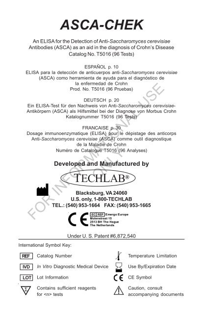
ASCA-CHEK - TechLab
ASCA-CHEK - TechLab
ASCA-CHEK - TechLab

You also want an ePaper? Increase the reach of your titles
YUMPU automatically turns print PDFs into web optimized ePapers that Google loves.
<strong>ASCA</strong>-<strong>CHEK</strong><br />
An ELISA for the Detection of Anti-Saccharomyces cerevisiae<br />
Antibodies (<strong>ASCA</strong>) as an aid in the diagnosis of Crohn’s Disease<br />
Catalog No. T5016 (96 Tests)<br />
ESPAÑOL p. 10<br />
ELISA para la detección de anticuerpos anti-Saccharomyces cerevisiae<br />
(<strong>ASCA</strong>) como herramienta de ayuda para el diagnóstico de<br />
la enfermedad de Crohn<br />
Prod. No. T5016 (96 Pruebas)<br />
DEUTSCH p. 20<br />
Ein ELISA-Test für den Nachweis von Anti-Saccharomyces cerevisiae-<br />
Antikörpern (<strong>ASCA</strong>) als Hilfsmittel bei der Diagnose von Morbus Crohn<br />
Katalognummer T5016 (96 Tests)<br />
FRANCAISE p. 30<br />
Dosage immunoenzymatique (ELISA) pour le dépistage des anticorps<br />
Anti-Saccharomyces cerevisiae (<strong>ASCA</strong>) comme outil diagnostique<br />
de la Maladie de Crohn<br />
Numéro de Catalogue T5016 (96 Analyses)<br />
International Symbol Key:<br />
Developed and Manufactured by<br />
TECHLAB ®<br />
Blacksburg, VA 24060<br />
U.S. only, 1-800-TECHLAB<br />
TEL.: (540) 953-1664 FAX: (540) 953-1665<br />
FOR INFORMATIONAL USE<br />
ONLY<br />
EC REP Emergo Europe<br />
Molenstraat 15<br />
2513 BH The Hague<br />
The Netherlands<br />
Under U. S. Patent #6,872,540<br />
REF<br />
IVD<br />
LOT<br />
Catalog Number<br />
In Vitro Diagnostic Medical Device<br />
Lot Information<br />
Contains sufficient reagents<br />
for tests<br />
Temperature Limitation<br />
Use By/Expiration Date<br />
CE Symbol<br />
Caution, consult<br />
accompanying documents
2<br />
<strong>ASCA</strong>-<strong>CHEK</strong><br />
INTENDED USE<br />
The <strong>ASCA</strong>-<strong>CHEK</strong> test is an enzyme-linked immunosorbent assay (ELISA) for the<br />
qualitative detection of human anti-S. cerevisiae antibodies (<strong>ASCA</strong>) in feces and serum.<br />
The test result is used as an aid in the diagnosis of Crohn’s disease in combination with<br />
clinical and other laboratory findings.<br />
FOR IN VITRO DIAGNOSTIC USE.<br />
EXPLANATION<br />
An estimated 1 million Americans suffer from inflammatory bowel disease (IBD) that<br />
is comprised of Crohn’s disease and ulcerative colitis (1). IBD is characterized by a<br />
chronic inflammatory response that results in histologic damage to the intestinal lining.<br />
Crohn’s disease may involve the entire gastrointestinal tract and include inflammation<br />
extending into the transmural mucosa, whereas ulcerative colitis only affects the large<br />
bowel and includes inflammation of the mucosal lining. These two distinct diseases<br />
require a rapid differential diagnosis for optimal treatment. Conventional methods<br />
utilizing multiple endoscopy examinations and histological analysis may take years to<br />
confirm a diagnosis (2). Test methods for determining the presence of serum <strong>ASCA</strong> as<br />
a marker of Crohn’s disease are currently available, with sensitivities ranging from 35 -<br />
70% (3-6).<br />
The <strong>ASCA</strong>-<strong>CHEK</strong> test is an ELISA for detecting <strong>ASCA</strong> in human feces and serum.<br />
The test provides a method utilizing antigens of Saccharomyces cerevisiae for<br />
measuring total fecal and serum <strong>ASCA</strong> as an aid to distinguish Crohn’s disease from<br />
other gastrointestinal illnesses such as ulcerative colitis and irritable bowel syndrome.<br />
PRINCIPLE OF THE TEST<br />
The <strong>ASCA</strong>-<strong>CHEK</strong> test detects anti-Saccharomyces cerevisiae antibodies (<strong>ASCA</strong>).<br />
The microassay wells supplied with the kit contain immobilized antigens of Saccharomyces<br />
cerevisiae. The Conjugate consists of anti-human immunoglobulin antibody<br />
conjugated to horseradish peroxidase. In the assay, an aliquot of either fecal or serum<br />
specimen is emulsified in the Diluent and the diluted specimen is transferred to the<br />
microassay well. If <strong>ASCA</strong> are present in the specimen, they will bind to the immobilized<br />
antigens. After incubation, the wells are washed and the Conjugate is added. The<br />
Conjugate binds to the <strong>ASCA</strong> captured by the immobilized antigens. A second series of<br />
wash steps removes any unbound material. Following the addition of Substrate, a<br />
color is detected due to the enzyme-antibody-antigen complexes that form in the<br />
presence of <strong>ASCA</strong>.<br />
FOR INFORMATIONAL USE<br />
ONLY<br />
REAGENTS<br />
DIL 10X 10X Diluent, 40 mL (10X concentrate of a phosphate buffered protein<br />
solution containing 0.2% thimerosal). The 1X Diluent is also to be used as<br />
the negative control (see TEST PROCEDURE).<br />
CONJ ENZ Conjugate, 7 mL (goat anti-human polyclonal immunoglobulin-HRP in a<br />
buffered solution containing 0.02% thimerosal)<br />
SUBS REAG<br />
CONTROL +<br />
Substrate, 14 mL (tetramethylbenzidine substrate and peroxide)<br />
Positive Control, 3.5 mL (human <strong>ASCA</strong> in a buffered protein solution<br />
containing 0.02% thimerosal)<br />
WASHBUF 20X Wash Buffer Concentrate, 50 mL (20X concentrate containing phosphate-buffered<br />
saline, detergent and 0.2% thimerosal)<br />
H 2<br />
SO 4<br />
MA<br />
0.6N<br />
PLT<br />
Stop Solution, 7 mL (0.6 N sulfuric acid). CAUTION: Avoid contact with<br />
skin. Flush with water immediately if contact occurs.<br />
Microassay Plate, 12 strips, 8 wells per strip, coated with antigens of<br />
Saccharomyces cerevisiae (stored with desiccant)
3<br />
PRECAUTIONS<br />
1. Reagents from different kits should not be mixed. Do not use the kit past the<br />
expiration date.<br />
2. Reagents should be at room temperature before use.<br />
3. Gently mix all reagents before use.<br />
4. Caps and tips are color coded; do not mix!<br />
5. When handling the microassay wells, avoid scratching the bottom of the wells<br />
because this may result in elevated absorbance readings.<br />
6. Hold dropper bottles vertically when dispensing reagent to ensure proper drop<br />
size.<br />
7. Handle specimens and used microassay wells as if capable of transmitting<br />
infectious agents. Wear gloves when doing the test.<br />
8. Reagents contain thimerosal as a preservative and should be handled with normal<br />
laboratory caution.<br />
9. The Stop Solution contains 0.6 N sulfuric acid. Flush with water immediately if<br />
contact occurs.<br />
10. Unused microassay wells must be placed inside the resealable pouch immediately<br />
with the desiccant to protect them from moisture.<br />
11. Perform the washing procedure as directed to avoid high background reactions.<br />
12. Use fecal and serum specimens within 48 hours and 7 days, respectively, of<br />
collection to obtain optimal results. Even though a single freeze-thaw is acceptable,<br />
frozen specimens (-20°C or lower) may lose activity due to multiple freezing<br />
and thawing.<br />
13. Do not freeze the reagents. Store the kit between 2° and 8°C.<br />
14. The Substrate is light sensitive and should be protected from direct sunlight or UV<br />
sources. If the Substrate is exposed to light and develops a color, it must be<br />
discarded.<br />
15. Optimal results are obtained by following the specified test procedure. The<br />
concentrations, incubation conditions, and processing specifications have been<br />
optimized for sensitivity and specificity. Alterations of the specified procedure and/<br />
or test conditions may affect the sensitivity and specificity of the test.<br />
16. The Positive Control contains <strong>ASCA</strong> that is a human derived material. Material has<br />
been tested and found negative for antibody to HIV-1, HIV-2, HCV, and HbsAg. No<br />
known test method can offer complete assurance that infectious agents are<br />
absent. ALL HUMAN SOURCE PRODUCTS SHOULD BE HANDLED AS POTENTIALLY<br />
INFECTIOUS MATERIAL. A procedure for handling biohazards is published in the<br />
CDC/NIH Manual of Biosafety in Microbiology & Biomedical Laboratories.<br />
17. Microbial contamination of reagents may decrease the accuracy of the assay.<br />
Avoid microbial contamination of reagents by using sterile disposable pipettes if<br />
removing aliquots from reagent bottles.<br />
FOR INFORMATIONAL USE<br />
ONLY<br />
PRELIMINARY PREPARATIONS<br />
1. All reagents must be at room temperature prior to use in the assay.<br />
2. Prepare 1X Wash Solution. The Wash Solution is supplied as a 20X concentrate<br />
(a precipitate may be noticed and is acceptable). Dilute to a total volume of 1<br />
liter by adding 50 mL of the concentrate to 950 mL of deionized water. Label the<br />
bottle. Store any unused 1X Wash Solution between 2° and 8°C.<br />
3. Prepare 1X Diluent. The Diluent is supplied as a 10X concentrate (a precipitate<br />
may be noticed and is acceptable). Dilute to a total volume of 400 mL by adding 40<br />
mL of the concentrate to 360 mL of deionized water. Label the bottle. Store any<br />
unused 1X Diluent between 2° and 8°C.<br />
4. Microassay Plate preparation. Each strip contains 8 microassay wells coated<br />
with antigens of Saccharomyces cerevisiae. Each specimen or control will require<br />
one of these coated wells. Avoid contact with the bottom of the wells because this<br />
is the optical window for ELISA readers. Microassay wells not used must be<br />
immediately returned to the plastic bag and carefully resealed with desiccant.
4<br />
5. All reagents, with the exception of the Wash Buffer Concentrate and the 10X<br />
Diluent, are supplied in ready-to-use bottles. Reagents can be dispensed directly<br />
from the dropper bottles or decanted for use with multichannel pipettes. If excess<br />
reagent has been decanted, the excess should be discarded. Do not pour back<br />
into the bottle. The Substrate should be stored in and used from the light protected<br />
bottle in which it is supplied. If an aliquot is removed from the original bottle for any<br />
reason, do not return unused Substrate to the original bottle.<br />
COLLECTION OF FECAL SPECIMENS AND PREPARATION OF DILUTIONS<br />
NOTE: Standard collection and handling procedures used in-house for fecal specimens<br />
for culture are appropriate. Do not use specimens that have been collected or stored in<br />
10% Formalin, Merthiolate Formalin, Sodium Acetate Formalin, or Polyvinyl Alcohol.<br />
Store specimens at -20°C, or lower , if the test cannot be performed within 48 hours of<br />
collection. Thaw specimens at room temperature and mix well prior to sample preparation.<br />
Store diluted specimens between 2° and 8°C up to 48 hours. Whenever possible,<br />
test fecal specimens that are less than 48 hours old. Mix (vortex) specimens<br />
thoroughly prior to performing the assay. This includes complete mixing of<br />
the specimen prior to transfer to 1X Diluent as well as complete mixing of<br />
the diluted specimen prior to performing the assay.<br />
1. Prepare Diluted Specimens:<br />
For Liquid Fecal Specimens: Set up a plastic tube for each specimen to be<br />
tested. For each specimen, add 450 µL of 1X Diluent to each tube. Use a transfer<br />
pipette to add 50 µL (flared section) of liquid fecal specimen to the tube containing<br />
1X Diluent for a 1:10 dilution and mix well using a vortex mixer.<br />
For Formed/Solid Fecal Specimens: Set up a plastic tube for each specimen to<br />
be tested. For each specimen, add 450 µL of 1X Diluent to each tube. Add 0.05 g<br />
(flared section) or weigh 0.05 g of solid fecal specimen to the tube containing 1X<br />
Diluent for a 1:10 dilution and mix well using a vortex mixer.<br />
2. Vortex the tubes for 10 seconds and store between 2° and 8°C until the test is<br />
performed. Vortex again before transferring diluted specimen to the microassay<br />
well. This ensures thorough mixing of the specimen.<br />
COLLECTION OF SERUM SPECIMENS AND PREPARATION OF DILUTIONS<br />
NOTE: Blood samples should be collected by venipuncture and allowed to clot<br />
naturally. Serum should be separated from the clot. Store undiluted specimens at -20°C,<br />
or lower, if the test cannot be performed within 7 days of collection. Thaw specimens<br />
at room temperature and mix well prior to sample preparation. Diluted specimens may<br />
be stored between 2° and 8°C for up to 7 days. Whenever possible, test serum<br />
specimens that are less than 7 days old. Mix (vortex) specimens thoroughly<br />
prior to performing the assay. This includes complete mixing of the specimen<br />
prior to transfer to 1X Diluent as well as complete mixing of the diluted<br />
specimen prior to performing the assay.<br />
1. Prepare Diluted Specimens:<br />
For Serum Specimens: Set up and label 3 plastic tubes for each serum<br />
specimen to be tested. For each specimen, add 450 µL of 1X Diluent to each of<br />
the 3 tubes. Use a transfer pipette to add 50 µL (flared section) of serum specimen<br />
to the first tube containing 1X Diluent for a 1:10 dilution and mix well using a vortex<br />
mixer.<br />
2. Next, transfer 50 µL (flared section) from the 1:10 dilution into the second tube<br />
containing Diluent for a 1:100 dilution and mix well using a vortex mixer.<br />
3. Next, transfer 50 µL from the tube containing the 1:100 dilution into the second tube<br />
containing Diluent for a 1:1000 dilution and mix well using a vortex mixer.<br />
Vortex the tubes for 10 seconds and store between 2° and 8°C until the test is<br />
performed. Vortex again before transferring diluted specimen to the microassay<br />
well. This ensures thorough mixing of the specimen.<br />
FOR INFORMATIONAL USE<br />
ONLY
5<br />
TEST PROCEDURE<br />
Materials provided<br />
2 Plastic adhesive sheets 100 Flared Transfer pipettes (flared section = 50 µL)<br />
Transfer Pipette: Bulb 300 µL 200 µL 100 µL 50 µL Flared tip<br />
Materials and equipment required but not provided<br />
Squirt bottle for 1X Wash Solution<br />
Vortex mixer<br />
Tubes for dilution of specimen<br />
Discard container/absorbent paper<br />
Refrigerator for storage<br />
Deionized or distilled water<br />
Bottle for 1X Diluent<br />
37°C incubator<br />
ELISA reader capable of reading 450 nm or 450/620 nm<br />
1. Determine the number of wells to be used. Add 1 drop of Positive Control (black<br />
cap) to a positive control well. Add 50 µL (flared section) of 1X Diluent to a<br />
negative control well. Add 100 µL (first mark past flared section) of diluted<br />
specimen to one well.<br />
2. Incubate the wells at 37°C ± 2°C for 30 minutes.<br />
3. Shake out the contents of the microassay wells into a discard pan.<br />
4. Wash each well using the 1X Wash Solution in a squirt bottle with a fine-tipped<br />
nozzle, directing the 1X Wash Solution to the bottom of the well with force. Fill the<br />
wells (approximately 400 µL when filled), then shake the 1X Wash Solution out of<br />
the well into a discard pan. Slap the inverted plate on a dry paper towel and repeat<br />
wash steps four times using a dry paper towel each time. If any particulate<br />
matter is seen in the wells, continue washing until all the matter is removed.<br />
5. Add 1 drop of Conjugate (red cap) to each well. Incubate the wells at 37°C ± 2°C<br />
for 30 minutes.<br />
6. Repeat Steps #3 and #4. Dispose of all paper towels and specimen containers<br />
properly.<br />
7. Add 2 drops of Substrate (blue cap) to each well. Gently tap the wells to mix the<br />
contents. Incubate the wells at room temperature for 15 minutes. Gently tap the<br />
wells 1 or 2 times during the incubation period.<br />
8. Add 1 drop of Stop Solution (yellow cap) to each well. Gently tap the wells and<br />
wait 2 minutes before reading. The addition of Stop Solution converts the blue<br />
color to a yellow color, which is detected by measuring the optical density at 450<br />
nm on an ELISA reader. Wipe the underside of each well to remove moisture<br />
before measuring the optical density. If a dual reader is used, read at 450 nm and<br />
reference 620 nm. Read within two to ten minutes after adding Stop Solution.<br />
9. Record absorbance values for the positive control, negative control, and each<br />
specimen tested.<br />
FOR INFORMATIONAL USE<br />
ONLY<br />
QUALITY CONTROL<br />
The positive and negative control must meet the following criteria or the test is<br />
invalid. The positive control well must be a visible yellow color. When read on a<br />
spectrophotometer, it must have an OD 450<br />
and OD 450/620<br />
>0.500. The negative control<br />
must have an OD 450<br />
6<br />
Negative for serum specimens = OD 450<br />
In a separate clinical evaluation of the <strong>ASCA</strong>-<strong>CHEK</strong> test using serum specimens,<br />
there were 218 patients with IBD, 39 IBS/non-Crohn’s disease patients and 94 healthy<br />
control persons enrolled at 5 separate study sites (4 clinical sites and 1 in-house site).<br />
In the IBD group, there were 172 patients with Crohn’s disease and 46 with ulcerative<br />
colitis. The male to female ratio was approximately 1:1 and a total of 40% of patients<br />
was less than 19 years old. A total of 62% of the patients with Crohn’s disease was<br />
positive by the <strong>ASCA</strong>-<strong>CHEK</strong> test. The combined analysis for all 5 sites showed a<br />
sensitivity and specificity of 62% and 93%, respectively with an overall correlation of<br />
78% to clinically confirmed Crohn’s disease. Tables 2 and 3 show results with the<br />
<strong>ASCA</strong>-<strong>CHEK</strong> test and a commercial <strong>ASCA</strong> test compared to clinical assessments.<br />
Table 2. <strong>ASCA</strong>-<strong>CHEK</strong> test results of 4 clinical study sites and 1 in-house site for<br />
distinguishing Crohn’s disease from non-Crohn’s disease using serum.<br />
Serum N=351<br />
Crohn’s disease<br />
Ulcerative colitis, IBS<br />
and Non-IBD<br />
<strong>ASCA</strong>-<strong>CHEK</strong> test Positive 107 13<br />
<strong>ASCA</strong>-<strong>CHEK</strong> test Negative 65 166<br />
95% Confidence<br />
Intervals<br />
Sensitivity 62% 55 – 69%<br />
Specificity 93% 88 – 96%<br />
Correlation 78% 73 – 82%<br />
Table 3. Commercial <strong>ASCA</strong> test results of 4 clinical study sites and 1 in-house site for<br />
distinguishing Crohn’s disease from non-Crohn’s disease using serum.<br />
Serum N=274<br />
Crohn’s disease<br />
Ulcerative colitis, IBS<br />
and Non-IBD<br />
Commercial <strong>ASCA</strong> test Positive 89 7<br />
Commercial <strong>ASCA</strong> test Negative 51 127<br />
95% Confidence<br />
Intervals<br />
Sensitivity 64% 55 – 71%<br />
FOR INFORMATIONAL USE<br />
ONLY<br />
Specificity 95% 89 – 98%<br />
Correlation 79% 74 – 83%<br />
7<br />
In a comparative study between serum <strong>ASCA</strong> and fecal <strong>ASCA</strong>, a total of 82 IBD<br />
patients (45% adult and 55% pediatric) were enrolled and paired fecal and serum<br />
specimens were collected from each enrolled subject. The serum specimens were<br />
tested on a commercial serum <strong>ASCA</strong> IgG EIA and the fecal specimens were analyzed<br />
by the <strong>ASCA</strong>-<strong>CHEK</strong> test. There were a total of 5 fecal <strong>ASCA</strong>-positive specimens that<br />
were serum <strong>ASCA</strong> IgG-negative of which 4 were from patients with Crohn’s disease.<br />
The comparative statistical analysis between the two assays showed a percent<br />
positive agreement of 70% and a percent negative agreement of 88% with an overall<br />
agreement of 79%. Table 4 shows the individual test results for the serum and fecal<br />
specimen comparative study.
8<br />
Table 4. Comparative study done between the <strong>ASCA</strong>-<strong>CHEK</strong> test and a commercial<br />
serum <strong>ASCA</strong> IgG ELISA using paired fecal and serum specimens, respectively.<br />
N=82<br />
Commercial Serum <strong>ASCA</strong><br />
IgG ELISA Positive<br />
Commercial Serum <strong>ASCA</strong><br />
IgG ELISA Negative<br />
Fecal <strong>ASCA</strong>-<strong>CHEK</strong> test Positive 28 5<br />
Fecal <strong>ASCA</strong>-<strong>CHEK</strong> test Negative 12 37<br />
95% Confidence Intervals<br />
Percent Positive Agreement 70% 53 – 83%<br />
Percent Negative Agreement 88% 74 – 96%<br />
Overall Percent Agreement 79% 70 – 86%<br />
LIMITATIONS OF THE PROCEDURE<br />
1. Fecal samples from the following patients should be excluded from use in the<br />
<strong>ASCA</strong>-<strong>CHEK</strong> test: patients with a history of HIV and/or Hepatitis B and C, patients<br />
with a history of infectious diarrhea (within 6 months), and patients having had a<br />
colostomy and/or ileostomy within 1 month.<br />
2. The dilutions recommended for fecal and serum specimens in the Package Insert<br />
have been evaluated in clinical trials and have been found to be optimal for test<br />
results. Therefore, only the dilutions recommended in the Package Insert should be<br />
used.<br />
3. Only fecal and serum specimens have been clinically evaluated using the <strong>ASCA</strong>-<br />
<strong>CHEK</strong> test.<br />
4. Fecal specimens that have been preserved in 10% Formalin, Merthiolate Formalin,<br />
Sodium Acetate Formalin, Polyvinyl Alcohol or contain barium enema cannot be<br />
used. Serum specimens with microbial contamination or that have been heat<br />
treated should not be tested.<br />
5. A positive <strong>ASCA</strong>-<strong>CHEK</strong> test indicates the presence of antibody to S. cerevisiae and<br />
without consideration of a patient’s clinical history or physical examination does not<br />
necessarily indicate the presence of Crohn’s disease.<br />
6. Samples from patients with chronic liver disease and hypergammaglobulinemia<br />
were not evaluated by the <strong>ASCA</strong>-<strong>CHEK</strong> test for potential presence of fecal <strong>ASCA</strong>.<br />
7. It is possible that the fecal and serum <strong>ASCA</strong> results may not agree.<br />
CROSS-REACTIVITY FOR FECAL SPECIMENS<br />
Various intestinal organisms were examined for cross-reactivity in the <strong>ASCA</strong>-<br />
<strong>CHEK</strong> test. For the analysis, media cultures were spiked into a <strong>ASCA</strong>-negative fecal<br />
specimen and tested in the <strong>ASCA</strong>-<strong>CHEK</strong> according to the Package Insert. None of the<br />
organisms below reacted in the <strong>ASCA</strong>-<strong>CHEK</strong> are listed in this Package Insert. The<br />
<strong>ASCA</strong>-<strong>CHEK</strong> test was not evaluated for potential cross-reactivity with enteric viruses.<br />
Aeromonas hydrophila<br />
Escherichia coli ETEC E2348169<br />
Bacillus cereus Escherichia coli 0157:H7<br />
Bacillus subtilis<br />
Escherichia coli<br />
Bacteroides fragilis<br />
Helicobacter pylori<br />
Campylobacter coli<br />
Klebsiella pneumoniae<br />
Campylobacter jejuni<br />
Porphyromonas asaccharolytica<br />
Candida albicans<br />
Proteus vulgaris<br />
Clostridium butyricum<br />
Pseudomonas aeruginosa<br />
Clostridium difficile (non-toxigenic) VPI 11186 Salmonella typhimurium<br />
Clostridium difficile (toxigenic) VPI 10463 Serratia liquefaciens<br />
Clostridium difficile (ToxA-/ToxB+) 8864 Shigella dysenteriae<br />
Clostridium perfringens Type A<br />
Shigella flexneri<br />
Clostridium septicum<br />
Shigella sonnei<br />
FOR INFORMATIONAL USE<br />
ONLY
Clostridium sordellii<br />
Clostridium sporogenes<br />
Enterobacter cloacae<br />
Escherichia coli EIEC SD67<br />
Staphylococcus aureus<br />
Staphylococcus epidermidis<br />
Streptococcus faecalis<br />
Peptostreptococcus anaerobius<br />
9<br />
INTERFERING SUBSTANCES FOR SERUM<br />
Potential interfering substances present in serum were tested in the <strong>ASCA</strong>-<strong>CHEK</strong><br />
test. Each substance was tested for potential interference with <strong>ASCA</strong>-positive and<br />
<strong>ASCA</strong>-negative serum specimens. No interference was observed in the serum<br />
specimens tested. Table 5 shows the concentrations and methods of measurement for<br />
each substance tested.<br />
Table 5. Interfering substances tested in the <strong>ASCA</strong>-<strong>CHEK</strong> test.<br />
Test Substance Method of analysis Serum Concentration<br />
Lipase Hitachi 917 > 20 U/L<br />
Cholesterol Olympus AU5000 504 mg/dL<br />
Hemolyzed blood Visual red color Hemolyzed<br />
Rheumatoid Factor (RF) Nephelometry 27.9 IU/mL<br />
Antinuclear Antibodies (ANA) Nephelometry 0.263 (ratio)<br />
Bilirubin F (free) Beckman LX20 19 mg/dL<br />
Bilirubin C (conjugated) Beckman LX20 18 mg/dL<br />
INCIDENCE OF SERUM <strong>ASCA</strong> IN NON-CROHN’S DISEASE GROUPS<br />
Sera from non-Crohn’s disease groups were tested in the <strong>ASCA</strong>-<strong>CHEK</strong> test and<br />
showed a positivity range of 4% to 13%. Table 6 shows the individual disease groups<br />
with the corresponding percent positive rate. Serum <strong>ASCA</strong> may also be detected in<br />
patients with other autoimmune disorders (10).<br />
Table 6. Incidence of serum <strong>ASCA</strong> in Non-Crohn’s disease groups.<br />
Non-Crohn’s Disease Group Number Tested Number Positive Percent Positive<br />
Ulcerative colitis 46 6 13%<br />
Irritable Bowel Syndrome 30 3 10%<br />
Constipation 1 0 0%<br />
Esophagitis 1 0 0%<br />
H. pylori 1 0 0%<br />
Alcoholic Cirrhosis 20 1 5%<br />
ANA-positive 3 0 0%<br />
Healthy 94 4 4%<br />
FOR INFORMATIONAL USE<br />
ONLY<br />
REPRODUCIBILITY AND PRECISION<br />
The inter-assay variation for fecal specimens was determined using 8 <strong>ASCA</strong>positive<br />
and 4 <strong>ASCA</strong>-negative fecal specimens tested three times over a 6-day period.<br />
During the testing period all positive samples remained positive and all negative samples<br />
remained negative. The intra-assay variation was determined using 18 fecal specimens,<br />
13 <strong>ASCA</strong>-positive and 5 <strong>ASCA</strong>-negative, tested in 8 replicates for each specimen<br />
for a single test run. During the testing period all positive samples remained positive<br />
and all negative samples remained negative.<br />
The inter-assay variation for serum specimens was determined using 4 <strong>ASCA</strong>positive<br />
and 4 <strong>ASCA</strong>-negative serum specimens tested eight times over a 2-day period.<br />
During the testing period all positive samples remained positive and all negative samples<br />
remained negative. The intra-assay variation for serum was determined using 4 <strong>ASCA</strong>positive<br />
and 4 <strong>ASCA</strong>-negative serum specimens, tested in 8 replicates for each<br />
specimen for a single test run. During the testing period all positive samples remained<br />
positive and all negative samples remained negative.
10<br />
<strong>ASCA</strong>-<strong>CHEK</strong> - ESPAÑOL<br />
USO PREVISTO<br />
El test <strong>ASCA</strong>-<strong>CHEK</strong> es un ensayo de inmunoabsorción ligado a enzimas (ELISA)<br />
para la detección cualitativa de anticuerpos humanos anti-S.cerevisiae (<strong>ASCA</strong>) en<br />
heces y suero. El resultado del test se utiliza como ayuda para el diagnóstico de la<br />
enfermedad de Crohn junto con los signos clínicos y parámetros analíticos.<br />
PARA USO DIAGNÓSTICO IN VITRO.<br />
FUNDAMENTO<br />
Se estima que un millón de estadounidenses padecen enfermedad inflamatoria<br />
intestinal (EII), que comprende la enfermedad de Crohn y la colitis ulcerosa (1). La EII<br />
se caracteriza por una respuesta inflamatoria crónica que causa daños histológicos en<br />
la mucosa intestinal. La enfermedad de Crohn puede afectar todo el tracto digestivo y<br />
consiste en una inflamación que se extiende hasta la mucosa transparietal, mientras la<br />
colitis ulcerosa sólo afecta al colon e incluye la inflamación de la mucosa intestinal.<br />
Estas dos enfermedades diferentes requieren un diagnóstico diferencial rápido para un<br />
tratamiento óptimo. Pueden transcurrir años hasta que se confirma el diagnóstico con<br />
los métodos convencionales, que incluyen múltiples exploraciones endoscópicas y<br />
análisis histológicos (2). En la actualidad se dispone de métodos de análisis para<br />
determinar la presencia de <strong>ASCA</strong> en suero como marcador de la enfermedad de Crohn,<br />
con sensibilidades que oscilan entre el 35% y el 70% (3-6).<br />
El test <strong>ASCA</strong>-<strong>CHEK</strong> es un ELISA para la detección de <strong>ASCA</strong> en heces y suero<br />
humano. Este test proporciona un método que utiliza los antígenos del Saccharomyces<br />
cerevisiae para la determinación de los <strong>ASCA</strong> totales en heces y suero como ayuda<br />
para distinguir la enfermedad de Crohn de otras enfermedades digestivas, como la<br />
colitis ulcerosa y el síndrome del colon irritable.<br />
PRINCIPIO DEL TEST<br />
El test <strong>ASCA</strong>-<strong>CHEK</strong> detecta los anticuerpos anti-Saccharomyces cerevisiae<br />
(<strong>ASCA</strong>). Los pocillos de microanálisis suministrados con este kit contienen antígenos<br />
inmovilizados de Saccharomyces cerevisiae. El Conjugado está compuesto por<br />
anticuerpos antiinmunoglobulinas humanas conjugadas con peroxidasa de rábano<br />
picante. En el análisis, se emulsiona una parte alícuota de una muestra sérica o fecal<br />
en el Diluyente y la muestra diluida se transfiere al pocillo del microanálisis. Si hay<br />
anticuerpos <strong>ASCA</strong> presentes en la muestra, se unirán a los antígenos inmovilizados.<br />
Después de la incubación, los pocillos se lavan y se añade el Conjugado. El Conjugado<br />
se une a los <strong>ASCA</strong> captados por los antígenos inmovilizados. Una segunda serie de<br />
pasos de lavado elimina cualquier material no unido. Tras la adición de un Substrato, se<br />
detecta un color debido a los complejos enzima-anticuerpo-antígeno que se forman en<br />
presencia de <strong>ASCA</strong>.<br />
REACTIVOS<br />
DIL 10X<br />
CONJ ENZ<br />
SUBS REAG<br />
CONTROL +<br />
WASHBUF 20X<br />
FOR INFORMATIONAL USE<br />
ONLY<br />
Diluyente 10X, 40 ml (concentrado 10x de una solución de proteína<br />
tamponada con fosfato que contiene un 0,2% de timerosal). El Diluyente<br />
1x también se usa como control negativo (véase PROCEDIMIENTO DEL<br />
TEST).<br />
Conjugado, 7 ml (anti-inmunoglobulina policlonal humana-HRP de cabra<br />
en una solución tamponada que contiene un 0,02% de timerosal)<br />
Substrato, 14 ml (substrato con tetrametilbenzidina y peróxido).<br />
Control Positivo, 3,5 ml (<strong>ASCA</strong> humanos en una solución de proteína<br />
tamponada que contiene un 0,02% de timerosal).<br />
Tampón de lavado concentrado, 50 ml (concentrado 20x que contiene<br />
suero fisiológico tamponado con fosfato, detergente y 0,2% de timerosal).
H 2<br />
SO 4<br />
0.6N<br />
MA<br />
PLT<br />
11<br />
Solución de Parada, 7 ml (ácido sulfúrico 0,6N) ATENCIÓN: Evite el<br />
contacto con la piel. Aclarar inmediatamente con agua si ocurre el<br />
contacto.<br />
Placa para microanálisis, 12 tiras, 8 pocillos por cada tira, recubiertos<br />
con antígenos de Saccharomyces cerevisiae (conservados con<br />
desecante)<br />
PRECAUCIONES<br />
1. No deben mezclarse los reactivos de kits diferentes. No utilizar los kits después de<br />
la fecha de caducidad.<br />
2. Los reactivos estarán a temperatura ambiente antes de su uso.<br />
3. Mezclar suavemente todos los reactivos antes de su uso.<br />
4. Los tapones y las puntas están codificados por colores y no deben mezclarse.<br />
5. Cuando se manipulen pocillos de microanálisis, evitar el rascado del fondo de los<br />
pocillos, ya que esto podría causar lecturas de absorbancia elevadas.<br />
6. Sostener verticalmente los frascos con gotero al dispensar el reactivo para<br />
garantizar el adecuado tamaño de la gota.<br />
7. Manipular las muestras y los pocillos de microanálisis utilizados como potenciales<br />
transmisores de agentes infecciosos. Utilizar guantes para realizar el test.<br />
8. Los reactivos contienen tiomersal como conservante y se manipularán según las<br />
normas habituales de precaución de laboratorio.<br />
9. La Solución de Parada contiene ácido sulfúrico 0,6N. Aclarar inmediatamente con<br />
agua si se produce el contacto.<br />
10. Los pocillos de microanálisis no utilizados deben colocarse inmediatamente en la<br />
bolsa resellable junto con el desecante para protegerlos de la humedad.<br />
11. Efectuar el procedimiento de lavado tal como se indica para evitar reacciones de<br />
fondo elevadas.<br />
12. Las muestras fecales y séricas deben analizarse en un plazo no superior a 48<br />
horas y 7 días, respectivamente, desde su recogida, para obtener resultados<br />
óptimos. Aunque se permite una única congelación-descongelación, las muestras<br />
congeladas (-20° C o inferior) pueden perder actividad por múltiples ciclos de<br />
congelación y descongelación.<br />
13. No congelar los reactivos. Conservar los kits entre 2° y 8° C.<br />
14. El Substrato es sensible a la luz y debe protegerse de la luz solar directa o de<br />
fuentes de UV. Si el Substrato se expone a la luz y aparece color, debe<br />
rechazarse.<br />
15. Se obtienen resultados óptimos siguiendo el procedimiento analítico especificado.<br />
Las concentraciones, las condiciones de incubación y las especificaciones de<br />
procesamiento están optimizadas para la sensibilidad y la especificidad del test.<br />
Las alteraciones del procedimiento especificado y/o de las condiciones del test<br />
pueden afectar a su sensibilidad y especificidad.<br />
16. El Control Positivo contiene <strong>ASCA</strong>, un material de origen humano. El material ha<br />
sido analizado y es negativo para los anticuerpos frente al VIH-1, VIH-2, VHC y<br />
HBsAg. Ningún método conocido de análisis puede garantizar completamente la<br />
ausencia de agentes infecciosos. TODOS LOS PRODUCTOS DE ORIGEN HUMANO<br />
DEBEN MANIPULARSE COMO MATERIAL POTENCIALMENTE INFECCIOSO. En el<br />
Manual de Bioseguridad en Microbiología y Laboratorios Biomédicos (Manual of<br />
Biosafety in Microbiology & Biomedical Laboratories) de los CDC/NIH se publica<br />
un procedimiento para la manipulación de los materiales biológicos peligrosos.<br />
17. La contaminación microbiológica de los reactivos puede menoscabar la precisión<br />
del análisis. Evitar la contaminación microbiológica de los reactivos utilizando<br />
pipetas estériles desechables para extraer partes alícuotas de los frascos de<br />
reactivos.<br />
FOR INFORMATIONAL USE<br />
ONLY
12<br />
PREPARACIONES PRELIMINARES<br />
1. Todos los reactivos deben estar a temperatura ambiente antes de su uso en el<br />
análisis.<br />
2. Preparar la Solución de Lavado 1x. El Solución de Lavado se suministra como<br />
concentrado 20x (puede apreciarse un precipitado, lo cual es aceptable). Diluir<br />
hasta un volumen total de 1 litro añadiendo 50 ml del concentrado a 950 ml de agua<br />
desionizada. Etiquetar el frasco. Conservar la Solución de Lavado 1X no utilizada<br />
entre 2° y 8° C.<br />
3. Preparar el Diluyente 1x. El Diluyente se suministra como concentrado 10x<br />
(puede apreciarse un precipitado, lo cual es aceptable). Diluir hasta un volumen<br />
total de 400 ml añadiendo 40 ml del concentrado a 360 ml de agua desionizada.<br />
Etiquetar el frasco. Conservar el Diluyente 1x no utilizado entre 2° y 8° C.<br />
4. Preparación de las Placas para Microanálisis. Cada tira contiene 8 pocillos<br />
de microanálisis recubiertos con antígenos de Saccharomyces cerevisiae. Se<br />
utilizará uno de estos pocillos recubiertos para cada muestra o control. Evitar el<br />
contacto con el fondo de los pocillos, ya que es la ventana óptica de los lectores<br />
de ELISA. Los pocillos de microanálisis no utilizados deben devolverse<br />
inmediatamente a la bolsa de plástico y resellarse con cuidado con desecante.<br />
5. Todos los reactivos, a excepción del Tampón de Lavado Concentrado y el<br />
Diluyente 10x, se suministran en frascos listos para usar. Los reactivos pueden<br />
dispensarse directamente de los frascos con gotero o decantarse para su uso con<br />
pipetas multicanal. Si se ha decantado una cantidad excesiva de reactivo, deberá<br />
desecharse el sobrante. No se reintroducirá en el frasco. El Substrato debe<br />
conservarse y utilizarse del frasco fotoprotegido en el que se suministra. Si, por<br />
cualquier motivo, se retira una parte alícuota del frasco original, no debe<br />
reintroducirse el Substrato no utilizado en el frasco original.<br />
RECOGIDA DE LAS MUESTRAS FECALES Y PREPARACIÓN DE LAS DILUCIONES<br />
NOTA: Los procedimientos internos estándar de recogida y manipulación de las<br />
muestras fecales para el cultivo son adecuados. No utilizar las muestras que se hayan<br />
recogido o almacenado en formol al 10%, formol mertiolato, acetato sódico-formol o<br />
alcohol polivinílico. Conservar las muestras congeladas a -20° C o a temperatura<br />
inferior si el test no puede realizarse en las 48 horas siguientes a la recogida de la<br />
muestra. Descongelar las muestras a temperatura ambiente y mezclar bien antes de<br />
preparar las muestras. Conservar las muestras diluidas entre 2° y 8° C hasta 48 horas.<br />
Siempre que sea posible, analizar muestras fecales en un plazo inferior a 48 horas<br />
desde su recogida. Mezclar (con vórtex) completamente las muestras antes<br />
del análisis. Esto incluye el mezclado completo de la muestra antes de su<br />
transferencia al Diluyente 1x, así como el mezclado completo de la muestra<br />
diluida antes de realizar el análisis.<br />
1. Preparar las muestras diluidas:<br />
Para muestras fecales líquidas: Asignar un tubo de plástico para cada muestra<br />
que se vaya a analizar. Para cada muestra, añadir 450 µl de Diluyente 1x a cada<br />
tubo. Utilizar una pipeta para añadir 50 µl (sección ancha) de la muestra fecal<br />
líquida al tubo que contiene el Diluyente 1x para obtener una dilución 1:10 y<br />
mezclar bien con un mezclador de tipo vórtex.<br />
Para muestras fecales formes/sólidas: Asignar un tubo de plástico para cada<br />
muestra que se vaya a analizar. Para cada muestra, añadir 450 µl de Diluyente 1x a<br />
cada tubo. Añadir 0,05 g (sección ancha) o pesar 0,05 g de la muestra fecal sólida<br />
al tubo que contiene el Diluyente 1x para obtener una dilución 1:10 y mezclar bien<br />
con un mezclador de tipo vórtex.<br />
2. Mezclar con un vórtex los tubos durante 10 segundos y almacenarlos a una<br />
temperatura entre 2° y 8° C hast a que realice el análisis. Volver a mezclar en un<br />
vórtex antes de transferir la muestra diluida al pocillo de microanálisis. De esta<br />
forma se garantiza que la muestra se mezcla bien.<br />
FOR INFORMATIONAL USE<br />
ONLY
13<br />
RECOGIDA DE LAS MUESTRAS SÉRICAS Y PREPARACIÓN DE LAS DILUCIONES<br />
NOTA: Las muestras de sangre deben obtenerse por venopunción y se permitirá su<br />
coagulación natural. Se separará el suero del coágulo. Conservar las muestras<br />
congeladas a -20° C o a temperatura inferior si el test no puede realizarse en los 7 días<br />
siguientes a la recogida de la muestra. Descongelar las muestras a temperatura<br />
ambiente y mezclar bien antes de preparar las muestras. Las muestras diluidas pueden<br />
conservarse a una temperatura de 2° y 8° C durante un plazo máximo de 7 días.<br />
Siempre que sea posible, analizar muestras séricas en un plazo inferior a los 7 días<br />
desde su recogida. Mezclar (con vórtex) completamente las muestras antes<br />
del análisis. Esto incluye el mezclado completo de la muestra antes de su<br />
transferencia al Diluyente 1x, así como el mezclado completo de la muestra<br />
diluida antes de realizar el análisis.<br />
1. Preparar las muestras diluidas:<br />
Para muestras séricas: Asignar y etiquetar 3 tubos de plástico para cada<br />
muestra de suero que se vaya a analizar. En cada muestra, añada 450 µl de<br />
Diluyente 1x a cada uno de los tres tubos. Utilizar una pipeta para añadir 50 µl<br />
(sección ancha) de la muestra sérica al primer tubo que contiene el Diluyente 1x<br />
para obtener una dilución 1:10 y mezclar bien con un mezclador de tipo vórtex.<br />
2. A continuación, transferir 50 µl (sección ancha) de la dilución 1:10 al segundo tubo<br />
que contiene el Diluyente para obtener una dilución 1:100 y mezclar bien con un<br />
mezclador de tipo vórtex.<br />
3. A continuación, transferir 50 µl del tubo que contiene la dilución 1:100 al segundo<br />
tubo que contiene el Diluyente para obtener una dilución 1:1000 y mezclar bien con<br />
un mezclador de tipo vórtex.<br />
Mezclar con un vórtex los tubos durante 10 segundos y almacenarlos a una<br />
temperatura entre 2° y 8° C hast a que realice el análisis. Volver a mezclar en un<br />
vórtex antes de transferir la muestra diluida al pocillo de microanálisis. De esta<br />
forma se garantiza que la muestra se mezcla bien.<br />
PROCEDIMIENTO DEL TEST<br />
Materiales suministrados<br />
Dos hojas adhesivas de plástico<br />
100 pipetas anchas (sección ancha = 50 µl)<br />
Pipeta: Bulbo 300 µL 200 µL 100 µL 50 µL Punta ancha<br />
Materiales y equipamiento necesarios no suministrados<br />
Frasco con rociador para la Solución de Lavado 1x Mezclador de tipo vórtex<br />
Tubos para la dilución de las muestras<br />
Nevera para conservación<br />
Contenedor desechable/papel absorbente<br />
Agua desionizada o destilada<br />
Frasco para el Diluyente 1x Incubadora a 37 °C<br />
Lector de ELISA con capacidad de lectura de 450 nm o 450/620 nm<br />
FOR INFORMATIONAL USE<br />
ONLY<br />
1. Determinar el número de pocillos que se va a utilizar. Añadir 1 gota de Control<br />
Positivo (tapón negro) al pocillo de control positivo. Añadir 50 µl (sección ancha)<br />
del Diluyente 1x al pocillo de control negativo. Añadir 100 µl (primera marca<br />
después de la sección ancha) de la muestra diluida a un pocillo.<br />
2. Incubar los pocillos a 37° C ± 2° C durante 30 minutos.<br />
3. Vaciar el contenido de los pocillos de microanálisis a un contenedor de desechos.<br />
4. Lavar cada pocillo con la Solución de Lavado 1x en un frasco con rociador con<br />
boquilla de punta fina, dirigiendo con fuerza la Solución de Lavado 1x al fondo del<br />
pocillo. Llenar los pocillos (aproximadamente 400 µl cuando están llenos) y vaciar la<br />
Solución de Lavado 1x de los pocillos a un contenedor de desechos. Golpear la<br />
placa invertida sobre una compresa de papel seca y repetir cuatro veces los
14<br />
pasos de lavado, con una compresa de papel seca cada vez. Si se observan<br />
partículas en los pocillos, seguir lavando hasta que se elimine esta materia.<br />
5. Añadir 1 gota de Conjugado (tapón rojo) a cada pocillo. Incubar los pocillos a 37° C<br />
± 2° C durante 30 minutos.<br />
6. Repetir los pasos 3 y 4. Eliminar adecuadamente las compresas de papel y los<br />
contenedores de muestras.<br />
7. Añadir dos gotas de Substrato (tapón azul) a cada pocillo. Percutir suavemente los<br />
pocillos para mezclar el contenido. Incubar los pocillos durante 15 minutos a<br />
temperatura ambiente. Percutir suavemente los pocillos 1 ó 2 veces durante la<br />
incubación.<br />
8. Añadir una gota de Solución de Parada (tapón amarillo) a cada pocillo. Percutir<br />
suavemente los pocillos y esperar 2 minutos antes de la lectura. La adición de la<br />
Solución de Parada convierte el color azul en amarillo, lo cual se detecta midiendo<br />
la densidad óptica a 450 nm en un lector de ELISA. Antes de determinar la<br />
densidad óptica, limpiar la parte inferior de cada pocillo para retirar la humedad. Si<br />
se utiliza un lector dual, leer a 450 nm y la referencia a 620 nm. Leer entre 2 y 10<br />
minutos tras añadir la Solución de Parada.<br />
9. Registrar los valores de absorbancia del control positivo, el control negativo y de<br />
cada muestra analizada.<br />
CONTROL DE CALIDAD<br />
Los controles positivo y negativo deben cumplir los siguientes criterios o el test no<br />
es válido. El pocillo del control positivo deberá tener un color amarillo visible. Cuando<br />
se lee en un espectrofotómetro, debe tener una OD450 y OD450/620 > 0,500. El<br />
control negativo debe tener una OD 450<br />
< 0,110 o una OD 450/620 < 0,080. Para garantizar<br />
que no se ha producido ningún efecto de arrastre, debe repetirse el análisis si una<br />
muestra da un resultado débilmente positivo (es decir, < 0,400) y está adyacente a un<br />
pocillo fuertemente positivo.<br />
INTERPRETACIÓN DE LOS RESULTADOS DEL TEST<br />
Las mediciones deben determinarse a 450 nm en un espectrofotómetro de onda única o<br />
a 450/620 nm en un espectrofotómetro de onda doble.<br />
1. Espectrofotómetro con longitud de onda única a 450 nm<br />
Negativo para las muestras fecales = OD 450<br />
sanos procedentes de Virginia, Indiana, Minnesota, Massachussets y Alemania. La<br />
edad de la población osciló entre 1 y 79 años. Para las 5 poblaciones regionales, la<br />
especificidad en las muestras fecales fue del 90% con un intervalo de confianza del<br />
95% del 85-94%. Para las 5 poblaciones regionales, la especificidad en las muestras<br />
séricas fue del 93% con un intervalo de confianza del 95% del 88-96%.<br />
15<br />
PERÍODO DE VALIDEZ Y CONSERVACIÓN<br />
La fecha de caducidad del kit se indica en la etiqueta exterior. Las fechas de<br />
caducidad de cada componente se indican en las etiquetas individuales. El kit que<br />
contiene los reactivos con el período de validez indicado debe conservarse entre 2° y<br />
8° C y debe reintegrarse al frigorífico tan pronto como sea posible después de su uso.<br />
CARACTERÍSTICAS DEL RENDIMIENTO<br />
En la evaluación clínica del test <strong>ASCA</strong>-<strong>CHEK</strong> en muestras fecales se incluyó a 250<br />
pacientes con EII, 45 pacientes con EII/sin enfermedad de Crohn y 58 personas sanas<br />
de control, procedentes de 5 centros de estudio independientes (4 centros clínicos y 1<br />
centro interno). En el grupo con EII, 142 pacientes presentaban enfermedad de Crohn y<br />
108 colitis ulcerosa. La proporción de varones y mujeres fue aproximadamente de 1:1 y<br />
el 40% de los pacientes tenía menos de 18 años. La consistencia de las heces fue de<br />
sólida a líquida y la reactividad analítica fue aceptable en todas las muestras. El 57% de<br />
los pacientes con enfermedad de Crohn presentó un test <strong>ASCA</strong>-<strong>CHEK</strong> positivo. El<br />
análisis combinado de los 5 centros demostró una sensibilidad y una especificidad del<br />
57% y el 91%, respectivamente, con una correlación global del 77% con la enfermedad<br />
de Crohn confirmada clínicamente. En la Tabla 1 se muestran los resultados del test con<br />
el análisis combinado de los 4 centros de estudio y el centro interno.<br />
Tabla 1. Resultados del test <strong>ASCA</strong>-<strong>CHEK</strong> en 4 centros clínicos y 1 centro interno para<br />
distinguir la enfermedad de Crohn de otras enfermedades utilizando heces.<br />
Heces N=353<br />
Enfermedad de<br />
Crohn<br />
Colitis ulcerosa, SCI<br />
y sin EII<br />
Positivos con el test <strong>ASCA</strong>-<strong>CHEK</strong> 81 20<br />
Negativos con el test <strong>ASCA</strong>-<strong>CHEK</strong> 61 191<br />
Intervalos de<br />
confianza del 95%<br />
Sensibilidad 57% 49 – 65%<br />
Especificidad 91% 86 – 94%<br />
FOR INFORMATIONAL USE<br />
ONLY<br />
Correlación 77% 73 – 81%<br />
En otra evaluación clínica del test <strong>ASCA</strong>-<strong>CHEK</strong> realizada en muestras séricas se<br />
incluyó a 218 pacientes con EII, 39 pacientes con SCI/sin enfermedad de Crohn y 94<br />
personas sanas de control en 5 centros de estudio independientes (4 centros clínicos<br />
y 1 centro ambulatorio). En el grupo con EII, 172 pacientes presentaban enfermedad de<br />
Crohn y 46 colitis ulcerosa. La proporción de varones y mujeres fue aproximadamente<br />
de 1:1 y el 40% de los pacientes tenía menos de 19 años. El 62% de los pacientes con<br />
enfermedad de Crohn presentó un test <strong>ASCA</strong>-<strong>CHEK</strong> positivo. El análisis combinado de<br />
los 5 centros demostró una sensibilidad y una especificidad del 62% y el 93%,<br />
respectivamente, con una correlación global del 78% con la enfermedad de Crohn<br />
confirmada clínicamente. En las Tablas 2 y 3 se muestran los resultados con el test<br />
<strong>ASCA</strong>-<strong>CHEK</strong> y con un test <strong>ASCA</strong> comercial, comparados con las evaluaciones clínicas.
16<br />
Tabla 2. Resultados del test <strong>ASCA</strong>-<strong>CHEK</strong> en 4 centros clínicos y 1 centro interno para<br />
distinguir la enfermedad de Crohn de otras enfermedades utilizando suero.<br />
Suero N=351<br />
Tabla 3. Resultados del test comercial de <strong>ASCA</strong> en 4 centros clínicos y 1 centro<br />
interno para distinguir la enfermedad de Crohn de otras enfermedades<br />
utilizando suero.<br />
Suero N=274<br />
Enfermedad de<br />
Crohn<br />
Enfermedad de<br />
Crohn<br />
Colitis ulcerosa, SCI y<br />
sin EII<br />
Positivo con el test <strong>ASCA</strong>-<strong>CHEK</strong> 107 13<br />
Negativo con el test <strong>ASCA</strong>-<strong>CHEK</strong> 65 166<br />
Intervalos de<br />
confianza del 95%<br />
Sensibilidad 62% 55 – 69%<br />
Especificidad 93% 88 – 96%<br />
Correlación 78% 73 – 82%<br />
Colitis ulcerosa, SCI y<br />
sin EII<br />
Positivos con el test <strong>ASCA</strong> comercial 89 7<br />
Negativos con el test <strong>ASCA</strong> comercial 51 127<br />
Intervalos de<br />
confianza del 95%<br />
Sensibilidad 64% 55 – 71%<br />
Especificidad 95% 89 – 98%<br />
Correlación 79% 74 – 83%<br />
En un estudio comparativo sobre los <strong>ASCA</strong> séricos y fecales en 82 pacientes con<br />
EII (adultos, 45%; niños, 55%) se recogieron muestras emparejadas de heces y suero<br />
de cada sujeto participante. Las muestras séricas se analizaron con un ensayo de<br />
inmunoabsorción enzimático (EIA) comercial de <strong>ASCA</strong> IgG séricos y las muestras<br />
fecales se analizaron con el test <strong>ASCA</strong>-<strong>CHEK</strong>. Cinco muestras fecales positivas para<br />
<strong>ASCA</strong> fueron negativas para los <strong>ASCA</strong> IgG séricos, 4 de ellas procedían de pacientes<br />
con enfermedad de Crohn. El análisis estadístico comparativo entre los dos análisis<br />
demostró una concordancia positiva del 70% y una concordancia negativa del 88% con<br />
una concordancia global del 79%. En la tabla 4 se muestran los resultados individuales<br />
del test en el estudio comparativo de muestras séricas y fecales.<br />
FOR INFORMATIONAL USE<br />
ONLY
17<br />
Tabla 4. Estudio comparativo entre el test <strong>ASCA</strong>-<strong>CHEK</strong> y un ELISA comercial de <strong>ASCA</strong><br />
IgG séricos con muestras séricas y fecales emparejadas, respectivamente.<br />
N=82<br />
Positivos con el test <strong>ASCA</strong>-<strong>CHEK</strong><br />
en muestras fecales<br />
Negativos con el test <strong>ASCA</strong>-<br />
<strong>CHEK</strong> en muestras fecales<br />
ELISA comercial de <strong>ASCA</strong><br />
IgG séricos Positivo<br />
ELISA comercial de <strong>ASCA</strong><br />
IgG séricos Negativo<br />
28 5<br />
12 37<br />
Intervalos de confianza del<br />
95%<br />
Concordancia positiva 70% 53 – 83%<br />
Concordancia negativa 88% 74 – 96%<br />
Concordancia global 79% 70 – 86%<br />
LIMITACIONES DEL PROCEDIMIENTO<br />
1. Se excluirán del uso del test <strong>ASCA</strong>-<strong>CHEK</strong> las muestras fecales de los siguientes<br />
pacientes: pacientes con antecedentes de VIH o hepatitis B y C, pacientes con<br />
antecedentes de diarrea infecciosa (en los 6 meses anteriores) y pacientes a los<br />
que se les ha practicado una colostomía o una ileostomía durante el mes<br />
precedente.<br />
2. Las diluciones de las muestras fecales y séricas recomendadas en el prospecto<br />
han sido evaluadas en estudios clínicos y se ha determinado que sus resultados<br />
son óptimos. Por tanto, sólo deben utilizarse las diluciones recomendadas en el<br />
prospecto.<br />
3. Sólo se han evaluado clínicamente muestras fecales y séricas con el test <strong>ASCA</strong>-<br />
<strong>CHEK</strong>.<br />
4. No se pueden utilizar las muestras fecales conservadas en formol al 10%, formol<br />
mertiolato, acetato sódico-formol o alcohol polivinílico o que contengan bario<br />
procedente de un enema opaco. No se deben evaluar muestras de suero con<br />
contaminación microbiológica o que hayan sido tratadas con calor.<br />
5. Un resultado positivo con el test <strong>ASCA</strong>-<strong>CHEK</strong> indica la presencia del anticuerpo<br />
frente al S. cerevisiae y, sin tener en cuenta la anamnesis o la exploración física<br />
del paciente, no indica necesariamente la presencia de la enfermedad de Crohn.<br />
6. No se evaluaron con el test <strong>ASCA</strong>-<strong>CHEK</strong> la posible presencia de <strong>ASCA</strong> fecales en<br />
las muestras de los pacientes con hepatopatías crónicas e<br />
hipergammaglobulinemia.<br />
7. Es posible que los resultados de <strong>ASCA</strong> de las muestras fecales y séricas no<br />
concuerden.<br />
FOR INFORMATIONAL USE<br />
ONLY<br />
REACTIVIDAD CRUZADA EN MUESTRAS FECALES<br />
Se ha evaluado la reactividad cruzada de diferentes microorganismos intestinales<br />
con el test <strong>ASCA</strong>-<strong>CHEK</strong>. Para el análisis, se inocularon medios de cultivo en una<br />
muestra fecal negativa para <strong>ASCA</strong> y se analizó en el test <strong>ASCA</strong>-<strong>CHEK</strong> de acuerdo con<br />
el prospecto. En este prospecto se citan los microorganismos que no son reactivos en<br />
el test <strong>ASCA</strong>-<strong>CHEK</strong>. No se evaluó la posible reactividad cruzada del test <strong>ASCA</strong>-<strong>CHEK</strong><br />
con virus intestinales.<br />
Aeromonas hydrophila<br />
Escherichia coli ETEC E2348169<br />
Bacillus cereus Escherichia coli 0157:H7<br />
Bacillus subtilis<br />
Escherichia coli<br />
Bacteroides fragilis<br />
Helicobacter pylori<br />
Campylobacter coli<br />
Klebsiella pneumoniae<br />
Campylobacter jejuni<br />
Porphyromonas asaccharolytica
18<br />
Candida albicans<br />
Clostridium butyricum<br />
Clostridium difficile (non-toxigenic) VPI 11186<br />
Clostridium difficile (toxigenic) VPI 10463<br />
Clostridium difficile (ToxA-/ToxB+) 8864<br />
Clostridium perfringens Type A<br />
Clostridium septicum<br />
Clostridium sordellii<br />
Clostridium sporogenes<br />
Enterobacter cloacae<br />
Escherichia coli EIEC SD67<br />
Proteus vulgaris<br />
Pseudomonas aeruginosa<br />
Salmonella typhimurium<br />
Serratia liquefaciens<br />
Shigella dysenteriae<br />
Shigella flexneri<br />
Shigella sonnei<br />
Staphylococcus aureus<br />
Staphylococcus epidermidis<br />
Streptococcus faecalis<br />
Peptostreptococcus anaerobius<br />
SUSTANCIAS INTERFERENTES PROCEDENTES DEL SUERO<br />
En el test <strong>ASCA</strong>-<strong>CHEK</strong> se analizaron sustancias interferentes potenciales<br />
presentes en el suero. Se evaluó la posible interferencia de cada sustancia con<br />
muestras de suero positivas y negativas para <strong>ASCA</strong>. No se observó ninguna<br />
interferencia en las muestras de suero analizadas. En la Tabla 5 se muestran las<br />
concentraciones y los métodos de determinación de cada sustancia evaluada.<br />
Tabla 5. Sustancias interferentes evaluadas en el test <strong>ASCA</strong>-<strong>CHEK</strong>.<br />
Sustancia evaluada Método de análisis Concentración sérica<br />
Lipasa Hitachi 917 > 20 U/l<br />
Colesterol Olympus AU5000 504 mg/dl<br />
Sangre hemolizada Color rojo visual Hemolizada<br />
Factor reumatoide (FR) Nefelometría 27,9 UI/ml<br />
Anticuerpos antinucleares (AAN) Nefelometría 0,263 (cociente)<br />
Bilirrubina F (libre) Beckman LX20 19 mg/dl<br />
Bilirrubina C (conjugada) Beckman LX20 18 mg/dl<br />
INCIDENCIA DE <strong>ASCA</strong> SÉRICOS EN GRUPOS SIN ENFERMEDAD DE CROHN<br />
Se analizaron los sueros de grupos sin enfermedad de Crohn con el test <strong>ASCA</strong>-<br />
<strong>CHEK</strong> y se observó un intervalo de positividad comprendido entre el 4% y el 13%. En la<br />
Tabla 6 se muestran los grupos de enfermedad con el porcentaje de positividad<br />
correspondiente. Los <strong>ASCA</strong> séricos también pueden detectarse en pacientes con<br />
otros trastornos autoinmunitarios (10).<br />
Tabla 6. Incidencia de <strong>ASCA</strong> séricos en grupos sin enfermedad de Crohn.<br />
Grupo sin enfermedad de<br />
Crohn<br />
FOR INFORMATIONAL USE<br />
ONLY<br />
Número de<br />
pacientes<br />
evaluados<br />
Número con<br />
resultado positivo<br />
Porcentaje de<br />
resultados<br />
positivos<br />
Colitis ulcerosa 46 6 13%<br />
Síndrome de colon irritable 30 3 10%<br />
Estreñimiento 1 0 0%<br />
Esofagitis 1 0 0%<br />
H. pylori 1 0 0%<br />
Cirrosis alcohólica 20 1 5%<br />
AAN positivos 3 0 0%<br />
Sanos 94 4 4%<br />
REPRODUCIBILIDAD Y PRECISIÓN<br />
Se determinó la variación interanalítica de las muestras fecales con 8 muestras<br />
fecales positivas para <strong>ASCA</strong> y 4 negativas para <strong>ASCA</strong>, analizadas tres veces, durante
un período de 6 días. Durante el periodo de análisis todas las muestras positivas<br />
continuaron siendo positivas y todas las muestras negativas continuaron siendo<br />
negativas. La variación intraanalítica se determinó utilizando 18 muestras fecales, 13<br />
positivas para <strong>ASCA</strong> y 5 negativas para <strong>ASCA</strong>, analizadas en 8 repeticiones para cada<br />
muestra durante una única serie analítica. Durante el periodo de análisis todas las<br />
muestras positivas continuaron siendo positivas y todas las muestras negativas<br />
continuaron siendo negativas.<br />
Se determinó la variación interanalítica de las muestras séricas con 4 muestras<br />
séricas positivas para <strong>ASCA</strong> y 4 negativas para <strong>ASCA</strong>, analizadas ocho veces,<br />
durante un período de 2 días. Durante el periodo de análisis todas las muestras<br />
positivas continuaron siendo positivas y todas las muestras negativas continuaron<br />
siendo negativas. La variación intraanalítica de las muestras séricas se determinó<br />
utilizando 4 muestras séricas positivas para <strong>ASCA</strong> y 4 negativas para <strong>ASCA</strong>,<br />
analizadas en 8 repeticiones para cada muestra durante una única serie analítica.<br />
Durante el periodo de análisis todas las muestras positivas continuaron siendo<br />
positivas y todas las muestras negativas continuaron siendo negativas.<br />
FOR INFORMATIONAL USE<br />
ONLY<br />
19
20<br />
<strong>ASCA</strong>-<strong>CHEK</strong> - DEUTSCH<br />
VERWENDUNGSZWECK<br />
Der <strong>ASCA</strong>-<strong>CHEK</strong>-Test ist ein Enzyme-linked Immunosorbent-Assay (ELISA) für den<br />
qualitativen Nachweis von menschlichen Anti-S. cerevisiae-Antikörpern (<strong>ASCA</strong>) im<br />
Stuhl und Serum. Das Testergebnis dient in Kombination mit klinischen und anderen<br />
Laborergebnissen als Hilfsmittel bei der Diagnose von Morbus Crohn.<br />
IN-VITRO-DIAGNOSTIKUM.<br />
ERKLÄRUNG<br />
Etwa 1 Million Menschen in den USA leiden an einer chronisch entzündlichen<br />
Darmerkrankung (CED), die sich entweder als Morbus Crohn oder Colitis ulcerosa<br />
manifestiert (1). Die CED zeichnet sich durch eine chronisch entzündliche Reaktion<br />
aus, die zur histologischen Schädigung der Darmschleimhaut führt. Während Morbus<br />
Crohn den gesamten Verdauungstrakt erfassen und die Entzündung transmural die<br />
Schleimhaut durchziehen kann, ist bei Colitis ulcerosa nur der Dickdarm betroffen und<br />
lediglich die Schleimhautoberfläche entzündet. Für eine optimale Behandlung ist eine<br />
rasche Differenzialdiagnose dieser beiden unterschiedlichen Erkrankungen erforderlich.<br />
Bei herkömmlichen Methoden, die auf mehrere endoskopische Untersuchungen und<br />
eine histologische Analyse zurückgreifen, kann es Jahre bis zur Bestätigung einer<br />
Diagnose dauern. (2). Zurzeit sind Testmethoden erhältlich, bei denen das<br />
Vorhandensein von <strong>ASCA</strong> im Serum als Marker für Morbus Crohn mit einer Sensitivität<br />
zwischen 35 und 70% bestimmt wird (3-6).<br />
Der <strong>ASCA</strong>-<strong>CHEK</strong>-Test ist ein ELISA für den Nachweis von <strong>ASCA</strong> in menschlichem<br />
Stuhl und Serum. Bei diesem Test wird über die Antigene aus Saccharomyces<br />
cerevisiae der Gesamtgehalt von <strong>ASCA</strong> im Stuhl und Serum gemessen, was als<br />
Hilfsmittel bei der Unterscheidung von Morbus Crohn und anderen Magen-Darm-<br />
Erkrankungen wie Colitis ulcerosa und Reizdarmsyndrom dient.<br />
TESTPRINZIP<br />
Der <strong>ASCA</strong>-<strong>CHEK</strong>-Test weist Anti-Saccharomyces cerevisiae-Antikörper (<strong>ASCA</strong>)<br />
nach. Die mit dem Kit mitgelieferten Mikrotiterkavitäten enthalten immobilisierte Antigene<br />
von Saccharomyces cerevisiae. Das Konjugat besteht aus Anti-Human-Immunglobulin-<br />
Antikörper, der mit Meerrettich-Peroxidase konjugiert ist. Bei dem Test wird ein Aliquot<br />
einer Stuhl- oder Serumprobe im Verdünnungspuffer emulgiert und die verdünnte Probe<br />
in die Mikrotiterplattenkavität übertragen. Wenn <strong>ASCA</strong> in der Probe vorhanden sind,<br />
binden sie an die immobilisierten Antigene. Nach der Inkubation werden die Kavitäten<br />
gewaschen und das Konjugat hinzugefügt. Das Konjugat bindet an die durch die<br />
immobilisierten Antigene eingefangenen <strong>ASCA</strong>. Nicht gebundenes Material wird durch<br />
eine zweite Serie von Waschschritten entfernt. Nach der Zugabe von Substrat wird<br />
aufgrund der Enzym-Antikörper-Antigen-Komplexe, die sich in Gegenwart von <strong>ASCA</strong><br />
bilden, eine Färbung nachgewiesen.<br />
REAGENZIEN<br />
DIL<br />
10X<br />
CONJ ENZ<br />
SUBS REAG<br />
CONTROL +<br />
FOR INFORMATIONAL USE<br />
ONLY<br />
10x-Verdünnungspuffer, 40 ml (10x-Konzentrat einer<br />
phosphatgepufferten Proteinlösung mit 0,2% Thimerosal). Der 1x-<br />
Verdünnungspuffer wird auch als negative Kontrolle verwendet (siehe<br />
TESTVERFAHREN).<br />
Konjugat, 7 ml (polyklonales Anti-Human-Immunglobulin von der Ziege,<br />
konjugiert mit Meerrettich-Peroxidase, in einer gepufferten Lösung mit<br />
0,02% Thimerosal)<br />
Substrat, 14 ml (Lösung aus Tetramethylbenzidin-Substrat und Peroxid).<br />
Positive Kontrolle, 3,5 ml (menschliche <strong>ASCA</strong> in einer gepufferten<br />
Proteinlösung mit 0,02% Thimerosal).
WASHBUF 20X<br />
H 2<br />
SO 4<br />
0.6N<br />
MA<br />
PLT<br />
Waschpuffer-Konzentrat, 50 ml (20x-Konzentrat mit<br />
phosphatgepufferter Kochsalzlösung, Detergenz und 0,2% Thimerosal).<br />
Stopplösung, 7 ml (0,6 N Schwefelsäure). VORSICHT: Kontakt mit der<br />
Haut vermeiden. Bei Kontakt sofort mit Wasser abspülen.<br />
Mikrotiterplatte, 12 Streifen, 8 Kavitäten pro Streifen, beschichtet mit<br />
Antigenen von Saccharomyces cerevisiae (mit Trockenmittel gelagert).<br />
VORSICHTSMASSNAHMEN<br />
1. Reagenzien aus verschiedenen Kits nicht mischen. Verwenden Sie das Kit nicht<br />
nach dem Verfallsdatum.<br />
2. Die Reagenzien müssen vor dem Gebrauch Raumtemperatur annehmen.<br />
3. Alle Reagenzien vor Gebrauch vorsichtig mischen.<br />
4. Verschlüsse und Spitzen sind farbkodiert; nicht vertauschen!<br />
5. Achten Sie bei der Handhabung der Kavitäten darauf, diese nicht zu verkratzen, da<br />
dies zu erhöhten Absorptionsmesswerten führen kann.<br />
6. Halten Sie die Tropfflaschen beim Dispensieren von Reagenz senkrecht, um die<br />
richtige Tropfengröße sicherzustellen.<br />
7. Behandeln Sie die Proben und gebrauchten Kavitäten als infektiöses Material.<br />
Tragen Sie während der Durchführung des Tests Handschuhe.<br />
8. Die Reagenzien enthalten Thimerosal als Konservierungsstoff und sind gemäß<br />
üblicher Laborpraxis mit Vorsicht zu behandeln.<br />
9. Die Stopplösung enthält 0,6 N Schwefelsäure. Bei Kontakt sofort mit Wasser<br />
abspülen.<br />
10. Nicht verwendete Mikrotiterkavitäten müssen sofort mit Trockenmittel zurück in den<br />
wiederverschließbaren Beutel gegeben werden, damit sie vor Feuchtigkeit<br />
geschützt sind.<br />
11. Führen Sie das Waschverfahren nach Anweisung durch, um starke<br />
Hintergrundreaktionen zu vermeiden.<br />
12. Verwenden Sie Stuhl- und Serumproben innerhalb von 48 Stunden bzw. 7 Tagen<br />
nach der Entnahme, um optimale Ergebnisse zu erzielen. Einmaliges Einfrieren und<br />
Auftauen ist erlaubt, bei wiederholtem Einfrieren und Auftauen können gefrorene<br />
Proben (-20 °C oder niedriger) jedoch an Aktivität verlieren.<br />
13. Reagenzien nicht einfrieren. Lagern Sie das Kit zwischen 2 °C und 8 °C.<br />
14. Das Substrat ist lichtempfindlich und muss vor direktem Sonnenlicht und UV-Quellen<br />
geschützt werden. Wenn das Substrat Licht ausgesetzt wird und eine Färbung<br />
entwickelt, muss es entsorgt werden.<br />
15. Optimale Ergebnisse werden erzielt, wenn das angegebene Testverfahren befolgt<br />
wird. Die Konzentrationen, Inkubationsbedingungen und Verfahrensdetails wurden<br />
in Bezug auf Sensitivität und Spezifität optimiert. Abweichungen vom angegebenen<br />
Verfahren und/oder Änderungen der Testbedingungen können die Sensitivität und<br />
Spezifität des Tests beeinflussen.<br />
16. Die positive Kontrolle enthält <strong>ASCA</strong> menschlicher Herkunft. Das Material wurde<br />
getestet und als negativ auf Antikörper gegen HIV-1, HIV-2, HCV und HbsAg<br />
befunden. Das Vorhandensein von Infektionsträgern kann jedoch mit keiner<br />
bekannten Testmethode vollständig ausgeschlossen werden. ALLE PRODUKTE<br />
MENSCHLICHER HERKUNFT SIND ALS POTENZIELL INFEKTIÖS ZU BEHANDELN. Im<br />
CDC/NIH-Handbuch für Biosicherheit in mikrobiologischen und biomedizinischen<br />
Labors ist die Verfahrensweise für den Umgang mit biologischen Gefahrstoffen<br />
veröffentlicht.<br />
17. Eine mikrobielle Kontamination der Reagenzien kann die Genauigkeit des Tests<br />
beeinträchtigen. Vermeiden Sie mikrobielle Kontaminationen der Reagenzien durch<br />
Verwendung steriler Einweg-Pipetten für die Reagenzienentnahme aus den<br />
Flaschen.<br />
FOR INFORMATIONAL USE<br />
ONLY<br />
21
22<br />
VORBEREITUNGEN<br />
1. Vor der Testverwendung müssen alle Reagenzien Raumtemperatur angenommen<br />
haben.<br />
2. Bereiten Sie die 1X-Waschlösung zu. Die Waschlösung wird als 20x-<br />
Konzentrat geliefert (möglicherweise ist ein Präzipitat sichtbar, was akzeptabel ist).<br />
Verdünnen Sie auf ein Gesamtvolumen von 1 Liter, indem Sie 50 ml Konzentrat zu<br />
950 ml entionisiertem Wasser hinzufügen. Beschriften Sie die Flasche. Lagern Sie<br />
nicht gebrauchte 1x-Waschlösung zwischen 2 °C und 8 °C.<br />
3. Bereiten Sie den 1x-Verdünnungspuffer zu. Der Verdünnungspuffer wird als<br />
10x-Konzentrat geliefert (möglicherweise ist ein Präzipitat sichtbar, was akzeptabel<br />
ist). Verdünnen Sie auf ein Gesamtvolumen von 400 ml, indem Sie 40 ml Konzentrat<br />
zu 360 ml entionisiertem Wasser hinzufügen. Beschriften Sie die Flasche. Lagern<br />
Sie nicht gebrauchten 1x-Verdünnungspuffer zwischen 2 °C und 8 °C.<br />
4. Vorbereitung der Mikrotiterplatte. Jeder Streifen enthält 8 Mikrotiterkavitäten,<br />
die mit Antigenen von Saccharomyces cerevisiae beschichtet sind. Für jede Probe<br />
bzw. Kontrolle ist eine dieser beschichteten Kavitäten erforderlich. Berühren Sie<br />
nicht den Boden der Kavitäten, da dieser das optische Fenster für das ELISA-<br />
Lesegerät darstellt. Nicht verwendete Mikrotiterkavitäten müssen sofort zurück in<br />
den Kunststoffbeutel gegeben und mit Trockenmittel gut verschlossen werden.<br />
5. Mit Ausnahme des Waschpuffer-Konzentrats und 10x-Verdünnungspuffers werden<br />
alle Reagenzien in gebrauchsfertigen Flaschen geliefert. Die Reagenzien können<br />
direkt aus den Tropfflaschen verteilt oder für Mehrkanalpipetten dekantiert werden.<br />
Wenn überschüssiges Reagenz dekantiert wird, muss der Überschuss entsorgt<br />
werden. Nicht zurück in die Flasche gießen. Das Substrat muss in der<br />
lichtgeschützten Flasche, in der es geliefert wurde, gelagert und direkt aus dieser<br />
verwendet werden. Wenn aus der Originalflasche aus irgendeinem Grund ein<br />
Aliquot entnommen wurde, darf nicht verwendetes Substrat nicht wieder zurück in<br />
die Originalflasche gegeben werden.<br />
STUHLPROBENNAHME UND ZUBEREITUNG DER VERDÜNNUNGEN<br />
BITTE BEACHTEN: Die üblichen Labormethoden für die Entnahme und Handhabung<br />
von Stuhlproben für Kulturen sind geeignet. Verwenden Sie keine Proben, die in 10%<br />
Formalin, Merthiolat-Formalin, Natriumacetat-Formalin oder Polyvinylalkohol gesammelt<br />
oder gelagert wurden. Lagern Sie die Proben bei -20 °C oder darunter , wenn der Test<br />
nicht innerhalb von 48 Stunden nach der Entnahme durchgeführt werden kann. Lassen<br />
Sie die Proben bei Raumtemperatur auftauen, und mischen Sie sie vor der Vorbereitung<br />
für den Test gut durch. Lagern Sie verdünnte Proben zwischen 2 °C und 8 °C bis zu 48<br />
Stunden. Testen Sie nach Möglichkeit nur Stuhlproben, die weniger als 48 Stunden alt<br />
sind. Mischen (vortexen) Sie die Proben gründlich vor der Durchführung des<br />
Tests. Es müssen sowohl die Probe vor der Übertragung in den 10x-<br />
Verdünnungspuffer als auch die verdünnte Probe vor der Testdurchführung<br />
vollständig gemischt werden.<br />
FOR INFORMATIONAL USE<br />
ONLY<br />
1. Zubereitung der verdünnten Proben:<br />
Für flüssige Stuhlproben: Bereiten Sie für jede zu testende Probe ein<br />
Kunststoffreagenzröhrchen vor. Geben Sie für jede Probe 450 µl des 1x-<br />
Verdünnungspuffers in das jeweilige Reagenzröhrchen. Fügen Sie mit einer<br />
Transferpipette 50 µl (konisch erweiterter Bereich) der flüssigen Stuhlprobe in das<br />
Reagenzröhrchen mit 1x-Verdünnungspuffer hinzu, um eine 1:10 Verdünnung zu<br />
erhalten, und mischen Sie gut mit einem Vortex-Schüttler.<br />
Für halbfeste/feste Stuhlproben: Bereiten Sie für jede zu testende Probe ein<br />
Kunststoffreagenzröhrchen vor. Geben Sie für jede Probe 450 µl des 1x-<br />
Verdünnungspuffers in das jeweilige Reagenzröhrchen. Geben Sie 0,05 g (konisch<br />
erweiterter Bereich oder Abwiegen) der festen Stuhlprobe in das<br />
Reagenzröhrchen mit 1x-Verdünnungspuffer, um eine 1:10 Verdünnung zu erhalten,<br />
und mischen Sie gut mit einem Vortex-Schüttler.
2. Vortexen Sie die Reagenzröhrchen 10 Sekunden und lagern Sie diese bis zur<br />
Testdurchführung zwischen 2 °C und 8 °C. V ortexen Sie erneut, bevor Sie die<br />
verdünnte Probe in die Mikrotiterkavität übertragen. Damit ist eine gründliche<br />
Durchmischung der Probe gewährleistet.<br />
23<br />
SERUMPROBENNAHME UND ZUBEREITUNG DER VERDÜNNUNGEN<br />
BITTE BEACHTEN: Blutproben müssen mittels Venenpunktion entnommen werden und<br />
natürlich gerinnen. Serum vom Gerinnsel trennen. Lagern Sie die Proben bei -20 °C<br />
oder darunter, wenn der Test nicht innerhalb von 7 Tagen nach der Entnahme<br />
durchgeführt werden kann. Lassen Sie die Proben bei Raumtemperatur auftauen, und<br />
mischen Sie sie vor der Vorbereitung für den Test gut durch. Verdünnte Proben können<br />
zwischen 2º und 8ºC für bis zu 7 Tage gelagert werden. Testen Sie nach Möglichkeit<br />
nur Serumproben, die weniger als 7 Tage alt sind. Mischen (vortexen) Sie die<br />
Proben gründlich vor der Durchführung des Tests. Es müssen sowohl die<br />
Probe vor der Übertragung in den 10x-Verdünnungspuffer als auch die<br />
verdünnte Probe vor der Testdurchführung vollständig gemischt werden.<br />
1. Zubereitung der verdünnten Proben:<br />
Für Serumproben: Bereiten Sie für jede zu testende Serumprobe drei<br />
Kunststoffreagenzröhrchen vor und beschriften Sie diese. Geben Sie für jede<br />
Probe 450 µl des 1x-Verdünnungspuffers in jedes der drei Reagenzröhrchen.<br />
Fügen Sie mit einer Transferpipette 50 µl (konisch erweiterter Bereich) der<br />
Serumprobe in das erste Reagenzröhrchen mit 1x-Verdünnungspuffer hinzu, um<br />
eine 1:10 Verdünnung zu erhalten, und mischen Sie gut mit einem Vortex-Schüttler.<br />
2. Übertragen Sie nun 50 µl (konisch erweiterter Bereich) der 1:10 Verdünnung in das<br />
zweite Reagenzröhrchen mit Verdünnungspuffer, um eine 1:100 Verdünnung zu<br />
erhalten, und mischen Sie gut mit einem Vortex-Schüttler.<br />
3. Übertragen Sie nun 50 µl aus dem Reagenzröhrchen der 1:100 Verdünnung in das<br />
dritte Reagenzröhrchen mit Verdünnungspuffer, um eine 1:1000 Verdünnung zu<br />
erhalten, und mischen Sie gut mit einem Vortex-Schüttler.<br />
Vortexen Sie die Reagenzröhrchen 10 Sekunden und lagern Sie diese bis zur<br />
Testdurchführung zwischen 2 °C und 8 °C. V ortexen Sie erneut, bevor Sie die<br />
verdünnte Probe in die Mikrotiterkavität übertragen. Damit ist eine gründliche<br />
Durchmischung der Probe gewährleistet.<br />
TESTVERFAHREN<br />
Packungsinhalt<br />
2 Kunststoff-Klebebögen<br />
100 Transferpipetten (konisch erweiterter Bereich = 50 µl)<br />
FOR INFORMATIONAL USE<br />
ONLY<br />
Transferpipette: Kolben 300 µL 200 µL 100 µL 50 µL konisch erweiterte Spitze<br />
Benötigte, aber nicht enthaltene Materialien<br />
Spritzflasche für 1x-Waschlösung<br />
Vortex-Schüttler<br />
Reagenzröhrchen zur Probenverdünnung Abfallbehälter/Saugpapier<br />
Kühlschrank für die Lagerung<br />
Entionisiertes oder destilliertes Wasser<br />
Flasche für 1x-Verdünnungspuffer<br />
37°C-Inkubator<br />
ELISA-Lesegerät für eine Wellenlänge von 450 nm oder 450/620 nm<br />
1. Legen Sie die Anzahl der zu verwendenden Kavitäten fest. Geben Sie einen<br />
Tropfen positive Kontrolle (schwarzer Verschluss) in eine Kavität für die positive<br />
Kontrolle. Geben Sie 50 µl (konisch erweiterter Bereich) des 1x-<br />
Verdünnungspuffers in eine Kavität für die negative Kontrolle. Geben Sie 100 µl<br />
(erste Markierung hinter dem konisch erweiterten Bereich) der verdünnten Probe in<br />
eine Kavität.
24<br />
2. Inkubieren Sie die Kavitäten bei 37 °C ± 2 °C 30 Minuten lang.<br />
3. Schütteln Sie den Inhalt der Mikrotiterkavitäten in eine Abfallschale aus.<br />
4. Waschen Sie jede Kavität mit der 1x-Waschlösung aus einer Spritzflasche mit<br />
feiner Düse, indem Sie die 1x-Waschlösung jeweils kraftvoll auf den Boden der<br />
Kavität richten. Füllen Sie die Kavitäten (nach Füllung etwa 400 µl), und schütteln<br />
Sie die 1x-Waschlösung aus der Kavität in eine Abfallschale aus. Schlagen Sie die<br />
umgedrehte Platte gegen ein trockenes Papiertuch, und wiederholen Sie die<br />
Waschschritte vier Mal, wobei Sie jedes Mal ein trockenes Papiertuch verwenden.<br />
Wenn in den Kavitäten Partikel sichtbar sind, fahren Sie mit dem Waschen fort, bis<br />
alle Partikel entfernt sind.<br />
5. Geben Sie einen Tropfen Konjugat (roter Verschluss) in jede Kavität. Inkubieren<br />
Sie die Kavitäten bei 37 °C ± 2 °C 30 Minuten lang.<br />
6. Wiederholen Sie die Schritte 3 und 4. Entsorgen Sie alle Papiertücher und<br />
Probenbehälter ordnungsgemäß.<br />
7. Geben Sie 2 Tropfen Substrat (blauer Verschluss) in jede Kavität. Klopfen Sie zum<br />
Mischen des Inhalts sanft gegen die Kavitäten. Inkubieren Sie die Kavitäten 15<br />
Minuten lang bei Zimmertemperatur. Klopfen Sie während der Inkubationszeit 1<br />
oder 2 Mal sanft gegen die Kavitäten.<br />
8. Geben Sie 1 Tropfen Stopplösung (gelber Verschluss) in jede Kavität. Klopfen Sie<br />
sanft gegen die Kavitäten, und warten Sie bis zum Ablesen 2 Minuten. Durch<br />
Zugabe der Stopplösung wandelt sich die blaue Färbung in eine gelbe Färbung um.<br />
Dieser Effekt wird durch Messung der optischen Dichte bei 450 nm mit einem<br />
ELISA-Lesegerät nachgewiesen. Wischen Sie vor Messung der optischen Dichte<br />
die Unterseite jeder Kavität trocken. Wenn Sie ein Lesegerät für zwei Wellenlängen<br />
benutzen, lesen Sie bei 450 nm ab und verwenden Sie den Wert bei 620 nm als<br />
Referenzwert. Lesen Sie innerhalb von zwei bis zehn Minuten nach Zugabe der<br />
Stopplösung ab.<br />
9. Messen Sie die Absorptionswerte für die positive und negative Kontrolle sowie für<br />
alle getesteten Proben.<br />
QUALITÄTSKONTROLLE<br />
Damit der Test gültig ist, müssen die positive und negative Kontrolle folgende<br />
Kriterien erfüllen. Die Kavität der positiven Kontrolle muss eine sichtbare gelbe Färbung<br />
aufweisen. Wenn mit einem Spektrophotometer abgelesen wird, müssen OD450 und<br />
OD > 0,500 sein. 450/620<br />
Für die negative Kontrolle muss OD 450<br />
< 0,110 oder OD <<br />
450/620<br />
0,080 sein. Um sicherzustellen, dass keine Verschleppung stattgefunden hat,<br />
wiederholen Sie den Test, wenn eine Probe, die neben einer stark positiven Kavität<br />
liegt, ein schwach positives Ergebnis (d. h. < 0,400) liefert.<br />
FOR INFORMATIONAL USE<br />
ONLY<br />
INTERPRETATION DER TESTERGEBNISSE<br />
Die Messungen müssen mit einem Spektrophotometer für eine Wellenlänge bei 450 nm<br />
oder mit einem Spektrophotometer für zwei Wellenlängen bei 450/620 nm vorgenommen<br />
werden.<br />
1. Spektrophotometrie mit einer Wellenlänge bei 450 nm<br />
Negativ für Stuhlproben = OD 450<br />
< 0,150<br />
Negativ für Serumproben = OD 450<br />
< 0,110<br />
Positiv für Stuhlproben = OD 450<br />
≥ 0,150<br />
Positiv für Serumproben = OD 450<br />
≥ 0,110<br />
2. Spektrophotometrie mit zwei Wellenlängen bei 450/620 nm<br />
Negativ für Stuhlproben = OD450/620
25<br />
ERWARTUNGSWERTE<br />
Die Prävalenz von Morbus Crohn wird auf 10-198 Personen/100.000 geschätzt und<br />
scheint anzusteigen. Morbus Crohn ist in der nordeuropäischen und<br />
nordamerikanischen Bevölkerung am meisten verbreitet (9). Männer und Frauen sind<br />
etwa zu gleichen Teilen betroffen.<br />
In 2 separaten Gruppen mit gesunden Personen lieferten insgesamt 98% (57/58)<br />
und 96% (90/94) ein negatives Ergebnis für <strong>ASCA</strong> im Stuhl bzw. Serum. Zwei<br />
Probenpanels mit Stuhlproben (211) und Serumproben (186) von nicht an Morbus Crohn<br />
leidenden Patienten und gesunden Personen aus Virginia, Indiana, Minnesota, Massachusetts<br />
und Deutschland wurden mit dem <strong>ASCA</strong>-<strong>CHEK</strong>-Test getestet. Das Alter der<br />
Population lag zwischen 1 und 79 Jahren. Für die 5 regionalen Populationen ergab sich<br />
eine Spezifität für Stuhlproben von 90% mit einem 95-%-Konfidenzintervall von 85-94%.<br />
Für die 5 regionalen Populationen ergab sich eine Spezifität für Serumproben von 93%<br />
mit einem 95-%-Konfidenzintervall von 88-96%.<br />
HALTBARKEIT UND LAGERUNG<br />
Das Verfallsdatum des Kits ist auf dem Packungsetikett angegeben. Das jeweilige<br />
Verfallsdatum für die einzelnen Bestandteile ist auf den einzelnen Etiketten angegeben.<br />
Das Kit mit den Reagenzien von bestimmter Haltbarkeit muss zwischen 2 °C und 8 °C<br />
gelagert und nach dem Gebrauch so schnell wie möglich wieder in den Kühlschrank<br />
gestellt werden.<br />
LEISTUNGSDATEN<br />
An der klinischen Prüfung des <strong>ASCA</strong>-<strong>CHEK</strong>-Tests mit Stuhlproben nahmen 250<br />
Patienten mit CED, 45 RDS-Patienten, die nicht an Morbus Crohn litten, sowie 58<br />
gesunde Kontrollpersonen an 5 separaten Studienstandorten (4 klinische und 1 interner<br />
Standort) teil. In der CED-Gruppe befanden sich 142 Patienten mit Morbus Crohn und<br />
108 Patienten mit Colitis ulcerosa. Männer und Frauen waren etwa im Verhältnis 1:1<br />
beteiligt, und insgesamt 40% der Patienten waren bis zu 18 Jahren alt. Die<br />
Stuhlkonsistenz reichte von fest bis flüssig und zeigte im Test eine akzeptable<br />
Reaktivität. Insgesamt 57% der Patienten mit Morbus Crohn waren beim <strong>ASCA</strong>-<strong>CHEK</strong>-<br />
Test positiv. Die kombinierte Analyse für alle 5 Standorte zeigte eine Sensitivität von<br />
57% und eine Spezifität von 91%, wobei die Gesamtkorrelation mit klinisch bestätigtem<br />
Morbus Crohn 77% betrug. Tabelle 1 zeigt die Testergebnisse für die kombinierte<br />
Analyse aller 4 Studienstandorte und des einen internen Standortes.<br />
Tabelle 1.<br />
Ergebnisse des <strong>ASCA</strong>-<strong>CHEK</strong>-Tests von 4 klinischen Studienstandorten<br />
und 1 internen Standort für die Unterscheidung von Morbus Crohn und<br />
nicht Morbus Crohn anhand von Stuhlproben.<br />
FOR INFORMATIONAL USE<br />
ONLY<br />
Anzahl der Stuhlproben = 353<br />
Morbus Crohn<br />
Colitis ulcerosa, RDS<br />
und nicht-CED<br />
Positiv bei <strong>ASCA</strong>-<strong>CHEK</strong> -Test 81 20<br />
Negativ bei <strong>ASCA</strong>-<strong>CHEK</strong> -Test 61 191<br />
95%-Konfidenz-<br />
Intervalle<br />
Sensitivität 57% 49 – 65%<br />
Spezifität 91% 86 – 94%<br />
Korrelation 77% 73 – 81%<br />
An einer separaten klinischen Prüfung des <strong>ASCA</strong>-<strong>CHEK</strong>-Tests mit Serumproben<br />
nahmen 218 Patienten mit CED, 39 RDS-Patienten, die nicht an Morbus Crohn litten,<br />
sowie 94 gesunde Kontrollpersonen an 5 separaten Studienstandorten (4 klinische und
26<br />
1 interner Standort) teil. In der CED-Gruppe befanden sich 172 Patienten mit Morbus<br />
Crohn und 46 Patienten mit Colitis ulcerosa. Männer und Frauen waren etwa im<br />
Verhältnis 1:1 beteiligt, und insgesamt 40% der Patienten waren bis zu 19 Jahren alt.<br />
Insgesamt 62% der Patienten mit Morbus Crohn waren beim <strong>ASCA</strong>-<strong>CHEK</strong>-Test positiv.<br />
Die kombinierte Analyse für alle 5 Standorte zeigte eine Sensitivität von 62% und eine<br />
Spezifität von 93%, wobei die Gesamtkorrelation mit klinisch bestätigtem Morbus Crohn<br />
78% betrug. Tabelle 2 und 3 zeigen die Ergebnisse des <strong>ASCA</strong>-<strong>CHEK</strong>-Tests sowie eines<br />
handelsüblichen <strong>ASCA</strong>-Tests im Vergleich zu klinischen Untersuchungen.<br />
Tabelle 2. Ergebnisse des <strong>ASCA</strong>-<strong>CHEK</strong>-Tests von 4 klinischen Studienstandorten und<br />
1 internen Standort für die Unterscheidung von Morbus Crohn und nicht<br />
Morbus Crohn anhand von Serumproben.<br />
Serum Anzahl = 351<br />
Morbus Crohn<br />
Colitis ulcerosa, RDS<br />
und nicht-CED<br />
Positiv bei <strong>ASCA</strong>-<strong>CHEK</strong> -Test 107 13<br />
Negativ bei <strong>ASCA</strong>-<strong>CHEK</strong> -Test 65 166<br />
95%-Konfidenz-<br />
Intervalle<br />
Sensitivität 62% 55 – 69%<br />
Spezifität 93% 88 – 96%<br />
Korrelation 78% 73 – 82%<br />
Tabelle 3. Ergebnisse des handelsüblichen <strong>ASCA</strong>-Tests von 4 klinischen<br />
Studienstandorten und 1 internen Standort für die Unterscheidung von<br />
Morbus Crohn und nicht Morbus Crohn anhand von Serumproben.<br />
Serum Anzahl = 274<br />
Positiv bei handelsüblichem<br />
<strong>ASCA</strong>-Test<br />
Negativ bei handelsüblichem<br />
<strong>ASCA</strong>-Test<br />
Morbus Crohn<br />
Colitis ulcerosa, RDS<br />
und nicht-CED<br />
89 7<br />
51 127<br />
95%-Konfidenz-<br />
Intervalle<br />
Sensitivität 64% 55 – 71%<br />
FOR INFORMATIONAL USE<br />
ONLY<br />
Spezifität 95% 89 – 98%<br />
Korrelation 79% 74 – 83%<br />
An einer Vergleichsstudie zwischen <strong>ASCA</strong> im Serum und <strong>ASCA</strong> im Stuhl nahmen<br />
insgesamt 82 CED-Patienten (45% Erwachsene und 55% Kinder) teil. Von jedem<br />
Teilnehmer wurden je eine Stuhl- und Serumprobe entnommen. Die Serumproben<br />
wurden mit einem handelsüblichen <strong>ASCA</strong>-IgG-EIA getestet, die Stuhlproben wurden mit<br />
dem <strong>ASCA</strong>-<strong>CHEK</strong>-Test analysiert. Insgesamt waren 5 <strong>ASCA</strong>-positive Stuhlproben IgGnegativ<br />
für <strong>ASCA</strong> im Serum, wobei 4 dieser Proben von Patienten mit Morbus Crohn<br />
stammten. Die statistische Vergleichsanalyse der beiden Tests zeigte eine prozentuale<br />
positive Übereinstimmung von 70% und eine prozentuale negative Übereinstimmung von<br />
88%. Die Gesamtübereinstimmung betrug 79%. Tabelle 4 zeigt die einzelnen<br />
Testergebnisse für die Vergleichsstudie mit Serum- und Stuhlproben.
Tabelle 4. Vergleichsstudie zwischen dem <strong>ASCA</strong>-<strong>CHEK</strong>-Test und einem<br />
handelsüblichen IgG-ELISA für <strong>ASCA</strong> im Serum mit gepaarten Stuhl- bzw.<br />
Serumproben.<br />
Anzahl = 82<br />
Handelsüblicher IgG-<br />
ELISA f. <strong>ASCA</strong> im Serum<br />
Positiv<br />
Handelsüblicher IgG-<br />
ELISA f. <strong>ASCA</strong> im Serum<br />
Negativ<br />
<strong>ASCA</strong>-<strong>CHEK</strong> -Test Positiv 28 5<br />
<strong>ASCA</strong>-<strong>CHEK</strong> -Test Negativ 12 37<br />
27<br />
95%-Konfidenz-Intervalle<br />
Positive Übereinstimmung in % 70% 53 – 83%<br />
Negative Übereinstimmung in % 88% 74 – 96%<br />
Gesamtübereinstimmung in % 79% 70 – 86%<br />
GRENZEN DES VERFAHRENS<br />
1. Stuhlproben folgender Patienten sollten nicht mit dem <strong>ASCA</strong>-<strong>CHEK</strong>-Test analysiert<br />
werden: Patienten mit HIV und/oder Hepatitis B und C in der Vorgeschichte,<br />
Patienten mit vorangegangener infektiöser Diarrhoe (innerhalb von 6 Monaten)<br />
sowie Patienten, die vor bis zu einem Monat einer Kolostomie und/oder Ileostomie<br />
unterzogen wurden.<br />
2. Die in der Packungsbeilage empfohlenen Verdünnungen für Stuhl- und Serumproben<br />
wurden in klinischen Studien geprüft und erwiesen sich als optimal für<br />
Testergebnisse. Daher sollten nur die empfohlenen Verdünnungen verwendet<br />
werden.<br />
3. Ausschließlich Stuhl- und Serumproben wurden mit dem <strong>ASCA</strong>-<strong>CHEK</strong>-Test klinisch<br />
evaluiert.<br />
4. Stuhlproben, die in 10% Formalin, Merthiolat-Formalin, Natriumacetat-Formalin oder<br />
Polyvinylalkohol konserviert oder nach einem Bariumeinlauf entnommen wurden,<br />
dürfen nicht verwendet werden. Serumproben mit mikrobieller Kontamination bzw.<br />
wärmebehandelte Serumproben dürfen nicht getestet werden.<br />
5. Ein positives Ergebnis des <strong>ASCA</strong>-<strong>CHEK</strong>-Tests weist darauf hin, dass Antikörper<br />
gegen S. cerevisiae vorhanden sind, und bedeutet ohne Berücksichtigung der<br />
klinischen Vorgeschichte des Patienten oder körperliche Untersuchung nicht<br />
notwendigerweise, dass Morbus Crohn vorliegt.<br />
6. Proben von Patienten mit chronischer Lebererkrankung und<br />
Hypergammaglobulinämie wurden nicht mit dem <strong>ASCA</strong>-<strong>CHEK</strong>-Test auf ein mögliches<br />
Vorhandensein von <strong>ASCA</strong> im Stuhl geprüft.<br />
7. Möglicherweise stimmen die Ergebnisse für <strong>ASCA</strong> im Stuhl und im Serum nicht<br />
überein.<br />
FOR INFORMATIONAL USE<br />
ONLY<br />
KREUZREAKTIVITÄT FÜR STUHLPROBEN<br />
Es wurden verschiedene im Darm angesiedelte Organismen auf Kreuzreaktivität mit<br />
dem <strong>ASCA</strong>-<strong>CHEK</strong>-Test untersucht. Für die Analyse wurde eine <strong>ASCA</strong>-negative<br />
Stuhlprobe mit Medienkulturen versetzt und gemäß der Packungsbeilage mit dem <strong>ASCA</strong>-<br />
<strong>CHEK</strong>-Test getestet. Keiner der folgenden Organismen reagierte im <strong>ASCA</strong>-<strong>CHEK</strong>-Test.<br />
Der <strong>ASCA</strong>-<strong>CHEK</strong>-Test wurde nicht auf mögliche Kreuzreaktivität mit enteralen Viren<br />
geprüft.<br />
Aeromonas hydrophila<br />
Escherichia coli ETEC E2348169<br />
Bacillus cereus Escherichia coli 0157:H7<br />
Bacillus subtilis<br />
Escherichia coli<br />
Bacteroides fragilis<br />
Helicobacter pylori<br />
Campylobacter coli<br />
Klebsiella pneumoniae
28<br />
Campylobacter jejuni<br />
Candida albicans<br />
Clostridium butyricum<br />
Clostridium difficile (non-toxigenic) VPI 11186<br />
Clostridium difficile (toxigenic) VPI 10463<br />
Clostridium difficile (ToxA-/ToxB+) 8864<br />
Clostridium perfringens Type A<br />
Clostridium septicum<br />
Clostridium sordellii<br />
Clostridium sporogenes<br />
Enterobacter cloacae<br />
Escherichia coli EIEC SD67<br />
Porphyromonas asaccharolytica<br />
Proteus vulgaris<br />
Pseudomonas aeruginosa<br />
Salmonella typhimurium<br />
Serratia liquefaciens<br />
Shigella dysenteriae<br />
Shigella flexneri<br />
Shigella sonnei<br />
Staphylococcus aureus<br />
Staphylococcus epidermidis<br />
Streptococcus faecalis<br />
Peptostreptococcus anaerobius<br />
INTERFERIERENDE SUBSTANZEN FÜR SERUM<br />
Mögliche interferierende Substanzen im Serum wurden im <strong>ASCA</strong>-<strong>CHEK</strong>-Test<br />
getestet. Jede Substanz wurde auf mögliche Interferenz mit <strong>ASCA</strong>-positiven und<br />
<strong>ASCA</strong>-negativen Serumproben getestet. Es wurde keine Interferenz in den getesteten<br />
Serumproben beobachtet. Tabelle 5 zeigt die Konzentrationen und Messverfahren für<br />
jede getestete Substanz.<br />
Tabelle 5. Im <strong>ASCA</strong>-<strong>CHEK</strong>-Test getestete interferierende Substanzen.<br />
Testsubstanz Analyseverfahren Serumkonzentration<br />
Lipase Hitachi 917 > 20 E/L<br />
Cholesterin Olympus AU5000 504 mg/dl<br />
Hämolysiertes Blut Sichtbare rote Farbe Hämolysiert<br />
Rheumafaktor (RF) Nephelometrie 27,9 IE/ml<br />
Antinukleäre Antikörper (ANA) Nephelometrie 0,263 (Ratio)<br />
Bilirubin F (frei) Beckman LX20 19 mg/dl<br />
Bilirubin C (konjugiert) Beckman LX20 18 mg/dl<br />
INZIDENZ VON <strong>ASCA</strong> IM SERUM BEI NICHT-MC-GRUPPEN<br />
Seren aus nicht-Morbus-Crohn-Gruppen wurden mit dem <strong>ASCA</strong>-<strong>CHEK</strong>-Test<br />
getestet und ergaben einen Positivitätsbereich von 4%-13%. Tabelle 6 zeigt die<br />
einzelnen Gruppen mit der entsprechenden Positivrate in %. <strong>ASCA</strong> im Serum kann auch<br />
bei Patienten mit anderen Autoimmunerkrankungen nachgewiesen werden (10).<br />
FOR INFORMATIONAL USE<br />
ONLY<br />
Tabelle 6. Inzidenz von <strong>ASCA</strong> im Serum bei nicht-MC-Gruppen.<br />
nicht-Morbus-CrohnGruppe Anzahlgetestete Anzahl positive %positiv<br />
Colitis ulcerosa 46 6 13%<br />
Reizdarmsyndrom 30 3 10%<br />
Obstipation 1 0 0%<br />
Ösophagitis 1 0 0%<br />
H. pylori 1 0 0%<br />
Alkoholbedingte Leberzirrhose 20 1 5%<br />
ANA-positiv 3 0 0%<br />
Gesund 94 4 4%
REPRODUZIERBARKEIT UND PRÄZISION<br />
Die Inter-Assay-Variation für Stuhlproben wurde mit 8 <strong>ASCA</strong>-positiven und 4<br />
<strong>ASCA</strong>-negativen Stuhlproben bestimmt, die in einem Zeitraum von 6 Tagen 3x getestet<br />
wurden. Während des Testzeitraums blieben alle positiven Proben positiv und alle<br />
negativen negativ. Die Intra-Assay-Variation wurde mit 18 Stuhlproben – 13 <strong>ASCA</strong>positiven<br />
und 5 <strong>ASCA</strong>-negativen – bestimmt, die jeweils in 8 Replikaten in einem<br />
einzelnen Testlauf getestet wurden. Während des Testzeitraums blieben alle positiven<br />
Proben positiv und alle negativen negativ.<br />
Die Inter-Assay-Variation für Serumproben wurde mit 4 <strong>ASCA</strong>-positiven und 4<br />
<strong>ASCA</strong>-negativen Serumproben bestimmt, die 8x in einem Zeitraum von 2 Tagen getestet<br />
wurden. Während des Testzeitraums blieben alle positiven Proben positiv und alle<br />
negativen negativ. Die Intra-Assay-Variation für Serum wurde mit 4 <strong>ASCA</strong>-positiven<br />
und 4 <strong>ASCA</strong>-negativen Serumproben bestimmt, die jeweils in 8 Replikaten in einem<br />
einzelnen Testlauf getestet wurden. Während des Testzeitraums blieben alle positiven<br />
Proben positiv und alle Negativen negativ.<br />
FOR INFORMATIONAL USE<br />
ONLY<br />
29
30<br />
<strong>ASCA</strong>-<strong>CHEK</strong> - FRANÇAIS<br />
UTILISATION PRÉVUE<br />
Le test <strong>ASCA</strong>-<strong>CHEK</strong> est un dosage immunoenzymatique (ELISA) destiné à la<br />
détection qualitative des anticorps anti-S. cerevisiae (<strong>ASCA</strong>) humains dans les selles<br />
et le sérum. Le résultat du test sert d’aide au diagnostic de la maladie de Crohn, en le<br />
combinant aux résultats cliniques et à ceux obtenus en laboratoire.<br />
POUR UNE UTILISATION DIAGNOSTIQUE IN VITRO.<br />
EXPLICATION<br />
Nous estimons à 1 million le nombre d’Américains souffrant d’une maladie<br />
inflammatoire chronique de l’intestin (MICI), qui englobe la maladie de Crohn et la rectocolite<br />
hémorragique (1). Les MICI se caractérisent par une réponse inflammatoire<br />
chronique qui entraîne des dommages histologiques de la paroi de l’intestin. La maladie<br />
de Crohn peut impliquer l’ensemble du canal gastro-intestinal, et l’inflammation peut<br />
s’étendre à la muqueuse transmurale, alors que la recto-colite hémorragique peut<br />
uniquement affecter le gros intestin et l’inflammation peut toucher la muqueuse. Ces<br />
deux pathologies distinctes nécessitent un diagnostic différentiel rapide pour un<br />
traitement optimal. Les méthodes conventionnelles s’appuyant sur des examens par<br />
endoscopie et des analyses histologiques peuvent prendre plusieurs années avant de<br />
permettre la confirmation d’un diagnostic (2). Les méthodes de test permettant de<br />
déterminer la présence d’<strong>ASCA</strong> dans le sérum sanguin en tant qu’indicateur de la<br />
maladie de Crohn sont actuellement disponibles, avec des sensibilités comprises entre<br />
35 et 70% (3-6).<br />
Le test <strong>ASCA</strong>-<strong>CHEK</strong> est un dosage immunoenzymatique permettant de dépister les<br />
<strong>ASCA</strong> dans les selles et le sérum humains. Le test propose un procédé utilisant des<br />
antigènes de Saccharomyces cerevisiae permettant de mesurer la quantité totale<br />
d’<strong>ASCA</strong> dans les selles et le sérum ; cet outil diagnostique peut permettre de distinguer<br />
la maladie de Crohn d’autres pathologies gastro-intestinales telles que la recto-colite<br />
hémorragique et le syndrome du côlon irritable.<br />
PRINCIPE DU TEST<br />
Le test <strong>ASCA</strong>-<strong>CHEK</strong> détecte les anticorps anti-Saccharomyces cerevisiae<br />
(<strong>ASCA</strong>). Les micropuits fournis dans le kit contiennent des antigènes immobilisés de<br />
Saccharomyces cerevisiae. Le Conjugué se compose d’anticorps d’antiimmunoglobuline<br />
humaine conjugués à de la peroxydase de raifort. Dans l’essai, une<br />
aliquote d’un échantillon de selles ou de sérum est émulsifiée dans le Diluant et<br />
l’échantillon dilué est transféré dans le micropuits. Si des <strong>ASCA</strong> sont présents dans<br />
l’échantillon, ils sont liés aux échantillons immobilisés. Après incubation, les puits sont<br />
lavés et le Conjugué est ajouté. Le Conjugué se lie aux <strong>ASCA</strong> captés par les antigènes<br />
immobilisés. Les produits non liés sont ensuite éliminés par une seconde série de<br />
lavages. L’adjonction du Substrat entraîne une modification de la coloration suite à la<br />
formation d’un complexe enzyme-anticorps-antigène en présence d’<strong>ASCA</strong>.<br />
REACTIFS<br />
DIL<br />
10X<br />
CONJ ENZ<br />
SUBS REAG<br />
CONTROL +<br />
FOR INFORMATIONAL USE<br />
ONLY<br />
Diluant 10X, 40 ml (solution concentrée 10X de protéine tamponnée au<br />
phosphate contenant du thimérosal à 0,2 %). Le Diluant 1X est également<br />
utilisé en tant que contrôle négatif (voir PROCÉDURE DE TEST).<br />
Conjugué, 7 ml (immunoglobuline polyclonale de chèvre anti-humaine -<br />
Peroxydase de raifort dans une solution tamponnée contenant du<br />
thimérosal à 0,02%)<br />
Substrat, 14 ml (substrat de tétraméthylbenzidine et peroxyde).<br />
Contrôle positif, 3,5 ml (<strong>ASCA</strong> humaine dans une solution de protéine<br />
tamponnée contenant du thimérosal à 0,02%).
31<br />
WASHBUF 20X Tampon de lavage concentré, 50 ml (concentré à 20X contenant une<br />
solution saline tamponnée au phosphate, un détergent et du thimérosal à<br />
0,2%).<br />
H 2<br />
SO 4<br />
MA<br />
0.6N<br />
PLT<br />
Solution d’arrêt, 7 ml (0,6 N d’acide sulfurique). ATTENTION : éviter tout<br />
contact avec la peau. En cas de contact, rincer immédiatement à l’eau.<br />
Microplaques, 12 bandes, 8 micropuits par bande, enduits d’antigènes<br />
de Saccharomyces cerevisiae (sous emballage contenant un agent de<br />
dessiccation)<br />
PRÉCAUTIONS À PRENDRE<br />
1. Les réactifs des différents kits ne doivent pas être mélangés. Ne pas utiliser le kit<br />
si la date d’expiration est dépassée.<br />
2. Les réactifs doivent être à température ambiante avant utilisation.<br />
3. Mélanger doucement les réactifs avant utilisation.<br />
4. Les capsules et les embouts sont classés par couleur. Ne pas les mélanger !<br />
5. Lors des manipulations, éviter d’érafler le fond des micropuits, toute dégradation<br />
pouvant entraîner une lecture élevée de l’absorbance.<br />
6. Tenir les compte-gouttes en position verticale de façon à dispenser des gouttes de<br />
réactif de taille adéquate.<br />
7. Manipuler les échantillons et micropuits utilisés comme tout facteur potentiel de<br />
transmission d’agents infectieux. Utiliser des gants pour effectuer les tests.<br />
8. Les réactifs contiennent du thimérosal qui est utilisé en tant que conservateur. Ils<br />
doivent donc être manipulés conformément aux consignes données par les<br />
laboratoires.<br />
9. La Solution d’arrêt contient 0,6 N d’acide sulfurique. En cas de contact, rincer<br />
immédiatement à l’eau.<br />
10. Les micropuits non utilisés doivent être réintroduits sans attendre dans leur<br />
emballage refermable contenant un dessiccateur pour les protéger de l’humidité.<br />
11. Le lavage doit être réalisé suivant les consignes indiquées afin d’éviter toute<br />
réaction hors essai.<br />
12. Utiliser les échantillons de selles et de sérum respectivement dans les 48 heures et<br />
les 7 jours suivant le prélèvement pour obtenir des résultats optimaux. Bien qu’un<br />
cycle de congélation-décongélation soit acceptable, les échantillons congelés<br />
(-20°C ou moins) peuvent perdre leur activité si les cycles de congélationdécongélation<br />
sont trop fréquents.<br />
13. Ne pas congeler les réactifs. Conserver le kit à une température comprise entre 2°<br />
et 8°C.<br />
14. Le Substrat est photosensible et ne doit pas être directement exposé à la lumière<br />
solaire ou à une source de rayons UV. Si une coloration apparaît suite à une<br />
exposition du Substrat à la lumière, celui-ci doit être éliminé.<br />
15. La procédure de test spécifiée permet d’obtenir les meilleurs résultats. Les<br />
concentrations, les conditions d’incubation et les spécifications de traitement ont<br />
été optimisées de façon à rendre le test sensible et spécifique. Toute modification<br />
de la procédure spécifiée et/ou des conditions de test peut altérer la sensibilité et la<br />
spécificité du test.<br />
16. Le Contrôle positif contient des <strong>ASCA</strong>, un produit humain dérivé. La substance a<br />
été testée et a présenté des résultats négatifs pour les anticorps anti-VIH-1, VIH-2,<br />
HCV et HbsAg. Aucune méthode de test connue ne garantit l’absence totale<br />
d’agents infectieux. L’ENSEMBLE DES PRODUITS HUMAINS DOIVENT ETRE<br />
MANIPULES EN TANT QUE PRODUITS POTENTIELLEMENT INFECTIEUX. Un procédé<br />
de protection contre les risques biologiques est publié dans le Manual of Biosafety<br />
in Microbiology & Biomedical Laboratories du CDC/NIH.<br />
17. La contamination microbienne des réactifs peut réduire l’exactitude de l’analyse.<br />
Éviter la contamination microbienne des réactifs en utilisant des pipettes stériles<br />
jetables pour extraire les aliquotes des flacons à réactifs.<br />
FOR INFORMATIONAL USE<br />
ONLY
32<br />
PREPARATIONS PRELIMINAIRES<br />
1. Tous les réactifs doivent se trouver à température ambiante avant application dans<br />
l’essai.<br />
2. Préparer la Solution de lavage à 1X. La Solution de lavage se trouve sous<br />
forme de concentré à 20X (un précipité peut être observé et est acceptable).<br />
Verser 50 ml de concentré dans 950 ml d’eau déionisée pour obtenir un volume total<br />
d’un litre. Étiqueter la bouteille. Conserver toute Solution de lavage 1X non utilisée<br />
à une température comprise entre 2° et 8°C.<br />
3. Préparer le Diluant 1X. Le Diluant est fourni sous forme de concentré à 10X<br />
(un précipité peut être observé et est acceptable). Verser 40 ml de concentré dans<br />
360 ml d’eau déionisée pour obtenir un volume total de 400 ml. Étiqueter la bouteille.<br />
Conserver tout Diluant 1X non utilisé à une température comprise entre 2° et 8°C.<br />
4. Préparation de la microplaque. Chaque bande contient 8 micropuits enduits<br />
d’antigènes de Saccharomyces cerevisiae. Chaque échantillon ou solution de<br />
contrôle nécessite un de ces micropuits enduits. Éviter tout contact avec le fond<br />
des micropuits, il s’agit de la partie utilisée par les lecteurs ELISA. Les micropuits<br />
inutilisés doivent être immédiatement remis dans le sachet plastique et ce dernier<br />
doit être fermé hermétiquement avec l’agent de dessiccation.<br />
5. Tous les réactifs, excepté le Tampon de lavage concentré et le Diluant 10X, sont<br />
fournis dans des flacons prêts à l’emploi. Les réactifs peuvent être dispensés<br />
directement à l’aide des compte-gouttes ; ils peuvent également être transvasés<br />
puis dispensés à l’aide de pipettes multicanaux. Après transvasement, tout surplus<br />
de réactif devra être éliminé. Ne pas réintroduire dans le flacon original. Le<br />
Substrat doit être maintenu dans son récipient opaque d’origine et extrait de ce<br />
récipient suivant besoin. Après extraction, le Substrat inutilisé ne doit pas être<br />
réintroduit dans son récipient d’origine.<br />
PRÉLÈVEMENT DES ÉCHANTILLONS FÉCAUX ET PRÉPARATION DE DILUTIONS<br />
REMARQUE : Les méthodes internes standard de prélèvement et de manipulation des<br />
échantillons de selles sont appropriées. Ne pas utiliser des échantillons collectés ou<br />
conservés dans 10 % de formol, de formol thimérosal, dans du formol d’acétate de<br />
sodium ou d’alcool polyvinylique. Conserver les échantillons à -20°C si le test ne peut<br />
être réalisé dans un délai de 48 heures après le prélèvement. Laisser décongeler les<br />
échantillons à température ambiante et bien mélanger avant de préparer l’échantillon.<br />
Conserver les échantillons dilués à une température comprise entre 2° et 8°C jusqu’à 48<br />
heures. Si possible, tester les échantillons de selles de moins de 48 heures. Bien<br />
mélanger (mixer) les échantillons avant l’essai. Ceci inclut le mélange<br />
complet de l’échantillon avant de le transférer sur le Diluant 1X ainsi que le<br />
mélange complet de l’échantillon dilué avant de réaliser l’essai.<br />
FOR INFORMATIONAL USE<br />
ONLY<br />
1. Préparer des échantillons dilués :<br />
Pour des échantillons de selles liquides : Préparer un tube plastique pour<br />
chaque échantillon à tester. Pour chaque échantillon, ajouter 450 µl de Diluant 1X à<br />
chaque tube. Utiliser une pipette de transfert pour ajouter 50 µl (élément évasé) de<br />
l’échantillon de selles liquide au tube contenant du Diluant 1X pour une dilution 1:10<br />
et bien mélanger en utilisant un agitateur vortex.<br />
Pour des échantillons de selles formées/solides : Préparer un tube<br />
plastique pour chaque échantillon à tester. Pour chaque échantillon, ajouter 450 µl<br />
de Diluant 1X à chaque tube. Ajouter 0,05 g (élément évasé) ou peser 0,05 g<br />
d’échantillon de selles solides au tube contenant du Diluant 1X pour une dilution 1 :<br />
10 et bien mélanger en utilisant un agitateur vortex.<br />
2. Agiter les tubes pendant 10 secondes et les conserver à une température<br />
comprise entre 2° et 8°C jusqu’à réalisation du test. Agiter une fois de plus avant<br />
de transférer l’échantillon dilué vers le micropuits. Cette opération garantit un bon<br />
mélange de l’échantillon.
PRÉLÈVEMENT DES ÉCHANTILLONS DE SÉRUM ET PRÉPARATION DE DILUTIONS<br />
REMARQUE : les échantillons sanguins doivent être prélevés par ponction veineuse et<br />
leur coagulation doit être naturelle. Séparer le sérum du caillot. Conserver les<br />
échantillons non dilués à -20°C si le test ne peut être réalisé dans un délai de 7 jours<br />
après le prélèvement. Laisser décongeler les échantillons à température ambiante et<br />
bien mélanger avant de préparer l’échantillon. Les échantillons dilués peuvent être<br />
conservés entre 2° et 8°C pendant un maximum de 7 jours. Si possible, tester les<br />
échantillons de sérum de moins de 7 jours. Bien mélanger (mixer) les<br />
échantillons avant l’essai. Ceci inclut le mélange complet de l’échantillon<br />
avant de le transférer sur le Diluant 1X ainsi que le mélange complet de<br />
l’échantillon dilué avant de réaliser l’essai.<br />
1. Préparer des échantillons dilués :<br />
Pour les échantillons de sérum : préparer et étiqueter 3 tubes en plastique<br />
pour chaque échantillon de sérum à tester. Pour chaque échantillon, ajouter 450 µl<br />
de Diluant 1X dans chacun des trois tubes. Utiliser une pipette de transfert pour<br />
ajouter 50 µl (élément évasé) de l’échantillon de sérum dans le premier tube<br />
contenant du Diluant 1X pour une dilution au 1/10 et bien mélanger en utilisant un<br />
agitateur vortex.<br />
2. Ensuite, transférer 50 µl (élément évasé) de la dilution au 1/10 dans le deuxième<br />
tube contenant le Diluant pour une dilution au 1/100 et bien mélanger à l’aide d’un<br />
agitateur vortex.<br />
3. Ensuite, transférer 50 µl du tube contenant la dilution au 1/100 dans le deuxième<br />
tube avec le diluant pour obtenir une dilution au 1/1000 et bien mélanger à l’aide<br />
d’un agitateur vortex.<br />
Agiter les tubes pendant 10 secondes et les conserver à une température<br />
comprise entre 2° et 8°C jusqu’à réalisation du test. Agiter une fois de plus avant de<br />
transférer l’échantillon dilué vers le micropuits. Cette opération garantit un bon<br />
mélange de l’échantillon.<br />
PROCÉDURE DE TEST<br />
Matériel fourni<br />
2 Films adhésifs plastiques<br />
100 pipettes de transfert évasées (élément évasé = 50 µl)<br />
Pipette de transfert:<br />
Ballon 300 µL 200 µL 100 µL 50 µL Embout évasé<br />
FOR INFORMATIONAL USE<br />
ONLY<br />
Matériel et équipement nécessaires mais non fournis<br />
Pulvérisateur pour Solution de lavage 1X<br />
Agitateur vortex<br />
Tubes de dilution de l’échantillon<br />
Réfrigérateur de stockage<br />
Réceptacle à déchets/Papier absorbant<br />
Eau déionisée ou distillée<br />
Bouteille de Diluant 1X Incubateur à 37°C<br />
Lecteur ELISA capable d’effectuer une lecture à 450 nm ou à 450/620 nm<br />
1. Déterminer le nombre de micropuits à utiliser. Ajouter 1 goutte de Contrôle Positif<br />
(capsule noire) au micropuits de contrôle positif. Ajouter 50 µl (élément évasé) de<br />
Diluant 1X dans un puits de contrôle négatif. Ajouter 100 µl (premier repère après<br />
la partie évasée) d’échantillon dilué dans un micropuits.<br />
2. Incuber les micropuits à 37°C ± 2°C pendant 30 minutes.<br />
3. Secouer le contenu hors des micropuits dans un réceptacle à déchets.<br />
33
34<br />
4. Nettoyer les micropuits en projetant énergiquement la Solution de lavage 1X à l’aide<br />
d’un pulvérisateur à jet continu vers le fond de chaque micropuits. Remplir les<br />
micropuits (environ 400 µl une fois remplis), puis secouer la Solution de lavage 1X<br />
hors du micropuits dans un réceptacle à déchets. Rabattre la plaque à l’envers sur<br />
une serviette en papier sèche et répéter quatre fois les étapes de lavage en<br />
utilisant une serviette en papier sèche à chaque fois. Si des particules sont<br />
visibles dans les micropuits, continuer le lavage jusqu’à disparition complète de ces<br />
dernières.<br />
5. Ajouter une goutte de Conjugué (capsule rouge) dans chaque micropuits. Incuber<br />
les micropuits à 37°C ± 2°C pendant 30 minutes.<br />
6. Reprendre les étapes 3 et 4. Jeter toutes les serviettes en papier et récipients à<br />
échantillons selon les règles établies.<br />
7. Verser 2 gouttes de Substrat (capsule bleue) dans chaque micropuits. Tapoter<br />
légèrement sur les micropuits pour mélanger le contenu. Laisser incuber les<br />
micropuits à température ambiante pendant 15 minutes. Tapoter légèrement les<br />
micropuits 1 à 2 fois pendant la période d’incubation.<br />
8. Verser 1 goutte de Solution d’arrêt (capsule jaune) dans chaque micropuits.<br />
Tapoter légèrement les micropuits et attendre 2 minutes avant d’effectuer la lecture.<br />
Lors de l’adjonction de la Solution d’arrêt, la couleur bleue vire au jaune. Cette<br />
modification peut être détectée en mesurant la densité optique à une longueur<br />
d’onde de 450 nm sur un lecteur ELISA. Essuyer le dessous de chaque micropuits<br />
pour enlever l’humidité avant de mesurer la densité optique. Pour les lecteurs à<br />
double longueur d’onde, effectuer la lecture à 450 nm et se référer à 620 nm.<br />
Effectuer la lecture entre deux et dix minutes après l’adjonction de la Solution<br />
d’arrêt.<br />
9. Enregistrer les valeurs d’absorbance pour le contrôle positif, le contrôle négatif et<br />
chaque échantillon testé.<br />
CONTRÔLE DE LA QUALITÉ<br />
Les contrôles positif et négatif doivent répondre aux critères suivants, faute de<br />
quoi le test est invalide. Le micropuits de contrôle doit être d’aspect jaune. Si la lecture<br />
se fait sur un spectrophotomètre, le résultat des DO 450<br />
et DO 450/620<br />
doit être supérieur à<br />
0,500. Le contrôle négatif doit être inférieur à 0,110 DO 450<br />
ou inférieur à 0,080 DO 450/620<br />
.<br />
Pour vérifier l’absence de contamination, réitérer le test si un échantillon présente un<br />
résultat positif contestable (soit inférieur à 0,400) et se trouve à proximité d’un<br />
micropuits dont la positivité est sans conteste.<br />
INTERPRÉTATION DES RÉSULTATS<br />
Les mesures doivent être effectuées à 450 nm sur un spectrophotomètre à longueur<br />
d’onde unique ou à 450/620 nm sur un spectrophotomètre à longueur d’onde double.<br />
1. Longueur d’onde spectrophotométrique simple à 450 nm<br />
Négatif pour les échantillons fécaux : DO 450<br />
35<br />
VALEURS ATTENDUES<br />
La prévalence de la maladie de Crohn est estimée de 10 à 198 personnes/100 000<br />
et semble en augmentation. La maladie de Crohn est plus courante dans les populations<br />
d’Europe du Nord et d’Amérique du Nord (9). Ceci affecte les hommes et les<br />
femmes dans des proportions approximativement équivalentes.<br />
Dans deux groupes distincts de sujets sains, 98 % (57/58) et 96 % (90/94) ont été<br />
testés positifs pour les <strong>ASCA</strong> respectivement dans les selles et dans le sérum. Deux<br />
panels d’échantillons de selles (211) et de sérum (186) prélevés chez des patients ne<br />
souffrant pas de la maladie de Crohn et des sujets sains des états de Virginie, de<br />
l’Indiana, du Minnesota, du Massachusetts et d’Allemagne ont été soumis au test <strong>ASCA</strong>-<br />
<strong>CHEK</strong>. L’âge de la population s’étendait d’1 à 79 ans. Pour les 5 populations régionales,<br />
la spécificité pour les échantillons fécaux était de 90 %, soit entre 85 et 94 % avec un<br />
intervalle de confiance de 95 %. Pour les 5 populations régionales, la spécificité pour<br />
les échantillons sériques était de 93 %, soit entre 88 et 96 % avec un intervalle de<br />
confiance de 95 %.<br />
DURÉE DE CONSERVATION ET STOCKAGE<br />
La date d’expiration du kit est indiquée sur l’étiquette extérieure. La date<br />
d’expiration de chaque composant est indiquée sur chaque étiquette. Le kit contenant<br />
les réactifs dont la durée de conservation est indiquée doit être conservé à une<br />
température comprise entre 2° et 8°C et remis au réfrigérateur aussitôt que possible<br />
après utilisation.<br />
EFFICACITÉ DU TEST<br />
Dans l’évaluation clinique du test <strong>ASCA</strong>-<strong>CHEK</strong> sur des échantillons de selles, 250<br />
patients souffrant d’une MICI, 45 patients souffrant d’une colopathie fonctionnelle hors<br />
maladie de Crohn et 58 sujets sains ont été recrutés sur 5 sites d’étude différents (4<br />
sites cliniques et 1 site interne). Dans le groupe MICI, 142 patients présentaient une<br />
maladie de Crohn et 108 souffraient de recto-colite hémorragique. Le rapport homme/<br />
femme était approximativement de 1:1 et 40 % des patients étaient âgés de 18 ans ou<br />
moins. La consistance des selles variait de solide à liquide et tous les échantillons ont<br />
présenté une réactivité acceptable à l’essai. 57% des patients souffrant de la maladie<br />
de Crohn se sont révélés positifs au test <strong>ASCA</strong>-<strong>CHEK</strong>. L’analyse combinée pour les 5<br />
sites a mis en avant une sensibilité et une spécificité de 57% et 91% respectivement,<br />
avec une corrélation globale de 77% pour maladie de Crohn confirmée cliniquement. Le<br />
tableau 1 montre les résultats aux tests pour l’analyse combinée des 4 sites d’étude et<br />
du site interne.<br />
Tableau 1. Résultats du test <strong>ASCA</strong>-<strong>CHEK</strong> sur 4 sites d’étude clinique et 1 site interne<br />
pour distinguer la maladie de Crohn d’autres maladies sur des selles.<br />
Selles N=353<br />
FOR INFORMATIONAL USE<br />
ONLY<br />
Maladie de Crohn<br />
Recto-colite hémorragique,<br />
syndrome du côlon irritable et<br />
sans MICI<br />
Test <strong>ASCA</strong>-<strong>CHEK</strong> positif 81 20<br />
Test <strong>ASCA</strong>-<strong>CHEK</strong> négatif 61 191<br />
Intervalles de confiance de 95%<br />
Sensibilité 57% 49 – 65%<br />
Spécificité 91% 86 – 94%<br />
Corrélation 77% 73 – 81%
36<br />
Dans une évaluation clinique distincte du test <strong>ASCA</strong>-<strong>CHEK</strong> sur des échantillons de<br />
sérum, 218 patients souffrant d’une MICI, 39 patients souffrant d’un syndrome du côlon<br />
irritable hors maladie de Crohn et 94 sujets sains ont été recrutés sur 5 sites d’étude<br />
différents (4 sites cliniques et 1 site interne). Dans le groupe MICI, 172 patients<br />
présentaient une maladie de Crohn et 46 souffraient de recto-colite hémorragique. Le<br />
rapport homme/femme était approximativement de 1:1 et 40 % des patients étaient âgés<br />
de 19 ans ou moins. 62 % des patients souffrant de la maladie de Crohn se sont<br />
révélés positifs au test <strong>ASCA</strong>-<strong>CHEK</strong>. L’analyse combinée pour les 5 sites a mis en<br />
avant une sensibilité et une spécificité de 62% et 93% respectivement, avec une<br />
corrélation globale de 78% pour maladie de Crohn confirmée cliniquement. Les tableaux<br />
2 et 3 montrent les résultats comparatifs entre le test <strong>ASCA</strong>-<strong>CHEK</strong> et un test<br />
commercialisé des <strong>ASCA</strong>, par rapport aux examens cliniques.<br />
Tableau 2. Résultats du test <strong>ASCA</strong>-<strong>CHEK</strong> sur 4 sites d’étude clinique et 1 site interne<br />
pour distinguer la maladie de Crohn d’autres maladies sur des échantillons<br />
de sérum.<br />
Sérum N=351<br />
Maladie de Crohn<br />
Recto-colite hémorragique,<br />
syndrome du côlon irritable et<br />
sans MICI<br />
Test <strong>ASCA</strong>-<strong>CHEK</strong> positif 107 13<br />
Test <strong>ASCA</strong>-<strong>CHEK</strong> négatif 65 166<br />
Intervalles de confiance de 95%<br />
Sensibilité 62% 55 – 69%<br />
Spécificité 93% 88 – 96%<br />
Corrélation 78% 73 – 82%<br />
Tableau 3. Résultats du test commercialisé des <strong>ASCA</strong> sur 4 sites d’étude clinique et<br />
1 site interne pour distinguer la maladie de Crohn d’autres maladies sur des<br />
échantillons de sérum.<br />
Sérum N=274<br />
Maladie de Crohn<br />
Recto-colite hémorragique,<br />
syndrome du côlon irritable et<br />
sans MICI<br />
Test commercialisé <strong>ASCA</strong> positif 89 7<br />
FOR INFORMATIONAL USE<br />
ONLY<br />
Test commercialisé <strong>ASCA</strong> négatif 51 127<br />
Intervalles de confiance de 95%<br />
Sensibilité 64% 55 – 71%<br />
Spécificité 95% 89 – 98%<br />
Corrélation 79% 74 – 83%<br />
Dans une étude comparative entre l’<strong>ASCA</strong> sanguin et l’<strong>ASCA</strong> retrouvé dans les<br />
selles, un total de 82 patients avec MICI (45 % de patients adultes et 55 % de patients<br />
pédiatriques) ont été recrutés et des échantillons appariés de selles et de sérum ont<br />
été prélevés auprès de chaque sujet. Les échantillons de sérum sanguin ont été testés<br />
sur un sérum sanguin commercial <strong>ASCA</strong> IgG EIA et les échantillons de selles ont été<br />
analysés à l’aide du test <strong>ASCA</strong>-<strong>CHEK</strong>. Un total de 5 échantillons de selles positifs à<br />
l’<strong>ASCA</strong> étaient négatifs à l’IgG d’<strong>ASCA</strong> sanguin, et quatre d’entre eux provenaient de
37<br />
patients souffrant de la maladie de Crohn. L’analyse statistique comparative entre les<br />
deux essais a montré un pourcentage de confirmation positif de 70% et un pourcentage<br />
de confirmation négatif de 88%, pour une confirmation globale de 79%. Le tableau 4<br />
présente les résultats détaillés du test pour l’étude comparative échantillon sanguin/<br />
prélèvement de selles.<br />
Tableau 4. Étude comparative effectuée entre le test <strong>ASCA</strong>-<strong>CHEK</strong> et un sérum<br />
sanguin commercialisé <strong>ASCA</strong> IgG EIA utilisant respectivement des<br />
échantillons de selles et de sérum sanguin appariés.<br />
N=82<br />
Sérum commercialisé<br />
<strong>ASCA</strong> IgG ELISA Positif<br />
Sérum commercialisé<br />
<strong>ASCA</strong> IgG ELISA Négatif<br />
Test <strong>ASCA</strong>-<strong>CHEK</strong> sur selles positif 28 5<br />
Test <strong>ASCA</strong>-<strong>CHEK</strong> sur selles négatif 12 37<br />
Intervalles de confiance<br />
de 95%<br />
Pourcentage d’Accord Positif 70% 53 – 83%<br />
Pourcentage d’Accord Négatif 88% 74 – 96%<br />
Pourcentage d’Accord Global 79% 70 – 86%<br />
LIMITES DE LA PROCÉDURE<br />
1. Les échantillons fécaux des patients suivants doivent être exclus du test <strong>ASCA</strong>-<br />
<strong>CHEK</strong> : patients présentant des antécédents de VIH et/ou d’Hépatite B et C,<br />
patients présentant une anamnèse de diarrhées infectieuses (au cours des 6 mois<br />
précédant le test), et patients ayant souffert d’une colostomie et/ou d’une iléostomie<br />
au cours du mois précédant le test.<br />
2. Les dilutions d’échantillons de selles et de sérum recommandées par la notice ont<br />
été évaluées lors d’essais cliniques et se sont montrées optimales pour les<br />
résultats aux tests. Ainsi, seules les dilutions recommandées dans la notice doivent<br />
être appliquées.<br />
3. Seuls les échantillons fécaux et sériques ont été cliniquement évalués à l’aide du<br />
test <strong>ASCA</strong>-<strong>CHEK</strong>.<br />
4. Les échantillons de selles conservés dans 10 % de formol, de formol thimérosal, de<br />
formol d’acétate de sodium, d’alcool polyvinylique ou contenant les restes d’un<br />
lavement baryté ne peuvent pas être utilisés. Les échantillons de sérum avec<br />
contamination microbienne ou ayant subi un traitement thermique ne doivent pas<br />
être testés.<br />
5. Un test <strong>ASCA</strong>-<strong>CHEK</strong> positif indique la présence d’anticorps anti-S. cerevisiae et<br />
n’indique pas forcément la présence de la maladie de Crohn, en l’absence d’étude<br />
des antécédents cliniques et d’examen physique du patient.<br />
6. Les échantillons provenant de patients souffrant de pathologies hépatiques<br />
chroniques et d’hypergammaglobulinémie n’ont pas été soumis au test <strong>ASCA</strong>-<strong>CHEK</strong><br />
pour évaluer la présence potentielle d’<strong>ASCA</strong> dans les selles.<br />
7. Il est possible d’obtenir des résultats divergents entre les mesures d’<strong>ASCA</strong> sanguin<br />
et dans les selles.<br />
FOR INFORMATIONAL USE<br />
ONLY<br />
RÉACTIVITÉ CROISÉE DES ÉCHANTILLONS DE SELLES<br />
Différents organismes retrouvés dans les intestins ont été examinés afin de<br />
déceler une réactivité croisée dans le test <strong>ASCA</strong>-<strong>CHEK</strong>. Pour l’analyse, des cultures<br />
de milieu ont été introduites dans un échantillon de selles négatif à l’<strong>ASCA</strong> et l’ensemble<br />
a été soumis à l’<strong>ASCA</strong>-<strong>CHEK</strong>, conformément à la notice jointe. Parmi les organismes<br />
cités ci-dessous dans cette notice, aucun n’a réagi avec le test <strong>ASCA</strong>-<strong>CHEK</strong>. Le test<br />
<strong>ASCA</strong>-<strong>CHEK</strong> n’a pas été soumis à réactivité croisée potentielle avec des entérovirus.
38<br />
Aeromonas hydrophila<br />
Escherichia coli ETEC E2348169<br />
Bacillus cereus Escherichia coli 0157:H7<br />
Bacillus subtilis<br />
Escherichia coli<br />
Bacteroides fragilis<br />
Helicobacter pylori<br />
Campylobacter coli<br />
Klebsiella pneumoniae<br />
Campylobacter jejuni<br />
Porphyromonas asaccharolytica<br />
Candida albicans<br />
Proteus vulgaris<br />
Clostridium butyricum<br />
Pseudomonas aeruginosa<br />
Clostridium difficile (non-toxigenic) VPI 11186 Salmonella typhimurium<br />
Clostridium difficile (toxigenic) VPI 10463 Serratia liquefaciens<br />
Clostridium difficile (ToxA-/ToxB+) 8864 Shigella dysenteriae<br />
Clostridium perfringens Type A<br />
Shigella flexneri<br />
Clostridium septicum<br />
Shigella sonnei<br />
Clostridium sordellii<br />
Staphylococcus aureus<br />
Clostridium sporogenes<br />
Staphylococcus epidermidis<br />
Enterobacter cloacae<br />
Streptococcus faecalis<br />
Escherichia coli EIEC SD67<br />
Peptostreptococcus anaerobius<br />
INTERFÉRENCES ANALYTIQUES POUR LE SÉRUM<br />
Les substances suivantes présentes dans le sérum et pouvant potentiellement<br />
interférer avec le test <strong>ASCA</strong>-<strong>CHEK</strong> ont été testées. Chaque substance a été testée<br />
relativement aux interférences potentielles avec des échantillons de sérum positifs et<br />
négatifs pour les <strong>ASCA</strong>. Aucune interférence n’a été observée dans les échantillons<br />
de sérum testés. Le tableau 5 montre les concentrations et méthodes de mesure de<br />
chaque substance testée.<br />
Tableau 5. Substances provoquant des interférences avec le test <strong>ASCA</strong>-<strong>CHEK</strong>.<br />
Substance testée Méthode d’analyse Concentrationsérique<br />
Lipase Hitachi 917 > 20 U/l<br />
Cholestérol Olympus AU5000 504 mg/dl<br />
Sang hémolysé Couleur rouge à l’œil Hémolysé<br />
Facteur rhumatoïde Néphélométrie 27,9 UI/ml<br />
Anticorps antinucléaires (ANA) Néphélométrie 0,263 (ratio)<br />
Bilirubine L (libre) Beckman LX20 19 mg/dl<br />
Bilirubine C (conjuguée) Beckman LX20 18 mg/dl<br />
INCIDENCE DES <strong>ASCA</strong> SÉRIQUES DANS LES GROUPES HORS MALADIE DE CROHN<br />
Les sérums des groupes hors maladie de Crohn ont été testés avec le test <strong>ASCA</strong>-<br />
<strong>CHEK</strong> et ont montré une fourchette de positivité de 4 à 13 %. Le tableau 6 montre les<br />
différents groupes de maladies avec le pourcentage de positifs correspondant. Les<br />
<strong>ASCA</strong> sériques peuvent aussi être dépistés chez les patients atteints d’autres<br />
pathologies auto-immunes (10).<br />
Tableau 6. Incidence des <strong>ASCA</strong> sériques dans les groupes hors maladie de Crohn.<br />
Groupe non associé à la<br />
maladie de Crohn<br />
FOR INFORMATIONAL USE<br />
ONLY<br />
Nombre testé<br />
Nombre de positifs<br />
Pourcentage de<br />
positifs<br />
Recto-colite hémorragique 46 6 13%<br />
Syndrome du côlon irritable 30 3 10%<br />
Constipation 1 0 0%<br />
Œsophagite 1 0 0%<br />
H. pylori 1 0 0%<br />
Cirrhose alcoolique 20 1 5%<br />
Positif aux ANA 3 0 0%<br />
Sain 94 4 4%
REPRODUCTIBILITÉ ET PRÉCISION<br />
La variation inter-essais pour les échantillons de selles a été déterminée en utilisant<br />
8 échantillons de selles positifs aux <strong>ASCA</strong> et 4 négatifs aux <strong>ASCA</strong>, qui ont été testés<br />
trois fois sur une période de 6 jours. Pendant la période de test, tous les échantillons<br />
positifs sont restés positifs et tous les échantillons négatifs sont restés négatifs. La<br />
variation intra-essais a été déterminée en utilisant 18 échantillons de selles, 13 d’entre<br />
eux étant positifs à l’<strong>ASCA</strong> contre 5 négatifs, et chaque échantillon étant testé 8 fois<br />
par cycle. Pendant la période de test, tous les échantillons positifs sont restés positifs<br />
et tous les échantillons négatifs sont restés négatifs.<br />
La variation inter-essais pour les échantillons de sérum a été déterminée en<br />
utilisant 4 échantillons de sérum positifs aux <strong>ASCA</strong> et 4 échantillons négatifs aux <strong>ASCA</strong>,<br />
qui ont été testés huit fois sur une période de 2 jours. Pendant la période de test, tous<br />
les échantillons positifs sont restés positifs et tous les échantillons négatifs sont restés<br />
négatifs. La variation intra-essais pour le sérum a été déterminée en utilisant 4<br />
échantillons de sérum positifs aux <strong>ASCA</strong> et 4 échantillons négatifs, chaque échantillon<br />
étant testé 8 fois par cycle. Pendant la période de test, tous les échantillons positifs<br />
sont restés positifs et tous les échantillons négatifs sont restés négatifs.<br />
FOR INFORMATIONAL USE<br />
ONLY<br />
39
40<br />
REFERENCES CITED<br />
1. Everhart, J.E. 1994. Digestive Diseases in the United States: Epidemiology and<br />
Impact. U.S. Department of Health and Human Services, National Institutes of<br />
Health, National Institute of Diabetes and Digestive and Kidney Diseases. U.S.<br />
Government Printing Office. NIH Publication no. 94-1447.<br />
2. Sartor, R.B. 1995. Microbial Agents in Pathogenesis, Differential Diagnosis, and<br />
Complications of Inflammatory Bowel Disease. In M. Blaser, P. Smith, J. Ravdin, H.<br />
Greenberg, and R. Guerrant (ed.), Infections of the Gastrointestinal Tract. Raven<br />
Press, New York, NY.<br />
3. Annese, V., A. Andreoli, A. Andriulli, R.D. Inca, P. Gionchetti, A. Latiano, G.<br />
Lombardi, A. Piepoli, D. Poulain, and B. Sendid. 2001. Familial Expression of Anti-<br />
Saccharomyces cerevisiae Mannan Antibodies in Crohn’s Disease and Ulcerative<br />
Colitis: A GISC Study. Am. J. Gastroenterology 96:2407-2412.<br />
4. Dubinsky, M.C., J.J. Ofman, M. Urman, S. Targan, and E.G. Seidman. 2001. Clinical<br />
Utility of Serodiagnosis Testing in Suspected Pediatric Inflammatory Bowel<br />
Disease. Am. J. Gastroenterology 96:758-765.<br />
5. Peeters, M., S. Joosens, S. Vermeire, R. Vlietinck, X. Bossuyt and P. Rutgeerts.<br />
2001. Diagnostic Value of Anti-Saccharomyces cerevisiae and Antineutrophil<br />
Cytoplasmic Autoantibodies in Inflammatory Bowel Disease. Am. J. Gastroenterology<br />
96:730-734.<br />
6. Sandborn, W.J., E.V. Loftus, J.F. Colombel, K.A. Fleming, F. Seibold, H.A.<br />
Homburger, B. Sendid, R.W. Chapman, W. J. Tremaine, D.K. Kaul, J. Wallace, W.S.<br />
Harmsen, A.R. Zinsmeister, and S. Targan. 2001. Evaluation of Serologic Disease<br />
Markers in a Population Based Cohort of Patients with Ulcerative Colitis and<br />
Crohn’s Disease. Inflamm. Bowel Dis. 7:192-201.<br />
7. Boone, J., Sandborn, W., Pelanne, L., and D. Lyerly. 2002. Measurement of Anti-<br />
Saccharomyces Cervisiae Antibodies in Human Feces as an Indicator of Crohn’s<br />
disease. Supplement to Am. J. Gastroenterol. 97:S253:771. Scientific presentation<br />
at the Annual Scientific Meeting of the American College of Gastroenterology.<br />
8. Buderus, S., Ringelmann, H., Lentze, M., Boone, J., and D. Lyerly. 2003. Fecal<br />
Anti-Saccharomyces Antibodies (<strong>ASCA</strong>) – Evaluation of a New Diagnostic Test in<br />
Pediatric Patients with Inflammatory Bowel Disease (IBD). Supplement to Gastroenterology.<br />
124:A-196:S1327. Scientific presentation at Digestive Disease Week.<br />
9. (www.niddk.nih.gov/health/digest/pubs/crohns/crohns.htm).<br />
10. Muratori P, Muratori L, et al. Anti-Saccharomyces cerevisiae antibodies (<strong>ASCA</strong>)<br />
and autoimmune liver diseases. Clin Exp Immunol 2003; 132:473-476.<br />
FOR INFORMATIONAL USE<br />
ONLY<br />
RMS #92-016-02 Issued: 03/2008