Terapia ablativa della fibrillazione atriale: l' integrazione ECO-RM è ...
Terapia ablativa della fibrillazione atriale: l' integrazione ECO-RM è ...
Terapia ablativa della fibrillazione atriale: l' integrazione ECO-RM è ...

You also want an ePaper? Increase the reach of your titles
YUMPU automatically turns print PDFs into web optimized ePapers that Google loves.
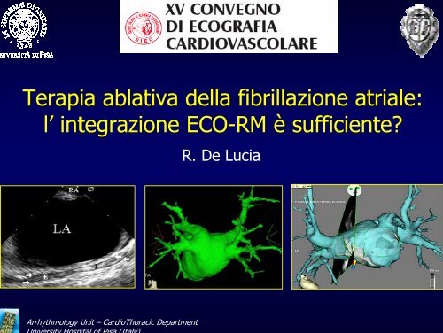
<strong>Terapia</strong> <strong>ablativa</strong> <strong>della</strong> <strong>fibrillazione</strong> <strong>atriale</strong>:<br />
l’ <strong>integrazione</strong> <strong>ECO</strong>-<strong>RM</strong> è sufficiente?<br />
R. De Lucia<br />
Arrhythmology Unit – CardioThoracic Department<br />
University Hospital of Pisa (Italy)
Ecocardiografia<br />
TEE<br />
ICE<br />
3D TEE<br />
Arrhythmology Unit – CardioThoracic Department<br />
U i it H it l f Pi (It l )
Mapping elettroanatomico<br />
L’ innovazione di tali sistemi consiste nella capacità di localizzare uno o più<br />
cateteri in uno spazio virtuale ricostruito e corrispondente alla camera<br />
cardiaca in esame, sovrapponendovi informazioni di natura elettrica.<br />
Arrhythmology Unit – CardioThoracic Department<br />
U i it H it l f Pi (It l )
Arrhythmology Unit – CardioThoracic Department<br />
U i it H it l f Pi (It l )<br />
CARTO XP
Arrhythmology Unit – CardioThoracic Department<br />
U i it H it l f Pi (It l )<br />
NavX
<strong>RM</strong> 3D Map Segmentazione<br />
1 Soglia di intensità<br />
Arrhythmology Unit – CardioThoracic Department<br />
U i it H it l f Pi (It l )<br />
3 Editing<br />
2 Segmentazione
<strong>RM</strong> 3D Map Segmentazione<br />
1 Soglia di intensità<br />
Arrhythmology Unit – CardioThoracic Department<br />
U i it H it l f Pi (It l )<br />
3 Editing<br />
2 Segmentazione
Mappaggio elettroanatomico<br />
Image Integrato<br />
MRI<br />
CT<br />
CartoSound<br />
Arrhythmology Unit – CardioThoracic Department<br />
U i it H it l f Pi (It l )
Ablazione del substrato dell’ FA<br />
STUDIO ANATOMICO<br />
PUNTURA TRANSETTALE<br />
NAVIGAZIONE ED ABLAZIONE<br />
SICUREZZA <strong>della</strong> PROCEDURA<br />
END-POINT ELETTROFISIOLOGICO e CLINICO<br />
Arrhythmology Unit – CardioThoracic Department<br />
U i it H it l f Pi (It l )
L’ <strong>integrazione</strong> <strong>ECO</strong>-<strong>RM</strong> è sufficiente a …<br />
STUDIO ANATOMICO<br />
- accurata valutazione preoperatoria?<br />
Arrhythmology Unit – CardioThoracic Department<br />
U i it H it l f Pi (It l )
Ecocardiografia<br />
STUDIO ANATOMICO<br />
TTE<br />
TEE<br />
ICE<br />
3D TEE<br />
Arrhythmology Unit – CardioThoracic Department<br />
U i it H it l f Pi (It l )
Arrhythmology Unit – CardioThoracic Department<br />
U i it H it l f Pi (It l )
Angio-<strong>RM</strong>N<br />
STUDIO ANATOMICO
“Tailored lesions”<br />
Arrhythmology Unit – CardioThoracic Department<br />
U i it H it l f Pi (It l )
Studio<br />
anatomico<br />
dell’ Atrio<br />
sinistro<br />
AP<br />
Arrhythmology Unit – CardioThoracic Department<br />
U i it H it l f Pi (It l )
Arrhythmology Unit – CardioThoracic Department<br />
U i it H it l f Pi (It l )
Arrhythmology Unit – CardioThoracic Department<br />
U i it H it l f Pi (It l )<br />
VCSS
PA<br />
VCSS<br />
Arrhythmology Unit – CardioThoracic Department<br />
U i it H it l f Pi (It l )
Studio anatomico dell’ Atrio sinistro<br />
VCSD<br />
VCSS<br />
SC<br />
Arrhythmology Unit – CardioThoracic Department<br />
U i it H it l f Pi (It l )
L’ <strong>integrazione</strong> <strong>ECO</strong>-<strong>RM</strong> è sufficiente a …<br />
STUDIO ANATOMICO<br />
- valutazione accurata preoperatoria? SI <strong>RM</strong> > <strong>ECO</strong><br />
Arrhythmology Unit – CardioThoracic Department<br />
U i it H it l f Pi (It l )
L’ <strong>integrazione</strong> <strong>ECO</strong>-<strong>RM</strong> è sufficiente a …<br />
STUDIO ANATOMICO<br />
- valutazione accurata preoperatoria?<br />
PUNTURA TRANSETTALE<br />
- guidare la procedura in sicurezza?<br />
Arrhythmology Unit – CardioThoracic Department<br />
U i it H it l f Pi (It l )
ICE nella puntura transettale<br />
“Safe approach”<br />
1) Consente una visione bidimensionale dettagliata del setto inter<strong>atriale</strong> e <strong>della</strong><br />
fossa ovale con una accuratezza del 100%, così come di eventuali anomalie<br />
strutturali a carico di questa regione 1.<br />
2) Una volta punto il SIA, consente una precoce somministrazione di eparina<br />
evitando formazioni trombotiche all’ interno <strong>della</strong> sheat transettale peraltro<br />
visualizabili con lo stesso ICE 2 .<br />
Cooper JM, Epstein LM, Use of Intracardiac Echocardiography to Guide Anlation of AF, Circulation, 2001;104:3010-3013.<br />
Klein AL. Role of transesophageal echocardiography-guided cardioversion of patients with atrial fibrillation. J Am Coll Cardiol 2001<br />
C di l I i i th M t f At i l Fib ill ti J A C ll C di l 2006
ICE nella puntura transettale<br />
Arrhythmology Unit – CardioThoracic Department<br />
University Hospital of Pisa (Italy)<br />
Hummel J Transseptal Catheterization with ICE Technology Focus 2004
Arrhythmology Unit – CardioThoracic Department<br />
University Hospital of Pisa (Italy)
<strong>RM</strong>N nella puntura transettale<br />
1) Consente lo studio preliminare dell’ anatomia cardiaca e quindi la<br />
documentazione ad es. di eventuali anomalie come l’ingrandimento<br />
<strong>atriale</strong>, la presenza di aneurismi atriali o la dilatazione dell’ aorta<br />
ascendente, oppure la pervietà del forame ovale etc…<br />
2) Non rappresenta però una metodica di imaging real-time.<br />
Cooper JM, Epstein LM, Use of Intracardiac Echocardiography to Guide Anlation of AF, Circulation, 2001;104:3010-3013.
L’ <strong>integrazione</strong> <strong>ECO</strong>-<strong>RM</strong> è sufficiente a …<br />
STUDIO ANATOMICO<br />
- valutazione accurata preoperatoria?<br />
PUNTURA TRANSETTALE<br />
- guidare la procedura in sicurezza?<br />
SI <strong>ECO</strong><br />
Arrhythmology Unit – CardioThoracic Department<br />
U i it H it l f Pi (It l )
L’ <strong>integrazione</strong> <strong>ECO</strong>-<strong>RM</strong> è sufficiente a …<br />
STUDIO ANATOMICO<br />
- valutazione accurata preoperatoria?<br />
PUNTURA TRANSETTALE<br />
- guidare la procedura in sicurezza?<br />
NAVIGAZIONE ED ABLAZIONE<br />
- rendere la navigazione comoda, affidabile e precisa?<br />
- erogare energia nel sito corretto muovendosi in modo rapido nella camera <strong>atriale</strong>?<br />
Arrhythmology Unit – CardioThoracic Department<br />
U i it H it l f Pi (It l )
Circulation, 2001<br />
ICE fornisce una visualizzazione diretta delle vene polmonari ed<br />
localizzazione <strong>della</strong> giunzione atrio-venosa.<br />
una precisa<br />
Esptein e colleghi hanno dimostrato come la localizzazione del catetere in atrio sinistro<br />
percepita mediante guida fluoroscopica differisce significativamente da quella<br />
confermata da ICE nel 25% dei casi1. ICE si è mostrato inoltre superiore alla<br />
angiografia nel visualizzare gli osti venosi polmonari 2 .<br />
Epstein LM, Comparative study of fluoroscopy, Circulation 1998;98:1796-1801<br />
A d M I t di h di h J A C ll C di l 2000
Arrhythmology Unit – CardioThoracic Department<br />
University Hospital of Pisa (Italy)
Circulation, 2001<br />
ICE può esser utilizzato per garantire un appropriato contatto dell’<br />
elettrodo e la sua stabilità, prima dell’ applicazione di energia a<br />
radiofrequenza 1 .<br />
Un buon contatto si traduce in una migliore erogazione d’energia e<br />
nella formazione di lesioni di maggiori dimensioni 2 .<br />
“ICE è in grado di fornire continue informazioni di carattere anatomico che<br />
aumentano l’accuratezza <strong>della</strong> lesioni create dal catetere riducendo<br />
considerevolmente il tempo di scopia utilizzato 3 ”<br />
Epstein LM, Comparative study of fluoroscopy, Circulation 1998;98:1796-1801<br />
Kalman JM, Biophysical characteristic of, Am Heart J. 1997;133:8-18
ICE per la navigazione<br />
Sebbene consenta un navigazione affidabile, precisa e real-time, ICE<br />
non offre immagini di tipo tridimensionale, e soprattutto non consente<br />
la gestione remota dello strumento stesso.<br />
Questo tipo di navigazione intraprocedurale può risultare pertanto più<br />
scomoda e di maggior durata temporale rispetto ad i sistemi EAMimage<br />
integrati.<br />
Arrhythmology Unit – CardioThoracic Department<br />
University Hospital of Pisa (Italy)
Studio condotto su 85 pazienti in anestesia generale.<br />
“ICE è in grado di fornire immagini di altrettanta buona qualità a<br />
supporto di tale procedura. Tuttavia esso richiede un accesso vascolare<br />
separato ed un catetere addizionale all’ interno del cuore. Risulta inoltre<br />
difficile visionare l’intera camera <strong>atriale</strong> sinistra mediante ICE”.<br />
Arrhythmology Unit – CardioThoracic Department<br />
University Hospital of Pisa (Italy)
Arrhythmology Unit – CardioThoracic Department<br />
U i it H it l f Pi (It l )<br />
Surface Registration
Statistica punti<br />
Allineamento viene definito<br />
ottimale quando tra le due<br />
mappe:<br />
• nessuno dei punti ha una<br />
distanza > 5 mm<br />
• la media <strong>della</strong> distanza dei<br />
punti risulta < 2 mm<br />
Arrhythmology Unit – CardioThoracic Department<br />
U i it H it l f Pi (It l )
Arrhythmology Unit – CardioThoracic Department<br />
U i it H it l f Pi (It l )
Arrhythmology Unit – CardioThoracic Department<br />
U i it H it l f Pi (It l )
Arrhythmology Unit – CardioThoracic Department<br />
U i it H it l f Pi (It l )
Arrhythmology Unit – CardioThoracic Department<br />
University Hospital of Pisa (Italy)
Arrhythmology Unit – CardioThoracic Department<br />
University Hospital of Pisa (Italy)
Heart Rhythm, 200<br />
“Ciascuno di questi step può produrre errori che portano ad una<br />
ricostruzione anatomica non corrispondente alla realtà”.<br />
Arrhythmology Unit – CardioThoracic Department<br />
University Hospital of Pisa (Italy)
Heart Rhythm, 200<br />
N° di punti di EAM: tale da<br />
coprire l’intero volume <strong>atriale</strong>.<br />
Contatto con la parete: la<br />
geometria <strong>della</strong> camera<br />
<strong>atriale</strong> costruita con l’EAM<br />
può rislutare distorta dalla<br />
manipolazione del catetere.
Registrazione accurata: il problema dei landmark o punti fiduciari.
Heart Rhythm, 200<br />
Movimento del paziente: altera completamente i riferimenti di navigazione.<br />
Kistler PM, Validation of three-dimensional cardiac, JCE 2006, 17:341-348
EAM Image integrato<br />
ECHO<br />
La Biosense Webster Inc ha sviluppato un nuovo catetere mappante<br />
entrato recentemente nel mercato americano.<br />
Per mezzo di un sensore di posizione montato sulla punta di un<br />
catatere per ecografia intracardiaca 2D, questo nuovo catetere è in<br />
grado di generare un volume 3D ottenuto da multiple immagini real<br />
time ecografiche 2D.<br />
Ciò potrà ovviare all’esecuzione preliminare <strong>della</strong> <strong>RM</strong> o <strong>della</strong> TC, ed agli<br />
errori durante la fase di registrazione.<br />
Arrhythmology Unit – CardioThoracic Department<br />
U i it H it l f Pi (It l )
EAM Image integrato<br />
CartoSound Biosense Webster<br />
Arrhythmology Unit – CardioThoracic Department<br />
U i it H it l f Pi (It l )
EAM Image integrato<br />
CartoSound Biosense Webster<br />
Arrhythmology Unit – CardioThoracic Department<br />
U i it H it l f Pi (It l )
Arrhythmology Unit – CardioThoracic Department<br />
U i it H it l f Pi (It l )
L’ <strong>integrazione</strong> <strong>ECO</strong>-<strong>RM</strong> è sufficiente a …<br />
STUDIO ANATOMICO<br />
- valutazione accurata preoperatoria?<br />
PUNTURA TRANSETTALE<br />
- guidare la procedura in sicurezza?<br />
NAVIGAZIONE ED ABLAZIONE<br />
- rendere la navigazione comoda, affidabile e precisa? SI <strong>RM</strong> > <strong>ECO</strong><br />
- erogare energia nel sito corretto muovendosi in modo rapido nella camera <strong>atriale</strong>?<br />
Arrhythmology Unit – CardioThoracic Department<br />
U i it H it l f Pi (It l )
L’ <strong>integrazione</strong> <strong>ECO</strong>-<strong>RM</strong> è sufficiente a …<br />
STUDIO ANATOMICO<br />
- valutazione accurata preoperatoria?<br />
PUNTURA TRANSETTALE<br />
- guidare la procedura in sicurezza?<br />
NAVIGAZIONE ED ABLAZIONE<br />
- rendere la navigazione comoda, affidabile e precisa?<br />
- erogare energia nel sito corretto muovendosi in modo rapido nella camera <strong>atriale</strong>?<br />
SICUREZZA <strong>della</strong> PROCEDURA<br />
- prevenire, rilevare precocemente e guidare il trattamento delle complicanze?<br />
Arrhythmology Unit – CardioThoracic Department<br />
U i it H it l f Pi (It l )
Arrhythmology Unit – CardioThoracic Department<br />
U i it H it l f Pi (It l )<br />
J Cardiovasc Electrophysiol, 200
L’ <strong>integrazione</strong> <strong>ECO</strong>-<strong>RM</strong> è sufficiente a …<br />
STUDIO ANATOMICO<br />
- valutazione accurata preoperatoria?<br />
PUNTURA TRANSETTALE<br />
- guidare la procedura in sicurezza?<br />
NAVIGAZIONE ED ABLAZIONE<br />
- rendere la navigazione comoda, affidabile e precisa?<br />
- erogare energia nel sito corretto muovendosi in modo rapido nella camera <strong>atriale</strong>?<br />
SICUREZZA <strong>della</strong> PROCEDURA SI <strong>ECO</strong><br />
- prevenire, rilevare precocemente e guidare il trattamento delle complicanze?<br />
Arrhythmology Unit – CardioThoracic Department<br />
U i it H it l f Pi (It l )
L’ <strong>integrazione</strong> <strong>ECO</strong>-<strong>RM</strong> è sufficiente a …<br />
STUDIO ANATOMICO<br />
- valutazione accurata preoperatoria?<br />
PUNTURA TRANSETTALE<br />
- guidare la procedura in sicurezza?<br />
NAVIGAZIONE ED ABLAZIONE<br />
- rendere la navigazione comoda, affidabile e precisa?<br />
- erogare energia nel sito corretto muovendosi in modo rapido nella camera <strong>atriale</strong>?<br />
SICUREZZA <strong>della</strong> PROCEDURA<br />
- prevenire, rilevare precocemente e guidare il trattamento delle complicanze?<br />
END-POINT ELETTROFISIOLOGICO e CLINICO<br />
- aumentare il grado di successo in termini di oucome clinico, sicurezza ed efficacia<br />
delle recidive?<br />
Arrhythmology Unit – CardioThoracic Department<br />
U i it H it l f Pi (It l )
325 pazienti sottoposti ad isolamento delle VP: (56 con tecnica Lasso; 107 sotto<br />
guida ICE; 152 sotto guida ICE con regolazione dell’ energia a RF basata sulla<br />
visualizzazione di microbolle)
PACE, 2007<br />
Outcome a 6 mesi<br />
Percentuale<br />
per gruppo<br />
Fallimento<br />
Successo<br />
completo<br />
Successo in tp<br />
antiaritmica
Arrhythmology Unit – CardioThoracic Department<br />
U i it H it l f Pi (It l )<br />
JCE October 200
L’ <strong>integrazione</strong> <strong>ECO</strong>-<strong>RM</strong> è sufficiente a …<br />
STUDIO ANATOMICO<br />
- valutazione accurata preoperatoria?<br />
PUNTURA TRANSETTALE<br />
- guidare la procedura in sicurezza?<br />
NAVIGAZIONE ED ABLAZIONE<br />
- rendere la navigazione comoda, affidabile e precisa?<br />
- erogare energia nel sito corretto muovendosi in modo rapido nella camera <strong>atriale</strong>?<br />
SICUREZZA <strong>della</strong> PROCEDURA<br />
- prevenire, rilevare precocemente e guidare il trattamento delle complicanze?<br />
END-POINT ELETTROFISIOLOGICO e CLINICO SI TC > <strong>ECO</strong><br />
- aumentare il grado di successo in termini di oucome clinico, sicurezza ed efficacia<br />
delle recidive?<br />
Arrhythmology Unit – CardioThoracic Department<br />
U i it H it l f Pi (It l )
Grazie…<br />
Arrhythmology Unit – CardioThoracic Department<br />
U i it H it l f Pi (It l )