- Page 1 and 2: PDF processed with CutePDF evaluati
- Page 3 and 4: NFKAR 2011
- Page 5 and 6: Då kör vi igång! NFKAR 2011
- Page 7 and 8: Spottkörtlarnas funktion Skydd - S
- Page 9 and 10: Parotid Space (PS) •Parotid gland
- Page 11 and 12: Anatomi Glandula Submandibularis
- Page 13 and 14: Anatomi Glandula Sublingualis • B
- Page 15 and 16: Sublingual space NFKAR 2011 T1FS+
- Page 17 and 18: Symptombild - differentialdiagnosti
- Page 19 and 20: Klinisk Presentation Persisterande
- Page 21 and 22: • 2-3 projektioner • Låg expon
- Page 23 and 24: CBCT munbotten • Sensitiv metod (
- Page 25 and 26: Sialografi-submandibularis • Caru
- Page 27 and 28: Sialografi submandibularis • 3 pr
- Page 29 and 30: Sialografi submandibularis • 3 pr
- Page 31 and 32: Sialografi submandibularis • 3 pr
- Page 33 and 34: Sialografi - Parotis • Papilla pa
- Page 35 and 36: Sialografi - Parotis • 4 projekti
- Page 37 and 38: Submentovertikal NFKAR 2011
- Page 39 and 40: • Problem: Intravenös kontrast
- Page 41 and 42: MR spottkörtlar • Expansiviteter
- Page 43 and 44: MR spottkörtlar-expansiviteter •
- Page 45 and 46: DWIBS (Diffusion-weighted Whole-bod
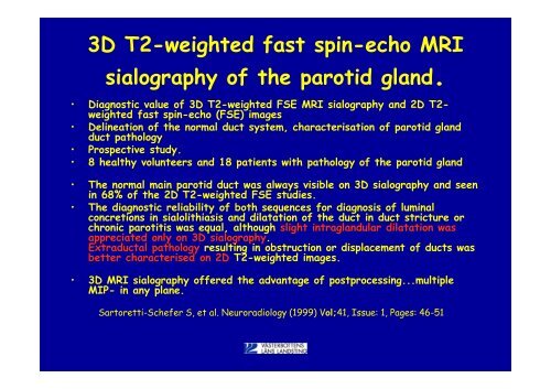
- Page 47: Teknik • 3D - CISS (Constructive
- Page 51 and 52: MR Sialografi - fördelar • Icke
- Page 53 and 54: MR sialografi • Philips Achieva 1
- Page 55 and 56: Fikadags? NFKAR 2011
- Page 57 and 58: Ultraljud • Ultraljud används in
- Page 59 and 60: B-mode Ultraljud • Riktning på e
- Page 61 and 62: Ultraljud Teknik - normalanatomi
- Page 63 and 64: Submandibularis - transversell Us-i
- Page 65 and 66: Parotis - transversell Us-image cou
- Page 67 and 68: Det är bra om man har kunskap att
- Page 69 and 70: Ultrasonography in head and neck sw
- Page 71 and 72: Dags för en tempohöjning! NFKAR 2
- Page 73 and 74: Klinisk Presentation Persisterande
- Page 75 and 76: • Icke-förkalkade konkrement •
- Page 77 and 78: Sialolith -sialografi NFKAR 2011 Se
- Page 79 and 80: • Sensitivt • Hög stråldos DT
- Page 81 and 82: • Sensitivt • Hög stråldos DT
- Page 83 and 84: sialolith1 NFKAR 2011
- Page 85 and 86: sialoith3 NFKAR 2011
- Page 87 and 88: • Konkrement- signal void • Ses
- Page 89 and 90: Dilaterade gångar - salivstas /sia
- Page 91 and 92: Mucusplugg - obstruction • Kontra
- Page 93 and 94: Sialointervention • Stenar kan av
- Page 95 and 96: NFKAR 2011 Instrument för minimalt
- Page 97 and 98: Bra att vara två som hjälps åt N
- Page 99 and 100:
Sialointervention NFKAR 2011
- Page 101 and 102:
Sialointervention NFKAR 2011
- Page 103 and 104:
Voilá NFKAR 2011
- Page 105 and 106:
• Värk o svullnad vä vid målti
- Page 107 and 108:
½årskoll NFKAR 2011 Tömning 1 mi
- Page 109 and 110:
CBCT-Sialografi NFKAR 2011 Images c
- Page 111 and 112:
Klinisk Presentation Persisterande
- Page 113 and 114:
Retrospective study of the effectiv
- Page 115 and 116:
Sjögrens syndrom NFKAR 2011
- Page 117 and 118:
Sjögrens? NFKAR 2011
- Page 119 and 120:
Eftergranskning 1år gammal DT NFKA
- Page 121 and 122:
Primärt sjögrens syndrom! NFKAR 2
- Page 123 and 124:
Kvinna 60 år • Recidiverande par
- Page 125 and 126:
Destruktiv förändring vä. Väsen
- Page 127 and 128:
Man 65 år • Man tid väs frisk f
- Page 129 and 130:
Målet är nära! NFKAR 2011
- Page 131 and 132:
Tumregel spottkörteltumörer • 8
- Page 133 and 134:
Recidiv eller körtel? • Pat op -
- Page 135 and 136:
Brunese L et al. Delayed CT enhance
- Page 137 and 138:
Man 85 år • Pleomorft adenom hö
- Page 139 and 140:
Split-bolus 90s NFKAR 2011
- Page 141 and 142:
Öl funderade över tecken på ev.
- Page 143 and 144:
85 yrs old male with thyroid cancer
- Page 145 and 146:
Habermann CR, et al. Diffusionsgewi
- Page 147 and 148:
NFKAR 2011 • DWI can differentiat
- Page 149 and 150:
CT fluoroscopy guided intervention
- Page 151 and 152:
NFKAR 2011
- Page 153 and 154:
man 26 år • Frisk, ung man som i
- Page 155 and 156:
MRI suggest tumour. DWI, ADC- map s
- Page 157 and 158:
Soft palate - value of ADC. T1 PD T
- Page 159 and 160:
• Frontal STIR visar halskörtlar
- Page 161 and 162:
AJCC- nodal classification •Struc
- Page 163 and 164:
I mål! NFKAR 2011
- Page 165 and 166:
Utan så här inspirerade! NFKAR 20
- Page 167 and 168:
Se på hela bilden! NFKAR 2011
- Page 169 and 170:
Man 50 år Tumor - annat? NFKAR 201
- Page 171 and 172:
Ranula/lymfangiom • Tid frisk man
- Page 173 and 174:
17-årig flicka Ej sublingualt! NFK
- Page 175 and 176:
Kvinna 56 år • I flera månader
- Page 177 and 178:
Kvinna 56 år MR 2½månad senare N
- Page 179 and 180:
Kvinna 35 år Ser ni något? NFKAR
- Page 181 and 182:
Kvinna 35 år Nu ser ni väl något
- Page 183 and 184:
15 yrs old girl - whartin, pleomorp
- Page 185 and 186:
Dam 75 år Vad ser ni? NFKAR 2011
- Page 187 and 188:
Dam 75 år Sten föreslås för ex,
- Page 189 and 190:
Kvinna 78 år • Sedan många år
- Page 191 and 192:
Kvinna 78 år DT NFKAR 2011
- Page 193 and 194:
60 år, man lokal svullnad NFKAR 20
- Page 195 and 196:
Man 60 år Flera körtlar, Även i
- Page 197 and 198:
55 årig kvinna • Sensibilitetsst
- Page 199 and 200:
55 årig kvinna Adenocystisk cancer
- Page 201 and 202:
60 år man. Diagnos? NFKAR 2011
- Page 203 and 204:
Kvinna 80 år Tumor - sialoadenit?
- Page 205 and 206:
• 1 år knöl under vänster mand
- Page 207 and 208:
Kvinna 90 år • Tandlös och prot
- Page 209 and 210:
Kvinna 70 år • 70-årig kvinna m
- Page 211 and 212:
Kvinna 70 år Vad ser ni? NFKAR 201
- Page 213 and 214:
Kvinna 41 år Vad ser ni? NFKAR 201
- Page 215 and 216:
Kvinna 60-års åldern • Sedan n
- Page 217 and 218:
Kvinna 60-års åldern Vä sida •
- Page 219 and 220:
Flicka 7 år Opereras för Ranula.
- Page 221 and 222:
Man 55 år Konkrement med ventileff
- Page 223 and 224:
Kvinna 48 år Diagnos? NFKAR 2011
- Page 225 and 226:
Kvinna i 50-årsåldern FNAC Juni 0
- Page 227 and 228:
Man 60 år • Man med lätt svulln
- Page 229 and 230:
Man 60 år Palpation - > DT NFKAR 2
- Page 231:
Summary •Knowledge of spatial ana