- Page 2: List of Boxes Clinical applications
- Page 6 and 7: F. A. Davis Company 1915 Arch Stree
- Page 8 and 9: To the Instructor A s the science a
- Page 10 and 11: To the Student This is your textboo
- Page 12 and 13: x To the Student We hope this textb
- Page 14 and 15: Contents CHAPTER 1 Organization and
- Page 16 and 17: xiv Contents Testes, 243 Other Horm
- Page 18 and 19: xvi Contents CHAPTER 22 An Introduc
- Page 20 and 21: CHAPTER 1 Chapter Outline Levels of
- Page 22 and 23: 4 Organization and General Plan of
- Page 24 and 25: 6 Organization and General Plan of
- Page 26 and 27: Muscular system Nervous system Skel
- Page 28 and 29: 10 Organization and General Plan of
- Page 30 and 31: 12 Organization and General Plan of
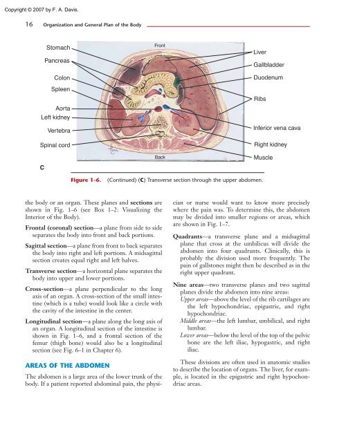
- Page 32 and 33: Table 1-3 TERMS OF LOCATION AND POS
- Page 36 and 37: 18 Organization and General Plan of
- Page 38 and 39: 20 Organization and General Plan of
- Page 40 and 41: CHAPTER 2 Chapter Outline Elements
- Page 42 and 43: 24 Some Basic Chemistry When you he
- Page 44 and 45: 26 Some Basic Chemistry Na + Cl = N
- Page 46 and 47: 28 Some Basic Chemistry A strand of
- Page 48 and 49: 30 Some Basic Chemistry BOX 2-1 BLO
- Page 50 and 51: 32 Some Basic Chemistry Figure 2-5.
- Page 52 and 53: 34 Some Basic Chemistry but the phy
- Page 54 and 55: Triglyceride Glycerol 3 Fatty acids
- Page 56 and 57: 38 Some Basic Chemistry A Amino aci
- Page 58 and 59: 40 Some Basic Chemistry Active site
- Page 60 and 61: 42 Some Basic Chemistry nucleotides
- Page 62 and 63: 44 Some Basic Chemistry 6. Acids, b
- Page 64 and 65: CHAPTER 3 Chapter Outline Cell Stru
- Page 66 and 67: 48 Cells All living organisms are m
- Page 68 and 69: 50 Cells Cilia Microvilli Golgi app
- Page 70 and 71: 52 Cells Table 3-1 Organelle Endopl
- Page 72 and 73: 54 Cells FACILITATED DIFFUSION The
- Page 74 and 75: 56 Cells pumping of the heart. Filt
- Page 76 and 77: 58 Cells Recall that in Chapter 2 y
- Page 78 and 79: 60 Cells CELL DIVISION Cell divisio
- Page 80 and 81: 62 Cells Nucleus Interphase Prophas
- Page 82 and 83: 64 Cells STUDY OUTLINE Human cells
- Page 84 and 85:
66 Cells FOR FURTHER THOUGHT 1. Ant
- Page 86 and 87:
CHAPTER 4 Chapter Outline Epithelia
- Page 88 and 89:
70 Tissues and Membranes A tissue i
- Page 90 and 91:
72 Tissues and Membranes bladder fi
- Page 92 and 93:
74 Tissues and Membranes BOX 4-1 CY
- Page 94 and 95:
76 Tissues and Membranes Cartilage
- Page 96 and 97:
78 Tissues and Membranes BOX 4-2 VI
- Page 98 and 99:
Smooth muscle Skeletal muscle Cardi
- Page 100 and 101:
82 Tissues and Membranes Neurons Ce
- Page 102 and 103:
84 Tissues and Membranes Table 4-5
- Page 104 and 105:
86 Tissues and Membranes 3. Special
- Page 106 and 107:
CHAPTER 5 Chapter Outline The Skin
- Page 108 and 109:
90 The Integumentary System The int
- Page 110 and 111:
Stratum corneum Langerhans cell Mel
- Page 112 and 113:
94 The Integumentary System Melanoc
- Page 114 and 115:
96 The Integumentary System or arre
- Page 116 and 117:
98 The Integumentary System However
- Page 118 and 119:
100 The Integumentary System Table
- Page 120 and 121:
102 The Integumentary System 6. Apo
- Page 122 and 123:
CHAPTER 6 Chapter Outline Functions
- Page 124 and 125:
106 The Skeletal System Imagine for
- Page 126 and 127:
108 The Skeletal System are attache
- Page 128 and 129:
Chondrocytes producing cartilage B
- Page 130 and 131:
112 The Skeletal System Several hor
- Page 132 and 133:
114 The Skeletal System Zygomatic a
- Page 134 and 135:
116 The Skeletal System Palatine pr
- Page 136 and 137:
Table 6-2 BONES OF THE SKULL—IMPO
- Page 138 and 139:
• 120 The Skeletal System Cervica
- Page 140 and 141:
122 The Skeletal System BOX 6-4 the
- Page 142 and 143:
• Acromial end Acromicon process
- Page 144 and 145:
Figure 6-13. Hip bones and sacrum.
- Page 146 and 147:
128 The Skeletal System Table 6-4 B
- Page 148 and 149:
Synovial membrane Bone Joint capsul
- Page 150 and 151:
132 The Skeletal System 3. Hormones
- Page 152 and 153:
134 The Skeletal System stomachs. S
- Page 154 and 155:
CHAPTER 7 Chapter Outline Muscle St
- Page 156 and 157:
138 The Muscular System Do you like
- Page 158 and 159:
140 The Muscular System motor areas
- Page 160 and 161:
142 The Muscular System is glycogen
- Page 162 and 163:
B Bundles of muscle cells Muscle ce
- Page 164 and 165:
146 The Muscular System CONTRACTION
- Page 166 and 167:
BOX 7-3 MUSCULAR DYSTROPHY Muscular
- Page 168 and 169:
150 The Muscular System Flexion Ext
- Page 170 and 171:
Trapezius Deltoid Biceps brachii Br
- Page 172 and 173:
Epicranial aponeurosis Orbicularis
- Page 174 and 175:
156 The Muscular System Deltoid Del
- Page 176 and 177:
Iliopsoas Pectineus Gluteus maximus
- Page 178 and 179:
160 The Muscular System tion of var
- Page 180 and 181:
162 The Muscular System 14. In term
- Page 182 and 183:
CHAPTER 8 Chapter Outline Nervous S
- Page 184 and 185:
166 The Nervous System Most of us c
- Page 186 and 187:
168 The Nervous System BOX 8-1 MULT
- Page 188 and 189:
170 The Nervous System TYPES OF NEU
- Page 190 and 191:
172 The Nervous System Table 8-2 St
- Page 192 and 193:
174 The Nervous System Table 8-3 MA
- Page 194 and 195:
176 The Nervous System BOX 8-3 SPIN
- Page 196 and 197:
178 The Nervous System Lateral vent
- Page 198 and 199:
180 The Nervous System In the human
- Page 200 and 201:
182 The Nervous System The auditory
- Page 202 and 203:
184 The Nervous System BOX 8-7 PARK
- Page 204 and 205:
186 The Nervous System Cranial meni
- Page 206 and 207:
188 The Nervous System Optic chiasm
- Page 208 and 209:
Sympathetic Parasympathetic Eye Cil
- Page 210 and 211:
192 The Nervous System STUDY OUTLIN
- Page 212 and 213:
194 The Nervous System 6. As tissue
- Page 214 and 215:
CHAPTER 9 Chapter Outline Sensory P
- Page 216 and 217:
198 The Senses Our senses constantl
- Page 218 and 219:
200 The Senses wood floor, concrete
- Page 220 and 221:
202 The Senses indistinct). There a
- Page 222 and 223:
204 The Senses Levator palpebrae su
- Page 224 and 225:
206 The Senses Pigment cells Choroi
- Page 226 and 227:
BOX 9-3 ERRORS OF REFRACTION Normal
- Page 228 and 229:
210 The Senses The importance of pu
- Page 230 and 231:
212 The Senses Semicircular canals
- Page 232 and 233:
214 The Senses BOX 9-5 DEAFNESS Dea
- Page 234 and 235:
216 The Senses ARTERIAL RECEPTORS T
- Page 236 and 237:
218 The Senses 7. Ciliary body (mus
- Page 238 and 239:
220 The Senses for lunch he decided
- Page 240 and 241:
CHAPTER 10 Chapter Outline Chemistr
- Page 242 and 243:
224 The Endocrine System We have al
- Page 244 and 245:
226 The Endocrine System Figure 10-
- Page 246 and 247:
228 The Endocrine System Oxytocin O
- Page 248 and 249:
230 The Endocrine System BOX 10-1 D
- Page 250 and 251:
232 The Endocrine System T and T 4
- Page 252 and 253:
234 The Endocrine System Bones Kidn
- Page 254 and 255:
236 The Endocrine System Hyperglyce
- Page 256 and 257:
238 The Endocrine System BOX 10-3 D
- Page 258 and 259:
240 The Endocrine System The functi
- Page 260 and 261:
242 The Endocrine System BOX 10-4 D
- Page 262 and 263:
244 The Endocrine System Other clai
- Page 264 and 265:
246 The Endocrine System STUDY OUTL
- Page 266 and 267:
248 The Endocrine System membrane,
- Page 268 and 269:
CHAPTER 11 Chapter Outline Characte
- Page 270 and 271:
252 Blood One of the simplest and m
- Page 272 and 273:
254 Blood BLOOD CELLS There are thr
- Page 274 and 275:
256 Blood A C B D Figure 11-3. Bloo
- Page 276 and 277:
258 Blood RBCs Circulate 120 days M
- Page 278 and 279:
A ABO blood types B Typing and cros
- Page 280 and 281:
262 Blood A normal WBC count (part
- Page 282 and 283:
264 Blood BOX 11-5 WHITE BLOOD CELL
- Page 284 and 285:
266 Blood Table 11-3 CHEMICAL CLOTT
- Page 286 and 287:
268 Blood and if it were inside a v
- Page 288 and 289:
270 Blood 4. Platelet plugs—ruptu
- Page 290 and 291:
CHAPTER 12 Chapter Outline Location
- Page 292 and 293:
274 The Heart In the embryo, the he
- Page 294 and 295:
276 The Heart Left subclavian arter
- Page 296 and 297:
278 The Heart Aorta Coronary sinus
- Page 298 and 299:
280 The Heart BOX 12-1 CORONARY ART
- Page 300 and 301:
282 The Heart BOX 12-3 ELECTROCARDI
- Page 302 and 303:
284 The Heart Table 12-2 PHYSIOLOGY
- Page 304 and 305:
286 The Heart AGING AND THE HEART T
- Page 306 and 307:
288 The Heart pulses because of the
- Page 308 and 309:
C CHAPTER 13 Chapter Outline Arteri
- Page 310 and 311:
292 The Vascular System The role of
- Page 312 and 313:
294 The Vascular System BOX 13-1 DI
- Page 314 and 315:
296 The Vascular System The amount
- Page 316 and 317:
298 The Vascular System Anterior fa
- Page 318 and 319:
300 The Vascular System Table 13-1
- Page 320 and 321:
302 The Vascular System Table 13-2
- Page 322 and 323:
304 The Vascular System BOX 13-3 PU
- Page 324 and 325:
306 The Vascular System Aorta Arter
- Page 326 and 327:
308 The Vascular System BOX 13-4 HY
- Page 328 and 329:
310 The Vascular System Adrenal gla
- Page 330 and 331:
312 The Vascular System Lung and va
- Page 332 and 333:
314 The Vascular System BOX 13-5 CI
- Page 334 and 335:
316 The Vascular System right atriu
- Page 336 and 337:
318 The Vascular System autonomic n
- Page 338 and 339:
CHAPTER 14 Chapter Outline Lymph Ly
- Page 340 and 341:
322 The Lymphatic System and Immuni
- Page 342 and 343:
324 The Lymphatic System and Immuni
- Page 344 and 345:
326 The Lymphatic System and Immuni
- Page 346 and 347:
328 The Lymphatic System and Immuni
- Page 348 and 349:
330 The Lymphatic System and Immuni
- Page 350 and 351:
332 The Lymphatic System and Immuni
- Page 352 and 353:
334 The Lymphatic System and Immuni
- Page 354 and 355:
336 The Lymphatic System and Immuni
- Page 356 and 357:
338 The Lymphatic System and Immuni
- Page 358 and 359:
340 The Lymphatic System and Immuni
- Page 360 and 361:
CHAPTER 15 Chapter Outline Division
- Page 362 and 363:
344 The Respiratory System Sometime
- Page 364 and 365:
346 The Respiratory System the aden
- Page 366 and 367:
348 The Respiratory System Frontal
- Page 368 and 369:
350 The Respiratory System A Red bl
- Page 370 and 371:
BOX 15-3 PNEUMOTHORAX Pneumothorax
- Page 372 and 373:
354 The Respiratory System Liters 6
- Page 374 and 375:
356 The Respiratory System Alveoli
- Page 376 and 377:
358 The Respiratory System Carbon d
- Page 378 and 379:
360 The Respiratory System O 2 CO2
- Page 380 and 381:
362 The Respiratory System SUMMARY
- Page 382 and 383:
364 The Respiratory System • Phys
- Page 384 and 385:
366 The Respiratory System 5. At a
- Page 386 and 387:
CHAPTER 16 Chapter Outline Division
- Page 388 and 389:
370 The Digestive System A hurried
- Page 390 and 391:
372 The Digestive System the roots
- Page 392 and 393:
374 The Digestive System Table 16-1
- Page 394 and 395:
376 The Digestive System thin layer
- Page 396 and 397:
378 The Digestive System ence of fo
- Page 398 and 399:
Inferior vena cava Right hepatic ve
- Page 400 and 401:
382 The Digestive System Hepatic du
- Page 402 and 403:
384 The Digestive System called the
- Page 404 and 405:
386 The Digestive System BOX 16-4 I
- Page 406 and 407:
388 The Digestive System The liver
- Page 408 and 409:
390 The Digestive System periodonta
- Page 410 and 411:
392 The Digestive System amino acid
- Page 412 and 413:
CHAPTER 17 Chapter Outline Body Tem
- Page 414 and 415:
396 Body Temperature and Metabolism
- Page 416 and 417:
398 Body Temperature and Metabolism
- Page 418 and 419:
400 Body Temperature and Metabolism
- Page 420 and 421:
402 Body Temperature and Metabolism
- Page 422 and 423:
404 Body Temperature and Metabolism
- Page 424 and 425:
406 Body Temperature and Metabolism
- Page 426 and 427:
408 Body Temperature and Metabolism
- Page 428 and 429:
410 Body Temperature and Metabolism
- Page 430 and 431:
Box 17-6 LEPTIN AND BODY-MASS INDEX
- Page 432 and 433:
414 Body Temperature and Metabolism
- Page 434 and 435:
416 Body Temperature and Metabolism
- Page 436 and 437:
CHAPTER 18 Chapter Outline Kidneys
- Page 438 and 439:
420 The Urinary System The first su
- Page 440 and 441:
422 The Urinary System Nephron Rena
- Page 442 and 443:
424 The Urinary System of the effer
- Page 444 and 445:
426 The Urinary System Proximal con
- Page 446 and 447:
428 The Urinary System plasma, diss
- Page 448 and 449:
430 The Urinary System Peritubular
- Page 450 and 451:
432 The Urinary System Parietal per
- Page 452 and 453:
434 The Urinary System BOX 18-5 BLO
- Page 454 and 455:
436 The Urinary System tubule that
- Page 456 and 457:
438 The Urinary System FOR FURTHER
- Page 458 and 459:
M CHAPTER 19 Chapter Outline Water
- Page 460 and 461:
442 Fluid-Electrolyte and Acid-Base
- Page 462 and 463:
444 Fluid-Electrolyte and Acid-Base
- Page 464 and 465:
446 Fluid-Electrolyte and Acid-Base
- Page 466 and 467:
448 Fluid-Electrolyte and Acid-Base
- Page 468 and 469:
450 Fluid-Electrolyte and Acid-Base
- Page 470 and 471:
452 Fluid-Electrolyte and Acid-Base
- Page 472 and 473:
CHAPTER 20 Chapter Outline Meiosis
- Page 474 and 475:
456 The Reproductive Systems The pu
- Page 476 and 477:
Stem cell Mitosis Oogonium New stem
- Page 478 and 479:
460 The Reproductive Systems Sperma
- Page 480 and 481:
462 The Reproductive Systems BOX 20
- Page 482 and 483:
464 The Reproductive Systems Fimbri
- Page 484 and 485:
466 The Reproductive Systems urethr
- Page 486 and 487:
468 The Reproductive Systems Table
- Page 488 and 489:
470 The Reproductive Systems Table
- Page 490 and 491:
472 The Reproductive Systems 3. Ute
- Page 492 and 493:
CHAPTER 21 Chapter Outline Human De
- Page 494 and 495:
476 Human Development and Genetics
- Page 496 and 497:
2 cell stage (36 hours) 4 cell stag
- Page 498 and 499:
480 Human Development and Genetics
- Page 500 and 501:
Table 21-2 GROWTH OF THE EMBRYO-FET
- Page 502 and 503:
484 Human Development and Genetics
- Page 504 and 505:
486 Human Development and Genetics
- Page 506 and 507:
488 Human Development and Genetics
- Page 508 and 509:
490 Human Development and Genetics
- Page 510 and 511:
492 Human Development and Genetics
- Page 512 and 513:
494 Human Development and Genetics
- Page 514 and 515:
CHAPTER 22 Chapter Outline Classifi
- Page 516 and 517:
498 An Introduction to Microbiology
- Page 518 and 519:
500 An Introduction to Microbiology
- Page 520 and 521:
502 An Introduction to Microbiology
- Page 522 and 523:
504 An Introduction to Microbiology
- Page 524 and 525:
506 An Introduction to Microbiology
- Page 526 and 527:
508 An Introduction to Microbiology
- Page 528 and 529:
510 An Introduction to Microbiology
- Page 530 and 531:
512 An Introduction to Microbiology
- Page 532 and 533:
514 An Introduction to Microbiology
- Page 534 and 535:
Table 22-3 DISEASES CAUSED BY BACTE
- Page 536 and 537:
Table 22-3 DISEASES CAUSED BY BACTE
- Page 538 and 539:
Table 22-4 Virus DISEASES CAUSED BY
- Page 540 and 541:
Table 22-4 Virus DISEASES CAUSED BY
- Page 542 and 543:
524 An Introduction to Microbiology
- Page 544 and 545:
526 An Introduction to Microbiology
- Page 546 and 547:
APPENDIX A Units of Measure 528
- Page 548 and 549:
APPENDIX B Abbreviations The use of
- Page 550 and 551:
APPENDIX C Normal Values for Some C
- Page 552 and 553:
APPENDIX E Eponymous Terms An epony
- Page 554 and 555:
536 Prefixes, Combining Word Roots,
- Page 556 and 557:
538 Prefixes, Combining Word Roots,
- Page 558 and 559:
540 Answers to Illustration Questio
- Page 560 and 561:
542 Answers to Illustration Questio
- Page 562 and 563:
544 Answers to Illustration Questio
- Page 564 and 565:
546 Answers to Illustration Questio
- Page 566 and 567:
548 Glossary teeth, tongue, salivar
- Page 568 and 569:
550 Glossary Anterior (an-TEER-ee-y
- Page 570 and 571:
552 Glossary Bacteria (bak-TEER-ee-
- Page 572 and 573:
554 Glossary Cartilage (KAR-ti-lidj
- Page 574 and 575:
556 Glossary Codon (KOH-don) The se
- Page 576 and 577:
558 Glossary the liver when excess
- Page 578 and 579:
560 Glossary Emulsify (e-MULL-si-fy
- Page 580 and 581:
562 Glossary cise for the colon and
- Page 582 and 583:
564 Glossary cose to glycogen to be
- Page 584 and 585:
566 Glossary Hyperglycemia (HIGH-pe
- Page 586 and 587:
568 Glossary Isotonic exercise (EYE
- Page 588 and 589:
570 Glossary pathogens, dead or dam
- Page 590 and 591:
572 Glossary Muscle tone (MUSS-uhl
- Page 592 and 593:
574 Glossary Oligodendrocyte (ah-li
- Page 594 and 595:
576 Glossary Pentose sugar (PEN-toh
- Page 596 and 597:
578 Glossary Projection (proh-JEK-s
- Page 598 and 599:
580 Glossary loop of Henle, distal
- Page 600 and 601:
582 Glossary that is a consequence
- Page 602 and 603:
584 Glossary sucrose to glucose and
- Page 604 and 605:
586 Glossary Transient flora (TRAN-
- Page 606 and 607:
588 Glossary Visceral (VISS-er-uhl)
- Page 608 and 609:
590 Index Anterior cerebral artery,
- Page 610 and 611:
592 Index Cellulose, 34 Cementum, 3
- Page 612 and 613:
594 Index Ethmoid bone, 113, 115f,
- Page 614 and 615:
596 Index Hypothermia, 400b, 402f H
- Page 616 and 617:
598 Index Motion sickness, 216b Mot
- Page 618 and 619:
600 Index Prostaglandins (PGs), 244
- Page 620 and 621:
602 Index Sub-clinical infection, 5
- Page 622:
12-2 Heart Murmur, 281 12-3 Electro