Mahidol University Annual Research Abstracts, Vol. 31
Mahidol University Annual Research Abstracts, Vol. 31
Mahidol University Annual Research Abstracts, Vol. 31

You also want an ePaper? Increase the reach of your titles
YUMPU automatically turns print PDFs into web optimized ePapers that Google loves.
<strong>Mahidol</strong> <strong>University</strong> <strong>Annual</strong> <strong>Research</strong> <strong>Abstracts</strong>, <strong>Vol</strong>. <strong>31</strong> 207<br />
EFFECT OF DIFFERENT CONCENTRATION OF<br />
COCONUT OIL, OLIVE OIL, SESAME OIL AND<br />
YEAST EXTRACT ON GROWTH OF PITYROS-<br />
PORUM SPECIES (NO. 548)<br />
Mansuang Wuthi-udomlert 1 and Omboon Luanratana 2<br />
1 Department of Microbiology, 2 Department of Pharmacognosy,<br />
Faculty of Pharmacy, <strong>Mahidol</strong> <strong>University</strong>.<br />
Key words : Pityrosporum spp., olive oil, coconut oil, sesame oil<br />
Direct examination is used to demonstrate the causative<br />
agent of pityriasis (tinea) versicolor and dandruff associated organism,<br />
Pityrosporum orbiculare (P. ovale or Malassezia furfur). In<br />
some particular cicumstance, this yeast requires a culture in which<br />
some kind of oil or supplements are substantially needed. In this<br />
study, local coconut oil (C), local sesame oil (S) and commercial<br />
olive oil (O) at 0.25% and 0.5% as well as yeast extract (Y) 2% and<br />
4% were incorporated into Sabouraud dextrose medium (SDA) to<br />
culture pure clinical isolates of Pityrosporum spp. Colonial size and<br />
characteristic were recorded macroscopically. After full growth of<br />
organisms were obtained, the length and width of yeast from Gram<br />
stain were measured and average, therefore, the cellular area were<br />
obtained. Growth enhancing ability of each medium with different<br />
concentration of oil were compared. The data concluded that using<br />
0.25% of each oil with 2% yeast extract, average width and average<br />
length of yeast cells obtained were statistically similar (p > 0.01).<br />
According to area data, local coconut oil can apparently be a substitution<br />
for conventional olive oil for culturing purpose of Pityrosporum<br />
spp.<br />
(Proceeding of The 41 th Kasetsart <strong>University</strong> <strong>Annual</strong> Conference 2003<br />
: 216-220.)<br />
INHIBITORY EFFECT OF LYOPHILIZED<br />
AQUEOUS EXTRACT OF THAI TRADITIONAL<br />
HERBS AGAINST PROPIONIBACTERIUM ACNES<br />
(NO. 549)<br />
Mansuang Wuthi-udomlert 1 , Sompop Prathanturarug 2 , and<br />
Juree Jearanaisilavong 3<br />
1 Department of Microbiology, 2 Department of Pharmaceutical<br />
Botany, Faculty of Pharmacy, 3 Department of Microbiology, Faculty<br />
of Medicine, Siriraj Hospital, <strong>Mahidol</strong> <strong>University</strong>.<br />
Key words : Propionibacterium acnes, anti-acnes, lyophilization<br />
Six Thai commercial herbs from local distributor were<br />
investigated for their efficiencies on bacteria associated in acne formation.<br />
Form volunteers, with various degree of acne, 42 strains of<br />
Propionibacterium acnes were isolated anaerobically, using modified<br />
blood agar medium. Lyopholization of aqueous extracts were<br />
obtained from Acacia concinna Wild. DC., Cardiospermum<br />
helicacabum L., Clerodendrum indicum L., Curcuma xanthorriza<br />
Roxb., Plumbagp zeylanica L. and Terminalia citrina Roxb., with<br />
20.36, 3.98, 1.86, 7.20, 1.95 and 27.58% yield (w/w), respectively.<br />
Herbal prohibition effects demonstrated as inhibitory zone diameters<br />
(IZD) by the use of agar diffusion method. Comparison with referenced<br />
drug, 10 mg per disc ampicillin, T. cirtrina exhibited statisti-<br />
cally similar effect against tested organisms (p > 0.01) as well as the<br />
effect of C. helicacabum but for different number of susceptible<br />
strains (42 and 4 strains, respectively).<br />
Active ingredient responsible to these inhibitions, local<br />
dermal irritation and possibility to formulate into external traditional<br />
drug are interesting for further study.<br />
(Proceeding of The 3 rd World Congress on Medicinal and Aromatic<br />
Plants for Human Welfare. Chiang Mai, Thailand, 2003 : 413.)<br />
ETHNOMEDICAL USES OF THAI ANNON-<br />
ACEOUS PLANTS (1) (NO. 550)<br />
Wongsatit Chuakul 1 , Noppamas Soonthornchareonnon 2<br />
1 Department of Pharmaceutical Botany, 2 Department of Pharmaceutical<br />
Pharmacognosy, Faculty of Pharmacy, <strong>Mahidol</strong> <strong>University</strong>.<br />
Key words : Ethnomedical, Thai Annonaceous Plants, Thailand.<br />
A survey on the utilization of medicinal plants at seventeen<br />
provinces; i.e. Nan. Sukhothai, Ubon Ratchathani, Si Sa Ket,<br />
Chaiyaphum, Yasothon, Nakhon Ratchasima, Surin, Saraburi,<br />
Kanchanaburi, Chanthaburi, Krabi, Surat Thani, Songkhla,<br />
Phattalung, Narathiwat and Yala were carried out by interviewing<br />
herbalists followed by collecting plant specimens and identifying<br />
the specimens. In addition, the plant specimens were compared with<br />
the authentic specimens at two herbaria: the Bangkok Herbarium<br />
(BK), Botany Section, Botany and Weed Science Division, Department<br />
of Agriculture, Ministry of Agriculture and Cooperatives and<br />
the Royal Forest Herbarium (BKF), National Park, Wildlife and Plant<br />
Conservation Department, Ministry of National Resources and Environment.<br />
Sixty-two medicinal plants of Thai annonnaceous plants<br />
were recordet from ten provinces in Thailand. Twenty-five genera<br />
of these ANNONACEAE; Alphonsea, Anaxagorea, Annona,<br />
Artabotris, Cananga, Cyathostemma, Dasymaschalon, Desmos,<br />
Ellipeia, Miliusa, Mitreflora, Monocarpia, Nervopetalum, Orophea,<br />
Polyalthia, Pseuduvaria, Rauwenhoffia, Stelechocarpus, Uvaria and<br />
Xylopia and all of these plants were ethomedicinal used.<br />
(Thai J Phytopharm 2003; 10(1): 25-32.)<br />
ETHNOMEDICAL USES OF THAI ZINGIBERA-<br />
CEOUS PLANTS (1) (NO. 551)<br />
Wongsatit Chuakul, Ampol Boonpleng<br />
Department of Pharmaceutical Botany, Faculty of Pharmacy,<br />
<strong>Mahidol</strong> <strong>University</strong><br />
Key words : Ethnomedical, Thai Zingiberaceous Plants, Thailand<br />
A survey on the utilization of medicinal plants at twentytwo<br />
provinces; i.e. Nan, Chiang Mai, Lampang, Sukhothai,<br />
Phetchabun, Chaiyaphum, Maha Sarakham, Ubon Ratchathani, Si<br />
Sa Ket, Yasothon, Surin, Buri Ram, Saraburi, Chanthaburi,<br />
Kanchanaburi, Phetchaburi, Surat Thani, Nakhon Si Thammarat,<br />
Phattalung, Trang, Krabi and Pattani were carried out by interviewing<br />
herbalists followed by collecting plant specimens and identify-
208<br />
ing the specimens. In addition, the plant specimens were compared<br />
with the authentic specimens at two herbaria: the Bangkok Herbarium<br />
(BK), Botany Section, Botany and Weed Science Division, Department<br />
of Agriculture, Minisry of Agriculture and Cooperatives and<br />
the Royal Forest Herbarium (BKF), National Park, Wildlife and Plant<br />
Conservation Department, Ministry of National Resources and Environment.<br />
Fifty-eight species of medicinal plants belonging to<br />
ZINGIBERACEAE family were recorded as medicinal plants. Twelth<br />
genera of these ZINGIBERACEAE; each each of 11 Curcuma and<br />
Zingiber, each of 8 Alpinia and Kaempferia, 6 Etlingera, 4<br />
Boesenbergia, each of 3 Amomum and Globba, 2 Elletariopsis and<br />
each of 1 Gagnepainia and Hedychium; and all of these plants were<br />
ethnomedicinal used.<br />
(Thai J Phytopharm 2003; 10(1): 33-9.)<br />
ETHNOMEDICAL USES OF THAI ORCHIDACEOUS<br />
PLANTS (NO. 552)<br />
Wongsatit Chuakul<br />
Department of Pharmaceutical Botany, Faculty of Pharmacy,<br />
<strong>Mahidol</strong> <strong>University</strong>.<br />
Key words : Ethnomedical, Thai Orchidaceous Plants, Thailand<br />
A survey on the utilization of medicinal plants at<br />
Sukhothai, Phitsanulok, Maha Sarakham, Ubon Ratchathani,<br />
Chaiyaphum, Yasothon, Surin, Kanchanaburi, Krabi, and Yala, were<br />
carried out by interviewing herbalists followed by collecting plant<br />
specimens and identifying the specimens. In addition, the plant specimens<br />
were compared with the authentic specimens at two herbaria:<br />
the Bangkok Herbarium, Botany Section, Botany and Weed Science<br />
Division, Department of Agriculture, Ministry of Agriculture and<br />
Cooperatives and the Forest Herbarium, Royal Forest Department,<br />
Ministry of Agriculture and Cooperatives. Forty-six medicinal plants<br />
of Thai Orchidaceous plants were recordet from ten provinces in<br />
Thailand. Twenty-six genera of these ORCHIDACEAE; Acampe,<br />
Apostasi, Bulbophyllum, Calanthe, Cirrhopetalum, Cleisostoma,<br />
Coelogyne, Cymbidium, Dendrobium, Doritis, Eria, Eulophia,<br />
Gastrochilus, Geodorum, Grammatophyllum, Habenaria, Luisia,<br />
Neuwiedia, Nervillia, Pecteilis, Robiquetia, Spathoglottis, Sunipia,<br />
Thrixspermum, Trias, and Vanilla and all of these plants were<br />
ethnomedicinal used.<br />
(<strong>Mahidol</strong> <strong>University</strong> Journal of Pharmaceutical Science 2002; 29<br />
(3-4), 41-5.)<br />
HIGH-FREQUENCY SHOOT MULTIPLICATION<br />
IN CURCUMA LONGA L. USING THIDIAZURON<br />
(NO. 553)<br />
Sompop Prathanturarug 1 , Noppamas Soonthornchareonnon 2 ,<br />
Wongsatit Chuakul 1 , Yuvaluk Phaidee 1 , Promjit Saralamp 1<br />
1 Department of Pharmaceutical Botany and 2 Department of Pharmacognosy,<br />
Faculty of Pharmacy, <strong>Mahidol</strong> <strong>University</strong>, E-mial<br />
:pyspr@mahidol.ac.th<br />
Key words : Curcuma longa, In vitro propagation, Thidiazuron, Turmeric<br />
Faculty of Pharmacy<br />
The effects of plant growth regulators, explant tpes, and<br />
culture regimens were investigated on in vitro shoot proliferation<br />
from terminal bud explants of Curcuma longa. Each bud was longitudinally<br />
divided into four equal pieces,each 1 cm in length, and<br />
used as explants. These were then cultured on MS medium supplemented<br />
with 18.17 mM tidiazuron for 4 weeks prior to transfer to<br />
MS medium without growth regulator for 8 weeks. Under these<br />
conditions, a shoot induction rate of 18.22+0.62 shoots/explant was<br />
obtained after 12 weeks of cultures. Spontaneous rooting was<br />
achieved. The regenerated plants were transferred to soil under<br />
greenhoust conditions and subsequently grown successfully in the<br />
field.<br />
(Acknowledgements: This work was supported by the National <strong>Research</strong><br />
Council of Thailand. Plant Cell Reports 21 (2003): 1054-<br />
1059.)<br />
IDENTIFICATION OF PUERARIA CANDOLLEI<br />
GRAHAM EXBENTH. VARIETIES USING RAPD<br />
ANALYSIS (NO. 554)<br />
Sompop Prathanturarug 1 , Noppamas Soonthornchareonnon 2 ,<br />
Wongsatit Chuakul 1 , Promjit Saralamp 1 , Takayuki Tohge 3 ,<br />
Hiroshi Sudo 3 , Mami Yamazaki 3 , Tsutomu Ishikawa 4 , Kazuki<br />
Saito 3<br />
1 Department of Pharmaceutical Botany and 2 Department of Pharmacognosy,<br />
Faculty of Pharmacy, <strong>Mahidol</strong> <strong>University</strong>, Thailand,<br />
3 Department of Molecular Biology and Biotechnology and 4 Department<br />
of Medicinal Organic Chemistry, Graduate school of<br />
Pharmaceutical Sciences, Chiba <strong>University</strong>, Japan, E-mail :<br />
pyspr@mahidol.ac.th<br />
Key words : Pueraria candollei, RAPD analysis, Metabolite profiling<br />
Kwao Keur is used in Thai traditional medicine for rejuvenation.<br />
Its tuberous root contains phytoestrogen, such as genistein,<br />
genistin, daidzein, daidzin, coumestrol, microestrol, and<br />
deoxymiroestrol. Pueraria candollei var. candollei and P. candollei<br />
var. mirifica was used as the source of Kwao Keur. Using botanical<br />
characters is difficult to separate one from another varieties. The<br />
aim of present investigation is to distinguish between P. candollei<br />
var. candollei and P. candollei var. mirifica using the random amplified<br />
polymorphic DNA (RAPD) analysis. Genetic variation among<br />
sixteen plants of P.candollei var. candollei and P. candollei var.<br />
mirifica collected from three areas of Thailand was analyzed using<br />
eleven oligonucleotide primers by RAPD method. The number of<br />
polymorphic/monomorphic bands among the pairwise combinations<br />
of the plants and the total number of bands were determined to categorize<br />
all the plants in different groups of similarity and dissimilarity<br />
at the genetic level. The procedure described also allowed the<br />
identification and discrimination of the varieties of Pueraria<br />
candollei. Furthermore, secondary metabolites of the tuberous roots<br />
of both varieties were profiled using LC-MS spectroscopy. Puerarin<br />
was identified as a major component of the tuberous root extracts.<br />
(This work was supported by the National <strong>Research</strong> Council of Thailand<br />
and NRCT-JSPS Core <strong>University</strong> Program. Presented at the<br />
sixth JSPS-NRCT Joint Seminar: Recent advances in Natural Medicine<br />
<strong>Research</strong>, December 2-6, 2003, Bangkok, Thailand.)
<strong>Mahidol</strong> <strong>University</strong> <strong>Annual</strong> <strong>Research</strong> <strong>Abstracts</strong>, <strong>Vol</strong>. <strong>31</strong> 209<br />
IN VITRO PROPAGATION OF MALLOTUS<br />
REPANDUS (WILLD.) MUELL. ARG. (NO. 555)<br />
Songsri Kaewsuwan 1,2 , Noppamas Soonthornchareonnon 2 ,<br />
Sompop Prathanturarug 1<br />
1 Department of Pharmaceutical Botany and 2 Department of Pharmacognosy,<br />
Faculty of Pharmacy, <strong>Mahidol</strong> <strong>University</strong>, E-mail :<br />
pyspr@mahidol.ac.th<br />
Key words : Mallotus repandus, in vitro propagation, medicinal<br />
plant, anti-inflamatory drug<br />
An in vitro propagation protocol has been developed for<br />
Mallotus repandus (Willd.) Muell. Arg. (Euphorbiaceae), a potential<br />
medicinal plant for anti-inflammatory drug development. Nodal<br />
segments and shoot tips from 2 month-old appearing shoots were<br />
aseptically cultured on solid Murashige and Skoog (MS) basal medium<br />
supplemented with three cytokinins alone or in combination<br />
with 1-naphthylacetic acid (NAA) to induce shoot formation for 8<br />
weeks. Of the three cytokinins tested (6-benzylaminopurine (BA),<br />
isopentenylaminopurine (2iP), and kinetin (Kin), BA induced shoot<br />
development most efficiently. The best shoot regeneration rate (3.36<br />
shoots/response explant) was obtained when the explants were cultured<br />
on MS medium supplemented with 4,44 μM BA. Addition of<br />
0.54 μM NAA to the media suppressed shoot induction rate, but<br />
induced callus formation. The regenerated shoots were cut and cultured<br />
on MS medium containing 10.74-53.71 μM NAA for rooting.<br />
The best root induction (73.08%) was revealed using MS medium<br />
supplemented with 32.23 μM NAA. Regenerated plants were successfully<br />
transferred to soil and grown under greenhouse conditions.<br />
The stem of the regenerants will be chemicall analysed to compared<br />
with their mother plants.<br />
(This work was supported by the National <strong>Research</strong> Council of Thailand.<br />
Presented at the 3rd World Congress on Medicinal and Aromatic<br />
Plants for Human Welfare, February 3-7, 2003, Chiangmai,<br />
Thailand.)<br />
MANAGENESE COMPLEXES OF CURCUMIN<br />
AND ITS DERIVATIVES : EVALUATION FOR<br />
THE RADICAL SCAVENGING ABILITY AND<br />
NEUROPROTECTIVE ACTIVITY (NO. 556)<br />
Opa Vajragupta 1 , Preecha Boonchong 1 , Hiroshi Watanabe 2 ,<br />
Michihisa Tohda 2 , Naparat Kummasud 1 , and Yaowared<br />
Sumanont 1<br />
1 Faculty fo Pharmacy, <strong>Mahidol</strong> <strong>University</strong>, Bangkok, Thailand<br />
and 2 Institute of Natural Medicine, Toyama Medical and Pharmaceutical<br />
<strong>University</strong>, Toyama, Japan<br />
Key words : Managanese complex, Superoxide dismutase mimic,<br />
2-Vesselocclusion, MPTP, Dopamine<br />
In this study, three managanese complexes of curcumin<br />
(Cp) and related compounds, diacetylcurcumin (AcyICp) and ethylenediamine<br />
derivative (CpED), were synthesized and evaluated in<br />
vitro for antilipid peroxidation and superoxide dismutase activity.<br />
The manganese complexes exhibited a great capacity to protect brain<br />
lipids against peroxidation with IC 50 of 6.3-26.3 μM. All manga-<br />
nese complexes showed much greater SOD activity than their corresponding<br />
antioxidant ligands as well as trolox with IC 50 values of<br />
8.9 - 29.9 μM. AcylCp and curcumin manganese complexes<br />
(AcylCpCpx and CpCpx) also gave the highest inhibitory activity to<br />
H 2 O 2 -induced cell damage (oxidative stress) at 0.1 μg/ml (0.2 μM)<br />
in NG108-15 cells, which were more potent than curcumin and related<br />
compounds. The neuropharmacological tests in mice supported<br />
the idea that the SOD mimicking complexes were able to penetrate<br />
to the brain as well as their role in the modulation of brain neurotransmitters<br />
under the aberrant conditions. The complexes significantly<br />
improved the learning and memory impairment induced<br />
by transient ischemic/reperfusion. AcylCpCpx, CpCpx, and<br />
CpEDCpx showed significant protection at 6.25, 25 and 50 mg/mg<br />
(i.p.), respectively, whereas managanese acetate and curcumin had<br />
no effect at doses of 50 mg/kg. In addition, treatment of AcylCpCpx<br />
and curcumin significantly attenuated MPTP-induced striatal dopamine<br />
depletion in mice, which was in accordance with the increase in<br />
the density of dopaminergic neurons when compared with MPTPtreated<br />
mice. These results support the important role of manganese<br />
in importing SOD activity and consequently, the enhancement of<br />
radical scavenging activity. AcylCpCpx and CpCpx seem to be the<br />
most promising neuroprotective agents for vascular dementia.<br />
(Free Rodical Biology & Medicine, 35(12), 1632-44, 2003.)<br />
MANGANESE-BASED COMPLEXES OF RADICAL<br />
SCAVENGERS AS NEUROPROTECTIVE AGENTS<br />
(NO. 557)<br />
Opa Vajragupta 1 , Preecha Boonchong 1 , Yaowared Sumanont 1 ,<br />
Hiroshi Watanabe 2 , Yuvadee Wongkrajanga and Naparat<br />
Kammasuda<br />
1 Faculty of Pharmacy, <strong>Mahidol</strong> <strong>University</strong>, 447 Sri-Ayudhya<br />
Road, Bangkok, Thailand, 2 Institute of Natural Medicine,<br />
Toyama Medical and Pharmaceutical <strong>University</strong>, 2630 Sugitani,<br />
Toyama 930-0194, Japan.<br />
Key words : Managese complex, Superoxide dismutase mimic, Radical<br />
scavengers<br />
Managese was incorporated in the structure of the selected<br />
antioxidants to mimic the superoxide dismutase (SOD) and to increase<br />
radical scavenging ability. Five manganese complexes (1-5)<br />
showed potent SOD activity in vitro with IC 50 of 1.18-1.84 mM and<br />
action against lipid peroxidation in vitro with IC 50 of 1.97-8.00 mM<br />
greater than their ligands and trolox. The manganese complexes<br />
were initially tested in vivo at 50 mg/kg for antagonistic activity on<br />
methamphetamine (MAP)-induced hypermotility resulting from<br />
dopamine release in the mice brain. Only manganese complexes of<br />
kojic acid (1) and 7-hydroxyflavone (3) exhibited the significant<br />
suppressions on MAP-induced hypermotility and did not significantly<br />
decrease the locomotor activity in normal condition. Manganese<br />
complex 3 also showed protective effects against learning and<br />
memory impairment in transient cerebral ischemic mice. These results<br />
supported the brain delivery and the role of manganese in SOD<br />
activity as well as in the modulation of brain neurotransmitters in<br />
the aberrant condition. Manganese complex 3 from 7-hydroxyflavone<br />
was the promising candidate for radical implicated neurodegenerative<br />
diseases.<br />
(Bioorganic & Medicinal Chemistry 2003, 11, 2329-2337.)
210<br />
CYTOPROTECTIVE AND CYTOTOXIC EFFECTS<br />
OF CURCUMIN: DUAL ACTION ON H 2 O 2 -INDUCED<br />
OXIDATIVE CELL DAMAGE IN NG 108-15 CELLS<br />
(NO. 558)<br />
Promote Mahakunakorn 1 , Michihisa Tohda 1 , Yukihisa<br />
Murakami, A Kinzo Matsumoto 1 , Hiroshi Watanabe 1 , Opa<br />
Vajaragupta 2<br />
1 Institute of Natural Medicine, Yoyama Medical and<br />
Parmaceutical <strong>University</strong>, Yoyama, Japan. 2 Faculty of Pharmacy,<br />
<strong>Mahidol</strong> <strong>University</strong>, Bangkok, Thailand.<br />
Key words : Manganese complex, Superoxide dismutase mimic,<br />
Radical scavengers<br />
The ability of curcumin, a natural antioxidant isolate from<br />
Curcuma longa, to inhibit hydrogen peroxide (H 2 -O 2 )-induced cell<br />
damage in NG 108-15 cells was examined. When added simultaneously<br />
with 500 mM H 2 -O 2 , curcumin (25-100 mM) effectively<br />
protected cells from oxidative damage. However, when the cells<br />
were pretreated with curcumin (25-100 mM) for 1.5 h before H 2 O 2<br />
exposure, curcumin was unable to inhibit H 2 O 2 -induced cell damage.<br />
Instead, it caused a significant concentration-dependent decrease<br />
in cell viability after H 2 O 2 exposure. This dual action of<br />
curcumin suggests that pretreatment with curcumin by itself did not<br />
have any significant effect on the viability of the NG 108-15 cells,<br />
but it sensitized them to oxidative damage induced by H 2 O 2 under<br />
our experimental conditions. It appears that these events may not<br />
relate to the antioxidant and free radical scavenging activities of<br />
curcumin.<br />
(Biol. Pharm Bull 26(5), 725-28, 2003.)<br />
SIMULTANEOUS DETERMINATION OF<br />
PARACETAMOL AND CHLORPHENIRAMINE<br />
MALEATE BY MICELLAR ELECTROKINETIC<br />
CHROMATOGRAPHY (NO. 559)<br />
Leena Suntornsuk 1 , Ongart Pipitharome 1 , Prapin Wilairat 2<br />
1 Department of Pharmaceutical Chemistry, Faculty of Pharmacy,<br />
2 Department of Chemistry, Faculty of Science, <strong>Mahidol</strong> <strong>University</strong>,<br />
Bangkok 10400, Thailand (pyll@mahidol.ac.th).<br />
Key words : micellar electrokinetic chromatography (MEKC),<br />
paracetamol (PARA), chlorpheniramine maleate (CPM), cold remedy<br />
ingredients<br />
A micella electrokinetic chromatography (MEKC) method<br />
was established for determination of paracetamol (PARA) and<br />
chlorpheniramine maleate (CPM) in cold tablets. Separation of both<br />
drugs as well as other seven cold remedy ingredients was achieved<br />
in 25.5 min using a sodium dihydrogenphosphate-sodium tetraborate<br />
buffer (10 mM, pH 9.0) containing sodium dodecyl sulfate (SDS)<br />
(50 mM) and acetonitrile (26% v/v). The effective capillary length<br />
of 50 cm, the separating voltage of 15 kV and the temperature of<br />
30 o C was optimized. Detection was by a diode array detector at 214<br />
nm. Method linerity was excellent (r 2 > 0.999) over the concentration<br />
tested (10-250 μg/mL) with good precision and accuracy. Recoveries<br />
were good (> 99%) with limits of detection of 0.4 and 0.5<br />
μg/mL and limits of quantitation of 2 (%RSD = 3.1%) and 4 (%RSD<br />
= 2.4%) μg/mL, for PARA and CPM, respectively. The developed<br />
method was applied for determination of ingredients in cold tablets<br />
and was found to be simple, rapid and efficient.<br />
(J Pharm. Biomed. Anal 2003, 33:441-449.)<br />
METHOD DEVELOPMENT FOR SEPARATION<br />
OF ACTIVE INGREDIENTS IN COLD MEDICINES<br />
BY MICELLAR ELECTROKINETIC CHROMATO-<br />
GRAPHY (NO. 560)<br />
Leena Suntornsuk 1 , Ongart Pipitharome 1 , Prapin Wilairat 2<br />
1 Department of Pharmaceutical Chemistry, Faculty of Pharmacy,<br />
<strong>Mahidol</strong> <strong>University</strong>, 447 Sri-Ayudhaya Rd., Rajathevee; 2 Department<br />
of Chemistry, Faculty of Science, <strong>Mahidol</strong> <strong>University</strong>, Rama<br />
VI Rd., Rajathevee, Bangkok 10400, Thailand (pylll@<br />
mahidol.ac.th)<br />
Key words : cold medicine ingredients, micellar electrokinetic chromatography<br />
(MEKC)<br />
Separation of nine commonly used active ingredients in<br />
cold medicines, were demonstrated by micellar electrokinetic chromatography.<br />
The ingredients included paracetamol, chlorpheniramine<br />
maleate, diphenhydramine hydrochloride, triprolidine hydrochloride,<br />
phenlpropanolamine hydrochloride, dextromethorphan<br />
hydrobromide, loratadine, aspirin and caffeine. Effects of buffer<br />
concentrations, pH, organic modifiers and capillary length were investigated.<br />
The optimum conditions were achieved in 10 mM sodium<br />
dihydrogenphosphate-sodium tetraborate buffer, pH 9.0, containing<br />
50 mM sodium dodecyl sulfate and 28% v/v acetonitrile using<br />
the effective length of 50 cm, the separating voltage of +15 kV<br />
and the capillary temperature of 30 o C. Separation of all peaks was<br />
obtained within 28.4 min with a resolution of 1.2.<br />
(Sci. Pharm. 2003, 71: 235-250.)<br />
Faculty of Pharmacy<br />
QUANTITATIVE ANALYSIS OF AGLYCONE<br />
QUERCETIN IN MULBERRY LEAVE (MORUS<br />
ALBA I.) BY CAPILLARY ZONE ELECTRO-<br />
PHORESIS (NO. 561)<br />
Leena Suntornsuk 1 , Saowapak Kasemsook 2 , Surapote Wongyai 2<br />
1 Department of Pharmaceutical Chemistry, Faculty of Pharmacy,<br />
<strong>Mahidol</strong> <strong>University</strong>, Bangkok, Thailand; 2 Faculty of Pharmacy,<br />
Rangsit <strong>University</strong>, Bangkok, Thailand. (pylll@mahidol.ac.th)<br />
Key words : Aglycone queretin, capillary electrophoresis, rutin<br />
A capillary zone electrophoresis method was established<br />
for analysis of aglycone quercetin in mulberry leaves (Morus alba<br />
L.). Influence of background electrolyte concentrations and pHs,<br />
surfactant concentrations, organic solvents, oven temperature and<br />
voltage on the separation of aglycone quercetin, other flavonoids<br />
with related structures and a phenolic acid, were systematically investigated.<br />
The optimum condition providing baseline separation<br />
of all compounds within 16.5 min was obtained in 150 mM boric<br />
acid (pH 10.0) using a fused silica capillary with an effective length<br />
of 42.5 cm (50 μm, inner diamete), oven temperature 0f 32 o C and
<strong>Mahidol</strong> <strong>University</strong> <strong>Annual</strong> <strong>Research</strong> <strong>Abstracts</strong>, <strong>Vol</strong>. <strong>31</strong> 211<br />
applied voltage of 15 kV. Linearity of the method was excellent (r 2<br />
> 0.999) over the concentration tested (40-160 μg/mL). The relative<br />
standard deviations (%RSDs) from injection, intraday and inter-day<br />
precision using rutin as an internal standard were less than 2.5%.<br />
Recoveries were good (= 100.0%, %RSD = 0.8%) with a limit of<br />
detection and limit of quantitation of 0.86 and 3.16 μg/mL (%RSD<br />
= 1.8%), respectively. The aglycone quercetin found in the mulberry<br />
leaves was 0.194 g/100 g (%RSD = 0.9%) on dry weight.<br />
(Electrophoresis 2003, 24: 1236-1241. 3 rd International Symposium<br />
on Separation in the BioScience 5-8 May 2003, Moscow, Russia.)<br />
FEATHER DEGRADATION BY BACILLUS SP.<br />
FS 46 IN SUBMERGED CULTIVATION (NO. 562)<br />
Worapot Suntornsuk 1 , Leena Suntornsuk 2<br />
1 Department of Microbiology, Faculty of Science, King Mongkut’s<br />
<strong>University</strong> of Technology Thonburi, Bangkok 10140, Thailand;<br />
2 Department of Pharmaceutical Chemistry, Faculty of Pharmacy,<br />
<strong>Mahidol</strong> <strong>University</strong>, Bangkok 10400, Thailand.<br />
Key words : Feather; Biodegradations; Bacillus sp.; Keratinase;<br />
Animal feed<br />
Cultivation conditions affecting feather degradation by<br />
Bacillus sp. FK 46 were investigated. The results showed that feather<br />
was almost completely degraded under the following conditions :<br />
1% whole chicken feather as a substrate at the initial medium pH of<br />
9 with 5% bacterial inoculum, at a temperature of 37 o C and a shaking<br />
speed of 250 rev/min. Glucose, methanol, Tween 80 and Triton<br />
X-100, however, had no effect on feather degradation. After feather<br />
was degraded, its residue and fermented broth would become a protein<br />
feed for animals.<br />
(Bioresouce Tech. 2002, 86 : 239-243.)<br />
ANTI-VENOM POTENTIAL OF BUTANOLIC-<br />
EXTRACT OF ECLIPTA PROSTRATA AGAINST<br />
MALAYAN PIT VIPER VENOM (NO. 563)<br />
Pimolpan Pithayanukul 1 , Sasitorn Laovachirasuwan 1 , Rapepol<br />
Bavovada 2 , Narumol Pakmanee 3 , Rutt Suttisri 2<br />
1 Faculty of Pharmacy, <strong>Mahidol</strong> <strong>University</strong>, Bangkok, Thailand;<br />
2 Faculty of Pahrmaceutical Sciences, Chulalongkorn <strong>University</strong>,<br />
Bangkok, Thailand. 3 Queen Saovabha Memorial Institute, Thai<br />
Red Cross Society, Bangkok, Thailand.<br />
Key words : Eclipta prostrata; Malayan Pti viper; Anti-snake venom<br />
The butanolic and purified butanolic extracts of Eclipta<br />
prostrata were evaluated for their anti-venom potential. Inhibition<br />
of letha, hemorrhagic, proteolytic, and phospholipase A 2 activites of<br />
Calloselasma rhodostoma (Malayan pit viper, MPV) venom by these<br />
extracts were determined. Demethylwedelolactone was indentified<br />
as their major constituent. The butanolic extract, at 2.5 mg/mouse,<br />
was able to completely neutralize teh lethal activity of 2LD 50 of<br />
MPV venom, but increasing the dose diminished the effect. The<br />
purified butanolic extract, at 1.5-4.5 mg/mouse,was able to neutralize<br />
the lethality o the venom at around 50-58%. Both extracts par-<br />
tially inhibited the hemorrhagic activity but displayed very low antiphospholipase<br />
A 2 activity and did not inhibit proteolytic activity of<br />
MPV vemon.<br />
(Journal of Ethnopharmacology 2004, 90(2-3): 347-352.)<br />
VARIATION OF ANTHRAQUINONE CONTENT<br />
IN SENNA SIAMEA (LAM.) IRWIN & BARNEBY<br />
LEAVES (NO. 564)<br />
Wandee Gritsanapan and Somsak Nualkaew<br />
Department of Pharmacognosy, Faculty of Pharmacy, <strong>Mahidol</strong><br />
<strong>University</strong>. E-mail : pywgs@mahidol.ac.th<br />
Key words : Senna siamea, Cassia siamea, Anthraquinone content<br />
Senna siamea (Lam.) Irwin & Barneby or Cassia siamea<br />
Lamk. which is a native plant of South East Asia is widely cultivated<br />
in all parts of Thailand. The young leaves and flowers of this<br />
plant were used to be recommended as a bitter tonic, mild laxative<br />
drug and sleeping aid. A sleeping aid action is due to CNS depressant<br />
activity of a major component, barakol while a laxative effect is<br />
due to anthraquinone components. Since glycosides of anthraquinones<br />
in plants are reported to be the active form of laxative action,<br />
plants containing higher yield of anthraquinone glycosides should<br />
have a better laxative potency. Thus, quantitative analysis of anthraquinone<br />
content in the leaves of S. siamea collected from different<br />
geographical areas of Thailand in all seasons were studied. By<br />
UV-vis spectrophotometric method, the content of total anthraquinones<br />
in the leaves collected from northern, northeastern, central and<br />
southern parts of Thailand in summer, rainy season and winter were<br />
found to be 0.07 - 0.20% while the content of total anthraquinone<br />
glycosides were 0.06-0.12% dry weight. The sample containing highest<br />
total anthraquinone content (0.20%) was the northern sample collected<br />
in summer while the highest total anthraquinone glycosides<br />
content (0.12%) was the central-winter sample. It was shown that<br />
total anthraquinone glycoside content in the leaves of S. siamea growing<br />
in all parts of Thailand was high in winter and summer. So,<br />
collection of the leaves of this plant for laxative drug should be done<br />
in winter and summer. However, the variation of both total anthraquinones<br />
and total anthraquinone glycosides in the leaves collected<br />
from several areas and seasons were not significantly different<br />
(p < 0.05). Due to anthraquinone glycoside content, S. siamea<br />
leaf is not a good source for anthraquinone laxative drug.<br />
(The research was partially granted by Plant Genetic Conservation<br />
Project, The Royal Chitralada Project, Dusit Palace. Proceeding<br />
Book of a Seminar on Thai Natural Resources : Nature of Life,<br />
Bangkok, 10-12 May 2003.)<br />
ANTIOXIDANT ACTIVITY OF AZADIRACHTA<br />
INDICA (NO. 565)<br />
Pongtip Sithisarn 1 , Rungtawan Supabphol 2 , and Wandee<br />
Gritsanapan 1<br />
1 Department of Pharmacognosy, Faculty of Pharmacy, <strong>Mahidol</strong><br />
<strong>University</strong>; 2 Department of Physiology, Faculty of Medicine,<br />
Srinakarintarawirot <strong>University</strong>. E-mail : pywgs@mahidol.ac.th
212<br />
Key words : Antioxidant activity, Azadirachta indica, Siamese neem<br />
tree<br />
Siamese neem tree (Azadirachta indica A. Juss var.<br />
siamensis Valeton, Meliaceae) is a tropical plant found in several<br />
countries such as Southern Laos, Western Cambodia and also in<br />
Thailand. There are many reports concerning its medicinal properties<br />
such as antipyretic, antidiarrhea, insecticidal and antioxidant<br />
activities. Antoixidant activity of the bark extract of this plant compared<br />
to standard antioxidant was reported to be high (TEAC =<br />
0.9130). It was also mentioned about cancer chemopreventive components<br />
of Siamese neem tree flowers and antioxidant activity of the<br />
seed extract. Thus, antioxidant activity of several parts of this plant<br />
are interesting to be investigated for more details. Several parts of<br />
Siamese need tree i.e. leaf, flower, ripe fruit rind, ripe fruit flesh, raw<br />
fruit, seed and stem bark were studied for antioxidant activity using<br />
DPPH assay. It was found that aqueous leaf extract,ethanol flower<br />
extract and ethanol stem bark extract showed quite high activities<br />
(EC 50 = 26.48, 27.91 and 30.55 μg/ml, respectively) but when using<br />
ABTS-metmyoglogin assay, the activities at 1 mg/ml concentration<br />
of the extracts were shown to be 0.9592, 0.9881 and 1.0640 mmol/<br />
L, respectively. The leaf samples are easy for collection in all seasons<br />
while the flower and bark samples can be collected in some<br />
seasons only. So, active components from the leaves of Siamese<br />
neem tree would be further separated and identified.<br />
(The research was partially granted by National Center for Genetic<br />
Engineering and Biotechnology (BIOTEC). Proceeding Book of<br />
Seminar on Thai Natural Resources : Nature of Life, Bangkok, 10-<br />
12 May 2003.)<br />
COMPARISON OF ANTIOXIDANT ACTIVITY OF<br />
TWO VARIETIES OF AZADIRACHTA INDICA<br />
(NO. 566)<br />
Pongtip Sithisarn 1 , Rungtawan Supabphol 2 , and Wandee<br />
Gritsanapan 1<br />
1 Department of Pharmacognosy, Faculty of Pharmacy, <strong>Mahidol</strong><br />
Univesity, 2 Department of Physiology, Faculty of Medicine,<br />
Srinakarintarawirot <strong>University</strong>. E-mail : pywgs@mahidol.ac.th<br />
Key words : Siamese neem tree, neem, antioxidant activity<br />
Three parts of neem and Siamese need tree i.e. leaf, flower<br />
and stem bark were extracted and tested for antioxidant activity using<br />
DPPH scavenging assay and total antioxidant activity determination.<br />
It was found that EC 50 of antioxidant activities tested by<br />
DPPH scavenging assay from leaf aqueous extract, flower ethanol<br />
extract and stem bark ethanol extract of Siamese neem tree were<br />
26.48, 27.91 and 30.55 μg/mL, respectively while the results from<br />
neem were 104.73, 89.04 and 11.05 μg/mL, respectively. The total<br />
antioxidant activities of leaf aqueous extract, flower ethanol extract<br />
and stem bark ethanol extract of Siamese neem tree tested by total<br />
antioxidant activity determination were 0.9592, 0.9881 and 1.0640<br />
mmol/L, respectively while the results from neem were 0.8122,<br />
0.6793 and 1.2<strong>31</strong>8 mmol/L, respectively.<br />
(The research was partially granted by National Center for Genetic<br />
Engineering and Biotechnology (BIOTEC). Proceeding Book of the<br />
3 rd Indochina Conference on Pharmaceutical Science III Bangkok,<br />
20-23 May 2003.)<br />
Faculty of Pharmacy<br />
PHENOLIC COMPONENTS FROM MELODORUM<br />
SIAMENSE (NO. 567)<br />
Junya Intaranongpai 1 and Wandee Gritsanapan 2<br />
1 Pharmaceutical Chemistry and Phytochemistry Graduate<br />
Programme, Faculty of Pharmacy, <strong>Mahidol</strong> <strong>University</strong>, Bangkok,<br />
Thailand; 2 Department of Pharmacognosy, Faculty of Pharmacy,<br />
<strong>Mahidol</strong> <strong>University</strong>, Bangkok, Thailand. E-mail : pywgs@<br />
mahidol.ac.th<br />
Key words : Melodorum siamense, Annonaceae, Nom maeo<br />
Melodorum siamense Scheff. (Rauwenhoffia siamensis<br />
Scheff., Annonanceae) which is called in Thai as Nom maeo (central<br />
is a climber usually found in the central part of Thailand (1-5). Its<br />
flowers have been traditionally used for cardiotonic agent while the<br />
wood is used as antipyretics (6). This plant is botanically related to<br />
M. fruiticosum Lour. which its leaves and branches were reported to<br />
contain butenolides with cytotoxic activity in several tumor cell lines<br />
(4). There is no report about chemical constituent and biological<br />
activity of M. siamense before. Thus, the aim of this study is to<br />
investigate the chemical constituents of the wood and stem bark of<br />
this plant.<br />
By column chromatographic technique, two phenolic<br />
compounds which are 1-(3,4,5-trimethoxy benzene)-1-methoxy-1ethylene-2-benzoate<br />
ester and 1,2-bezenedi carboxylic acid bis(5methylheptyl)ester<br />
were isolated from the n-hexane extract of the<br />
wood and ethyl acetate extract of the stem bark, respectively. The<br />
structure elucidations of the compounds were done based on spectroscopic<br />
and reported data. The compounds should be further investigated<br />
for cytotoxic activity to compare with reported activity of<br />
M. fruiticosum.<br />
(Proceeding Book of the 3 rd Indochina Conference on Pharmaceutical<br />
Science III Bangkok, 20-23 May 2003.)<br />
ANTIOXIDATIVE ACTIVITY OF EXTRACTS<br />
FROM NEEM AND SIAMESE NEEM LEAF,<br />
FLOWER AND STEM BARK (NO. 568)<br />
Pongtip Sithisarn 1 , Rungtawan Supabphol 2 , and Wandee<br />
Gritsanapan 1<br />
1 Department of Pharmacognosy, Faculty of Pharmacy, <strong>Mahidol</strong><br />
<strong>University</strong>; 2 Department of Physiology, Faculty of Medicine,<br />
Srinakarintarawirot <strong>University</strong>. E-mail : pywgs@mahidol.ac.th<br />
Key words : antioxidative activity, Neem, Siamense neem<br />
Neem (Azadirachta indica A. Juss var. indica) and<br />
Siamese neem tree (Azadirachta indica A. Juss var. siamensis) are<br />
plants in Meliaceae family. Siamese neem tree is usually found in<br />
Thailand, Southern Lao and Western Cambodia while neem is always<br />
found in India, Pakistan, Indonesia, Africa and also Thailand.<br />
There are many reports indicating their biological activities such as<br />
antipyretic, antimalarial, cancer chemopreventive, insecticidal and<br />
also antioxidant activities. Because of their close taxonomic relationship,<br />
so the comparison of their antioxidant activities is interesting<br />
for investigation.
<strong>Mahidol</strong> <strong>University</strong> <strong>Annual</strong> <strong>Research</strong> <strong>Abstracts</strong>, <strong>Vol</strong>. <strong>31</strong> 213<br />
Three parts of neem and Siamese neem tree i.e. leaf, flower<br />
and stem bark were extracted and tested for antioxidant activities<br />
using DPPH assay. It was found that EC 50 of antioxidant activities<br />
from leaf aqueous extract, flower ethanol extract and stem bark ethanol<br />
extract of Siamese neem tree were 26.48, 27.91 and 30.55 μg/<br />
mL, respectively while the results from neem were 104.73, 89.04<br />
and 11.05 μg/mL, respectively.<br />
The results show that all extracts from Siamese neem tree<br />
have quite high antioxidant activities while neem extracts show lower<br />
activities except the stem bark ethanol extract. From these results,<br />
antioxidant activity of both plants should be confirmed by other<br />
methods. Separation, purification and identification of active components<br />
should be further investigated.<br />
(The research was partially granted by National Center for Genetic<br />
Engineering and Biotechnology (BIOTEC). Presented at RGJ Seminar<br />
Series XXIV : Pharmaceutics and Pharmaceutical Technology,<br />
Bangkok, 15-16 October, 2003.)<br />
COMPARISON OF ANTIOXIDANT ACTIVITIES<br />
OF THE LEAVES AND FLOWERS OF SIAMESE<br />
NEEM TREE (NO. 569)<br />
Pongtip Sithisarn 1 , Wandee Gritsanapan 1 , and Rungtawan<br />
Supabphol 2<br />
1 Department of Pharmacognosy, Faculty of Pharmacy, <strong>Mahidol</strong><br />
<strong>University</strong>, 2 Department of Physiology, Faculty of Medicine,<br />
Srinakarintarawirot <strong>University</strong>.<br />
Key words : antioxidant, antioxidant activity, Siamense Neem Tree,<br />
Azadirachta indica<br />
Siamese neem tree (Azadirachta A. Juss var. siamensis<br />
Valeton) is the plant that young leaves and flowers have been popularly<br />
used as vegetables of bitter tonic. The leaf aqueous and flower<br />
ethanol extracts of Siamese neem tree were tested for free radical<br />
scavenging activity using DPPH scavenging assay, total antioxidant<br />
activity determination and confirmed for antioxidant activity on lipid<br />
peroxidation in bronchogenic cancer cell culture using TBARS<br />
method. The results of DPPH scavenging assay showed that leaf<br />
aqueous and flower ethanol extracts exhibited antioxidant activities<br />
with EC 50 of 26.48 + 3.63 and 27.91 + 2.73 μg/ml, respectively while<br />
their antioxidant activities at 1 mg/ml were 0.9592 and 0.9881 mmol/<br />
l when tested by total antioxidant activity determination. On bronchogenic<br />
cancer cell culture, treatment of both extracts at concentration<br />
100 μg/ml could decrease MDA level significantly from 148.72%<br />
to 100.97% and from 148.72 to 105.53%, respectively. The results<br />
show that both leaves and flowers exhibit high antioxidant activity.<br />
So, consuming these parts of Siamese neem tree as vegetables should<br />
be beneficial for the body and confirm traditional use of this plant as<br />
bitter tonic.<br />
(The research was partially granted by National Center for Genetic<br />
Engineering and Biotechnology (BIOTEC). Presented at The 6 th<br />
JSPS-NRCT Joint Seminar “Resent Advances in Natural Medicine<br />
<strong>Research</strong>” Bangkok, 2-4 December 2003.)<br />
ACUTE TOXICITY OF A THAI TRADITIONAL<br />
PREPARATION : PRASAPLAI (NO. 570)<br />
Somsak Nualkaew 1 , Wandee Gritsanapan 1 , and Chongkol<br />
Tiangda 2<br />
1 Department of Pharmacognosy, Faculty of Pharmacy, <strong>Mahidol</strong><br />
<strong>University</strong>, 2 Department of Pharmacology, Faculty of Pharmacy,<br />
<strong>Mahidol</strong> <strong>University</strong>. E-mail : pywgs@mahidol.ac.th<br />
Key words : Prasaplai, traditional medicine, acute toxicity<br />
Administration of the water suspension of the Prasaplai<br />
preparation, a Thai traditional medicine, up to 2.5 g/kg and the water<br />
extract of up to 20 g/kg (calculated as powdered drug) which was<br />
the maximum dose for rats, did not produce any signs and symptoms<br />
of toxicity in rats during the first 24 hrs. and no rats died at these<br />
dose levels. After observation for a further 14 days, no toxic symtom<br />
nor dead rats were observed.<br />
(The research was partially granted by The Royal Golden Jubilee<br />
Ph.D. Program, The Thailand <strong>Research</strong> Fund. Presented at RGJ<br />
Seminar Series XXIV : Pharmaceutics and Pharmaceutical Technology,<br />
Bangkok, 15-16 Octover, 2003.)<br />
ANTISPASMODIC EFFECT OF A THAI<br />
TRADITIONAL PREPARATION : PRASAPLAI<br />
(NO. 571)<br />
Somsak Nualkaew 1 , Wandee Gritsanapan 1 and Chongkol<br />
Tiangda 2<br />
1 Department of Pharmacognosy, Faculty of Pharmacy, <strong>Mahidol</strong><br />
<strong>University</strong>, 2 Department of Pharmacology, Faculty of Pharmacy,<br />
<strong>Mahidol</strong> <strong>University</strong>. E-mail : pywgs@mahidol.ac.th<br />
Key words : Antispasmodic effect, Prasaplai, Dysmenorrhea<br />
The aqueous extract of Prasaplai, a Thai traditional medicine<br />
was studied on the isolated rat uterus in the estrous stage. The<br />
uterine contraction was induced by different agonists. The results<br />
showed that the aqueous extract of Prasaplai could inhibit the contraction<br />
of uterine muscle induced by submaximal doses of acetylcholine<br />
(2.04 x 10 -4 mg/ml), oxytocin (1.54 x 10 -4 mg/ml) and prostaglandin<br />
E 2 (PGE 2 ) (6.00 x 10 -4 mg/ml). The inhibition was concentration-dependent<br />
and reversible by tissue washing. The IC 50 of<br />
Prasplai extract expressed as milligram of powdered preparation per<br />
ml of perfusion solution for acetylcholine, oxytocin and PGE 2 were<br />
11.70 mg/ml 10.00 mg/ml and 5.75 mg/ml, respectively. These data<br />
suggest that the Prasaplai has an antispasmodic effect against uterine<br />
contraction with various mechanisms and corroborate the traditional<br />
use in the treatment of dysmenorrhea.<br />
(The research was partially granted by The Royal Golden Jubilee<br />
Ph.D. Program, The Thailand <strong>Research</strong> Fund. Presented at The 6 th<br />
JSPS-NRCT Joint Seminar “Resent Advances in Natural Medicine<br />
<strong>Research</strong>” Bangkok, 2-4 December 2003.)
214<br />
PHYTOCHEMISTRY AND ANTI-MALARIAL,<br />
ACTIVITY OF EUPATORIUM ODORATUM L.<br />
(NO. 572)<br />
Rungnapa Ongkana 1 , Peerapan Tan-ariya 2 , Weena<br />
Jiratchariyakul 1 and Aimon Somanabandhu 1<br />
1 Department of Pharmacognosy, Faculty of Pharmacy, <strong>Mahidol</strong><br />
<strong>University</strong>, 2 Department of Microbiology, Faculty of Science,<br />
<strong>Mahidol</strong> <strong>University</strong>. E-mail : pyasm@mahidol.ac.th<br />
Key words : Eupatorium odoratum, antimalarial constituents<br />
Eupatorium odoratum L. is a common scadent shrub, native<br />
to central and tropical South America as well as to Southeast<br />
Asia. In a preliminary study, Eupatorium odoratum L., a plant traditionally<br />
used in Thailand to treat fever and malaria, was found to<br />
have in vitro antiplasmodial property against Plasmodium falciparum.<br />
the present study is designed to search for active compounds and to<br />
confirm the anti-malarial activity of this plant. The leaves of Eupatorium<br />
odoratum L. were collected from roadsides in Ratchaburi.<br />
The chloroform extract was found to be active against P. falciparum<br />
with IC 50 value of 9 μg/ml. Column fractionation of chloroform<br />
extract gave six fractions, three of which were found to be active.<br />
The compounds isolated from active fraction(s) were identified using<br />
spectroscopic techniques and were further tested for their antimalarial<br />
activity.<br />
(The research was partially supported by a research grant from<br />
<strong>Mahidol</strong> <strong>University</strong>. Presented at Joint International Tropical Medicine<br />
Meeting 2002 “The 8 th Chamlong-Tranakchit Harinasuta Lecture”<br />
Montien Riverside Hotel, Bangkok, Thailand, 20-22 November<br />
2002.)<br />
PHYTOCHEMICAL AND BIOLOGICAL ACTIVITY<br />
OF ANOMIANTHUS DULCIS STEM BARK. (NO. 573)<br />
Lersak Ubonopas 1 , Noppamas Soonthornchareonnon 1 , Khanit<br />
Suwanborirux 2 , Prapai Wongsinkongman 3 , Wongsatit Chuakul 4<br />
1 Department of Pharmacognosy, Faculty of Pharmacy, <strong>Mahidol</strong><br />
<strong>University</strong>, Bangkok 10400, Thailand; 2 Department of Pharmacognosy,<br />
Faculty of Pharmaceutical Sciences, Chulalongkorn <strong>University</strong>,<br />
Bangkok 10330, Thailand; 3 Department of Medical Sciences,<br />
Institute of Medicinal Plant <strong>Research</strong>, Ministry of Public<br />
Health, Nondhaburi, 11000 Thailand; 4 Department of Pharmaceutical<br />
Botany, Faculty of Pharmacy, <strong>Mahidol</strong> <strong>University</strong>,<br />
Bangkok 10400, Thailand. E-mail : pynsk@mahidol.ac.th<br />
Key words : cytotoxicity, Anomianthus dulcis, 10-methoxyliriodenine<br />
Anomianthus dulcis (Dun.) J. Sincl. is an annonaceous<br />
plant. It has been used as a folklore medicine in Thailand for fever<br />
treatment. Alcoholic extract (F001) of the stem bark was partitioned<br />
between dichloromethane and water (1:1), providing the water crude<br />
extract (F002) and dichloromethane crude extract (F003). F003 was<br />
further partitioned between hexane and 90% methanol (1:1), providing<br />
the methanol crude extract (F006) and hexane crude extract<br />
(F007), respectively. F002 was further partitioned with ethyl acetate<br />
to afford the ethyl acetate crude extract (F005) and residue<br />
(F004), and the fractions were concentrated under vacuum. The bio-<br />
Faculty of Pharmacy<br />
logical activity of fractions were screened for cytotoxicity by using<br />
brine shrimp lethality test (BST) and human tumor cell line panel.<br />
F006 actively exhibited in BST (LD 50 0.33 μg/ml) and selectively<br />
cytotoxic to breast cancer cell line (MCF-7) and vincristine resistant<br />
cancer cell line (HCT-8) at the concentration 20 μg/ml. Bioassayguided<br />
fractionation led to teh isolation of two known compounds<br />
which were oxoaporphine alkaloids namely 9-methoxyliriodenine<br />
(1) by spectral data, as well as comparison with literature data. Both<br />
compounds were the first discovery in this plant and this study, was<br />
the first report of cytotoxic activity of the extract from A. dulcis<br />
stem bark.<br />
(The research was partially granted by <strong>Mahidol</strong> <strong>University</strong>. The paper<br />
was presented at The Sixth JSPS-NRCT Join Seminar : Recent<br />
Advances in Natural Medicine <strong>Research</strong>, Faculty of Pharmaceutical<br />
Sciences, Chulalongkorn <strong>University</strong>, December 2-4, 2003.)<br />
STUDIES OF PHYTOCHEMISTRY AND ACARI-<br />
CIDAL ACTIVITY FOR HOUSE DUST MITE<br />
FROM TRIGONOSTEMON REIDIOIDES CRAIB<br />
ROOTS (NO. 574)<br />
Monnun Sakayarojkul 1 , Noppamas Soonthorncharoennon 1 ,<br />
Masahiko Isaka 2 , Vanna Mahakittikun 3 and Wongsatit Chuakul 4<br />
1 Department of Pharmacognosy, Faculty of Pharmacy, <strong>Mahidol</strong><br />
<strong>University</strong>, Bangkok 10400, Thailand. 2 National Center for Genetic<br />
Engineering and Biotechnology (BIOTEC), Nation Science<br />
and Technology Development Agency (NSTDA), Pathumthani<br />
12120, Thailand. 3 Department of Parasitology, Faculty of Medicine<br />
Siriraj Hospital, <strong>Mahidol</strong> <strong>University</strong>, Bangkok 10700, Thailand.<br />
4 Department of Pharmaceutical Botany, Faculty of Pharmacy,<br />
<strong>Mahidol</strong> <strong>University</strong>, Bangkok 10400, Thailand. E-mail :<br />
pynsk@mahidol.ac.th<br />
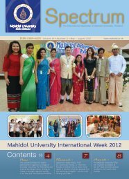
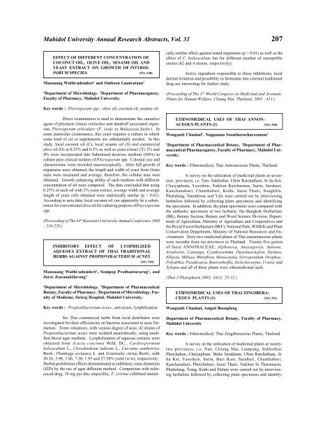
Key words : acaricidal activity, Trigonostemon reidioides, 2demethyl<br />
rediocide A<br />
Acaricidal activity of T. reidioides root extracts was investigated<br />
on Dermatophagoides pteronyssinus (D. p), the most com<br />
mon house dust mite in Thailand.<br />
Benzyl benzoate was used as posi<br />
tive acaricidal compound which<br />
showed LC 50 = 7.20x10 -3 mg/cm 2<br />
after 3 days of incubaion time.<br />
Bioassay-guided fractionation of<br />
the hexane crude extract (LC 50 =<br />
0.60 mg/cm 2 ) led to the isolation<br />
of fraction II which composed of a<br />
novel compound, 2-demethyl<br />
rediocide A, together with the<br />
known compound, rediocide A,<br />
and two unidentified compounds.<br />
Fraction II (LC 50 = 3.27x10 -3 mg/<br />
cm 2 ) showed almost 2-fold more active than benzyl benzoate. The<br />
structural elucidations were based on analysis of the spectroscopic<br />
and literature data.<br />
(The research was partially granted by <strong>Mahidol</strong> <strong>University</strong>. The paper<br />
was presented at The Sixth JSPS-NRCT Join Seminar : Recent<br />
Advances in Natural Medicine <strong>Research</strong>, Faculty of Pharmaceutical<br />
Sciences, Chulalongkorn <strong>University</strong>, December 2-4, 2003.)