Friesia XI, 5
Friesia XI, 5
Friesia XI, 5

Create successful ePaper yourself
Turn your PDF publications into a flip-book with our unique Google optimized e-Paper software.
- 268-<br />
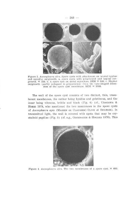
Figure 3. A scosphaera atra. Spore cysts with attachment an la teral hyphae<br />
and mycelial out g ro wth. a. spore cysts with attachment and hyphal outgrowth.<br />
X 250. b. A spore cyst an aerial mycelium. SEM X 500. c. Hyphal<br />
outg r owth (partly coUapsed in preparation) from the tube-shaped extension<br />
af the sp ore cyst membrane. SEM X 2000.<br />
The wall of the spore cyst consists of two distinct, t hin, translucent<br />
membranes, the outher being hyaline and gelatinous, and the<br />
inner being vitreous, brittie and black (Fig. 4) (cf., UDAGAWA &<br />
HORIE 1974, who mentio ned the two membranes in the spore cysts<br />
of Ascosphaera apis ( MASSE N ex CLAUSSEN) OLIVE et SPILTom). In<br />
transmitted light, the wall is covered with sp ots that may be crystalloid<br />
papillae (Fig. 5) (cf. e.g., GOCHNAUER & HUGHES 1976). This<br />
F ig ur e 4. Ascosphaera atra. The two membranes af a spore cyst. X 600.