Liebe Kolleginnen und Kollegen! - ÖGDV
Liebe Kolleginnen und Kollegen! - ÖGDV
Liebe Kolleginnen und Kollegen! - ÖGDV
Erfolgreiche ePaper selbst erstellen
Machen Sie aus Ihren PDF Publikationen ein blätterbares Flipbook mit unserer einzigartigen Google optimierten e-Paper Software.
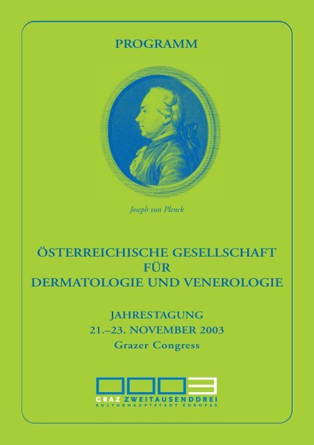
PROGRAMM<br />
Joseph von Plenck<br />
ÖSTERREICHISCHE GESELLSCHAFT<br />
FÜR<br />
DERMATOLOGIE UND VENEROLOGIE<br />
JAHRESTAGUNG<br />
21.–23. NOVEMBER 2003<br />
Grazer Congress
<strong>Liebe</strong> <strong>Kolleginnen</strong> <strong>und</strong> <strong>Kollegen</strong>!<br />
Jede Jahrestagung stellt eine besondere Herausforderung an die Verantwortlichen<br />
dar: Wie motiviert man die Mitglieder der Gesellschaft, aus der Vielzahl<br />
an angebotenen Veranstaltungen die eigene auszuwählen, gerade diese zu<br />
besuchen?<br />
Der Ort ist wichtig: Graz, die Kulturhauptstadt Europas 2003, hat Vieles zu<br />
bieten, der Grazer Congress ist ein würdiger Veranstaltungsrahmen.<br />
Im Programm muss der Trapezakt zwischen „hoher Wissenschaft“ <strong>und</strong><br />
Praxisrelevanz gelingen: Neben den traditionellen Diakliniken, den freien<br />
Vorträgen <strong>und</strong> der Posterausstellung sind heuer erstmals die Arbeitsgruppen<br />
aktiv in das wissenschaftliche Programm eingeb<strong>und</strong>en: Höhepunkte <strong>und</strong><br />
Neues aus den jeweiligen Spezialgebieten sollen gut verdaulich dargeboten<br />
werden. Für die Spezialvorlesungen (Hebra <strong>und</strong> Plenck) konnten „hochkarätige“<br />
Redner gewonnen werden. Die Rede zur Mitgliederversammlung <strong>und</strong> der<br />
Abschlussvortrag werden von Nicht-Dermatologen, Koryphäen ihres Faches,<br />
gehalten.<br />
Qualitätsmanagement ist wohl ein Reizwort des beginnenden Jahrtausends: ein<br />
Spezialsymposium widmet sich ausgesuchten Themenkreisen. Dazupassend<br />
seien die neu aufgelegten Leitlinien der DDG in Erinnerung gerufen; sie sind<br />
in Buchform von der Fa. Hermal erhältlich <strong>und</strong> zeitgemäß auch im Internet<br />
abrufbar (www.awmf.de). In jeder Subkommission haben <strong>ÖGDV</strong>-Mitglieder<br />
mitgearbeitet, diese Leitlinien haben somit auch für uns verbindlichen<br />
Charakter. Und schließlich sei auf ein Seminar am Freitag zu Tagungsbeginn<br />
hingewiesen, in dem die Umsetzung des Fachwissens im Praxisalltag –<br />
„Marketing in der Arztpraxis“ – gefördert werden soll – wir werden uns in<br />
Zukunft auch mit diesem Themenkreis verstärkt auseinandersetzen müssen.<br />
Referenten, Organisatoren <strong>und</strong> auch die zahlreich vertretenen Firmen freuen<br />
sich auf Ihr Kommen. Falls Sie noch nicht gebucht haben: Ein Besuch unserer<br />
Homepage (www.oegdv.at) sollte Ihnen diese Aufgabe leicht machen!<br />
Univ.-Prof. Dr. Elisabeth Aberer Univ.-Prof. Dr. Werner Aberer<br />
Schriftführerin der <strong>ÖGDV</strong> Präsident der <strong>ÖGDV</strong><br />
1
Donnerstag, 20. November 2003<br />
19.00–21.00 Präsidiumssitzung<br />
PROGRAMM<br />
Freitag, 21. November 2003<br />
Administrative Sitzungen<br />
9.00–10.00 Wissenschaftlicher Ausschuss Blauer Salon<br />
9.00–10.00 Administrative Sitzung der AG Seminarraum<br />
Humangenetik <strong>und</strong> molekulare Parterre (hinter der<br />
Therapie Registrierung)<br />
9.00–10.00 Administrative Sitzung der AG STD<br />
<strong>und</strong> Dermatologische Mikrobiologie<br />
Seminarraum K7<br />
9.00–13.00 Administrative Sitzung der AG Mikroskopiersaal des<br />
Dermatohistopathologie mit Institutes für<br />
Schnittseminar Pathologie<br />
Auenbrugger Platz 25<br />
Klinikum-LKH Graz<br />
10.00–13.00 Vorstandssitzung Blauer Salon<br />
18.00–18.30 Administrative Sitzung der AG<br />
Allergologie<br />
Seminarraum K7<br />
18.00–19.30 Administrative Sitzung der AG<br />
Melanom <strong>und</strong> dermatologische<br />
Onkologie<br />
Blauer Salon<br />
Workshop<br />
10.00–13.45 Praxis-Erfolgssystem<br />
P. Eibl-Schober<br />
„Marketing – die neue Dimension<br />
in der Arztpraxis“<br />
Seminarraum K7<br />
2
PROGRAMM<br />
Tagungsbeginn<br />
13.45–14.00 Begrüßung Saal Steiermark<br />
Freitag, 21. November 2003<br />
14.00–16.00 Wissenschaftliche Sitzung der AG STD <strong>und</strong><br />
Dermatologische Mikrobiologie<br />
Neue <strong>und</strong> wiederentdeckte Erreger in der<br />
Dermato-Venerologie<br />
Vorsitz: G. Stingl<br />
R. Müllegger (Graz)<br />
Neueste Erkenntnisse zu zeckenübertragenen<br />
Infektionskrankheiten<br />
W. Graninger (Graz)<br />
Mikroorganismen <strong>und</strong> Autoimmunität<br />
A. Salat (Wien)<br />
Squamous intraepithelial lesions: Ätiopathogenese, Diagnostik<br />
<strong>und</strong> Therapie<br />
K. Rappersberger (Wien)<br />
Hepatitis-Viren als (un)mittelbare Verursacher von<br />
Hautkrankheiten<br />
16.00–16.30 P ause<br />
3
PROGRAMM<br />
Freitag, 21. November 2003<br />
16.30–17.30 Freie Vorträge I<br />
Vorsitz: H. Hintner, H. J. Rauch<br />
K. Holubar, St. Fatovic-Ferencic (Wien; Zagreb/Kroatien)<br />
Lorenz Matthäus Carl Rigler (1815–1862) – ein Grazer<br />
„Josephiner“ an der Hohen Pforte<br />
Alexandra Geusau, N. Sandor, S. Rödler, A. Bodgalian,<br />
A. Zuckermann (Wien)<br />
Haut- <strong>und</strong> Schleimhautmanifestationen bei<br />
organtransplantierten, immunsupprimierten Patienten<br />
H. Maier, P. Donath, A. Cabaj, S. Geiger, H. Hönigsmann (Wien)<br />
Erfolgreiche Behandlung von anogenitalen Warzen mit einem<br />
Blitzlampen-gepumpten gepulsten Farbstofflaser<br />
A. Schneeberger, E. Messeritsch, F. Karlhofer, H. Maier,<br />
H. Hönigsmann, G. Stingl, S. N. Wagner (Wien)<br />
Blasenbildung nach Tumorbestrahlung<br />
J. H. Wilkens (Wildau/Deutschland)<br />
Wirksamkeit der Dermodyne ® UV-freien Phototherapie bei<br />
Kindern <strong>und</strong> jungen Erwachsenen<br />
M. Glatz, M. Golestani, H. Kerl, R. R. Müllegger (Graz)<br />
Serologisches Follow-up von Patienten mit Erythema migrans<br />
nach antibiotischer Therapie<br />
G. J. Sturm, A. Heinemann, W. Aberer (Graz)<br />
Diagnostik der Insektengiftallergie: Der Basophilen-<br />
Aktivierungstest (BAT) im Vergleich zu Hauttest <strong>und</strong><br />
spezifischem IgE<br />
M. Misir (Osijek/Kroatien)<br />
Information on AIDS in Biomedical Journals in Croatia<br />
Christina M. Reinisch, W. Weninger, Ch. Mayer, K. Paiha,<br />
H. Lassmann, E. Tschachler (Wien; Neuilly/Frankreich;<br />
Boston/USA)<br />
Das Nervenendorgan der menschlichen Haut – neue Einblicke<br />
4
PROGRAMM<br />
Freitag, 21. November 2003<br />
17.30–18.00 Wissenschaftliche Sitzung der AG Allergologie<br />
Vorsitz: W. Aberer<br />
B. Kränke (Graz)<br />
Allergologisches Qualitätsmanagement in der<br />
dermatologischen Praxis<br />
ab 19.30 Gesellschaftsabend der Fa. Fujisawa<br />
(Programm siehe Seite 16)<br />
5
PROGRAMM<br />
Samstag, 22. November 2003<br />
8.30– 9.30 Diaklinik I<br />
Vorsitz: N. Sepp, J. Hutterer<br />
Tamara Kopp, D. Zillikens, G. Stingl, F. Karlhofer<br />
(Wien; Würzburg/Deutschland)<br />
Konfluierende Pusteln: IgA Pemphigus mit dualer<br />
Antigen-Reaktivität<br />
Kerstin Beringer-Jäger (Wien)<br />
Pemphigus vulgaris – Therapie <strong>und</strong> Verlauf<br />
Van Anh Nguyen (Innsbruck)<br />
Bullöses Pemphigoid<br />
Ana Benedicic Pilih, A. Vizjak, A. Kansky, M. Bercic,<br />
A. Pejovnik Pustinek, J. Arzensek, F. Wojnarowska<br />
(Celje/Slowenien; Oxford/GB)<br />
Linear IgA bullous dermatosis<br />
Jutta Popp-Habeler (Wels)<br />
Polychondritis recidivans et atrophicans<br />
M. Wilhelm (Innsbruck)<br />
Antiphospholipidsyndrom<br />
Alina Weiss (Wien)<br />
Generalisierte Herpes simplex Infektion bei Morbus Darier<br />
Birgit Raffier (Linz)<br />
Staphylococcal scalded skin syndrome<br />
J. Jabkowski (Linz)<br />
Leukozyten Adhäsions Defizienz<br />
A. Bacher (St. Pölten)<br />
Livedo fulminans<br />
Ulrike Hein (Wien)<br />
Ausgeprägte Arzneimittelreaktion bei einem Patienten mit<br />
unklarem, pulmonalem Infiltrat <strong>und</strong> Nierenversagen<br />
6
PROGRAMM<br />
Samstag, 22. November 2003<br />
9.30–10.30 Ferdinand von Hebra-Gedächtnisvorlesung<br />
Vorsitz: G. Stingl<br />
A. Y. Finlay (Cardiff/Wales)<br />
Was benötigt der dermatologische Patient wirklich?<br />
10.30–11.00 P ause<br />
11.00–13.00 Symposium: „F<strong>und</strong>amente der modernen Medizin“<br />
Vorsitz: B. Volc-Platzer, H. Kerl<br />
T. Schäfer (Lübeck)<br />
Epidemiologie ist mehr als nur zählen!<br />
T. L. Diepgen (Heidelberg)<br />
Die Cochrane Library, nicht nur eine Bibliothek!<br />
J. Steurer (Zürich)<br />
Evidence based medicine; hilfreich oder hinderlich, glauben<br />
oder wissen?<br />
W. Sterry (Berlin)<br />
ISO-Zertifizierung, Qualitätsmanagement <strong>und</strong> andere<br />
Gütesiegel; behandeln wir deshalb <strong>und</strong> damit Patienten besser?<br />
Dieses Symposium wird durch einen großzügigen Grant der<br />
Fa. NOVARTIS – ohne Einflussnahme auf das Programm –<br />
unterstützt.<br />
13.00–14.00 M ittagsbuffet im Kammermusiksaal<br />
7
PROGRAMM<br />
Samstag, 22. November 2003<br />
14.00–15.00 Freie Vorträge II<br />
Vorsitz: H. Kofler, D. Maurer<br />
Tamara Jandl, F. Koszik, H. Korschan, H. Kittler, T. Berger,<br />
P. Mischer, H. Kehrer, C. Wolber, R. Strohal, B. Volc-Platzer,<br />
S. N. Wagner, G. Stingl, A. Schneeberger<br />
(Wien, Wels, Linz, Feldkirch)<br />
Estimation of the minimal tumor load detected by the<br />
serological melanoma markers S100B and MIA<br />
R. Loewe, H. Kittler, G. Fischer, I. Faé, K. Wolff, P. Petzelbauer<br />
(Wien)<br />
Mutations in the BRAF kinase gene correlate with rapid<br />
melanocytic lesion growth<br />
S. N. Wagner, G. Goess, A. Schneeberger, S. Koller, A. M.Krieg,<br />
J. Smolle, G. Stingl (Wien, Graz; Wellesley/USA)<br />
Melanoma immunotherapy with immunostimulatory CpG<br />
oligodeoxynucleotides–first clinical experience<br />
W. Hötzenecker, J. G. Meingassner, R. Ecker, G. Stingl,<br />
A. Stuetz, A. Elbe-Buerger (Wien)<br />
Corticosteroids affect viability, maturation and immune<br />
function of murine epidermal Langerhans cells in contrast to<br />
pimecrolimus<br />
M. Mildner, V. Mlitz, C. Ballaun, E. Tschachler<br />
(Wien; Neuilly/Frankreich)<br />
UVA radiation induces HGF/SF in dermal fibroblasts which<br />
protects them from UVA induced apoptosis<br />
Caterina Barresi, H. Rossiter, E. F. Wagner, E. Tschachler<br />
(Wien; Neuilly/Frankreich)<br />
Keratin5-CRE/LOXP mediated inactivation of VEGF<br />
sensitizes mouse skin to UVB-induced photo-damage<br />
8
PROGRAMM<br />
Samstag, 22. November 2003<br />
M. Schmuth, Ch. Haqq, T. Willson, Ch. Schoonjans, J. Auwerx,<br />
P. Chambon, P. M. Elias, K. R. Feingold (Innsbruck;<br />
Illkirch/Frankreich; San Francisco/USA)<br />
PPAR-delta Effekte auf die Epidermis<br />
P. Mrass, M. Rendl, M. Mildner, C. Ballaun, B. Lengauer,<br />
E. Tschachler (Wien; Neuilly/Frankreich)<br />
All-trans-retinoic-acid treatment leads to an upregulation of<br />
pro-apoptotic caspases, and p53, and sensitizes primary<br />
keratinocytes to UVB-induced apoptosis<br />
Alessandra Handisurya, S. Shafti-Keramat, O. Forsl<strong>und</strong>,<br />
R. Kirnbauer (Wien; Malmö/Schweden)<br />
Rekombinante Virushüllen von HPV92, ein neuer<br />
Papillomvirustyp isoliert aus einem Basaliom<br />
9
PROGRAMM<br />
Samstag, 22. November 2003<br />
15.00–16.30 Mitgliederversammlung, Verleihung der Preise /<br />
Diplome <strong>und</strong> Vortrag<br />
Vorsitz: W. Aberer<br />
W. Rauch (Graz)<br />
Der Arzt <strong>und</strong> die Informationsgesellschaft<br />
16.30–17.00 Pause<br />
17.00–17.30 Wissenschaftliche Sitzung der AG Humangenetik <strong>und</strong><br />
molekulare Therapie<br />
Vorsitz: J. Bauer<br />
J. Thalhammer (Salzburg)<br />
Genetische Vakzinierung: Wo stehen wir heute?<br />
17.30–19.00 Wissenschaftliche Sitzung der AG Ästhetische<br />
Dermatologie <strong>und</strong> Kosmetologie<br />
Kosmetische Produkte: Entwicklung – Galenik –<br />
EU-Kosmetikrecht<br />
Vorsitz: E. M. Kokoschka<br />
W. Schlocker (Innsbruck)<br />
GALENIK – Gr<strong>und</strong>lagen <strong>und</strong> ihre technischen Aspekte<br />
E. Leitner (Graz)<br />
Kosmetische Zubereitungen <strong>und</strong> Produkthygiene<br />
G. Gribl (Wien)<br />
Kosmetikrecht <strong>und</strong> Produktsicherheit<br />
17.30–19.30 Gesellschaftsabend der Fa. Schering<br />
(Programm siehe Seite 16)<br />
10
PROGRAMM<br />
Sonntag, 23. November 2003<br />
08.30–09.30 Diaklinik II<br />
Vorsitz: H. Hönigsmann, U. Längle<br />
G. Wickenhauser (Wien)<br />
Ungewöhnliches Exanthem bei einem Kleinkind<br />
N. Tomi, Ch. Szolar-Platzer (Graz)<br />
Allergie auf ein Antiallergikum<br />
Carola Wolber (Feldkirch)<br />
Therapieresistente zentrofaciale Schwellung im Jugendalter<br />
B. Leinweber (Graz)<br />
Prurigo pigmentosa<br />
Eva Messeritsch, A. Geusau (Wien)<br />
Vaginale Erosionen – es muss nicht immer eine STD sein<br />
Birgit Lederer (Klagenfurt)<br />
Pityriasis rubra pilaris<br />
Karin Kaindl (Salzburg)<br />
Therapie der Psoriasis pustulosa mit Infliximab<br />
A. Vucinic Dugonik, M. Belic, J. Miljkovic (Maribor/Slowenien)<br />
Eosinophilic fasciitis<br />
F. Weihsengruber (Wien)<br />
Generalisiertes mucocutanes Pyoderma gangränosum<br />
Constanze Jonak, J. Schwarzmeier (Wien)<br />
Inflammierte papulosquamöse Herde bei einer onkologischen<br />
Patientin unter systemischer Chemotherapie<br />
09.30–10.30 Josef von Plenck-Gedächtnisvorlesung<br />
Vorsitz: K. Wolff<br />
J. Ring (München)<br />
Haut, Allergie <strong>und</strong> Umwelt<br />
10.30–11.00 P ause<br />
11
PROGRAMM<br />
Sonntag, 23. November 2003<br />
11.00–11.30 Wissenschaftliche Sitzung der AG Operative Dermatologie,<br />
Lasertherapie <strong>und</strong> W<strong>und</strong>heilungsforschung<br />
Vorsitz: J. Koller<br />
J. Koller (Salzburg)<br />
Hautchirurgie im Armentarium der Dermatologie<br />
K. Böhler-Sommeregger (Wien)<br />
Überblick über das erweiterte therapeutische Spektrum der<br />
Phlebologie<br />
11.30–12.00 Wissenschaftliche Sitzung der AG Melanom <strong>und</strong><br />
dermatologische Onkologie<br />
Vorsitz: H. Pehamberger<br />
A. Schneeberger (Wien)<br />
Wert <strong>und</strong> Unwert von Tumormarkern beim Melanom<br />
Ingrid H. Wolf (Graz)<br />
Klinische Erfahrungen mit Imiquimod zur Melanomtherapie<br />
G. Weinlich (Innsbruck)<br />
Erfahrungen mit DC-Vakzination bei Melanompatienten<br />
Ch. Höller (Wien)<br />
Genasense–from bench to bedside<br />
12.00–13.00 Festsitzung<br />
Vorsitz: T. Luger, G. Schuler<br />
H. Piza (Innsbruck)<br />
Was sind die Voraussetzungen für Spitzenleistungen?<br />
Überreichung der Goldmedaille der <strong>ÖGDV</strong> an<br />
Prof. Dr. K. Wolff<br />
ca. 13.15 E nde der Jahrestagung<br />
Posterausstellung: Die Poster hängen während der ganzen Jahrestagung im<br />
Gang vor dem Saal Steiermark (Postergröße 90 cm breit, 200 cm hoch) aus.<br />
Industrieausstellung: Während der ganzen Jahrestagung im Foyer 1. Stock<br />
12
POSTER<br />
P1 M. Alalmi, Y. Al-Kordofani, S. M. Al-Awlaqi, K. A. Hussien, P. Soyer,<br />
H. Starz, D. Ghazarian, H. Grossmann (Sanaà/Yemen; Graz)<br />
Ashy dermatosis and lichen planus pigmentosus. A clinicopathological<br />
study in the republic of Yemen<br />
P2 Ch. Bangert, G. Stary, G. Stingl, T. Kopp (Wien)<br />
Preferential occurrence of different inflammatory dendritic cell subsets<br />
in contact hypersensitivity and atopic dermatitis<br />
P3 B. Binder, T. Kern, G. Ginter-Hanselmayer (Graz)<br />
Onychomykose <strong>und</strong> Paronychie durch Candida albicans bei einem<br />
Säugling mit Trisomie 21<br />
P4 P. Donath, H. Maier, S. Geiger, A. Cabaj, H. Hönigsmann (Wien)<br />
Behandlung von Mollusca contagiosa mit einem gepulsten<br />
Farbstofflaser<br />
P5 A. Hofer, A. S. Hassan, F. J. Legat, H. Kerl, P. Wolf (Graz)<br />
Behandlung der Vitiligo mit Excimer Laser (308NM)<br />
P6 D. Kopera, R. Kokol (Graz)<br />
685-NM Low-Energy-Lasers zur Behandlung des Ulcus cruris:<br />
Präsentation der Ergebnisse an 44 Patienten<br />
P7 D. Linder, G. Obermoser, G. Schiesari, P. Fritsch (Venedig/Italien;<br />
Innsbruck)<br />
Noduläres Pemphigoid<br />
P8 M. Unkauf, J. L. Kienzler, J. Magnette, C. Queille-Roussel,<br />
A. Sanchez-Ponton, J. P. Ortonne (Frankfurt/Deutschland;<br />
Nice, Nimes/Frankreich; Nyon/Schweiz)<br />
Low dose Diclofenac-Na gel is effective in reducing the pain and<br />
inflammation associated with exposure to ultraviolet light – results of<br />
two randomised clinical studies<br />
P9 P. Lührs, T. Biedermann, W. Schmidt, G. Stingl, M. Röcken,<br />
A. Schneeberger (Wien; Tübingen/Deutschland)<br />
Early interleukin 4 administration promotes TH1-driven, protective<br />
anti-cancer immunity<br />
13
POSTER<br />
P10 H. Maier, P. Donath, M. Tirant, D. Relic, S. Farokhnia, H. Hönigsmann,<br />
A. Tanew (Wien)<br />
Prospektive, randomisierte, Placebo-kontrollierte Doppelblind-Studie<br />
über die Psoriasis-Therapie mit pflanzlichen Wirkstoffen<br />
P11 S. Mayr-Kanhäuser, G. Ginter-Hanselmayer (Graz)<br />
Malassezia-assoziierte Gesichtsdermatosen–erste therapeutische<br />
Erfahrungen mit Itraconazol<br />
P12 E. Ch. Prandl, M. V. Schintler, S. Spendel, G. Wittgruber, B. Hellbom,<br />
E. Scharnagl (Graz)<br />
Follow up nach KTP (532nm)-Laser-Therapie von Besenreisern<br />
P13 E. Sadler, G. Pohla-Gubo, H. Hintner, J. W. Bauer (Salzburg)<br />
Autoimmune bullöse Dermatosen: Klinisches Management von<br />
187 Patienten über 13 Jahre<br />
P14 R. Schöllnast, B. Kränke, W. Aberer (Graz)<br />
Erukismus<br />
P15 M. Shekari Yazdi (Wien)<br />
Traditional Persian medicine and its use in dermatology<br />
P16 A. Uthman, M. Dockal, J. Söltz-Szöts, E. Tschachler (Wien)<br />
Fluconazole upregulates SconC expression and downregulates sulphur<br />
metabolism in M. canis<br />
P17 St. Wöhrl, M. Focke, G. Hinterhuber, G. Stingl, M. Binder (Wien)<br />
Anaphylaxie auf Patent blau V<br />
P18 C. Wolber, M. Schelling, G. Hartmann, U. Gruber, F. Offner, M. Takacs,<br />
W. Jurecka, R. Strohal (Feldkirch, Wien)<br />
Nanokristalline Silberauflagen als neue MRSA Barriere- <strong>und</strong><br />
Therapieoption?<br />
P19 I. Zalaudek, G. Argenziano, B. Leinweber, L. Ciatrella,<br />
R. Hofmann-Wellenhof, J. Malvehy, S. Puig, M. A. Pizzichetta,<br />
L. Thomas, H. P. Soyer, H. Kerl (Graz; Aviano, Rom, Neapel/Italien;<br />
Barcelona/Spanien; Lyon/Frankreich)<br />
Auflichtmikroskopische Strukturen des Morbus Bowen<br />
14
POSTER<br />
P20 M. Schmuth, D. D. Bikle, T. Willson, D. J. Mangelsdorf, P. M. Elias,<br />
K. R. Feingold (Innsbruck; Dallas, San Francisco/USA)<br />
Wirkmechanismen der Aktivierung von LXR durch Oxysterole in<br />
Keratinozyten<br />
P21 G. Hinterhuber, K. Cauza, K. Brugger, R. Dingelmaier-Hovorka,<br />
R. Horvat, K. Wolff, D. Foedinger (Wien)<br />
RPE65 in human keratinocytes–a putative retinol binding protein<br />
receptor<br />
P22 M. Laimer, A. Klausegger, W. Aberer, V. Wally, C. M. Lanschuetzer,<br />
H. Hintner, J. W. Bauer (Salzburg, Graz)<br />
Deletion within the 3’-UTR of C1-INH-gene as a causative factor of<br />
hereditary angioedema?<br />
P23 K. Cauza, A. Grassauer, G. Hinterhuber, R. Horvat, K. Rappersberger,<br />
K. Wolff, D. Foedinger (Wien)<br />
FcgRI, II and III expression in cultured human keratinocytes and their<br />
response to interferon-g treatment<br />
P24 P. Petzelbauer, Y. Miyazaki, P. Friedl, G. Wickenhauser, M. Pillinger,<br />
P. Zacharowsk, M. Gröger, K. Wolff, K. Zacharowski (Wien)<br />
The fibrin-derived peptide Bb15-42 reduces myocardial reperfusion injury<br />
P25 St. Wagner, Ch. Hafner, D. Allwardt, J. Jasinska, Ch. Zielinski,<br />
O. Scheiner, K. Wolff, U. Wiedermann, H. Breiteneder, H. Pehamberger<br />
(Wien)<br />
Vaccination with a mimotope of the high molecular weight melanoma<br />
accociated antigen induces antibodies inhibiting melanoma tumor cell<br />
growth<br />
P26 C. Hoeller, D. Fink, E. Heere-Ress, F. Roka, T. Lucas, V. Sexl, V. Wacheck,<br />
M. Gleave, K. Wolff, H. Pehamberger, B. Jansen (Wien)<br />
Clusterin is associated with treatment resistance and regulation of the<br />
anti-apoptotic Bcl-2 family member Bcl-xL in human melanoma<br />
P27 C. Hoeller, C. Thallinger, B. Pratscher, E. Heer-Ress, V. Wacheck, V. Sexl,<br />
K. Wolff, H. Pehamberger (Wien)<br />
Expression of the non-receptor associated tyrosine kinase syk influences<br />
the metastatic behaviour of melanoma cells<br />
15
GESELLSCHAFTSPROGRAMM<br />
Freitag, 21. November 2003<br />
19.30 Festabend der Fa. Fujisawa<br />
Ein Kunstgenuss der besonderen Art<br />
Seifenfabrik Veranstaltungszentrum<br />
Angergasse 41–43, A-8010 Graz<br />
Genügend Parkmöglichkeiten;<br />
Busse fahren direkt vom Grazer Congress<br />
um 19.20, 19.30 <strong>und</strong> 19.40<br />
Beim Fujisawa-Kongressstand erhalten Sie Ihre persönliche<br />
Eintrittskarte – mit Gewinn-Los!<br />
Samstag, 22. November 2003<br />
19.30 Festabend der Fa. Schering<br />
Empfangscocktail im Kunsthaus Graz („Die Blase“)<br />
mit anschließender Führung durch die Ausstellung:<br />
Einbildung – Das Wahrnehmen in der Kunst<br />
Dinner im Hotel Wiesler<br />
Anmeldung erbeten bis zum 14. November 2003 unter:<br />
(+43/1) 970 37-331 oder<br />
e-mail: beate.duchek@schering.at<br />
16
ALLGEMEINE HINWEISE<br />
Tagungsort:<br />
Grazer Congress<br />
Kongress-Ausstellungs- <strong>und</strong> Kommunikationszentrum<br />
Sparkassenplatz, A-8010 Graz<br />
Tel.: (+43/316) 80 49-0<br />
Tagungsgebühren:<br />
Mitglieder der <strong>ÖGDV</strong> mit Praxis € 130,–<br />
Mitglieder der <strong>ÖGDV</strong> ohne Praxis € 100,–<br />
Mitglieder der <strong>ÖGDV</strong> in Ausbildung* € 80,–<br />
Nichtmitglieder € 210,–<br />
Nichtmitglieder in Ausbildung* € 100,–<br />
Studenten/Dissertanten* frei<br />
*) Bitte bei der Anmeldung Tätigkeitsnachweis/Inskriptionsbestätigung<br />
mitschicken.<br />
Zahlungsmodalitäten:<br />
Banküberweisung: Steiermärkische Bank <strong>und</strong> Sparkassen AG,<br />
A-8011 Graz, „Dermatologentagung 2003“ Konto-Nr.: 00000-981480,<br />
BLZ 20815, „Spesenfrei für den Empfänger“<br />
ODER<br />
Bitte belasten Sie meine Kreditkarte:<br />
❑ VISA ❑ EURO/MASTERCARD ❑ DINERS ❑ AMEX<br />
Kreditkartennummer: ..................../..................../..................../....................<br />
Ablaufdatum: ............./............. (MM/JJ)<br />
Ort, Datum: ................................. Unterschrift: ..................................................<br />
Mit Ihrer Unterschrift bestätigen Sie die o. g. Bedingungen.<br />
17
ALLGEMEINE HINWEISE<br />
Wissenschaftliches <strong>und</strong> Administratives Sekretariat:<br />
Univ.-Prof. Dr. Werner & Univ.-Prof. Dr. Elisabeth Aberer<br />
Univ.-Klinik für Dermatologie<br />
Auenbruggerplatz 8, A-8036 Graz<br />
Tel.: (+43) 316 385 3926, Fax.: (+43) 316 385 3782<br />
e-mail: werner.aberer@uni-graz.at<br />
e-mail: elisabeth.aberer@uni-graz.at<br />
Wiener Medizinische Akademie<br />
Alserstraße 4, A-1090 Wien<br />
Tel.: (+43/1) 405 13 83-20, Fax: (+43/1) 405-13 83-23<br />
e-mail: associations@medacad.org<br />
Kongressanmeldung <strong>und</strong> Zimmerreservierung:<br />
Graz-Tourismusgesellschaft mbH<br />
Kaiserfeldgasse 15, A-8011 Graz<br />
Tel.: (+43) 316 8075-62, Fax: (+43) 316 8075-55<br />
e-mail: gr@graztourismus.at, oder: www.oegdv.at<br />
anklicken des Kulturhauptstadtsymbols<br />
Fachausstellung:<br />
Medizinische Ausstellungs- <strong>und</strong> Werbegesellschaft<br />
Freyung 6, A-1010 Wien<br />
Tel.: (+43/1) 536 63-33, Fax: (+43/1) 535 60 16<br />
e-mail: maw@media.co.at<br />
Wichtiger Hinweis: Die Teilnahme an den wissenschaftlichen<br />
Sitzungen der Jahrestagung 2003 der Österreichischen Gesellschaft für<br />
Dermatologie <strong>und</strong> Venerologie entspricht, gemäß Approbation durch<br />
das Fortbildungsreferat der Österreichischen Ärztekammer für das Fach<br />
Haut- <strong>und</strong> Geschlechtskrankheiten, der Absolvierung von 16 St<strong>und</strong>en<br />
im Rahmen des Diplomfortbildungsprogramms (DFP).<br />
18
VERZEICHNIS DER REFERENTEN UND VORSITZENDEN<br />
ABERER Werner, Prof. Dr.<br />
Univ.-Hautklinik, Abteilung für Umweltdermatologie <strong>und</strong> Venerologie, Graz<br />
ALMALMI Mohammed, Dr.<br />
Department of Dermatology and Venerology, Sana’a, Yemen<br />
BACHER Albert, Dr.<br />
LKH, Dermatologische Abteilung, St. Pölten<br />
BANGERT Christine, Dr.<br />
Univ.-Hautklinik, Abteilung für Imm<strong>und</strong>ermatologie <strong>und</strong> Infektiöse Hautkrankheiten,<br />
Wien<br />
BARRESI Caterina, Dr.<br />
Univ.-Hautklinik, Abteilung für Imm<strong>und</strong>ermatologie <strong>und</strong> Infektiöse Hautkrankheiten,<br />
Wien<br />
BAUER Johann, Doz. Dr.<br />
LKH, Dermatologische Abteilung, Salzburg<br />
BENEDICIC Pilih Ana, Dr.<br />
Dermatoveneroloski oddelek, Spiosna bolnisnica Celj, Oblakova 5, 3000 Celje<br />
BERINGER-JÄGER Kerstin, Dr.<br />
Wilhelminenspital, Dermatologische Abteilung, Wien<br />
BINDER Barbara, OA Dr.<br />
Univ.-Hautklinik Graz, Abteilung für Allgemeine Dermatologie, Graz<br />
BÖHLER-SOMMEREGGER Kornelia, Prof. Dr.<br />
Univ.-Hautklinik, Abteilung für Allgemeine Dermatologie, Wien<br />
CAUZA Karla, Dr.<br />
Univ.-Hautklinik, Abteilung für Allgemeine Dermatologie, Wien<br />
DIEPGEN Thomas L., Prof. Dr.<br />
Universitätsklinikum, Abteilung Klinische Sozialmedizin, Bergheimerstrasse 58,<br />
D-69115 Heidelberg<br />
DONATH Peter, Dr.<br />
Univ.-Hautklinik, Abteilung für Spezielle Dermatologie <strong>und</strong> Umweltdermatosen, Wien<br />
EIBL-SCHOBER Petra, dipl. Coach <strong>und</strong> Trainerin im Ges<strong>und</strong>heitswesen<br />
Mittelgasse 11/10, 1060 Wien<br />
FINLAY Andrew Y., Prof. Dr.<br />
University of Wales College of Medicine, Department of Dermatology, Health Park,<br />
Cardiff CF14, 4XN, UK<br />
GEUSAU Alexandra, Prof. Dr.<br />
Univ.-Hautklinik, Abteilung für Imm<strong>und</strong>ermatologie <strong>und</strong> Infektiöse Hautkrankheiten,<br />
Wien<br />
GLATZ Martin, Dr.<br />
Univ.-Hautklinik, Abteilung für Allgemeine Dermatologie, Graz<br />
19
VERZEICHNIS DER REFERENTEN UND VORSITZENDEN<br />
GRANINGER Winfried, Prof. Dr.<br />
Univ.-Klinik für Innere Medizin, Abteilung für Rheumatologie, Graz<br />
GRIBL Gerhard, Ing.<br />
Kosmetik Transparent, Wienerbergerstraße 7, PF 77, A-1130 Wien<br />
HANDISURYA Alessandra, Dr.<br />
Univ.-Hautklinik, Abteilung für Imm<strong>und</strong>ermatologie <strong>und</strong> Infektiöse Hautkrankheiten,<br />
Wien<br />
HEIN Ulrike, Dr.<br />
Donauspital SMZ Ost, Dermatologische Abteilung, Wien<br />
HINTERHUBER Gabriele, Dr.<br />
Univ.-Hautklinik, Abteilung für Allgemeine Dermatologie, Wien<br />
HINTNER Helmut, Prof. Dr.<br />
LKH, Dermatologische Abteilung, Salzburg<br />
HOELLER Christoph, Dr.<br />
Univ.-Hautklinik, Abteilung für Allgemeine Dermatologie, Wien<br />
HÖNIGSMANN Herbert, Prof. Dr.<br />
Univ.-Hautklinik, Abteilung für Spezielle Dermatologie <strong>und</strong> Umweltdermatosen, Wien<br />
HÖTZENECKER Wolfram, Dr.<br />
Univ.-Hautklinik, Abteilung für Imm<strong>und</strong>ermatologie <strong>und</strong> Infektiöse Hautkrankheiten,<br />
Wien<br />
HOFER Angelika, OA Dr.<br />
Univ.-Hautklinik, Abteilung für Allgemeine Dermatologie, Graz<br />
HOLUBAR Karl, Prof. Dr.<br />
Institut für Geschichte der Medizin, Wien<br />
HUTTERER Judith, Dr.<br />
Hautarztpraxis, Wien<br />
JABKOWSKI Jörg, OA Dr.<br />
KH der Elisabethinen, Dermatologische Abteilung, Linz<br />
JANDL Tamara, Dr.<br />
Univ.-Hautklinik, Abteilung für Imm<strong>und</strong>ermatologie <strong>und</strong> Infektiöse Hautkrankheiten,<br />
Wien<br />
JONAK Constanze, Dr.<br />
Univ.-Hautklinik, Abteilung für Spezielle Dermatologie <strong>und</strong> Umweltdermatosen, Wien<br />
KERL Helmut, Prof. Dr.<br />
Univ.-Hautklinik, Abteilung für Allgemeine Dermatologie, Graz<br />
KOFLER Heinz, Doz. Dr.<br />
Allergieambulatorium, Hall<br />
KOKOSCHKA Eva Maria, Prof. Dr.<br />
Univ.-Hautklinik, Abteilung für Allgemeine Dermatologie, Wien<br />
20
VERZEICHNIS DER REFERENTEN UND VORSITZENDEN<br />
KOLLER Josef, OA Dr.<br />
LKH, Dermatologische Abteilung, Salzburg<br />
KOPERA Daisy, Prof. Dr.<br />
Univ.-Hautklinik, Abteilung für Allgemeine Dermatologie, Graz<br />
KOPP Tamara, Dr.<br />
Univ.-Hautklinik, Abteilung für Imm<strong>und</strong>ermatologie <strong>und</strong> Infektiöse Hautkrankheiten,<br />
Wien<br />
KRÄNKE Birger, Prof. Dr.<br />
Univ.-Hautklinik, Abteilung für Umweltdermatologie <strong>und</strong> Venerologie, Graz<br />
LÄNGLE Udo, Dr.<br />
Hautarztpraxis, Dornbirn<br />
LAIMER Martin, Dr.<br />
LKH, Abteilung für Dermatologie, Salzburg<br />
LEDERER Birgit, Dr.<br />
LKH, Dermatologische Abteilung, Klagenfurt<br />
LEINWEBER Bernd, Dr.<br />
Univ.-Hautklinik, Abteilung für Allgemeine Dermatologie, Graz<br />
LEITNER Erich, Dr.<br />
Technische Universität, Institut für Lebensmittelchemie <strong>und</strong> -technologie,<br />
Petersgasse 12/II, A-8010 Graz<br />
LINDER Dennis, Dr.<br />
Ospedale SS. Giovanni & Paolo, Venedig<br />
LOEWE Robert, Dr.<br />
Univ.-Hautklinik, Abteilung für Allgemeine Dermatologie, Wien<br />
LÜHRS Petra, Dr.<br />
Univ.-Hautklinik, Abteilung für Imm<strong>und</strong>ermatologie <strong>und</strong> Infektiöse Hautkrankheiten,<br />
Wien<br />
LUGER Thomas, Prof. Dr.<br />
Univ.-Hautklinik, Münster<br />
MAIER Harald, Dr.<br />
Univ.-Hautklinik, Abteilung für Spezielle Dermatologie <strong>und</strong> Umweltdermatosen, Wien<br />
MAURER Dieter, Prof. Dr.<br />
Univ.-Hautklinik, Abteilung für Imm<strong>und</strong>ermatologie <strong>und</strong> Infektiöse Hautkrankheiten,<br />
Wien<br />
MAYR-KANHÄUSER Sigrid, OA Dr.<br />
Univ.-Hautklinik, Abteilung für Umweltdermatologie <strong>und</strong> Venerologie, Graz<br />
MESSERITSCH Eva, Dr.<br />
Univ.-Hautklinik, Abteilung für Imm<strong>und</strong>ermatologie <strong>und</strong> Infektiöse Hautkrankheiten,<br />
Wien<br />
21
VERZEICHNIS DER REFERENTEN UND VORSITZENDEN<br />
MILDNER Michael, Ing.<br />
Univ.-Hautklinik, Abteilung für Imm<strong>und</strong>ermatologie <strong>und</strong> Infektiöse Hautkrankheiten,<br />
Wien<br />
MISIR Mihael, Dr.<br />
Osijek, Kroatien<br />
MRASS Paul, Dr.<br />
Univ.-Hautklinik, Abteilung für Imm<strong>und</strong>ermatologie <strong>und</strong> Infektiöse Hautkrankheiten,<br />
Wien<br />
MÜLLEGGER Robert, Prof. Dr.<br />
Univ.-Hautklinik, Abteilung für Allgemeine Dermatologie, Graz<br />
NGUYEN Van Anh, Dr.<br />
Univ.-Hautklinik, Innsbruck<br />
PETZELBAUER Peter, Prof. Dr.<br />
Univ.-Hautklinik, Abteilung für Allgemeine Dermatologie, Wien<br />
PIZA Hildeg<strong>und</strong>e, Prof. Dr.<br />
Univ.-Klinik für Plastische Chirurgie, Anichstraße 35, A-6020 Innsbruck<br />
POPP-HABELER Jutta, Dr.<br />
KH der Barmherzigen Schwestern vom heiligen Kreuz, Dermatologische Abteilung, Wels<br />
PRANDL Eva Ch., Dr.<br />
Univ.-Klinik für Chirurgie, Abteilung für Plastische Chirurgie, Graz<br />
RAFFIER Birgit, Dr.<br />
AKH, Dermatologische Abteilung, Linz<br />
RAPPERSBERGER Klemens, Prof. Dr.<br />
Krankenanstalt Rudolfstiftung, Dermatologische Abteilung, Wien<br />
RAUCH Hans-Jörg, Dr.<br />
Hautarztpraxis, Wien<br />
RAUCH Wolf, Prof. Dr. Mag.<br />
KF-Universität, Institut für Informationswissenschaft, Universitätsstraße 15/F3,<br />
A-8010 Graz<br />
REINISCH Christina M., Dr.<br />
Univ.-Hautklinik, Abteilung für Imm<strong>und</strong>ermatologie <strong>und</strong> Infektiöse Hautkrankheiten,<br />
Wien<br />
RING Johannes, Prof. DDr.<br />
Technische Universität, Klinik für Dermatologie <strong>und</strong> Allergologie,<br />
Biedersteinerstraße 29, D-80802 München<br />
SADLER Elke, Dr.<br />
LKH, Dermatologische Abteilung, Salzburg<br />
SALAT Andreas, Dr.<br />
Universitätsklinik für Chirurgie, Abteilung für Allgemeine Chirurgie, Wien<br />
SCHÄFER Torsten, Prof. Dr. MPH<br />
Universitätsklinikum, Institut für Sozialmedizin, Beckergrube 43-47, D-23552 Lübeck<br />
22
VERZEICHNIS DER REFERENTEN UND VORSITZENDEN<br />
SCHLOCKER Wolfgang, Prof. Dr.<br />
Universität, Institut für Pharmazeutische Technologie, Innrain 52, A-6020 Innsbruck<br />
SCHMUTH Matthias, Dr.<br />
Univ.-Hautklinik, Innsbruck<br />
SCHNEEBERGER Achim, Dr.<br />
Univ.-Hautklinik, Abteilung für Imm<strong>und</strong>ermatologie <strong>und</strong> Infektiöse Hautkrankheiten,<br />
Wien<br />
SCHÖLLNAST Renate, Dr.<br />
Univ.-Hautklinik, Abteilung für Umweltdermatologie <strong>und</strong> Venerologie, Graz<br />
SCHULER Gerold, Prof. Dr.<br />
Univ.-Hautklinik, Erlangen<br />
SEPP Norbert, Prof. Dr.<br />
Univ.-Hautklinik, Innsbruck<br />
SHEKARI YAZDI Mohammad, DDr.<br />
Institut für Geschichte der Medizin, Abteilung Ethnomedizin, Wien<br />
STERRY Wolfram, Prof. Dr.<br />
Universitätsklinikum Charité, Klinik für Dermatologie, Schumannstraße 20,<br />
D-10117 Berlin<br />
STEURER Johann, Prof. Dr.<br />
Horten-Zentrum für praxisorientierte Forschung <strong>und</strong> Wissenstransfer, Postfach Nord,<br />
CH-8091 Zürich<br />
STINGL Georg, Prof. Dr.<br />
Univ.-Hautklinik, Abteilung für Imm<strong>und</strong>ermatologie <strong>und</strong> Infektiöse Hautkrankheiten,<br />
Wien<br />
STURM Gunter, Dr.<br />
Univ.-Hautklinik, Abteilung für Umweltdermatologie <strong>und</strong> Venerologie, Graz<br />
THALHAMMER Josef, Prof. Dr.<br />
Universität, Institut für Chemie <strong>und</strong> Biochemie, Hellbrunnerstraße 34,<br />
A-5020 Salzburg<br />
TOMI Nordwig, Dr.<br />
Univ.-Hautklinik, Abteilung für Umweltdermatologie <strong>und</strong> Venerologie, Graz<br />
UNKAUF Markus, Dr.<br />
c/o Edelman Gmbh, Bettinastrasse 64, D-60325 Frankfurt<br />
UTHMAN Aumaid, Dr.<br />
Univ.-Hautklinik, Abteilung für Imm<strong>und</strong>ermatologie <strong>und</strong> Infektiöse Hautkrankheiten,<br />
Wien<br />
VOLC-PLATZER Beatrix, Prof. Dr.<br />
Donauspital SMZ Ost, Dermatologische Abteilung, Wien<br />
VUCINIC Dugonik A., Dr.<br />
Department of Dermatovenerology, Splosna bolnisnica Maribor, Ljubljanska 5,<br />
2000 Maribor<br />
23
VERZEICHNIS DER REFERENTEN UND VORSITZENDEN<br />
WAGNER Stefan, Dr.<br />
Univ.-Hautklinik, Abteilung für Allgemeine Dermatologie, Wien<br />
WAGNER Stephan N., Dr.<br />
Univ.-Hautklinik, Abteilung für Imm<strong>und</strong>ermatologie <strong>und</strong> Infektiöse Hautkrankheiten,<br />
Wien<br />
WEIHSENGRUBER Felix, Dr.<br />
Krankenanstalt Rudolfstiftung, Dermatologische Abteilung, Wien<br />
WEISS Alina, Dr.<br />
Krankenhaus Wien-Lainz, Dermatologische Abteilung, Wien<br />
WICKENHAUSER Georg, Dr.<br />
Univ.-Hautklinik, Abteilung für Allgemeine Dermatologie, Wien<br />
WILHELM Manuel, Dr.<br />
Univ.-Hautklinik, Innsbruck<br />
WILKENS Jan H., Dr.<br />
OptoMed Licht Klinik AG, Wildau/Deutschland<br />
WÖHRL Stefan, Mag. Dr.<br />
Univ.-Hautklinik, Abteilung für Imm<strong>und</strong>ermatologie <strong>und</strong> Infektiöse Hautkrankheiten,<br />
Wien<br />
WOLBER Carola, Dr.<br />
LKH, Dermatologische Abteilung, Feldkirch<br />
ZALAUDEK Iris, Dr.<br />
Univ.-Hautklinik, Abteilung für Allgemeine Dermatologie, Graz<br />
24
SPONSOREN<br />
FUJISAWA<br />
NOVARTIS<br />
PROCTER & GAMBLE<br />
SCHERING<br />
UCB<br />
25
AUSSTELLERVERZEICHNIS<br />
ÄRZTEZENTRALE, Wien<br />
AB-CONSULT Pharma, Wien<br />
AESCA, Traiskirchen<br />
ALK-ABELLÓ Allergie-Service, Linz<br />
ALLERGOPHARMA, Wien<br />
BIOGLAN Pharma, Greifswald – Insel Riems (Deutschland)<br />
CANDELA Laser Deutschland, Neu-Isenburg (Deutschland)<br />
COSMETIQUE ACTIVE Österreich, Wien<br />
DDD Medizintechnik, Mainz (Deutschland)<br />
DERMAPHARM, Wien<br />
DERMATICA Exclusiv, Köln (Deutschland)<br />
DERMOSAN, Wien<br />
PIERRE FABRE Dermo-Cosmetique, Wien<br />
FUJISAWA, Wien<br />
GEBRO Pharma, Fieberbrunn<br />
GENZYME Austria, Wien<br />
GLAXOSMITHKLINE Pharma, Wien<br />
GMT GRUBHOLZ Medizin-Technik, Graz<br />
HAL-ALLERGY, Wien<br />
HELTSCHL GmbH, Schlüsslberg<br />
HERMAL BOOTS HEALTHCARE PRODUCTS (Austria), Wien<br />
ICN Pharmaceuticals Austria, Wels<br />
JOHNSON & JOHNSON, Hallein<br />
Dr. KOLASSA + MERZ, Wien<br />
KPH Medizinprodukte, Grießkirchen<br />
LEO Pharma, Wien<br />
MEDILAS, Wien<br />
A. MENARINI Pharma, Wien<br />
Ferdinand MENZL, Wien<br />
MERZ Pharmaceuticals, Frankfurt (Deutschland)<br />
NOVARTIS Pharma, Wien<br />
PELPHARMA, Wien<br />
PREVAL Dermatica, Tangstedt/Pi. (Deutschland)<br />
PROCTER & GAMBLE Austria, Wien<br />
DR. MICHAELS RICA, Traiskirchen<br />
SANOVA Pharma, Wien<br />
SCHERING Wien, Wien<br />
Dr. A & L. SCHMIDGALL, Wien<br />
SMITH & NEPHEW, Schwechat<br />
SPIRIG Pharma, Linz<br />
SYNERON Österreich, Mödling<br />
TEACHSCREEN Software, Passau (Deutschland)<br />
UCB Pharma, Wien<br />
WAVEGUIDE laser systems, Wien<br />
LOUIS WIDMER, Salzburg<br />
(Stand bei Drucklegung)<br />
26
KURZFASSUNGEN<br />
27
FREIE VORTRÄGE I<br />
LORENZ MATTHÄUS CARL RIGLER (1815–1862) – EIN GRAZER<br />
„JOSEPHINER“ AN DER HOHEN PFORTE<br />
Karl Holubar <strong>und</strong> Stella Fatovic-Ferencic, Wien <strong>und</strong> Zagreb<br />
LMC Rigler wurde am 20. Spetmeber 1815 in Graz geboren <strong>und</strong> starb am 15. dieses<br />
Monats 1862 in Graz. Promoviert im Josephinum in Wien als Dr. <strong>und</strong> Magister,<br />
machte er in Wien seine Ausbildung <strong>und</strong> arbeitete dann 14 Jahre an der Hohen<br />
Pforte, operierte den Grossherrn <strong>und</strong> kam nur nach mehrmaliger Aufforderung<br />
nach Österreich zurück. In Graz war er Professor an der damaligen med.-chirurg.<br />
Lehranstalt, Vorgängerin der Fakultät. Sein vergessenes Grab liegt am St. Peters-<br />
Friehof, Nachkommen konnten nicht eruiert werden. Für die Dermatologie war er<br />
als Maler tätig.<br />
28
FREIE VORTRÄGE I<br />
Haut- <strong>und</strong> Schleimhautmanifestationen bei organtransplantierten, immunsupprimierten<br />
Patienten<br />
Geusau A., Sandor N., *Rödler S., *Bodgalian A., *Zuckermann A.<br />
Abt. für Imm<strong>und</strong>ermatologie <strong>und</strong> Infektiöse Hautkrankheiten, *Univ.-Klinik für<br />
Dermatologie, Univ.-Klinik für Herz-Thoraxchirurgie, Wien<br />
Hintergr<strong>und</strong>: Organtransplantierte Patienten benötigen eine lebenslange immunsuppressive<br />
Therapie. Dies kann zu einer Reaktivierung von Herpesvirusinfektionen,<br />
kutanen Infektionen mit atypischen oder opportunistischen bakteriellen,<br />
viralen oder fungalen Erregern führen, oder zu atypischen Präsentationen<br />
bekannter Dermatosen. Außerdem besteht ein erhöhtes Risiko, Hauttumoren oder<br />
virus-induzierte Warzen, zu entwickeln. Deshalb werden diese Patienten seit drei<br />
Jahren in Kooperation mit der Herz-Thoraxchirurgie in einer Spezialsprechst<strong>und</strong>e<br />
dermatologisch betreut <strong>und</strong> ihre Daten erfasst.<br />
Methoden: Insgesamt wurden bisher 353 Patienten (270 Männer, 83 Frauen) bei insgesamt<br />
632 Besuchen gesehen. Gr<strong>und</strong> der Zuweisung war entweder eine routinemäßige<br />
Vorstellung, oder ein akutes oder chronisches Hautproblem. Es wurde ein<br />
genauer Haut- <strong>und</strong> Schleimhaut-status erhoben, verdächtige Hautveränderungen<br />
entweder biopsiert oder einer Exzision zugeführt, mögliche Infektionen kulturell<br />
oder mittels molekularbiologischer Methoden abgeklärt.<br />
Ergebnisse: 288 Patienten waren herz-, 32 lungen-, 37 nieren- bzw. lebertransplantiert.<br />
Im Mittel waren die Patienten bei der Erstvorstellung 54 Jahre alt, im Schnitt<br />
seit 68 Monaten transplantiert. Die häufigsten dermatol. Diagnosen waren<br />
Onychomykose (30% der Patienten), Gingivahyperplasie (20%), virale oder seborrhoische<br />
Warzen (25% bzw. 27%), Talgdrüsenhyperplasie (19%), aktinische<br />
Keratosen (15%). Drei<strong>und</strong>sechzig Patienten (18%) hatten zumindest einen von insgesamt<br />
120 exzidierten ‚Nonmelanoma skin cancers’ (NMSC) (40 Plattenepithel-<br />
Karzinome, 38 Basaliome, 27 M. Bowen, 15 Bowen Karzinome).<br />
Konklusion: Verglichen zur Normalbevölkerung besteht bei immun-supprimierten,<br />
organtransplantierten Patienten eine etwa 20-fach erhöhte Inzidenz von NMSC, bei<br />
Patienten mit chronischem Sonnenschaden ist diese sogar höher. In Anbetracht<br />
steigender Zahlen langzeitüberlebender, transplantierter Patienten gewinnt die dermatologische<br />
Betreuung <strong>und</strong> Krebsfrüherkennung an Bedeutung, ebenso wie die<br />
Aufklärung über Präventivmassnahmen.<br />
29
FREIE VORTRÄGE I<br />
30
FREIE VORTRÄGE I<br />
BLASENBILDUNG NACH TUMORBESTRAHLUNG<br />
A. Schneeberger (1), E. Messeritsch (1), F. Karlhofer (1), H. Maier (2), H.<br />
Hönigsmann (2), G. Stingl (1), S.N. Wagner (1). Univ.-Klinik für Dermatologie, (1)<br />
Abtlg. f. Imm<strong>und</strong>ermatologie <strong>und</strong> Infektiöse Hautkrankheiten, (2) Abtlg. f.<br />
Umweldermatologie, Wien<br />
Wir berichten über 3 Patienten, die eine besondere Verlaufsform eines bullösen<br />
Pemphigoids (BP) zeigten. Bei allen kam es initial zu einer Lokalisierung der Blasen<br />
in der Umgebung der bestrahlten bzw. Laser-behandelten Tumorstelle. Bei der<br />
ersten Patientin traten 3 Monate nach Resektion beidseitiger Mammakarzinome<br />
<strong>und</strong> nachfolgender adjuvanter Kobaltbestrahlung (GD: 62 Gy) pralle Blasen an beiden<br />
Mammae auf. Zur Behandlung eines Hypopharynxkarzinoms erhielt der zweite<br />
Patient eine Chemotherapie (Cisplatin) sowie gleichzeitig eine Teleradiatio (GD<br />
72Gy). Bereits unter dieser Therapie traten Läsionen im Bereich der Regio colli<br />
lateralis auf. Im Rahmen eines Rezidivs kam es zu einer generalisierten<br />
Blasenbildung. Der dritte Patient entwickelte zunächst ein Plattenepithelkarzinom<br />
der Glans penis bei vorbestehendem Lichen sclerosus et atrophicans welches operativ<br />
entfernt wurde. Zwei Jahre später trat ein Mb. Bowen auf, der mittels CO2-Laser<br />
abgetragen wurde. 8 Monate später wurde der Patient mit Blasen vorstellig, die<br />
zunächst im Bereich der Glans penis <strong>und</strong> später auch am Skrotum lokalisiert waren.<br />
Bei allen drei Patienten fanden sich in der histologischen sowie der direkten<br />
Immunfluoreszenzuntersuchung die Charakteristika des bullösen Pemphigoids.<br />
Außerdem fand sich in den Seren, sowie teilweise in der Blasenflüssigkeit, eine<br />
Reaktivität im ELISA mit BP180, nicht jedoch mit Desmoglein 1 oder 3. Unter einer<br />
Therapie mit Dapson sowie der lokalen Anwendung von Steroiden konnte bei allen<br />
drei Patienten eine Abheilung erreicht werden. Hinsichtlich der Pathogenese dieser<br />
lokalisierten Blasenbildung bei Vorhandensein von Autoantikörpern im Serum<br />
scheinen zwei Mechanismen von besonderer Bedeutung: Demaskierung des BP<br />
Antigens durch die Bestrahlung sowie Epitop-„Spreading“. Das lokalisierte BP<br />
nach Tumorbestrahlung ist eine seltene Erkrankung, die in der Literatur hauptsächlich<br />
beim Mammakarzinom beschrieben wurde. Unsere Fälle zeigen jedoch, dass<br />
der Dermatologe hinkünftig bei lokalisierten Blasen nach Radiatio verschiedenster<br />
Tumoren das BP in sein Spektrum an differential-diagnostischen Überlegungen einbeziehen<br />
muß.<br />
31
FREIE VORTRÄGE I<br />
32
FREIE VORTRÄGE I<br />
33
FREIE VORTRÄGE I<br />
34
INFORMATION ON AIDS IN BIOMEDICAL JOURNALS IN CROATIA<br />
Mihael Misir, Osijek, Kroatien<br />
FREIE VORTRÄGE I<br />
Transfer, impact and the quality of information regarding AIDS since its first appearance<br />
till today, in Croatian biomedical journals was analysed. All together 26<br />
Croatian biomedical journals were investigated. The first information on AIDS was<br />
compared with information in other European journals. It was concluded that biomedical<br />
journals played an important role in preparing the medical community, to<br />
face the threat of an AIDS epidemic long before the first patients appeared in the<br />
hospitals.<br />
35
FREIE VORTRÄGE I<br />
DAS NERVENENDORGAN DER MENSCHLICHEN HAUT –<br />
NEUE EINBLICKE<br />
Christina M. Reinisch*, Wolfgang Weninger*^, Christoph Mayer*, Karin Paiha § ,<br />
Hans Lassmann & , <strong>und</strong> Erwin Tschachler* #<br />
*Univ.-Klinik für Dermatologie <strong>und</strong> & Institut für Hirnforschung, Universität Wien,<br />
§ Institut für Molekulare Pathologie, Wien, # Centre de Recherches et<br />
d´Investigations Épidermiques et Sensorielles (CE.R.I.E.S.), Neuilly, Frankreich,<br />
^The Center for Blood Research, Department of Pathology, Harvard Medical<br />
School, Boston, MA<br />
Als Sitz des sensorischen Nervenendorgans stellt die Haut Kontakt zu unserer<br />
Umgebung her. Bisher wurden zur Untersuchung des cutanen Nervensystems meist<br />
Hautschnitte verwendet. Nachdem diese aber nur eine begrenzte Analyse des<br />
hauptsächlich horizontal oriententierten cutanen Nervensystems zulassen, präsentieren<br />
wir eine Methode, die eine umfassendere Darstellung des terminalen dermalen<br />
Nervenplexus <strong>und</strong> der terminalen nicht myeliniserenden Schwann Zellen ermöglicht.<br />
Die Methode basiert auf der immunchemischen Färbung von dermalen<br />
"Häutchenpräparationen" <strong>und</strong> deren Analyse mittels Licht-, Laser scanning- <strong>und</strong><br />
Elektronenmikroskopie. Wir verwendeten Antikörper gegen PgP9.5 <strong>und</strong><br />
NCAM/CD56, welche beide ein regelmäßiges Netzwerk von Fasern über die gesamte<br />
oberflächliche Dermis zeigten. Diese Fasern endeten frei innerhalb von 25mm<br />
unterhalb der dermo-epidermalen Junktionszone als freie Nervenendigungen. Die<br />
NCAM/CD56 Färbung wies im Faserverlauf Protrusionen mit einem Durchmesser<br />
von 5 bis 15mm auf, die Zellkernen entsprachen. In weiteren immunelektronenmikroskopischen<br />
Analysen konnten wir ultrastrukturelle Merkmale von<br />
Schwannschen Zellen, die terminale Nervenendigungen umhüllen, darstellen. Je<br />
nach Körperregion konnten wir zwischen 140 <strong>und</strong> mehr als 300 einzelne terminale<br />
Schwannsche Zellen pro mm 2 Hautoberfläche nachweisen. In einer Doppelfärbung<br />
für NCAM/CD56 <strong>und</strong> vWF konnten wir die topographischen Beziehungen des cutanen<br />
Nervensystems zum Gefäßsystem analysieren. Im Gegensatz zu konventionellen<br />
Hautschnitten ermöglicht uns diese Technik zum ersten Mal eine umfassende<br />
dreidimensionale Darstellung der Komplexizität des cutanen Nervensystems über<br />
eine Fläche mehrerer cm 2 . Durch ihre Anwendung lassen sich wichtige Erkenntnisse<br />
über das cutane Nervensystem sowohl unter physiologischen Bedingungen als auch<br />
im Verlauf von Erkrankungen gewinnen <strong>und</strong> dermale Schwannsche Zellen können<br />
im Detail in situ studiert werden.<br />
36
FREIE VORTRÄGE II<br />
ESTIMATION OF THE MINIMAL TUMOR LOAD DETECTED BY THE<br />
SEROLOGICAL MELANOMA MARKERS S100B AND MIA<br />
Tamara Jandl 1 , Frieder Koszik 1 , Heidemarie Korschan 1 , Harald Kittler 2 , Thomas<br />
Berger 3 , Paul Mischer 3 , Helmut Kehrer 4 , Carola Wolber 5 , Robert Strohal 5 , Beatrix<br />
Volc-Platzer 6 , Stephan N. Wagner 1 , Georg Stingl 1 , Achim Schneeberger 1<br />
1 Univ.-Klin. f. Dermatologie, Abt. f. Imm<strong>und</strong>ermatologie, Wien, 2 Abt. f. Allg.<br />
Dermatologie, Wien, 3 AKH Wels, 4 AKH Linz, 5 LKH Feldkirch, 6 Donauspital,<br />
Wien.<br />
Melanoma cells express S100B and Melanoma inhibitory activity (MIA). Both of<br />
them can be detected in sera of melanoma patients and have been proposed as melanoma<br />
markers. In the present study, we made an effort to estimate the minimal<br />
tumor load detectable by the use of either marker.<br />
To this end, serum samples of 334 melanoma and 224 control patients were tested<br />
for their S100B and MIA concentrations. Results obtained were compared to the<br />
S100B- and MIA production rates of 15 early passage human melanoma lines. We<br />
fo<strong>und</strong> that the mean S100B/MIA concentrations and the prevalence of elevated<br />
S100B/MIA levels correlate with the stage of the disease. In the group of patients<br />
with stage IV disease, 84% were fo<strong>und</strong> to test positive for S100B and 76% for MIA.<br />
Evaluation of the melanoma lines’ S100B- and MIA production rates revealed that<br />
12 (80%) produced S100B which ranged from 282 to 16,680 mg/10 6 cells /72h. Eleven<br />
(73%) were shown to secrete significant amounts of MIA ranging from 12.9 to<br />
2311.4 ng/10 6 cells /72 h. S100B was primarily released from dying cells while MIA<br />
was fo<strong>und</strong> to be secreted actively. Provided that the S100B- and MIA expression of<br />
the melanoma lines is representative of the patient’s melanoma cells both in terms<br />
of expression level and pattern, the minimal tumor load detectable by S100B and<br />
MIA was estimated to range from 1.01x10 7 to 2.5x10 10 and from 6.7x10 8 to 2.5x10 10<br />
cells, respectively.<br />
We conclude that melanoma cells of various patients differ with regard to their<br />
S100B and MIA production rates and propose that the latter represent one factor<br />
determining a given patient’s serum S100B and MIA level.<br />
37
FREIE VORTRÄGE II<br />
Mutations in the BRAF kinase gene correlate with rapid melanocytic lesion growth<br />
Robert Loewe, Harald Kittler, Gottfried Fischer, Ingrid Faé, Klaus Wolff, Peter<br />
Petzelbauer<br />
Department of Dermatology and Department of Blood Group Serology, University<br />
of Vienna Medical School<br />
Mutations in the BRAF-gene have been fo<strong>und</strong> in benign and malignant melanocytic<br />
lesions and the biological impact of this mutation is still unclear. In cell culture,<br />
the BRAF V599E mutation results in a constitutively active kinase function and in<br />
increased cell proliferation. We therefore addressed the question whether the occurrence<br />
of this mutation in vivo is associated with a rapid growth of melanocytic lesions.<br />
Using the digital epiluminescence image archive of the Pigmented Lesion Unit<br />
of the department we selected 49 melanocytic lesions, which did not meet the criteria<br />
of melanoma at the initial presentation. These lesions were excised 3–4 months<br />
later because of an increase in size or a change in structure. For comparison 35 lesions<br />
with no clinically visible changes during the same follow up period were randomly<br />
selected (which also were excised at the second visit for other reasons).<br />
BRAF mutations were identified by PCR followed by sequencing. Among the 35<br />
lesions without changes of size or structure, which were all identified as nevi by<br />
histology, a BRAF V599E mutation was fo<strong>und</strong> in 2 lesions. Among 13 lesions with<br />
structural changes, BRAF mutations were fo<strong>und</strong> in 3 melanomas and 1 nevus.<br />
Among 36 lesions with an increase in size BRAF mutations were fo<strong>und</strong> in 11 melanomas<br />
and 5 nevi. In other words it is 7 times more probable for a pigmented lesion<br />
with structural changes to have a mutation within the BRAF gene. The odds for a<br />
rapid growing pigmented lesion to have the BRAF V599E mutation are 13 times higher<br />
than to have the wild-type gene. We conclude that the somatic BRAF V599E mutation<br />
is significantly associated with a rapid growth of melanocytic lesions. Mechanisms<br />
controlling growth stop in benign lesions despite the presence of the BRAF mutation<br />
are <strong>und</strong>er investigation.<br />
38
FREIE VORTRÄGE II<br />
Melanoma Immunotherapy with Immunostimulatory CpG Oligodeoxynucleotides –<br />
First Clinical Experience<br />
Gerda Goëss 1 , Achim Schneeberger 1 , Silvia Koller 2 , Arthur M. Krieg 3 , Josef<br />
Smolle 2 , Georg Stingl 1 , Stephan N. Wagner 1<br />
1 Division of Immunology, Allergy and Infectious Diseases, Dept. of Dermatology,<br />
Medical University of Vienna, 2 Department of Dermatology, University of Graz,<br />
3 Coley Pharmaceutical Group, Inc., Wellesley, MA<br />
Stimulation of Toll-like receptor (TLR) 9 by pathogen-derived compo<strong>und</strong>s leads to<br />
activation of human antigen presenting plasmacytoid dendritic cells (pDC) and B<br />
cells. Stimulation of TLR9 directly activates B cells and pDC and indirectly dramatically<br />
increases cytotoxic T and natural killer cell responses as well as antigen-specific<br />
antibody levels, indicating that ligands for TLR9 are promising candidates for<br />
the development of drug formulations to stimulate anti-tumor immunity. Synthetic<br />
oligodeoxynucleotides containing CpG motifs (CpG-ODN) specifically interact with<br />
TLR9 and are strong activators of both innate and specific immunity. CPG 7909 is<br />
an ODN that has been optimized for stimulating human TLR9, but also has activity<br />
in mouse models due to crossreaction with the mouse TLR9. In preclinical models,<br />
CPG 7909 has shown impressive antitumor activity when used alone or in combination<br />
with tumor antigens, monoclonal antibodies, and dendritic cells. Currently,<br />
several clinical trials are on-going worldwide and in several of these trials, encouraging<br />
clinical responses have been seen after administration of CPG 7909.<br />
In a clinical phase II trial in stage IV melanoma patients, we applied CPG 7909 s.c.<br />
at weekly intervals. To particularly trigger tumor-specific adoptive immunity, we<br />
preferentially injected CPG 7909 in the surro<strong>und</strong>ing of peripheral lymph nodes draining<br />
tumor-bearing skin areas. So far, this treatment was fo<strong>und</strong> to induce objective<br />
tumor responses as assessed by the EORTC-RECIST guidelines. Adverse events<br />
included transient injection site reactions (erythema, swelling, induration), fever,<br />
and arthralgias. Hematological and non-hematological toxicities were limited. We<br />
conclude that CPG 7909 monotherapy exerts anti-tumor activity in melanoma<br />
patients, is well-tolerated and can be applied safely.<br />
39
FREIE VORTRÄGE II<br />
CORTICOSTEROIDS AFFECT VIABILITY, MATURATION AND IMMUNE<br />
FUNCTION OF MURINE EPIDERMAL LANGERHANS CELLS IN CON-<br />
TRAST TO PIMECROLIMUS<br />
Wolfram Hoetzenecker, *Josef G. Meingassner, † Rupert Ecker, Georg Stingl,<br />
*Anton Stuetz, Adelheid Elbe-Buerger;<br />
DIAID, Department of Dermatology, University of Vienna Medical School,<br />
Austria; *Novartis Research Institute, Vienna, Austria; † Competence Center for Bio<br />
Molecular Therapeutics, Vienna, Austria.<br />
Given the importance of dendritic cells in the immune response, we investigated the<br />
effect of corticosteroids (CS) on the integrity, survival and function of murine<br />
Langerhans cells (LC) in comparison with pimecrolimus, a novel anti-inflammatory<br />
drug for the topical treatment of atopic dermatitis. BALB/c mice were treated twice<br />
on one day with ethanolic solutions of the compo<strong>und</strong>s. Twenty four – 72 h after the<br />
last application, we observed fragmented DNA, caspase-3 activity and an upregulation<br />
of CD95 expression in LC from mice treated with CS but not in LC of<br />
pimecrolimus – or vehicle-treated animals. CS-epidermal cell (EC) supernatants but<br />
not pimecrolimus-EC supernatants contained significantly lower amounts of soluble<br />
factors which are required for LC survival and maturation than EC supernatants<br />
from vehicle-treated mice. With regard to LC maturation, CS but not pimecrolimus<br />
inhibited the expression of CD25, CD205 and costimulatory molecules. In line with<br />
this, LC from pimecrolimus-treated mice were similar to LC from vehicle-treated<br />
mice in their capacity to stimulate antigen-presenting function and migration,<br />
whereas LC from CS-treated mice were greatly impaired in these abilities. In summary,<br />
our data show for the first time that CS but not pimecrolimus induce apoptosis<br />
in LC in situ and that pimecrolimus has a more selective mode of action than<br />
CS supporting a higher safety of topical pimecrolimus in the treatment of inflammatory<br />
skin diseases.<br />
40
FREIE VORTRÄGE II<br />
UVA RADIATION INDUCES HGF/SF IN DERMAL FIBROBLASTS WHICH<br />
PROTECTS THEM FROM UVA INDUCED APOPTOSIS<br />
Michael Mildner 1 , Veronika Mlitz 1 , Claudia Ballaun1 and Erwin Tschachler 1,2<br />
1 Division of Immunology, Allergy and Infectious Diseases, Department of<br />
Dermatology, Vienna Medical School, Austria, 2 Centre de Recherches et<br />
d’Investigations Epidermiques et Sensorielles (CE.R.I.E.S.), Neuilly, France<br />
We have recently reported that hepatocyte growth factor/scatter factor (HGF/SF) is<br />
able to inhibit UVB induced apoptosis in human keratinocytes (KC) via the PI-3-<br />
Kinase/ Akt pathway. Since dermal fibroblasts (FB) are the major source for<br />
HGF/SF in the skin we proposed that FB derived HGF/SF might provide a survival<br />
signal not only to KC but also to FB.<br />
In the present study we investigated whether irradiation of FB with UVA (340–390<br />
nm) and UVB (290–330 nm) leads to a direct activation of HGF/SF in these cells.<br />
The exposure of FB to UVB (8 to 32 mJ/cm 2 ) showed no effect on the production of<br />
HGF/SF. In contrast to UVB, we observed a strong induction of HGF/SF after<br />
UVA-irradiation (10 to 40 J/cm 2 ). Although we fo<strong>und</strong> a rapid induction of HGF/SF<br />
mRNA production, the secreted protein into the culture supernatant was not detected<br />
until 48 hours after exposure to UVA. The late time point suggested that the<br />
secreted HGF/SF was immediately bo<strong>und</strong> to its receptor and that this binding could<br />
initiate an anti-apoptotic signalling cascade as in KC. To investigate this theory, we<br />
added an anti-HGF/SF antibody to UVA irradiated FB and analyzed the histone<br />
release. The addition of 5mg/ml anti-HGF/SF dramatically increased the number of<br />
apoptotic cells after exposure to UVA. Furthermore we could show that as in KC,<br />
in FB the PI-3-Kinase/ Akt pathway is also involved in this anti-apoptotic pathway,<br />
since the addition of specific PI-3-Kinase inhibitors also strongly increased the number<br />
of apoptotic cells after exposure to UVA. In contrast the addition of a MAP-<br />
Kinase inhibitor showed no effect.<br />
Our finding that HGF/SF-production in FB is induced after UVA irrdiation, and<br />
protects these cells from UVA induced apoptosis, together with our previous<br />
finding, that HGF/SF inhibits UVB-induced apoptosis of KC might represent an<br />
important autocrine and paracrine loop supporting epidermal homeostasis after<br />
UV-injury.<br />
41
FREIE VORTRÄGE II<br />
KERATIN5-CRE/LOXP MEDIATED INACTIVATION OF VEGF SENSITI-<br />
ZES MOUSE SKIN TO UVB-INDUCED PHOTO-DAMAGE<br />
C. Barresi, H. Rossiter, E. F. Wagner and E. Tschachler<br />
D.I.A.I.D., Univ. of Vienna Medical School, Vienna; I.M.P. Vienna, Austria,<br />
CE.R.I.E.S., Neuilly, France<br />
Excessive exposure of skin to sunlight results in erythema, dilation of dermal blood<br />
vessels and vascular hyperpermeability, suggesting that these changes in the dermal<br />
vasculature are necessary components of the protective response to UV-induced<br />
photodamage. Vascular Endothelial Growth Factor (VEGF) is one of several proangiogenic<br />
factors that are induced in skin after UVB irradiation, and epidermal<br />
keratinocytes (KC) are the major source of VEGF in skin. Using the Cre/LoxP<br />
system <strong>und</strong>er the control of the keratin5 promoter, we have generated mice in which<br />
VEGF has been inactivated in epidermal KC (VEGF-A ∆k5-cre/∆k5-cre ), and used these<br />
animals to study the contribution of KC-derived VEGF to acute and chronic UVBinduced<br />
photodamage. We fo<strong>und</strong> that these mice developed burn-like lesions after<br />
a single UVB irradiation, at a dose at which the control mice were unaffected. At<br />
higher doses, at which the control mice also showed acute photo-damage, the<br />
VEGF-A ∆k5-cre/∆k5-cre mice developed the lesions earlier and healed more slowly.<br />
Microscopic examination of the irradiated skin revealed massive inflammation, with<br />
complete loss of the epidermis in the mutant but not in the control mice, and impared<br />
vascularization in the upper dermis of mutant mice. Active caspase 3 staining<br />
revealed more apoptotic cells in the dermis, but lower numbers in the epidermis of<br />
mutant mice, suggesting that the latter may have died by necrosis. Proliferating cells<br />
were reduced in the epidermis of the mutant mice. We also performed quantitative<br />
analysis of cutaneous blood vessels of mice after 10 weeks of UVB irradiation.<br />
Cutaneous vascularization was greatly diminished in mutant mice with a prominent<br />
effect on large-sized vessels.<br />
In the absence of functional VEGF in epidermal KC, the skin is extremely sensitive<br />
to UVB-induced photo-damage, characterized by a reduction of subepidermal blood<br />
vessels.<br />
Whether VEGF protects keratinocytes directly or indirectly via modulation of blood<br />
vessel density is still <strong>und</strong>er investigation.<br />
42
FREIE VORTRÄGE II<br />
PPAR-DELTA EFFEKTE AUF DIE EPIDERMIS<br />
Matthias Schmuth, Christopher Haqq # , Tim Willson*, Christina Schoonjans F , Johan<br />
Auwerx F , Pierre Chambon F , Peter M. Elias + , Kenneth R. Feingold #+<br />
Universitätsklinik für Dermatologie <strong>und</strong> Venerologie, Innsbruck; Nuclear Receptor<br />
Discovery Research*, GlaxoSmithKline, Research Triangle Park; Institut de<br />
Génetique et Biologie Cellulaire et Moléculaire (IGBMC) F , CNRS/<br />
INSERM/Université Louis Pasteur, Illkirch, France; Departments of Medicine # and<br />
Dermatology + , University of California San Francisco<br />
Peroxisome Proliferator Activated Rezeptoren (PPAR) gehören zur Familie der<br />
nukleären Hormonrezeptoren. Nach Bindung eines Liganden <strong>und</strong> Heterodimerisierung<br />
mit dem Retinoid X Rezeptor (RXR)-alpha beeinflussen sie die<br />
Transkription von Schlüsselgenen des zellulären Stoffwechsels. Weil die pharmakologische<br />
Manipulation anderer Mitglieder dieser Molekülfamilie (Steroidrezeptoren,<br />
Retinoidrezeptoren, Vitamin D Rezeptor) sich in der dermatologischen<br />
Therapie bewährt hat, wurde in der vorliegenden Untersuchung die Auswirkung<br />
einer Aktivierung von PPAR-delta auf die Epidermis untersucht. Die topische<br />
Applikation eines hochselektiven, synthetischen PPAR-delta Liganden zeigte in<br />
Mäusehaut eine anti-inflammatorische Wirkung, induzierte den epidermalen<br />
Lipidmetabolismus, verbesserte die epidermale Regenerationsfähigkeit <strong>und</strong> steigerte<br />
die Keratinozytendifferenzierung während epidermale Proliferations- <strong>und</strong><br />
Zelltodvorgänge unbeeinflußt blieben. Die fördernde Wirkung auf die<br />
Differenzierung war in Mäusen, denen RXR-alpha fehlte, vermindert, jedoch in<br />
PPAR-alpha defizienten Mäusen erhalten, was auf die Rezeptorspezifität der<br />
Wirkung hinweist (PPAR-delta defiziente Mäuse sind nicht überlebensfähig). In der<br />
Keratinozytenkultur wurde die Induktion von Genen nachgewiesen, denen die antiinflammatorische<br />
Wirkung (IkappaBalpha), die differenzierungs-fördernde<br />
Wirkung (Cornified Envelope Proteine) <strong>und</strong> die Wirkung auf den<br />
Lipidmetabolismus (Adipose Differentiation Related Protein) zugeschrieben werden<br />
kann. Weil PPAR-delta Liganden keine atrophogene Wirkung auf die<br />
Epidermis besitzen <strong>und</strong> darüberhinaus günstige Effekte auf epidemiologisch wichtige<br />
Systemerkrankungen bestehen (anti-diabetisch, Steigerung von HDL<br />
Cholesterin), könnten derartige Substanzen in Hinblick auf das<br />
Nebenwirkungsprofil für die dermatologische Therapie von Vorteil sein.<br />
43
FREIE VORTRÄGE II<br />
All-trans-retinoic-acid treatment leads to an upregulation of pro-apoptotic caspases,<br />
and p53, and sensitizes primary keratinocytes to UVB-induced apoptosis<br />
Paul Mrass 1 , Michael Rendl 1 , Michael Mildner 1 , Claudia Ballaun 1 , Barbara<br />
Lengauer 1 , and Erwin Tschachler 1,2<br />
1 Division of Immunology, Allergy and Infectious Diseases, Department of<br />
Dermatology, Vienna Medical School, Austria; 2 Centre de Recherches et<br />
d’Investigations Epidermiques et Sensorielle (CE.R.I.E.S.), Neuilly, France<br />
Retinoids are derivatives of vitamin A, which are widely used for dermatologic<br />
therapy, and have a wide variety of effects on proliferation and differentiation of<br />
keratinocytes (KC) in vitro. Prompted by our observation that in skin equivalent<br />
cultures retinoids lead to spontaneous apoptosis of keratinocytes, we investigated<br />
the mechanisms involved. Using confluent monolayer culture of KC, as well as in<br />
vitro reconstructed human epidermis, we fo<strong>und</strong> that in addition to the moderate<br />
induction of spontaneous apoptosis, retinoids sensitized keratinocytes to the lethal<br />
effects of UVB-irradiation. To elucidate the molecular basis of this phenomenon, we<br />
investigated the expression of proteins involved in apoptosis-pathways. We fo<strong>und</strong><br />
that ATRA-treatment strongly increased expression of the pro-apoptotic caspases-3,<br />
-6, -7, and, -9, and of the tumor-supressor p53 at the mRNA-, and the proteinlevel.<br />
Conversely, pharmacological inhibition of caspase-activity, and p53 led to a<br />
reduction of UVB-induced cell-death. In conclusion, our data demonstrate that retinoids<br />
sensitize confluent, primary KC to UVB-induced apoptosis by upregulation of<br />
pro-apoptotic molecules. Future investigations will be essential to determine<br />
whether similar mechanisms are responsible for the therapeutic effects of retinoids<br />
in vivo.<br />
44
FREIE VORTRÄGE II<br />
45
POSTER<br />
ASHY DERMATOSIS AND LICHEN PLANUS PIGMENTOSUS. A CLINICO-<br />
PATHOLOGICAL STUDY IN THE REPUBLIC OF YEMEN<br />
Almalmi M., Al-Kordofani Y., Al-Awlaqi S. M., Hussien K. A., Soyer P., Starz H.,<br />
Ghazarian D., Grossmann H. Al-Kuwait University Hospital, Department of<br />
Dermatology and Venerology, Sana’a, Yemen<br />
Ashy Dermatosis (AD) and Erythema Dyschromicum Perstans (EPD) is a clinical<br />
syndrome of unknown origin which was reported first in El-Salvador in 1957, later<br />
in Venezuela in 1961 (Latin America) and in the USA in 1966. In Japan it was<br />
diagnosed as Lichen Planus Pigmentosus (LPP) in 1956.<br />
This descriptive prospective study was done in the Dermatology Out Patients<br />
Department of Sana’a University Faculty of Medicine, Al-Kuwait University<br />
Hospital, Sana’a, Republic of Yemen.<br />
The objectives of the study are: 1. To assess the clinical and histopatho-logical characteristics<br />
of patients with AD and LPP, and define differences or similarities of the<br />
study populations of 33. 2. To find out the relation of socioeconomic status in<br />
Yemeni patients with AD and LPP. 3. To compare the proportion of AD and LPP<br />
in Republic of Yemen with the other studies fulfilled in Latin America, North<br />
America, Europe, Japan, and India.<br />
33 Yemeni patients, 11 females and 22 males, 6–70 years old presented with itchy<br />
and non itchy gray-blue, dark brown hypo- and hyperpigmented maculo-papulopatches<br />
skin eruptions in the face, neck, trunk and upper and lower limbs, of<br />
several months duration. Complete blood image, tests for syphilis, skin scraping for<br />
fungi, Wood’s light, rheumatoid factor, stool and urine analyses showed no abnormalities.<br />
A skin biopsy with subse-quent histopathological examination was done on<br />
all selected patients.<br />
The clinical data, investigations and the histopathological findings showed that 23<br />
patients (5 females and 18 males) suffered from AD and 10 (6 females <strong>und</strong> 3 males)<br />
of LPP.<br />
AD or EDP (0,087%) and LPP (0,038%) are very rare in our population. These two<br />
diseases are different clinically but similar histopathologically. The exact etiology is<br />
unknown but racial, nutritional and environmental factors are suspected. This is<br />
because of the fact that most of those patients were poor and working as farmers and<br />
street vendors exposed to sunlight, and persons having skin type III were more<br />
affected as in Latin America.<br />
46
POSTER<br />
Preferential occurrence of different inflammatory dendritic cell subsets in contact<br />
hypersensitivity and atopic dermatitis<br />
Christine Bangert, Georg Stary, Georg Stingl, Tamara Kopp. Department of<br />
Dermatology, DIAID, University of Vienna Medical School, Vienna, Austria.<br />
By investigating the number and the distribution of different dendritic cell (DC)<br />
subsets in contact hypersensitivity and atopic dermatitis (AD), we sought to obtain<br />
information about the nature of the pathogenetically relevant antigen-presenting<br />
cells in these two diseases. To pursue this aim, we compared, by immunofluorescence,<br />
DC subpopulations of biopsies obtained from 72h epicutaneous patch tests<br />
(EPT; n=10), AD lesions (n=5) and normal human skin (NS; n=5). Langerin was<br />
used as marker for Langerhans cells (LCs), CD1c for dermal dendritic cells (DDCs),<br />
CD1a/CD1c for inflammatory dendritic epidermal/dermal cells (IDECs/IDDCs)<br />
and CD123/BDCA-2 for plasmacytoid dendritic cells (pDCs).<br />
In NS, Langerin + cells were located predominantly in the epidermal (25.9/mm basement<br />
membrane) and CD1c + cells in the dermal compartment (28.6/mm 2 ). Only few<br />
cells were CD1a + /CD1c + and CD123 + /BDCA-2 + . In EPT and AD lesions the number<br />
of dermal Langerin + cells (14.5/mm 2 and 10.5/mm 2 ) was augmented at the<br />
expense of their epidermal counterpart (18.1/mm and 16.4/mm), and CD1c + cells<br />
represented the major dermal population (100.8/mm 2 and 44.7/mm 2 ). Among the<br />
non-indigenous DC subsets of human skin, pDCs were the dominant cell population<br />
in EPT lesions (49.5/mm 2 dermis) as opposed to IDECs/IDDCs in AD (50%<br />
of dermal and 86% of epidermal CD1c + cells).<br />
Our results demonstrate profo<strong>und</strong> differences in the content and number of different<br />
DC subpopulations in eczema when compared to NS. The predominance of<br />
pDCs in EPT – and IDECS/IDDCs in AD lesions may speak for the existence of<br />
diverse DC-driven T-cell differentiation pathways in these skin diseases.<br />
47
POSTER<br />
48
POSTER<br />
49
POSTER<br />
50
POSTER<br />
685-NM LOW-ENERGY-LASERS ZUR BEHANDLUNG DES ULCUS<br />
CRURIS: PRÄSENTATION DER ERGEBNISSE AN 44 PATIENTEN<br />
Daisy Kopera, Rok Kokol, Univ.-Klinik für Dermatologie, Graz<br />
Crurale Ulcera gehören zu den häufigsten chronischen Erkrankungen der Haut im<br />
höheren Lebensalter. Erfahrungsgemäß handelt es sich um schlecht <strong>und</strong> langsam<br />
heilende W<strong>und</strong>en, deren Behandlung oft Monate <strong>und</strong> Jahre in Anspruch nimmt.<br />
Regelmäßige Verbandwechsel durch geschultes Personal <strong>und</strong> große Mengen an<br />
Verbandstoffen sind dazu erforderlich.<br />
Niedrig energetischem Laserlicht – sog. Softlaser – wird eine biostimulatorische <strong>und</strong><br />
somit w<strong>und</strong>heilungsfördernde Wirkung nachgesagt, jedoch bisher kaum klinisch<br />
objektiviert oder placebokontrolliert bestätigt.<br />
Zum Vergleich an Größenreduktion der zu behandelnden cruralen Ulcera mit <strong>und</strong><br />
ohne Lasertherapie wurde an der Univ.-Klinik für Dermatologie in Graz eine derartige<br />
Studie initiiert. Dazu werden 2 Behandlungsgruppen (die Gruppen Laser <strong>und</strong><br />
Placebo) gebildet; eine dritte Gruppe (Standard) dient zur Quantifizierung des<br />
Placeboeffekts. Alle Patienten erhalten die Standardtherapie.<br />
Patientengruppen: Laser: 17 Probanden: behandelt mit Lokaltherapie, zusätzlich<br />
Softlasertherapie; Placebo: 17 Probanden: behandelt mit Lokaltherapie, zusätzlich<br />
Placebo-Laser; Standard: 10 Patienten: behandelt mit Lokaltherapie als Kontrollen.<br />
Der Placebo-Laser, der zum Einsatz kommt ist ein Gerät, das äusserlich mit dem<br />
Verum-Laser ident ist <strong>und</strong> auch vom behandelnden Arzt nicht unterschieden<br />
werden kann.<br />
Hauptzielgröße ist die Wirksamkeit des Softlasers, gemessen an der Reduktion der<br />
Fläche der cruralen Ulcera, besser als die des Placebolasers? (Vergleich der<br />
Ulcusreduktion zwischen den Gruppen Laser <strong>und</strong> Placebo). Die Ulcusgröße wird<br />
am Beginn (Tag 1) <strong>und</strong> am Ende der Therapie (Tag 28) planimetrisch vermessen;<br />
daraus wird als Zielgröße die relative Differenz berechnet.<br />
Nebenzielgröße: Beeinflußt die Verwendung eines Placebolasers die W<strong>und</strong>heilung?<br />
Die Studie soll zeigen ob die zusätzliche Anwendung von niedrig energetischem<br />
Laserlicht in der Behandlung cruraler Ulcera die W<strong>und</strong>heilung beschleunigt <strong>und</strong> ob<br />
Ulcera damit rascher sowie unter Einsparung von Zeit <strong>und</strong> Verbandmaterial zur<br />
Abheilung gebracht werden können.<br />
51
NODULÄRES PEMPHIGOID<br />
POSTER<br />
Linder* M. D., Obermoser G., Schiesari § G., Fritsch P. / Ospedale SS. Giovanni &<br />
Paolo* – Venedig (Italien), Ospedale Civile di Mirano § (Venedig, Italien),<br />
Universitätsklinik für Dermatologie <strong>und</strong> Venerologie, Innsbruck<br />
Die 66-jährige Patientin wird an der Innsbrucker Klinik wegen einer seit ca. 2 Jahren<br />
bestehenden, exzessiv juckenden Dermatose vorstellig. Sie war zuvor in ihrem<br />
Heimatland mehrere Monate mit Prednisolon, Methotrexat <strong>und</strong> zuletzt Dapson<br />
ohne wesentliche Besserung behandelt worden. Die in mehreren Hautabteilungen<br />
erwogenen Diagnosen waren bullöses Pemphigoid, lineare IgA-Dermatose <strong>und</strong><br />
kutanes Eosinophiliesyndrom.<br />
Es finden sich am gesamten Integument zahlreiche excoriierte Papeln <strong>und</strong> Prurigo<br />
nodularis-ähnliche Knoten, jedoch keine Blasen; früher hätten einzelne solche<br />
bestanden. Hautnahe Schleimhäute sind frei. Zwei Histologien zeigen eine akanthotische,<br />
hyper-parakeratotische Epidermis mit dichten, eosinophilenreichen<br />
R<strong>und</strong>zellinfiltraten, Spaltbildung fehlt. Direkte Immunfluoreszenz mehrmals negativ,<br />
Anti-BMZ-Ak-Titer 1:640, Split-Skin an Normalhaut: IgG am Blasendach.<br />
Immunpräzipitation: Banden bei 230 kD sowie (zart) bei 180 kD. Weitere<br />
Untersuchungen sind außer einer peripheren Eosinophilie (12%) <strong>und</strong> Gesamt-IgE<br />
1810U/ml unauffällig. In Zusammenschau von Klinik <strong>und</strong> Bef<strong>und</strong>en wird ein<br />
noduläres Pemphigoid diagnostiziert.<br />
Wegen anamnestischer Unverträglichkeit von Azathioprin wird, neben Fortführung<br />
der Prednisolontherapie (10mg/Tag), Ciclosporin angesetzt (Initialdosis 5mg/kg<br />
KG), begleitet von lokalen Steroiden. Der Juckeiz nimmt schnell ab, sistiert jedoch<br />
nicht völlig <strong>und</strong> rezidiviert häufig attackenartig; analoges gilt für die<br />
Prurigoläsionen. Nach ca. 12 Monaten wird Ciclosporin abgesetzt, die immer noch<br />
vorhandene Symptomatik kann durch Tacrolimus Salbe (0,1%) beherrscht werden.<br />
Die Anti BMZ-Ak-Titer bleiben konstant (1:640/1:1280).<br />
Das noduläre Pemphigoid ist eine seltene Variante des bullösen Pemphigoids, die<br />
wegen exzessiven Juckreizes <strong>und</strong> einer Prurigo nodularis-ähnlichen Klinik diagnostisch<br />
<strong>und</strong> therapeutisch schwierig ist.<br />
52
POSTER<br />
LOW DOSE DICLOFENAC-Na GEL IS EFFECTIVE IN REDUCING THE<br />
PAIN AND INFLAMMATION ASSOCIATED WITH EXPOSURE TO<br />
ULTRAVIOLET LIGHT – RESULTS OF TWO RANDOMISED CLINICAL<br />
STUDIES<br />
J. L. Kienzler/Novartis Consumer Health/Nyon/Switzerland; J. Magnette/Novartis<br />
Consumer Health/Nyon/Switzerland; C. Queille-Roussel/Centre de Pharmacologie<br />
Clinique Appliquée à la Dermatologie CPCAD/Nice/France; A. Sanchez-<br />
Ponton/Biostatem/Nîmes/France; J. P. Ortonne/Hôpital l’Archet/Nice/France.<br />
The potential of a diclofenac-Na Emulgel gel (diclofenac gel) to alleviate the pain<br />
and associated symptoms caused by sunburn has been evaluated. Sunburn was induced<br />
on the buttock skin of healthy adult Caucasian male subjects by irradiation with<br />
UVA+B rays. Investigational products were applied 6 h and 10 h after irradiation<br />
and efficacy assessed on the basis of spontaneous and provoked pain, erythema and<br />
oedema and skin colour and temperature. The minimal efficacious concentration<br />
evaluated in a first concentration – finding study (0.1% diclofenac gel versus 0.25%<br />
diclofenac gel versus vehicle gel) was 0.1% and was efficacious in alleviating pain<br />
(spontaneous and provoked) as well as reducing erythema, oedema and skin temperature.<br />
In a second study, a single versus two application comparison study, a single<br />
application of 0.1% gel was sufficient to alleviate the pain and accompanying<br />
symptoms of sunburn with an onset of action 2 h after the first application. A second<br />
application of gel 4 hours after the first application maintained the analgesia and<br />
reduction of other symptoms for a period of up to 48 hours after irradiation.<br />
53
POSTER<br />
EARLY INTERLEUKIN 4 ADMINISTRATION PROMOTES TH1-DRIVEN,<br />
PROTECTIVE ANTI-CANCER IMMUNITY<br />
Petra Lührs*, Tilo Biedermann § , Walter Schmidt & , Georg Stingl* Martin Röcken § ,<br />
Achim Schneeberger*; *DIAID, Dept. of Dermatology, Univ. of Vienna Med.<br />
School, Austria; § Dept. of Dermatology, Univ. of Tübingen Med. School, Germany;<br />
& Intercell, Vienna, Austria<br />
Identifying agents that boost antigen-specific immune responses is an important goal<br />
of researchers involved in cancer immunotherapy. Recently, we fo<strong>und</strong> that the<br />
co-administration of the polycation poly-L-arginine (pR) and bgalactosidase (bgal;<br />
thereafter referred to as pR-PV) yields substantially higher numbers of specific,<br />
IFNg-secreting, CD8+ T cells than s.c. bgal-application. In addition, there are studies<br />
showing that IL-4 is capable of promoting the activation of TH1 lymphocytes provided<br />
that it is applied early after the induction of the immune response and at high<br />
doses. Based on these observations, we asked whether IL-4 can be used to augment<br />
the immunological as well as clinical efficacy of our pR-PV. To test this hyothesis,<br />
BALB/c mice were injected s.c. with the pR-PV either alone or admixed with IL-4.<br />
Seven days later, CD8+ T cells producing either IFNg or IL-4 in response to bgal<br />
were quantified by ELISPOT analysis. Results obtained showed that coadministration<br />
of IL-4 significantly enhances the number of vaccine-induced, specific CD8+ T<br />
lymphocytes that produce IFNg. In addition, we fo<strong>und</strong> the IL-4 treatment used to<br />
shift the specific immune response towards a TH1 phenotype. To investigate the<br />
effect of IL-4 aministration on the clinical efficacy of the pR-PV, BALB/c mice were<br />
injected on days 0 and 14 with pR-PV +/– IL-4 and challenged on day 24 by the s.c.<br />
inoculation bgal-expressing RENCA cells. None of the naïve controls (n=6) rejected<br />
the tumor inoculum. S.c. administration of the pR-PV protected 3/6 (50%) animals.<br />
The highest protection rate (5/6; 83%) was obtained by the combined use of the pR-<br />
PV and IL-4.<br />
Together, these results demonstrate that IL-4 has the potential to amplify protective<br />
anti-cancer immune responses. Clarification of its mode of action will eventually<br />
enable us to optimize dose and schedule of IL-4 application.<br />
54
POSTER<br />
55
POSTER<br />
Malassezia-assoziierte Gesichtsdermatosen – erste therapeutische Erfahrungen mit<br />
Itraconazol<br />
S. Mayr-Kanhäuser, G. Ginter-Hanselmayer<br />
Univ.-Klinik für Dermatologie <strong>und</strong> Venerologie Graz, Österreich<br />
Obwohl lipophile Hefen seit 150 Jahren beschrieben <strong>und</strong> Gegenstand zahlreicher<br />
Untersuchungen sind, ist ihre pathogenetische Bedeutung nicht endgültig geklärt.<br />
Derzeit werden 7 (9) Malassezia-Spezies unterschieden, sie können in unterschiedlicher<br />
Häufigkeit <strong>und</strong> teils speziesspezifischer Abhängigkeit am Integument des Menschen<br />
gef<strong>und</strong>en werden.<br />
Die Bedeutung von Malassezia (Pityrosporum)-Hefen beim Menschen ist durch eine bislang<br />
nicht detailliert geklärte Biodiversität des Keims charakterisiert: Während sich der<br />
Keim in seiner saprophytären Phase als Kommensale auf der Haut <strong>und</strong> im Ohr<br />
Ges<strong>und</strong>er findet, können Pityrosporum-Hefen als Pathogen die Pityriasis versicolor<br />
sowie die Malassezia-Follikulitis als auch weitere Krankheitsbilder induzieren. Der<br />
Einfluss von Pityrosporum-Hefen bei Gesichtsdermatosen des seborrhoischen<br />
Formenkreises (seborrhoische Dermatitis, Rosacea, periorale Dermatitis, Akne) als<br />
auch der atopischen Dermatitis ist, obwohl ätiologisch ungeklärt, wahrscheinlich. Da<br />
erfahrungsgemäß eine Verringerung der Keimzahl bzw. Kolonisation der Haut mit einer<br />
Besserung des klinischen Erscheinungsbildes korreliert, erscheint der Einsatz von<br />
Pityrosporum-wirksamen Antimykotika gerechtfertigt.<br />
Methodik:<br />
In einer offenen, nicht randomisierten klinischen Studie erhielten bislang 27 Patienten<br />
mit therapeutisch hartnäckigen, sog. „austherapierten“ Gesichtsdermatosen des seborrhoischen<br />
Formenkreises als Induktionstherapie Itraconazol in einer Tagesdosis von 200<br />
mg über einen Zeitraum von 14 Tagen, gefolgt von einer Erhaltungstherapie von 200 mg<br />
Itraconazol einmal wöchentlich über weitere 4–8 (12) Wochen.<br />
Bisherige Ergebnisse:<br />
Bei 7 (7/27) Patienten wurde eine völlige Remission der Gesichtsdermatose erzielt, bei 8<br />
(8/27) Patienten konnte eine geringgradige bis deutliche Besserung der Gesichtshauterkrankung<br />
erreicht werden. 5 (5/27) Patienten zeigten bisher keine Veränderung, ein<br />
vorzeitiger Abbruch der Therapie (non-compliance n=3, Verschlechterung n=1) erfolgte<br />
bei 4 (4/27) Patienten, 3 weitere Patienten wurden erst kürzlich inkludiert. Eine<br />
UEW wurde nur bei einem Patienten (Diarrhoe) beobachtet.<br />
Schlussfolgerung:<br />
Mit Itraconazol kann bei Patienten mit therapierefraktären Gesichtsdermatosen bevorzugt<br />
des seborrhoischen Formenkreises fakultativ eine komplette Heilung oder zumindest<br />
eine Besserung erzielt werden. Auch Stagnation oder Exazerbation der<br />
Hautveränderungen wurden beobachtet. Obwohl die derzeit behandelte Patientengruppe<br />
für eine statistische Evaluation noch zu klein ist, kann das Triazol Itraconazol als<br />
interessante therapeutische Alternative erachtet werden.<br />
Keywords: Malassezia-Hefen, Gesichtsdermatosen, Itraconazol<br />
56
POSTER<br />
FOLLOW UP NACH KTP (532nm)-LASER – THERAPIE VON<br />
BESENREISERN<br />
E.-Ch. Prandl, M. V. Schintler, S. Spendel, G. Wittgruber, B. Hellbom,<br />
E. Scharnagl<br />
Universitätsklinik für Chirurgie, Klin. Abtlg. f. Plastische Chirurgie Graz<br />
Gr<strong>und</strong>lagen: Besenreiservarizen sind intrakutan gelegene Gefäßektasien mit einem<br />
Durchmesser zwischen 0,1 <strong>und</strong> 1,0 mm. Sie stellen vor allem für Frauen, bei denen<br />
sie häufiger auftreten als bei Männern, ein kosmetisches Problem ohne hämodynamische<br />
Relevanz dar.<br />
Methodik: Wir beziehen unsere Nachuntersuchung auf die von uns im Jahr 2001<br />
durchgeführte Studie, bei der wir 70 weibliche Patienten mit Besenreiservarizen an<br />
der unteren Extremität drei Mal im Abstand von 6 Wochen mit dem KTP (532nm)-<br />
Laser (Energiedichte 15–16 J/cm 2 , Spotdurchmesser 1 mm, Pulsdauer 10 msec)<br />
laserten <strong>und</strong> 18 Wochen nach der letzten Laserung auswerteten. Das Ziel der Studie<br />
war es, herauszufinden, welchen Einfluß der KTP (532nm)-Laser auf Besenreiservarizen<br />
in Abhängigkeit des Gefäßdurchmessers hat <strong>und</strong> inwieweit mit sichtbaren<br />
Hautveränderungen gerechnet werden muss. 56 Patienten, welche die Studie abgeschlossen<br />
haben, wurden entsprechend der Größe der Gefäßdurchmesser in zwei<br />
Gruppen geteilt. In der Gruppe 1 (Gefäßdurchmesser ≤0,6 mm) waren 30 Wochen<br />
nach der ersten Laserung bei 33% der Patienten die gelaserten Besenreiservarizen<br />
nicht mehr sichtbar, bei 40% ließ sich eine Verkleinerung der Gefäßdurchmesser<br />
gegenüber der Ausgangsgröße feststellen <strong>und</strong> bei 27% blieb die Größe unverändert.<br />
In der Gruppe 2 (Gefäßdurchmesser 0,7–1,0 mm) waren bei allen Patienten die gelaserten<br />
Besenreiservarizen sichtbar, bei 12% kam es zu einer Verkleinerung der<br />
Gefäßdurchmesser <strong>und</strong> 88% der Patienten zeigten keine Größenänderung. Bei 13<br />
Patienten trat eine Hyperpigmentierung des gelaserten Areals auf.<br />
Ergebnisse: Von den 56 Patienten, welche die Studie 2001 abgeschlossen haben,<br />
kommen 41 Patienten zur Nachuntersuchung. Sowohl Patienten der Gruppe 1 als<br />
auch der Gruppe 2 zeigen keine Größenänderung der gelaserten Besenreiser. Es<br />
wird festgestellt, dass es insgesamt zu einer Zunahme an Besenreiservarizen an der<br />
unteren Extremität kommt. Aufgetretene Pigmentverschiebungen sind 2 Jahre nach<br />
Laserung in 90% nicht mehr sichtbar.<br />
Schlussfolgerung: Der KTP (532 nm)-Laser ist langfristig eine effektive, nicht-invasive<br />
Methode für die Behandlung von bestehenden Besenreiservarizen an der unteren<br />
Extremität mit Gefäßdurchmessern von kleiner als 0,7 mm. Auftretende<br />
Pigmentverschiebungen nach Laserung sind temporär begrenzt.<br />
57
POSTER<br />
58
POSTER<br />
E R U K I S M U S<br />
R. Schöllnast, B. Kränke, W. Aberer<br />
Univ.-Klinik für Dermatologie <strong>und</strong> Venerologie, Abt. für Umweltdermatologie, Graz<br />
Ein 7-jähriges Mädchen mit atopischer Diathese, aber ohne spezifische Allergieanamnese, sowie<br />
Hauttyp I wurde in gutem Allgemeinzustand nach einem Badetag im Juni mit papulovesikulösen<br />
Hautveränderungen in unserer Allergieambulanz vorgestellt. Nachdem die Mutter des Kindes ihre<br />
Tochter zu Hause gebadet hatte, traten am selben Tag Pruritus <strong>und</strong> am nächsten Tag gleichartige<br />
Effloreszenzen am Oberschenkel <strong>und</strong> am Unterbauch der Mutter im Sinne von<br />
„Abklatschläsionen“ auf. Aufgr<strong>und</strong> der offensichtlichen Übertragbarkeit <strong>und</strong> einer erfolglosen<br />
Milbensuche, entschlossen wir uns zu einer Stanzbiopsie am Oberschenkel der Mutter, deren histologische<br />
Aufarbeitung allerdings einen unspezifischen Bef<strong>und</strong> zeigte. Erst im Rahmen einer exakten<br />
Nachanamnese hinsichtlich Nahrungsmittelaufnahme sowie etwaiger Kontaktstoffe wie<br />
Sonnencremes, Pflanzen oder Tierkontakte stellte sich heraus, dass das Kind im Rahmen des<br />
Badenachmittags auch mit kleinen grünlich-bräunlichen, haarigen Raupen unter Eichen gespielt<br />
hatte. Somit war es schlüssig, die juckende Dermatose auf den Raupenkontakt zurückzuführen.<br />
Mit milder topischer Steroidtherapie <strong>und</strong> einem kühlenden Externum heilten die Effloreszenzen<br />
innerhalb einer Woche ab.<br />
Die „Raupendermatitis“ ist im englischen Sprachraum als „caterpillar dermatitis“ bereits seit<br />
Jahrzehnten bekannt. Dieses Phänomen wurde erstmals 1967 als „Erucism“ beschrieben.<br />
Es können sowohl die Haut, Atemwege als auch die Augen betroffen sein <strong>und</strong> selten kann es sogar<br />
zu systemischer Manifestation bis hin zum anaphylaktischen Schock kommen. In unserem Bericht<br />
beschränkt sich die Manifestation lediglich auf die Haut. Sie kann in sehr unterschiedlicher Morphe<br />
auftreten – urtikarielle Exantheme, lokalisierte oder generalisierte Ekzeme, Papulovesikeln im<br />
Sinne einer toxisch-irritativen Dermatitits – <strong>und</strong> pathogenetisch sowohl eine IgE-vermittelte als<br />
auch nicht-IgE-vermittelte Hautreaktion darstellen. Ursache ist in jedem Fall der direkte oder indirekte<br />
Kontakt mit einem Protein, nämlich Thaumetopoein, ein von Raupen(gift)haaren<br />
(Brennhaaren, Setae) produziertes Gift. Thaumetopoein wirkt als Histaminliberator bzw. hat<br />
selbst histaminische Wirkung.<br />
In Deutschland <strong>und</strong> in Österreich kommt in wärmeren Gebieten der Eichenprozessionsspinner<br />
(Thaumetopoea processionae linnaeus, Flugzeit Ende Juli bis Mitte September), im Norden <strong>und</strong><br />
Osten auch der Kiefernprozessionsspinner (Thaumetopoea pinivora, Flugzeit Juni / Juli) vor. Der<br />
Pinienprozessionsspinner (Thaumetopoea pityocampa) ist sehr wärmebedürftig <strong>und</strong> tritt daher nur<br />
im Süden Europas, vor allem in den mediterranen Ländern, auf.<br />
In unserem Bericht handelt es sich bei der von der Patientin beschriebenen kleinen haarigen Raupe<br />
um den Eichenprozessionsspinner (Thaumetopoea processionae linnaeus), welcher – wie es der<br />
Name auch sagt – Eichen bewohnt. Bemerkenswert bei dieser Form der Kontaktdermatitis ist<br />
nicht nur das Auftreten von Quaddeln <strong>und</strong> Papulovesikeln nach direktem Kontakt mit<br />
Raupenhaaren (Brennhaaren, Setae), sondern dass indirekter Kontakt, zum Beispiel über kontaminierte<br />
Kleidung bzw. über die Luft – sog. Air borne dermatitis – zu Beschwerden führt.<br />
Die Therapiemöglichkeiten bei lokalen Manisfestationen beschränken sich neben topischen<br />
Steroiden <strong>und</strong> blander Lokaltherapie lediglich auf systemisch verabreichte Antihistaminika. Auch<br />
Zinklotio bzw. Natriumbikarbonat – Lösung sind gut wirksam. Von großer Wichtigkeit ist es aber,<br />
die Härchen mittels intensiver Spülung mit Wasser bzw. anschließend einem Klebeband mechanisch<br />
zu entfernen. Bei Auftreten anaphylaktischer Reaktionen ist nach den etablierten Richtlinien<br />
zur Schockbehandlung vorzugehen.<br />
Wir beabsichtigen mit dieser Kasuistik über seropapulös-urtikarielle Hauteffloreszenzen, welche<br />
anamnestisch eindeutig mit haarigen Raupen in Zusammenhang stehen, eine in unseren Breiten<br />
immer häufiger vorkommende „Sommerdermatose“ näher zu bringen, die nicht zuletzt wegen<br />
ihres epidemieartigen Auftretens bereits zum Sperren einiger beliebter Ausflugsziele in der<br />
Umgebung Wiens geführt hat.<br />
Im Allgemeinen gelten die Reaktionen als harmlos, sofern nicht Auge oder Atemwege mitbetroffen<br />
sind oder es zu Systemmanifestationen kommt. Über allem steht jedoch die Prävention, nämlich<br />
die Aufklärung der Patienten bezüglich Übertragungsmodus <strong>und</strong> Verhaltensmaßnahmen.<br />
59
POSTER<br />
TRADITIONAL PERSIAN MEDICINE AND ITS USE IN DERMATOLOY<br />
Mohammad Shekari Yazdi / Institut für die Geschichte der Medizin der Universität<br />
Wien, Abteilung Ethnomedizin / Wien<br />
Persia due to its geographical location and its long history and civilisation has an old<br />
tradition of medicine. Unfortunately till recently in the most of the medical history<br />
books, it was represented as Arabic or Islamic medicine. In fact the late Persian<br />
medicine was a syncretic medicine of Indian, Unani and Persian origin. During my<br />
field study in Iran as part of my Dr. Med. Sc. Ethnomedical research, I fo<strong>und</strong> the use<br />
of three methods of Persian medicine very popular between traditional doctors of<br />
Iran. These three methods are leech therapy, cupping and phlebotomy. Every of<br />
these modalities were used in very different conditions and especially in diverse dermatological<br />
diseases. For example leech therapy was used for the treatment of acne<br />
vulgaris and leg ulcers. I saw its use for the treatment of Angina Pectoris too.<br />
Cupping was used as a method for the increasing of the overall immunity of the<br />
patient and locally for the improvement of the blood circulation. For example it was<br />
used for the treatment of a burn ulcer. They had also a special form of cupping<br />
called allergic cupping that was used for desensitization with good results.<br />
Phlebotomy was used in Aphtosis and lower leg complaints. In Lumbago it had very<br />
good results. The above mentioned were only some examples of the contemporary<br />
use of traditional Iranian medicine in the field of dermatology. By this poster<br />
presentation, I will try to outline the theoretical base of such kind of medicine with<br />
many dermatological examples, supplemented by corresponding pictures.<br />
60
POSTER<br />
FLUCONAZOLE UPREGULATES SCONC EXPRESSION AND DOWNRE-<br />
GULATES SULPHUR METABOLISM IN M. CANIS<br />
Aumaid Uthman 1 , Michael Dockal 2 , Josef Söltz-Szöts 1 , and Erwin Tschachler 1,2<br />
Ludwig Boltzmann Institute for venero-dermatological infection 1 , and Department<br />
of Dermatology, University of Vienna Medical School 2 , Vienna, Austria<br />
Azole antifungals are widely used to treat infections with dermatophyte fungi.<br />
Whereas it is well established that this class of drugs interferes with fungal ergosterol<br />
synthesis, little is known about its potential other biological effects. We used<br />
differential display to detect gene regulations in dermatophyte (M.canis) treated<br />
with fluconazole. The regulated genes were isolated from cDNA library. Recently<br />
we reported that fluconazole downregulates the expression of the fungal metallothionein<br />
gene. Here we isolated and determined the structural organization of<br />
M.canis SconC gene (sulphur metabolism negative regulator), which is upregulated<br />
by fluconazole. Since this effect occurred within 30 minutes after exposure of the<br />
fungus to fluconazole, it is unlikely that it is due to impaired ergosterol synthesis.<br />
Farther more we demonstrate that fluconazole inhibit sulphur metabolism in<br />
M.canis. which may be due to SconC upregulation. Our data demonstrate that in<br />
addition to its action on ergosterol synthesis, fluconazole acts on other biological<br />
pathways in fungal cells. This work opens the possibility to search for ways to optimize<br />
the antifungal action of azoles.<br />
61


