Florian Fässler - Berner Fachhochschule Technik und Informatik
Florian Fässler - Berner Fachhochschule Technik und Informatik
Florian Fässler - Berner Fachhochschule Technik und Informatik

Sie wollen auch ein ePaper? Erhöhen Sie die Reichweite Ihrer Titel.
YUMPU macht aus Druck-PDFs automatisch weboptimierte ePaper, die Google liebt.
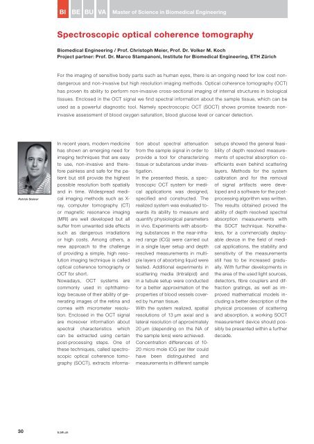
Patrick Steiner<br />
30 ti.bfh.ch<br />
BI BE BU VA Master of Science in Biomedical Engineering<br />
Spectroscopic optical coherence tomography<br />
Biomedical Engineering / Prof. Christoph Meier, Prof. Dr. Volker M. Koch<br />
Project partner: Prof. Dr. Marco Stampanoni, Institute for Biomedical Engineering, ETH Zürich<br />
For the imaging of sensitive body parts such as human eyes, there is an ongoing need for low cost nondangerous<br />
and noninvasive but high resolution imaging methods. Optical coherence tomography (OCT)<br />
has proven its ability to perform noninvasive crosssectional imaging of internal structures in biological<br />
tissues. Enclosed in the OCT signal we find spectral information about the sample tissue, which can be<br />
used as a powerful diagnostic tool. Namely spectroscopic OCT (SOCT) shows promise towards noninvasive<br />
assessment of blood oxygen saturation, blood glucose level or cancer detection.<br />
In recent years, modern medicine<br />
has shown an emerging need for<br />
imaging techniques that are easy<br />
to use, noninvasive and therefore<br />
painless and safe for the patient<br />
but still provide the highest<br />
pos sible resolution both spatially<br />
and in time. Widespread medical<br />
imaging methods such as Xray,<br />
computer tomography (CT)<br />
or magnetic resonance imaging<br />
(MRI) are well developed but all<br />
suffer from unwanted side effects<br />
such as dangerous irradiations<br />
or high costs. Among others, a<br />
new approach to the challenge<br />
of providing a simple, high resolution<br />
imaging technique is called<br />
optical coherence tomography or<br />
OCT for short.<br />
Nowadays, OCT systems are<br />
commonly used in ophthalmology<br />
because of their ability of generating<br />
images of the retina and<br />
cornea with micrometer resolution.<br />
Enclosed in the OCT signal<br />
are moreover information about<br />
spectral characteristics which<br />
can be extracted using certain<br />
postprocessing steps. One of<br />
these techniques, called spectroscopic<br />
optical coherence tomography<br />
(SOCT), extracts informa<br />
tion about spectral attenuation<br />
from the sample signal in order to<br />
provide a tool for characterizing<br />
tissue or substances <strong>und</strong>er investigation.<br />
In the presented thesis, a spectroscopic<br />
OCT system for medical<br />
applications was designed,<br />
specified and constructed. The<br />
realized system was evaluated towards<br />
its ability to measure and<br />
quantify physiological parameters<br />
in vivo. Experiments with absorbing<br />
substances in the nearinfrared<br />
range (ICG) were carried out<br />
in a single layer setup and depth<br />
resolved measurements in multiple<br />
layers of absorbing liquid were<br />
tested. Additional experiments in<br />
scattering media (Intralipid) and<br />
in a tubule setup were conducted<br />
for a better approximation of the<br />
properties of blood vessels covered<br />
by human tissue.<br />
With the system realized, spatial<br />
resolutions of 13 µm axial and a<br />
lateral resolution of approximately<br />
20 µm (depending on the NA of<br />
the sample lens) were achieved.<br />
Concentration differences of 10<br />
20 micro mole ICG per liter could<br />
have been distinguished and<br />
measurements in different sample<br />
setups showed the general feasibility<br />
of depth resolved measurements<br />
of spectral absorption coefficients<br />
even behind scattering<br />
layers. Methods for the system<br />
calibration and for the removal<br />
of signal artifacts were developed<br />
and a software for the postprocessing<br />
algorithm was written.<br />
The results obtained proved the<br />
ability of depth resolved spectral<br />
absorption measurements with<br />
the SOCT technique. Nonetheless,<br />
for a commercially deployable<br />
device in the field of medical<br />
applications, the stability and<br />
sensitivity of the measurements<br />
still has to be increased gradually.<br />
With further developments in<br />
the area of the used light sources,<br />
detectors, fibre couplers and diffraction<br />
gratings, as well as improved<br />
mathematical models including<br />
a better description of the<br />
physical processes of scattering<br />
and absorption, a working SOCT<br />
measurement device should possibly<br />
be presented within a further<br />
decade.