Aberrant migration of Ascaridia galli in a Myna (Acridotheres tristis)
Aberrant migration of Ascaridia galli in a Myna (Acridotheres tristis)
Aberrant migration of Ascaridia galli in a Myna (Acridotheres tristis)

Create successful ePaper yourself
Turn your PDF publications into a flip-book with our unique Google optimized e-Paper software.
Sci Parasitol 13(3):129-131, September 2012<br />
ISSN 1582-1366<br />
129<br />
SHORT RESEARCH NOTE<br />
<strong>Aberrant</strong> <strong>migration</strong> <strong>of</strong> <strong>Ascaridia</strong> <strong>galli</strong> <strong>in</strong> a <strong>Myna</strong> (<strong>Acridotheres</strong> <strong>tristis</strong>)<br />
H. Borji 1� , J. Razmyar 2<br />
1 – Ferdowsi University <strong>of</strong> Mashhad, School <strong>of</strong> Veter<strong>in</strong>ary Medic<strong>in</strong>e, Department <strong>of</strong> Pathobiology, Iran, P.O. Box<br />
9177948974.<br />
2 – Ferdowsi University <strong>of</strong> Mashhad, School <strong>of</strong> Veter<strong>in</strong>ary Medic<strong>in</strong>e, Department <strong>of</strong> Cl<strong>in</strong>ical Science, Iran.<br />
Correspondence: Tel. 0098-511-8788944, Fax 0098-511-8763302, E-mail hborji@um.ac.ir<br />
Abstract. The normal habitat <strong>of</strong> the parasitic stages <strong>of</strong> <strong>Ascaridia</strong> <strong>galli</strong> is <strong>in</strong> the small <strong>in</strong>test<strong>in</strong>e <strong>of</strong> poultry but the<br />
exact life cycle is poorly understood. At necropsy, <strong>in</strong> retroperitoneal <strong>of</strong> a common <strong>Myna</strong> (<strong>Acridotheres</strong> <strong>tristis</strong>) that<br />
was presented for autopsy, high number larvae <strong>of</strong> nematodes were found. After tak<strong>in</strong>g samples, the larvae were<br />
subjected to parasitological exam<strong>in</strong>ation. Larvae were identified <strong>Ascaridia</strong> <strong>galli</strong> that characterized by the<br />
presence <strong>of</strong> three large lips and the oesophagus has no posterior bulb. This report clearly showed that few larvae<br />
<strong>of</strong> <strong>Ascaridia</strong> <strong>galli</strong> could penetrate the small <strong>in</strong>test<strong>in</strong>e and were positioned <strong>in</strong> the abdom<strong>in</strong>al cavity at post<br />
<strong>in</strong>fection. It was far more common that the larvae were localized with<strong>in</strong> the epithelium or <strong>in</strong> the lumen <strong>of</strong> the<br />
crypts. It is therefore suggested that at least <strong>in</strong> this case “extra <strong>in</strong>test<strong>in</strong>al migratory phase” is a more appropriate<br />
term to be used for the A. <strong>galli</strong> larval localization as compared to the term “non migratory phase” currently used<br />
<strong>in</strong> many textbooks.<br />
Keywords: <strong>Ascaridia</strong> spp.; <strong>Acridotheres</strong> <strong>tristis</strong>; <strong>Aberrant</strong> <strong>migration</strong>.<br />
Introduction<br />
<strong>Ascaridia</strong> spp. has been found to be a common<br />
parasite <strong>of</strong> various birds <strong>in</strong> most parts <strong>of</strong> the<br />
world because its direct life cycle and the<br />
environmental resistance <strong>of</strong> its eggs favour<br />
<strong>in</strong>fections under these conditions (Soulsby,<br />
1982). Infections with <strong>Ascaridia</strong> spp. may<br />
cause, <strong>in</strong> addition to direct losses, reductions <strong>in</strong><br />
growth rate, weight loss, reduced feed<br />
conversion rates and damage to the <strong>in</strong>test<strong>in</strong>al<br />
mucosa, lead<strong>in</strong>g to blood loss and secondary<br />
<strong>in</strong>fections (Ackert and Herrick, 1928). In<br />
addition to these serious consequences,<br />
<strong>in</strong>fections may <strong>in</strong>terfere with behavior, social<br />
Received 05/08/2012. Accepted 01/09/2012.<br />
rank. <strong>Ascaridia</strong> spp. may also play a role <strong>in</strong><br />
transmission <strong>of</strong> Salmonella <strong>in</strong>fections result<strong>in</strong>g<br />
<strong>in</strong> disease and economic losses (Kaufmann,<br />
1996). The lifecycle <strong>of</strong> <strong>Ascaridia</strong> spp. is direct<br />
and the <strong>in</strong>fective stage is eggs and <strong>in</strong>fection<br />
takes place by <strong>in</strong>gestion <strong>of</strong> the <strong>in</strong>fective eggs<br />
which with food or water. The eggs hatch <strong>in</strong> the<br />
<strong>in</strong>test<strong>in</strong>e <strong>of</strong> the host and the larvae reach<br />
maturity <strong>in</strong> six to eight weeks <strong>in</strong> the <strong>in</strong>test<strong>in</strong>al<br />
mucosa with no out- <strong>migration</strong> <strong>of</strong> <strong>in</strong>test<strong>in</strong>al<br />
tract when they are swallowed by the birds<br />
(Ackert, 1931). On this background, this article<br />
describes an aberrant <strong>migration</strong> <strong>of</strong> <strong>Ascaridia</strong><br />
<strong>galli</strong> <strong>in</strong> a <strong>Myna</strong>.
Sci Parasitol 13(3):129-131, September 2012<br />
ISSN 1582-1366<br />
Case History<br />
A two year-old common <strong>Myna</strong> (<strong>Acridotheres</strong><br />
<strong>tristis</strong>) was presented for autopsy to Veter<strong>in</strong>ary<br />
Teach<strong>in</strong>g Hospital, School <strong>of</strong> Veter<strong>in</strong>ary<br />
Medic<strong>in</strong>e, Ferdowsi University <strong>of</strong> Mashhad. The<br />
history presented by the owner revealed that<br />
the bird was caged alone; its diet consisted <strong>of</strong><br />
commercial chicken layer’s pellets, fruits and<br />
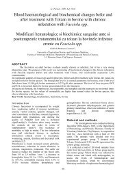
vegetables. Necropsy was done and <strong>in</strong><br />
abdomen cavity high number nematodes was<br />
found (figure 1). The worms were fixed <strong>in</strong> 10%<br />
buffered formal<strong>in</strong> and submitted to the<br />
Parasitology exam<strong>in</strong>ation. The nematodes were<br />
cleared immediately and identified up to genus<br />
level (Yamaguti, 1961). Moreover, to identify<br />
whether the larvae or adult worm found <strong>in</strong> the<br />
gastro<strong>in</strong>test<strong>in</strong>al tracts, the proventriculus to<br />
the cloaca were removed and were also<br />
subjected to parasitological exam<strong>in</strong>ation.<br />
Figure 1. Larvae <strong>of</strong> <strong>Ascaridia</strong> <strong>galli</strong><br />
<strong>in</strong> abdom<strong>in</strong>al cavity <strong>of</strong> <strong>Myna</strong><br />
Result and discussion<br />
The nematodes that were collected <strong>in</strong> this case<br />
were identified as <strong>Ascaridia</strong> <strong>galli</strong> by the<br />
presence <strong>of</strong> three large lips and the oesophagus<br />
has no posterior bulb. After open<strong>in</strong>g <strong>of</strong><br />
gastro<strong>in</strong>test<strong>in</strong>al tract by scissor, few number <strong>of</strong><br />
A. <strong>galli</strong> adult were recovered <strong>in</strong> small <strong>in</strong>test<strong>in</strong>e.<br />
Despite substantial research on other aspects<br />
<strong>of</strong> <strong>Ascaridia</strong> <strong>galli</strong>, the early larval phase <strong>of</strong> the<br />
life cycle is still not fully understood. There is<br />
agreement that the small <strong>in</strong>test<strong>in</strong>e is the<br />
normal habitat <strong>of</strong> the larvae, but whether or<br />
not the larvae have an <strong>in</strong>vasive phase <strong>in</strong> the<br />
lam<strong>in</strong>a propria <strong>of</strong> the tunica mucosa, i.e. a true<br />
130<br />
SHORT RESEARCH NOTE<br />
histotrophic phase, is not clear. Only few<br />
researchers have exam<strong>in</strong>ed the early larval<br />
phase (Ackert, 1923; Ackert, 1931; Todd and<br />
Crowdus, 1952; Tugwell and Ackert, 1952;<br />
Herd and McNaught, 1975). In the study by<br />
Todd and Crowdus (1952) larvae were located<br />
<strong>in</strong> both lumen and the mucosa, whereas Ackert<br />
(1923) found that larvae <strong>of</strong> A. <strong>galli</strong> were<br />
localized deeply among the <strong>in</strong>test<strong>in</strong>al villi and<br />
penetrated the <strong>in</strong>test<strong>in</strong>al Lieberkühn glands,<br />
but the exact age <strong>of</strong> these larvae was not stated.<br />
Moreover, the duration <strong>of</strong> this <strong>in</strong>itial phase has<br />
been a subject <strong>of</strong> discussion. Some authors<br />
mention that the histotrophic phase has a<br />
duration <strong>of</strong> 3–54 days before the larvae move<br />
to a f<strong>in</strong>al phase <strong>in</strong> the lumen (Perm<strong>in</strong> and<br />
Hansen, 1998), while other authors mentioned<br />
that the larvae live <strong>in</strong> the mucosa for several<br />
days up to weeks (Ackert, 1923; Tugwell and<br />
Ackert, 1952; Herd and McNaught, 1975). This<br />
was supported <strong>in</strong> the textbooks by Soulsby<br />
(1982) and Kaufmann (1996). On the other<br />
hand Lapage (1956) described <strong>in</strong> his book that<br />
the newly hatched larvae live <strong>in</strong> the contents <strong>of</strong><br />
the small <strong>in</strong>test<strong>in</strong>e, but later bury their head <strong>in</strong><br />
the <strong>in</strong>test<strong>in</strong>al crypts, and afterwards leave the<br />
<strong>in</strong>test<strong>in</strong>al mucosa to live <strong>in</strong> the <strong>in</strong>test<strong>in</strong>al lumen<br />
as adults.<br />
This report clearly showed that few larvae <strong>of</strong><br />
<strong>Ascaridia</strong> <strong>galli</strong> could penetrate the small<br />
<strong>in</strong>test<strong>in</strong>e and were positioned <strong>in</strong> the abdom<strong>in</strong>al<br />
cavity at post <strong>in</strong>fection. It was far more<br />
common that the larvae were localized with<strong>in</strong><br />
the epithelium or <strong>in</strong> the lumen <strong>of</strong> the crypts. It<br />
is therefore suggested that at least <strong>in</strong> this case<br />
“extra <strong>in</strong>test<strong>in</strong>al migratory phase” is a more<br />
appropriate term to be used for the A. <strong>galli</strong><br />
larval localization as compared to the term<br />
“non migratory phase” currently used <strong>in</strong> many<br />
textbooks.<br />
To achieve a better understand<strong>in</strong>g <strong>of</strong> the life<br />
cycle <strong>of</strong> A. <strong>galli</strong>, studies <strong>of</strong> the localization <strong>of</strong> the<br />
larvae <strong>in</strong> the <strong>in</strong>itial phase <strong>of</strong> the <strong>in</strong>fection are<br />
needed. This knowledge may be relevant for<br />
studies <strong>of</strong> population biology and immunity,<br />
e.g. parasite turnover after <strong>in</strong>fection with<br />
genetically marked cohorts <strong>of</strong> eggs as<br />
suggested by Katakam et al. (2010), or local<br />
immune response <strong>in</strong> the <strong>in</strong>test<strong>in</strong>e e.g. Degen et<br />
al. (2005) and Schwarz et al. (2011).
Sci Parasitol 13(3):129-131, September 2012<br />
ISSN 1582-1366<br />
Acknowledgements<br />
This work was carried out at the Poultry<br />
Reference Centre and Parasitology Section,<br />
Faculty <strong>of</strong> Veter<strong>in</strong>ary Medic<strong>in</strong>e, Ferdowsi<br />
University <strong>of</strong> Mashhad.<br />
References<br />
Ackert J.E. 1923. On the habitat <strong>of</strong> <strong>Ascaridia</strong><br />
perspicillum (Rud). Anat. Rec. 26:101-104.<br />
Ackert J.E. 1931. The morphology and life history <strong>of</strong><br />
the fowl nematode <strong>Ascaridia</strong> l<strong>in</strong>eate<br />
(Schneider). Parasitology 23:360-379.<br />
Ackert J.E., Herrick Ch.A. 1928. Effects <strong>of</strong> the<br />
nematode <strong>Ascaridia</strong> l<strong>in</strong>eate (Schneider) on<br />
grow<strong>in</strong>g. J. Parasitol. 15:1-13.<br />
Degen G.J.W., Daal N., Rothwell L., Kaiser P., Schijns<br />
V. 2005. Th1/Th2 polarization by viral and<br />
helm<strong>in</strong>th <strong>in</strong>fection <strong>in</strong> birds. Vet. Microbiol.<br />
105:163-167.<br />
Herd R.P., McNaught D.J. 1975. Arrested<br />
development and the histotrophic phase <strong>of</strong><br />
<strong>Ascaridia</strong> <strong>galli</strong> <strong>in</strong> the chicken. Int. J. Parasitol.<br />
5:401-406.<br />
Katakam K.K., Nejsum P., Kyvsgaard N.C., Jørgensen<br />
C.B., Thamsborg S.M. 2010. Molecular and<br />
parasitological tools for the study <strong>of</strong> <strong>Ascaridia</strong><br />
<strong>galli</strong> population dynamics <strong>in</strong> chickens. Avian<br />
Pathol. 39:81-85.<br />
131<br />
SHORT RESEARCH NOTE<br />
Kaufmann J. 1996. Parasitic Infections <strong>of</strong> Domestic<br />
Animals: A Diagnostic Manual. Birkhauser<br />
Verlag, Basel, pp. 357-358.<br />
Lapage G. 1956. Veter<strong>in</strong>ary Parasitology. Oliver and<br />
Boyd, Ed<strong>in</strong>burgh/London, p. 173.<br />
Perm<strong>in</strong> A., Hansen J.W. 1998. The Epidemiology,<br />
Diagnosis and Control <strong>of</strong> Poultry Parasite. FAO<br />
Animal Health Manual. Food and Agriculture<br />
Organization <strong>of</strong> the United Nations, Rome, Italy.<br />
Schwarz A., Gauly M., Abel H., Das G., Humburg J.,<br />
Rohn K., Breves G., Rautenschle<strong>in</strong> S. 2011.<br />
Immunopathogenesis <strong>of</strong> <strong>Ascaridia</strong> <strong>galli</strong> <strong>in</strong>fection<br />
<strong>in</strong> layer chicken. Dev. Comp. Immunol. 35:774-<br />
784.<br />
Soulsby E.J.L. 1982. Helm<strong>in</strong>ths, Arthropods and<br />
Protozoa <strong>of</strong> Domesticated Animals, 7th ed.<br />
Balliere T<strong>in</strong>dall. The English Language Book<br />
Society, London.<br />
Todd A.C., Crowdus D.H. 1952. On the life history <strong>of</strong><br />
<strong>Ascaridia</strong> <strong>galli</strong>. Trans. Am. Microscopical Soc.<br />
3:282-287.<br />
Tugwell R.L., Ackert J.E. 1952. On the tissue phase <strong>of</strong><br />
the life cycle <strong>of</strong> the fowl nematode <strong>Ascaridia</strong><br />
<strong>galli</strong> (Schrank). J. Parasitol. 4:277-288.<br />
Yamaguti S. 1961. Systema helm<strong>in</strong>thum. In: The<br />
Nematodes <strong>of</strong> vertebrates. Vol. 3, Part 4,<br />
Interscience Publishers Ltd., London.