Krassilovianthus gen. nov., a new staminate inflorescence with ...
Krassilovianthus gen. nov., a new staminate inflorescence with ...
Krassilovianthus gen. nov., a new staminate inflorescence with ...

You also want an ePaper? Increase the reach of your titles
YUMPU automatically turns print PDFs into web optimized ePapers that Google loves.
2 N.P. Maslova et al. / Review of Palaeobotany and Palynology 180 (2012) 1–14<br />
et al., 1990). Shilin recognized Asplenium dicksonianum Heer, Gleichenia<br />
sp., Sphenopteris sp., Sequoia heterophylla Vele<strong>nov</strong>sky, Cedrus sp.,<br />
Platanus pseudoguillelmae Krassilov, P. cuneiformis Krassilov,<br />
Dalbergites simplex (Newberry) Seward and Ilex sp. Frumin and<br />
Friis (1996, 1999) reported members of the Ranunculales, Urticales,<br />
Rosales, Myrtales, Celastrales, Platanaceae, Illiciaceae and<br />
Magnoliaceae from the flora.<br />
Four <strong>inflorescence</strong>s were studied. Photos of the <strong>inflorescence</strong>s<br />
were taken using a Leica M165 stereomicroscope equipped <strong>with</strong> a<br />
Leica DFC420 digital camera. The <strong>gen</strong>eral morphology of the<br />
<strong>inflorescence</strong>s was studied by a CamScan scanning electron microscope<br />
(SEM) after cleaning flowers <strong>with</strong> hydrofluoric acid. Some fragments of<br />
the <strong>inflorescence</strong>s were macerated <strong>with</strong> Schulze's solution and alkali<br />
andthenobserved<strong>with</strong>aCamScanSEM.<br />
For anatomical examination the <strong>inflorescence</strong>s were cleaned <strong>with</strong><br />
hydrofluoric acid, then gradually dehydrated and embedded in Kulzer's<br />
Tech<strong>nov</strong>it 2100 (2-hydroxyethyl methacrylate) as described in<br />
Igersheim and Cichocki (1996). The embedded material was serially<br />
sectioned at 6–10 μm <strong>with</strong> a rotary microtome. Sections were<br />
mounted unstained in BioMount mounting medium. Sections<br />
were examined under a Carl Zeiss Axioplan-2 bright field light<br />
photomicroscope (LM). Digital photomicrographs of sections<br />
were taken using a Leica DFC420 camera.<br />
Individual pollen grains and fragments of microsporangia were<br />
studied and photographed <strong>with</strong> the same LM and CamScan SEM at<br />
Borissiak Paleontological Institute (PIN) and CamScan and JSM SEMs<br />
and Jeol 100 B and Jeol 1011 transmission electron microscopes<br />
(TEMs) at Lomonosov Moscow State University. Standard methods<br />
for TEM studies were followed after Meyer-Melikyan et al. (2004).<br />
Ultrathin sections were made <strong>with</strong> an LKB Ultratome V and stained<br />
after Reynolds (1963).<br />
Collections # 417, 419 are housed at the Institute of Botany and<br />
Plant Introduction of the Committee of Science and Education of the<br />
Republic of Kazakhstan, Almaty, Kazakhstan.<br />
3. Systematic descriptions<br />
Phylum Magnoliophyta<br />
Order and Family Incertae Sedis<br />
Genus <strong>Krassilovianthus</strong> N. Maslova, Tekleva et Remizowa, <strong>gen</strong>. <strong>nov</strong>.<br />
Etymology: in honour of the palaeobotanist Professor Valentin<br />
Krassilov.<br />
Type species: <strong>Krassilovianthus</strong> sarbaensis N. Maslova, Tekleva et<br />
Remizowa, <strong>gen</strong>. et sp. <strong>nov</strong>.<br />
Diagnosis: Sessile <strong>staminate</strong> heads <strong>with</strong> numerous flowers per<br />
head. Flower <strong>with</strong> a well-developed perianth, exceeding stamen length.<br />
Androecium consists of two sessile stamens. Anthers bisporangiate and<br />
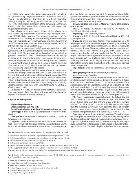
Plate I. <strong>Krassilovianthus</strong> sarbaensis <strong>gen</strong>. et sp. <strong>nov</strong>. 1–3 – under reflected light, 4–8 – SEM.<br />
1. Inflorescence architecture. Macerated 3-D preserved <strong>inflorescence</strong>. Specimen no. 417/106.<br />
2. Inflorescence architecture. Macerated 3-D preserved <strong>inflorescence</strong>. Specimen no. 417/102.<br />
3. Fragment of the <strong>inflorescence</strong> in the rock. Specimen no. 417/103.<br />
4. Inflorescence architecture. Holotype no. 417/101.<br />
5. Flower, two stamens (white arrows) and perianth (black arrows) are visible. Holotype no. 417/101.<br />
6. Flower, perianth elements (black arrows), pollen sacs (white arrows). Specimen no. 417/102.<br />
7, 8. Fragment of <strong>inflorescence</strong> axis, cells walls of the ordinary parenchyma <strong>with</strong> round pores are seen. Specimen no. 417/102.<br />
dithecate. Pollen sacs narrow, elongated. Connective indistinguishable.<br />
Epidermis of pollen sac apices bears stomata and rare trichome bases.<br />
Pollen small, tricolporate, finely reticulate. Lumina forming labyrinthine<br />
pattern. Aperture membrane granular.<br />
<strong>Krassilovianthus</strong> sarbaensis N. Maslova, Tekleva et Remizowa,<br />
<strong>gen</strong>. et sp. <strong>nov</strong>.<br />
Plate I,1–8; Plate II,1–6; Plate III,1–11; Plate IV,1–6; Plate V,1–6;<br />
Plate VI, 1–6; Plate VII — 1–10.<br />
Etymology: From the Sarbay locality.<br />
Holotype: # 417/101; Borissiak Paleontological Institute RAS, (Pl. I,<br />
4), designed here.<br />
Diagnosis: Sessile <strong>staminate</strong> heads 2–3 mm in diameter. Up to 40<br />
flowers per head. Perianth well-developed, exceeding stamen length.<br />
Epidermis of outer and inner perianth elements differs. Flowers <strong>with</strong><br />
two stamens. Stamen filaments lacking. Anthers bisporangiate and<br />
dithecate. Pollen sacs narrow, elongated, <strong>with</strong> almost parallel<br />
margins, occasionally confluent basally, but free for most of their<br />
length. Connective indistinguishable. Epidermis of pollen sac apices<br />
bears stomata and rare trichome bases. Pollen small, tricolporate<br />
and finely reticulate. Lumina varying in shape and size and forming<br />
labyrinthine pattern. Colpi length about 2/3 of polar axis; aperture<br />
membrane granular.<br />
Type locality: Western Kazakhstan, Sarbay locality, near Rudnyi<br />
city.<br />
Stratigraphic position: Zhirkindekskaya Formation.<br />
Age: Cenomanian–Turonian.<br />
Description: The <strong>staminate</strong> <strong>inflorescence</strong> consists of a rather massive<br />
longitudinally striate axis about 1 mm in diameter <strong>with</strong> heads<br />
of 2.5–3.0 mm in diameter (Plate I, 1, 2). Parenchyma cells of the<br />
axis are rectangular, 40–60 μm longand10–20 μm wide; the cell walls<br />
<strong>with</strong> small rounded pits (Plate I, 7, 8). Only fragmentary <strong>inflorescence</strong>s<br />
were found. Each fragment bears only a single head and the number<br />
of heads per axis is unknown. Heads are rather loose, <strong>with</strong> the central<br />
core of about 1 mm in diameter and up to 40 flowers per head.<br />
The flower microstructure was studied <strong>with</strong> the help of SEM and<br />
also in anatomical sections in transmitted light. The flowers are<br />
about 850 μm long and 350–560 μm wide, <strong>with</strong> two stamens<br />
(Plate I, 5,6;Plate III, 1–5; Plate VI, 1,2).Theperianthislonger<br />
than the stamens; however, in most cases the apices of perianth<br />
elements were broken during fossilisation due to their fragility<br />
and loose arrangement (Plate I, 4,5;Plate V, 2;Plate VI, 1,2).<br />
Two types of perianth elements were revealed. The epidermis of<br />
the outer perianth elements differs in the basal and apical parts of<br />
the elements (Plate II, 2). Basally, it consists of strongly elongated cells arranged<br />
in longitudinal rows (Plate II, 3), whereas apically the cells vary in<br />
shape and size from rectangular and spindle-shaped to triangular and polygonal<br />
(Plate II, 4). The walls of epidermal cells of the outer perianth<br />
Plate II. <strong>Krassilovianthus</strong> sarbaensis <strong>gen</strong>. et sp. <strong>nov</strong>., specimen no. 417/102, SEM.1–4. Outer perianth elements: 1, 3 — non-macerated cuticle, 2, 4 — macerated cuticle; 1–3 — view<br />
from the outside, 4 — view from the outside.<br />
1. Outer perianth element in apical part of flower.<br />
2. Cuticle of outer perianth element, view from the outside.<br />
3. Outer perianth element in basal part of flower.<br />
4. Cuticle of apical part of outer perianth element, view from the inside.<br />
5. Basal part of inner perianth element.<br />
6. Apical part of inner perianth element.