Krassilovianthus gen. nov., a new staminate inflorescence with ...
Krassilovianthus gen. nov., a new staminate inflorescence with ...
Krassilovianthus gen. nov., a new staminate inflorescence with ...

Create successful ePaper yourself
Turn your PDF publications into a flip-book with our unique Google optimized e-Paper software.
8 N.P. Maslova et al. / Review of Palaeobotany and Palynology 180 (2012) 1–14<br />
long axis of the sac (Plate III, 11). The cuticle of the pollen sac tips is<br />
preserved fragmentarily, only endothecium cells are present here<br />
(Plate IV, 1). Preserved cuticle fragments show ordinary epidermal<br />
cells that are square in the apical part of the pollen sacs, from 10 to<br />
20 μm, <strong>with</strong> a finely striate outer periclinal wall (Plate IV, 5,6).There<br />
are stomata about 10 μm long (Plate IV, 2–5) and trichomes. The<br />
stomatal type was not determined, but the subsidiary cells are narrower<br />
than ordinary epidermal cells, about 5 μm wide, rather strongly cutinized<br />
<strong>with</strong> cuticle folds situated perpendicular to the stoma axis (Plate IV, 3).<br />
Trichomes are about 8 μm indiameter(Plate IV, 6), <strong>with</strong> broken apices.<br />
Preserved trichome fragments reach 5 μm in length. Endothecium cells<br />
<strong>with</strong> thickened walls are clearly seen in sections (Plate VI, 3,5,6).<br />
Pollen grains are small, <strong>with</strong> the polar axis of 13.3 (12.0–14.6) μmand<br />
equatorial diameter of 10.3 (8.0–12.3) μm, tricolporate; ora are not always<br />
clearly seen (Plate VII, 1–4). The sculpture is finely reticulate <strong>with</strong><br />
lumina varying in shape and size, occasionally elongated, curved or<br />
slit-like, from small to rather large (probably resulting from openended<br />
lumina); this is especially well seen in the meso- and apocolpium<br />
regions (Plate VII, 2–7). A combination of small and large lumina<br />
results in a labyrinthine pattern (Plate VII, 3, 7). The<br />
difference in lumina size is almost indistinct near the aperture margin<br />
where the lumina are very small and almost fused in a continuous<br />
rim in some pollen grains or not fused in others. The colpus<br />
length is about 2/3 of the polar axis. The aperture membrane is<br />
granular (Plate VII, 3,5,6).<br />
The exine is semitectate; the ectexine is less electron dense than<br />
the endexine (Plate VII, 8–10). The non-apertural ectexine is 0.94<br />
(0.67–1.08) μm thick <strong>with</strong> the tectum 0.29 (0.24–0.35) μm thick,<br />
columellae 0.24 (0.2–0.33) μm high and 0.1 (0.08–0.13) μm wide,<br />
and the foot layer 0.43 (0.35–0.48) μm thick (Plate VII, 8, 9). The<br />
endexine is two-layered <strong>with</strong> the layers separating from each other,<br />
especially in the aperture region (Plate VII, 8, 10). The outer endexine<br />
layer is almost homo<strong>gen</strong>eous, being 0.06 (0.03–0.08) μm thick in the<br />
non-aperture region and becoming thinner in the aperture region. It<br />
tightly adjoins the ectexine throughout the pollen perimeter. The<br />
inner endexine layer is granular, 0.18 (0.15–0.2) μm thick in the<br />
non-aperture region; it separates from the outer endexine layer and<br />
may slightly thicken towards the aperture region. Orbicules occur.<br />
Comparison: The <strong>new</strong> <strong>gen</strong>us shows a unique combination of<br />
characters. <strong>Krassilovianthus</strong> shares <strong>with</strong> extant Platanus L. the <strong>gen</strong>eral<br />
architecture of the capitate <strong>inflorescence</strong>, fibrous endothecium, and<br />
tricolporate pollen. Also the two <strong>gen</strong>era are similar in the reduction<br />
of the stamen filament. Anthers are sessile in <strong>Krassilovianthus</strong>, while<br />
Platanus possesses a short stamen filament. The listed characters<br />
occur in some other fossil platanoids <strong>with</strong> a well-developed perianth.<br />
The late Paleocene-early Eocene Chemurnautia N. Maslova is similar<br />
to the <strong>new</strong> <strong>gen</strong>us in having rather loosely arranged pollen sacs, partly<br />
confluent at the base, and a well-developed fibrous layer, and in<br />
lacking a pronounced apical extension of the connective (Maslova,<br />
2002). However, flowers in the <strong>inflorescence</strong> of Chemurnautia are<br />
packed less tightly, the perianth is almost lacking and the pollen<br />
grains differ being tricolpate (not tricolporate) <strong>with</strong> a different<br />
reticulum pattern.<br />
<strong>Krassilovianthus</strong> differs from all species of Platanaceae by the floral<br />
structure. The perianth of <strong>Krassilovianthus</strong> is considerably longer than the androecium,<br />
which consists of two bisporangiate and dithecate stamens <strong>with</strong><br />
free pollen sacs, so the connective is indistinguishable. A well-developed<br />
perianth has been described in the extinct subfamily Gynoplatananthoideae<br />
(Platanaceae). However, in Gynoplatananthoideae,<br />
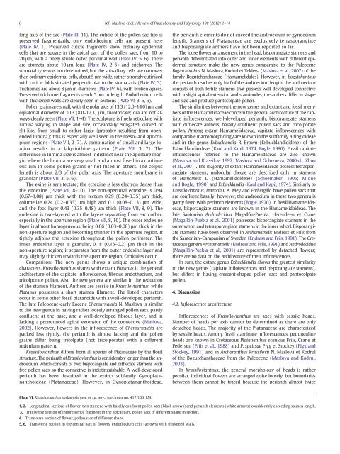
Plate VI. <strong>Krassilovianthus</strong> sarbaensis <strong>gen</strong>. et sp. <strong>nov</strong>., specimen no. 417/106. LM.<br />
the perianth elements do not exceed the androecium or gynoecium<br />
length. Stamens of Platanaceae are exclusively tetrasporangiate<br />
and bisporangiate anthers have not been reported so far.<br />
The loose flower arrangement in the head, bisporangiate stamens and<br />
perianth differentiated into outer and inner elements <strong>with</strong> different epidermal<br />
structure make the <strong>new</strong> <strong>gen</strong>us comparable to the Paleocene<br />
Bogutchanthus N. Maslova, Kodrul et Tekleva (Maslova et al., 2007)ofthe<br />
family Bogutchanthaceae (Hamamelidales). However, in Bogutchanthus<br />
the perianth reaches only half of the androecium length, the androecium<br />
consists of both fertile stamens that possess well-developed connective<br />
<strong>with</strong> a slight apical extension and staminodes, the anthers differ in shape<br />
and size and produce pantocolpate pollen.<br />
The similarities between the <strong>new</strong> <strong>gen</strong>us and extant and fossil members<br />
of the Hamamelidaceae concern the <strong>gen</strong>eral architecture of the capitate<br />
<strong>inflorescence</strong>s, well-developed perianth, bisporangiate stamens<br />
<strong>with</strong> dithecate anthers, basally confluent pollen sacs and tricolporate<br />
pollen. Among extant Hamamelidaceae, capitate <strong>inflorescence</strong>s <strong>with</strong><br />
comparable macromorphology are known in the subfamily Altingioideae<br />
and in the <strong>gen</strong>us Exbucklandia R. Brown (Exbucklandioideae) of the<br />
Exbucklandioideae (Kaul and Kapil, 1974; Bogle, 1986). Fossil capitate<br />
<strong>inflorescence</strong>s referred to the Hamamelidaceae are also known<br />
(Maslova and Krassilov, 1997; Maslova and Golovneva, 2000a,b; Zhou<br />
et al., 2001). The majority of extant Hamamelidaceae possess tetrasporangiate<br />
stamens; unilocular thecae are described only in stamens<br />
of Hamamelis L. (Hamamelidoideae) (Schoemaker, 1905; Mione<br />
and Bogle, 1990) andExbucklandia (Kaul and Kapil, 1974). Similarly to<br />
<strong>Krassilovianthus</strong>, Parrotia C.A. Mey and Fothergilla have pollen sacs that<br />
are confluent basally; however, the androecium in these two <strong>gen</strong>era is<br />
partly fused <strong>with</strong> perianth elements (Bogle, 1970). In fossil Hamamelidaceae,<br />
bisporangiate stamens are known in the Hamamelidoideae. The<br />
late Santonian Androdecidua Magallón-Puebla, Herendeen et Crane<br />
(Magallón-Puebla et al., 2001) possessesbisporangiatestamensinthe<br />
outer whorl and tetrasporangiate stamens in the inner whorl. Bisporangiate<br />
stamens have been observed in Archamamelis Endress et Friis from<br />
the Santonian–Campanian of Sweden (Endress and Friis, 1991). The Cretaceous<br />
<strong>gen</strong>era Archamamelis (Endress and Friis, 1991)andAndrodecidua<br />
(Magallón-Puebla et al., 2001) are represented by detached flowers;<br />
there are no data on the architecture of their <strong>inflorescence</strong>s.<br />
In sum, the extant <strong>gen</strong>us Exbucklandia shows the greatest similarity<br />
to the <strong>new</strong> <strong>gen</strong>us (capitate <strong>inflorescence</strong>s and bisporangiate stamens),<br />
but differs in having crescent-shaped pollen sacs and pantocolpate<br />
pollen.<br />
4. Discussion<br />
4.1. Inflorescence architecture<br />
Inflorescences of <strong>Krassilovianthus</strong> are axes <strong>with</strong> sessile heads.<br />
Number of heads per axis cannot be determined as there are only<br />
detached heads. The majority of the Platanaceae are characterized<br />
by sessile heads. Among fossil <strong>staminate</strong> <strong>inflorescence</strong>s, pedunculate<br />
heads are known in Cretaceous Platananthus scanicus Friis, Crane et<br />
Pedersen (Friis et al., 1988) and P. speirsae Pigg et Stockey (Pigg and<br />
Stockey, 1991) and in Archaranthus krassilovii N. Maslova et Kodrul<br />
of the Bogutchanthaceae from the Paleocene (Maslova and Kodrul,<br />
2003).<br />
In <strong>Krassilovianthus</strong>, the <strong>gen</strong>eral morphology of heads is rather<br />
peculiar. Individual flowers are arranged quite loosely, but boundaries<br />
between them cannot be traced because the perianth almost twice<br />
1, 2. Longitudinal sections of flower, two stamens <strong>with</strong> basally confluent pollen sacs (black arrows) and perianth elements (white arrows) considerably exceeding stamen length.<br />
3. Transverse section of <strong>inflorescence</strong> fragment in the apical part, pollen sacs of different shape in section.<br />
4. Transverse section of flower, pollen sacs of different shape.<br />
5, 6. Transverse section in the central part of flowers, endothecium cells (arrows) <strong>with</strong> thickened walls.