Autofluorescent Proteins with Excitation in the Optical ... - Tsien
Autofluorescent Proteins with Excitation in the Optical ... - Tsien
Autofluorescent Proteins with Excitation in the Optical ... - Tsien

Create successful ePaper yourself
Turn your PDF publications into a flip-book with our unique Google optimized e-Paper software.
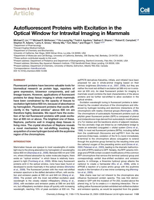
Chemistry & Biology<br />
Article<br />
<strong>Autofluorescent</strong> <strong>Prote<strong>in</strong>s</strong> <strong>with</strong> <strong>Excitation</strong> <strong>in</strong> <strong>the</strong><br />
<strong>Optical</strong> W<strong>in</strong>dow for Intravital Imag<strong>in</strong>g <strong>in</strong> Mammals<br />
Michael Z. L<strong>in</strong>, 1,5,6 Michael R. McKeown, 1,5 Ho-Leung Ng, 4 Todd A. Aguilera, 1 Nathan C. Shaner, 1,7 Robert E. Campbell, 1,8<br />
Stephen R. Adams, 1 Larry A. Gross, 1 Wendy Ma, 4 Tom Alber, 4 and Roger Y. <strong>Tsien</strong>1,2,3,* 1Department of Pharmacology<br />
2Howard Hughes Medical Institute<br />
3Department of Chemistry and Biochemistry<br />
University of California, San Diego, 9500 Gilman Drive, La Jolla, CA 92093, USA<br />
4Department of Molecular and Cell Biology, University of California, Berkeley, 356 Stanley Hall, Berkeley, CA 94720, USA<br />
5These authors contributed equally to this work<br />
6Present address: Department of Pediatrics and Department of Bioeng<strong>in</strong>eer<strong>in</strong>g, Stanford University, Palo Alto, CA 94305, USA<br />
7Present address: Monterey Bay Aquarium Research Institute, 7700 Sandholdt Road, Moss Land<strong>in</strong>g, CA 95039, USA<br />
8Present address: Department of Chemistry, University of Alberta, Edmonton, Alberta T6G 2G2, Canada<br />
*Correspondence: rtsien@ucsd.edu<br />
DOI 10.1016/j.chembiol.2009.10.009<br />
SUMMARY<br />
Fluorescent prote<strong>in</strong>s have become valuable tools for<br />
biomedical research as prote<strong>in</strong> tags, reporters of<br />
gene expression, biosensor components, and cell<br />
l<strong>in</strong>eage tracers. However, applications of fluorescent<br />
prote<strong>in</strong>s for deep tissue imag<strong>in</strong>g <strong>in</strong> whole mammals<br />
have been constra<strong>in</strong>ed by <strong>the</strong> opacity of tissues to<br />
excitation light below 600 nm, because of absorbance<br />
by hemoglob<strong>in</strong>. Fluorescent prote<strong>in</strong>s that excite efficiently<br />
<strong>in</strong> <strong>the</strong> ‘‘optical w<strong>in</strong>dow’’ above 600 nm are<br />
<strong>the</strong>refore highly desirable. We report here <strong>the</strong> evolution<br />
of far-red fluorescent prote<strong>in</strong>s <strong>with</strong> peak excitation<br />
at 600 nm or above. The brightest one of <strong>the</strong>se,<br />
Neptune, performs well <strong>in</strong> imag<strong>in</strong>g deep tissues <strong>in</strong><br />
liv<strong>in</strong>g mice. The crystal structure of Neptune reveals<br />
a novel mechanism for red-shift<strong>in</strong>g <strong>in</strong>volv<strong>in</strong>g <strong>the</strong><br />
acquisition of a new hydrogen bond <strong>with</strong> <strong>the</strong> acylim<strong>in</strong>e<br />
region of <strong>the</strong> chromophore.<br />
INTRODUCTION<br />
Mammalian tissues are opaque to most wavelengths of visible<br />
light due to <strong>the</strong> strong absorption by hemoglob<strong>in</strong> of wavelengths<br />
below 600 nm (Stamatas et al., 2006). Between 600 nm and 1200<br />
nm, where <strong>in</strong>frared absorption by water molecules beg<strong>in</strong>s, <strong>the</strong>re<br />
exists an ‘‘optical w<strong>in</strong>dow’’ <strong>in</strong> which tissue is relatively transparent<br />
to light (Tromberg et al., 2000). While many fluorescent<br />
prote<strong>in</strong>s emit <strong>in</strong> <strong>the</strong> optical w<strong>in</strong>dow, none have been found or<br />
developed to excite maximally <strong>in</strong> <strong>the</strong> optical w<strong>in</strong>dow (Shcherbo<br />
et al., 2009). The fluorescent prote<strong>in</strong> <strong>with</strong> <strong>the</strong> most red-shifted<br />
emission spectrum is <strong>the</strong> dsRed derivative mPlum, <strong>with</strong> excitation<br />
and emission peaks at 590 nm and 649 nm (Wang et al.,<br />
2004). The prote<strong>in</strong> <strong>with</strong> <strong>the</strong> most red-shifted excitation peak<br />
wavelength is <strong>the</strong> closely related mRaspberry (Wang et al.,<br />
2004), <strong>with</strong> excitation and emission peaks at 598 nm and 625<br />
nm, but mRaspberry excitation drops off quickly <strong>with</strong> <strong>in</strong>creas<strong>in</strong>g<br />
wavelength, reach<strong>in</strong>g 10% of peak excitation at 630 nm. The<br />
eqFP578 derivatives Katushka, mKate, and mKate2 have been<br />
proposed for use <strong>in</strong> whole-animal imag<strong>in</strong>g based on <strong>the</strong>ir<br />
<strong>in</strong>tr<strong>in</strong>sic brightness (Shcherbo et al., 2007, 2009), but <strong>the</strong>y are<br />
nei<strong>the</strong>r <strong>the</strong> most red-shifted <strong>in</strong> excitation (at 588 nm) nor <strong>in</strong> emission<br />
(at 635 nm). An ideal fluorescent prote<strong>in</strong> for imag<strong>in</strong>g <strong>in</strong><br />
mammals would comb<strong>in</strong>e <strong>the</strong> best characteristics of <strong>the</strong> above<br />
prote<strong>in</strong>s and be fur<strong>the</strong>r red-shifted to allow excitation <strong>in</strong> <strong>the</strong><br />
optical w<strong>in</strong>dow.<br />
<strong>Excitation</strong> wavelength tun<strong>in</strong>g <strong>in</strong> fluorescent prote<strong>in</strong>s is determ<strong>in</strong>ed<br />
by <strong>the</strong> covalent structure of <strong>the</strong> chromophore and <strong>in</strong>fluenced<br />
by hydrogen bond<strong>in</strong>g and electronic <strong>in</strong>teractions of <strong>the</strong><br />
chromophore <strong>with</strong> nearby chemical groups (Rem<strong>in</strong>gton, 2006).<br />
The chromophore of autocatalytic fluorescent prote<strong>in</strong>s such as<br />
jellyfish green fluorescent prote<strong>in</strong> (GFP) is composed of phenol<br />
and imidazol<strong>in</strong>one r<strong>in</strong>gs derived from autocatalytic modifications<br />
of a Tyr residue and <strong>the</strong> backbone atoms of adjacent residues.<br />
The two aromatic r<strong>in</strong>gs are l<strong>in</strong>ked by an methylidene bridge to<br />
create an extended conjugated p orbital system (Ormo et al.,<br />
1996). In most red fluorescent prote<strong>in</strong>s (RFPs), <strong>in</strong>clud<strong>in</strong>g dsRed<br />
from <strong>the</strong> corallimorph Discosoma and eqFP611 from <strong>the</strong> sea<br />
anemone Entacmaea, oxidation of <strong>the</strong> N-Ca bond immediately<br />
N-term<strong>in</strong>al to <strong>the</strong> chromophore allows conjugation to extend<br />
through <strong>the</strong> acylim<strong>in</strong>e group formed by <strong>the</strong> result<strong>in</strong>g im<strong>in</strong>e and<br />
<strong>the</strong> carbonyl oxygen of <strong>the</strong> preced<strong>in</strong>g am<strong>in</strong>o acid (Gross et al.,<br />
2000; Petersen et al., 2003), lead<strong>in</strong>g to <strong>the</strong> dramatic bathochromic<br />
shift of RFPs relative to GFP. Notably, <strong>the</strong> RFP variants mOrange<br />
and zFP538 conta<strong>in</strong> covalent modifications of this common<br />
RFP chromophore that remove <strong>the</strong> term<strong>in</strong>al carbonyl group and<br />
correspond<strong>in</strong>gly exhibit blue-shifted excitation and emission<br />
spectra. In mOrange, a threon<strong>in</strong>e hydroxyl group attacks <strong>the</strong><br />
carbonyl to create a hemiacetal (Shu et al., 2006), while <strong>in</strong><br />
zFP538 a reaction of a lys<strong>in</strong>e am<strong>in</strong>o group <strong>with</strong> <strong>the</strong> acylim<strong>in</strong>e<br />
results <strong>in</strong> <strong>the</strong> formation of a new im<strong>in</strong>e-conta<strong>in</strong><strong>in</strong>g r<strong>in</strong>g (Rem<strong>in</strong>gton<br />
et al., 2005).<br />
Side cha<strong>in</strong>s near but not <strong>in</strong>herent to <strong>the</strong> chromophore also<br />
<strong>in</strong>fluence fluorescent prote<strong>in</strong> spectra. This was first demonstrated<br />
<strong>in</strong> GFP <strong>with</strong> <strong>the</strong> mutation of Thr-203, located above <strong>the</strong><br />
phenol r<strong>in</strong>g, to Tyr to create a p-p stack<strong>in</strong>g <strong>in</strong>teraction. The result<strong>in</strong>g<br />
yellow fluorescent prote<strong>in</strong> exhibited red-shifted excitation<br />
and emission spectra, as would be expected from <strong>the</strong> greater<br />
Chemistry & Biology 16, 1169–1179, November 25, 2009 ª2009 Elsevier Ltd All rights reserved 1169
Table 1. Characteristics of Far-Red Fluorescent <strong>Prote<strong>in</strong>s</strong><br />
polarizability of <strong>the</strong> stacked p orbital system (Ormo et al., 1996).<br />
A cation-p <strong>in</strong>teraction between a histid<strong>in</strong>e side cha<strong>in</strong> and <strong>the</strong><br />
chromophore has <strong>the</strong> opposite effect of creat<strong>in</strong>g a hypsochromic<br />
shift <strong>in</strong> teal fluorescent prote<strong>in</strong>. This can be expla<strong>in</strong>ed by preferential<br />
stabilization of <strong>the</strong> ground state due to stabilization of<br />
negative charge over <strong>the</strong> chromophore phenolate (Henderson<br />
et al., 2007). Preferential stabilization of <strong>the</strong> ground state would<br />
<strong>in</strong>crease <strong>the</strong> energy difference between ground and excitation<br />
states, requir<strong>in</strong>g <strong>the</strong> absorption of higher-energy photons for<br />
excitation.<br />
We set out to create fluorescent prote<strong>in</strong>s that can be excited <strong>in</strong><br />
<strong>the</strong> optical w<strong>in</strong>dow at 600 nm and above. We carried out a<br />
directed evolution strategy start<strong>in</strong>g from Discosoma and Entacmaea<br />
family RFPs <strong>in</strong>volv<strong>in</strong>g site-directed and random mutagenesis.<br />
One of <strong>the</strong> result<strong>in</strong>g prote<strong>in</strong>s, Neptune, has excitation and<br />
emission peaks at 600 and 650 nm and performs well <strong>in</strong> deeptissue<br />
imag<strong>in</strong>g <strong>in</strong> live mammals us<strong>in</strong>g excitation light <strong>in</strong> <strong>the</strong><br />
optical w<strong>in</strong>dow. Our analysis of <strong>the</strong> structure of Neptune<br />
revealed multiple mechanisms contribut<strong>in</strong>g to <strong>the</strong> long wavelengths<br />
of Neptune, <strong>in</strong>clud<strong>in</strong>g enhanced chromophore coplanarity<br />
and <strong>the</strong> addition of a novel water-chromophore hydrogen<br />
bond to <strong>the</strong> acylim<strong>in</strong>e oxygen of <strong>the</strong> chromophore.<br />
RESULTS<br />
<strong>Excitation</strong><br />
Peak a<br />
Emission<br />
Peak a<br />
Evolution of Far-Red Fluorescent <strong>Prote<strong>in</strong>s</strong><br />
<strong>with</strong> <strong>Excitation</strong> <strong>in</strong> <strong>the</strong> <strong>Optical</strong> W<strong>in</strong>dow<br />
We first attempted to create red-shifted fluorescent prote<strong>in</strong>s<br />
based on mRFP1 by <strong>in</strong>troduc<strong>in</strong>g a tyros<strong>in</strong>e side cha<strong>in</strong> near <strong>the</strong><br />
chromophore to create a p-p stack<strong>in</strong>g <strong>in</strong>teraction similar to <strong>the</strong><br />
3 b<br />
f c<br />
Brightness<br />
Excited at Peak d<br />
Brightness<br />
Excited at 633 nm e<br />
Prote<strong>in</strong><br />
pKa<br />
dTomato g<br />
554 581 69,000 0.69 48 0 64 4.7<br />
mCherry g<br />
587 610 72,000 0.22 16 0.084 68
Chemistry & Biology<br />
Fluorescent <strong>Prote<strong>in</strong>s</strong> <strong>with</strong> Far-Red <strong>Excitation</strong><br />
Figure 1. Neptune Spectra and Sequence<br />
(A) Absorbance spectra of oxyhemoglob<strong>in</strong> (red), deoxyhemoglob<strong>in</strong> (dotted<br />
red), mKate (green dashed), mKate S158C (orange), mKate2 (dotted black),<br />
Neptune (dashed blue), and mNeptune (light blue). Units are M 1 cm 1 versus<br />
nm. Hemoglob<strong>in</strong> spectra are previously published (Stamatas et al., 2006).<br />
(Inset) Purified mCherry, mKate, S158C, and Neptune prote<strong>in</strong>s at 0.5 mg/ml<br />
<strong>in</strong> visible light.<br />
(B) Normalized excitation (left) and emission (right) spectra of mKate (green<br />
dashed), mKate S158C (orange), Neptune (dashed blue), and mNeptune (light<br />
blue). mKate2 excitation and emission spectra are identical to mKate, as<br />
described previously (Shcherbo et al., 2009).<br />
(C) Sequence of mNeptune aligned <strong>with</strong> its parent, mKate. Changes responsible<br />
for <strong>the</strong> red-shift are highlighted <strong>in</strong> red. The additional monomerization<br />
mutation to create mNeptune from Neptune is highlighted <strong>in</strong> green.<br />
Ser-158 to Cys or Ala improved <strong>the</strong> peak ext<strong>in</strong>ction coefficient of<br />
mKate (Figure 1A andTable 1). In <strong>the</strong> course of this work, mKate<br />
S158A (Pletnev et al., 2008) and its improved fold<strong>in</strong>g variant<br />
mKate2 (Shcherbo et al., 2009) were described. mKate and its<br />
S158A and S158C mutants all exhibit residual green fluorescent<br />
components (Figure S2A). While TagRFP (Shaner et al., 2008),<br />
TagRFP-T (Shaner et al., 2008), mKate, and mKate S158A<br />
(Figure S2A) bleached <strong>with</strong> complex k<strong>in</strong>etics, mKate S158C<br />
bleached <strong>in</strong> a monoexponential manner <strong>with</strong> a normalized halflife<br />
of 220 s, mak<strong>in</strong>g it <strong>the</strong> most photostable fluorescent prote<strong>in</strong><br />
<strong>with</strong> monoexponential photobleach<strong>in</strong>g. This rare attribute makes<br />
mKate S158C well suited for quantitative time-lapse experiments.<br />
We next attempted to fur<strong>the</strong>r red-shift mKate S158C by adopt<strong>in</strong>g<br />
<strong>the</strong> mechanism of emission red-shift<strong>in</strong>g <strong>in</strong> mPlum. In mPlum,<br />
Glu-13 hydrogen bonds to <strong>the</strong> term<strong>in</strong>al carbonyl of <strong>the</strong> conjugated<br />
bond system of <strong>the</strong> chromophore, but <strong>the</strong> <strong>in</strong>teraction is<br />
dependent on chromophore excitation and <strong>the</strong>refore primarily<br />
affects <strong>the</strong> emission spectrum (Abbyad et al., 2007; Shu et al.,<br />
2009b). We hypo<strong>the</strong>sized that a stable hydrogen bond<strong>in</strong>g <strong>in</strong>teraction<br />
<strong>with</strong> <strong>the</strong> term<strong>in</strong>al carbonyl <strong>in</strong> <strong>the</strong> ground state could cause<br />
a red-shift <strong>in</strong> excitation as well. Screen<strong>in</strong>g of libraries <strong>with</strong> all<br />
possible substitutions at position 13 and nearby positions 28,<br />
41, and 65, which are not conserved between mKate and<br />
mPlum, did not recover an obvious hydrogen bond donor at<br />
one of <strong>the</strong>se positions. However, we found that mutation of<br />
Met-41 to Gly alone resulted <strong>in</strong> red-shifted excitation and emission<br />
(Table 1). Two rounds of random mutagenesis by errorprone<br />
PCR <strong>the</strong>n yielded Y194F and S61C mutations, result<strong>in</strong>g<br />
<strong>in</strong> a prote<strong>in</strong> <strong>with</strong> 600 nm excitation and 650 nm emission<br />
(Figure 1B and Table 1). Because this prote<strong>in</strong> (mKate M41G<br />
S61C S158C Y194F; Figure 1C) looks blue <strong>in</strong> ambient light<strong>in</strong>g<br />
(Figure 1A) and has fluorescence wavelengths fur<strong>the</strong>st away<br />
from our start<strong>in</strong>g po<strong>in</strong>t among mKate derivatives, we named it<br />
Neptune.<br />
Neptune is <strong>the</strong> first bright fluorescent prote<strong>in</strong> (ext<strong>in</strong>ction coefficient<br />
72,000 M 1 cm 1 , quantum yield 0.18) <strong>with</strong> an excitation<br />
peak reach<strong>in</strong>g 600 nm. Compared to its parent, mKate, its<br />
absorption and excitation peaks are 18 nm redder (600 versus<br />
582 nm) and its peak ext<strong>in</strong>ction coefficient 71% larger (72,000<br />
versus 42,000 M 1 cm 1 ), result<strong>in</strong>g <strong>in</strong> more efficient excitation<br />
beyond 600 nm (Figure 1A and Table 1). Neptune exhibits a photobleach<strong>in</strong>g<br />
half-life longer than EGFP (Figure S2A), a pKa of 5.8,<br />
and fast maturation (half-maximal <strong>in</strong> 35 m<strong>in</strong>; Figure S2C). Like<br />
mKate and <strong>the</strong> brighter mKate S158A and S158C variants,<br />
Neptune shows a residual green component upon excitation at<br />
470 nm (Figure S2C).<br />
Visualization of Neptune <strong>in</strong> Cells and Deep<br />
Tissues of Liv<strong>in</strong>g Mammals<br />
Compared to previous fluorescent prote<strong>in</strong>s, Neptune has <strong>the</strong><br />
largest ext<strong>in</strong>ction coefficient at 633 nm, a wavelength well <strong>with</strong><strong>in</strong><br />
<strong>the</strong> optical w<strong>in</strong>dow and a common laser l<strong>in</strong>e used for excit<strong>in</strong>g<br />
<strong>the</strong> organic fluorophore Cy5 (Figure 1A and Table 1). We tested<br />
<strong>the</strong> ability to detect Neptune or mKate <strong>in</strong> cells <strong>with</strong> 633 nm excitation.<br />
Neptune was well detected <strong>in</strong> liver sections follow<strong>in</strong>g<br />
adenovirus-mediated gene transfer <strong>in</strong> mice (Figure 2A) <strong>with</strong><br />
532 or 633 nm laser excitation, while mKate was only detected<br />
above background <strong>with</strong> <strong>the</strong> 532 nm laser (Figure 2B). Thus <strong>the</strong><br />
bathochromic shift of Neptune relative to its parent mKate has<br />
conferred improved excitability at 633 nm, well <strong>with</strong><strong>in</strong> <strong>the</strong> optical<br />
w<strong>in</strong>dow.<br />
The ability to be excited at 630 nm makes Neptune potentially<br />
suitable for fluorescence imag<strong>in</strong>g <strong>in</strong> liv<strong>in</strong>g mammals us<strong>in</strong>g<br />
exist<strong>in</strong>g Cy5 filter sets. The liver <strong>in</strong> particular is a heavily vascularized<br />
organ where hemoglob<strong>in</strong> absorbance h<strong>in</strong>ders imag<strong>in</strong>g<br />
(Col<strong>in</strong> et al., 2000). Us<strong>in</strong>g an available Cy5 filter set (610–630<br />
nm excitation, 660–700 nm emission), Neptune fluorescence <strong>in</strong><br />
livers of liv<strong>in</strong>g mice follow<strong>in</strong>g adenovirus-mediated gene transfer<br />
was easily visible, <strong>with</strong> <strong>the</strong> brightest areas display<strong>in</strong>g 44-fold<br />
contrast over un<strong>in</strong>fected tissue autofluorescence (Figure 2C).<br />
We recently eng<strong>in</strong>eered a bacterial phytochrome <strong>in</strong>to a biliverd<strong>in</strong>-dependent<br />
<strong>in</strong>frared fluorescent prote<strong>in</strong>, IFP1.1, <strong>with</strong> excitation<br />
and emission maxima at 660 nm and 700 nm (Shu et al.,<br />
2009a). To see whe<strong>the</strong>r Neptune could be visualized orthogonally<br />
to IFP1.1, we imaged liv<strong>in</strong>g mice express<strong>in</strong>g ei<strong>the</strong>r Neptune<br />
or IFP1.1, us<strong>in</strong>g filter sets separately optimized for Cy5 (far-red<br />
Chemistry & Biology 16, 1169–1179, November 25, 2009 ª2009 Elsevier Ltd All rights reserved 1171
Figure 2. Performance of Neptune <strong>in</strong> Cells and Animals<br />
(A) Design of a monocistronic adenovirus vector for coexpression of GFP and Neptune (Ad-Neptune) or (Ad-mKate) <strong>in</strong> hepatocytes.<br />
(B) Sections of liver were removed from a mouse <strong>in</strong>jected <strong>with</strong> Ad-Neptune or Ad-mKate and imaged at red or far-red wavelengths on a confocal microscope. GFP<br />
was also imaged to confirm expression of <strong>the</strong> cistron. Fluorescence <strong>in</strong>tensities are shown <strong>in</strong> <strong>the</strong> chart. Scale bar, 50 mm.<br />
(C) Epifluorescence of Neptune <strong>in</strong> <strong>the</strong> liver of a liv<strong>in</strong>g mouse acquired on a Maestro imag<strong>in</strong>g system <strong>with</strong> excitation and emission at 610–630 nm and 660–700 nm,<br />
respectively. Scale bar, 5 mm.<br />
(D) Dual fluorescence imag<strong>in</strong>g <strong>in</strong> <strong>the</strong> optical w<strong>in</strong>dow is possible <strong>with</strong> Neptune and <strong>in</strong>frared fluorescent prote<strong>in</strong>. Images of live mice express<strong>in</strong>g Neptune or IFP1.1 <strong>in</strong><br />
liver were acquired <strong>with</strong> Cy5 and Cy5.5 channels. <strong>Excitation</strong>/emission wavelengths used were 610–630/660–700 nm for Cy5 and 625–675/700–730 nm for Cy5.5.<br />
In <strong>the</strong> right column, spectral unmix<strong>in</strong>g was used to correct for Neptune cross-excitation and -detection <strong>in</strong> <strong>the</strong> Cy5.5 channel. Restrict<strong>in</strong>g <strong>in</strong>frared fluorescent<br />
prote<strong>in</strong> excitation to above 650 nm <strong>with</strong> custom filters would produce similar results.<br />
emission) and Cy5.5 (<strong>in</strong>frared emission), respectively. Indeed<br />
liver-expressed Neptune was selectively imaged <strong>in</strong> <strong>the</strong> Cy5<br />
channel while IFP1.1 was preferentially imaged <strong>in</strong> <strong>the</strong> Cy5.5<br />
channel (Figure 2D). A small amount of cross-detection of<br />
Neptune occurred us<strong>in</strong>g our Cy5.5 excitation filter due to its<br />
broad excitation range (625–675 nm). This signal can be subtracted<br />
by l<strong>in</strong>ear unmix<strong>in</strong>g (Figure 2D) or preferably elim<strong>in</strong>ated<br />
by <strong>the</strong> future design of an appropriately narrow excitation filter<br />
for IFP1.1. We also confirmed that excitation wavelengths <strong>in</strong><br />
<strong>the</strong> optical w<strong>in</strong>dow detected liver fluorescence more efficiently<br />
than shorter wavelengths for both Neptune and its parent mKate<br />
(Figure S3 and Table S2).<br />
1172 Chemistry & Biology 16, 1169–1179, November 25, 2009 ª2009 Elsevier Ltd All rights reserved<br />
Chemistry & Biology<br />
Fluorescent <strong>Prote<strong>in</strong>s</strong> <strong>with</strong> Far-Red <strong>Excitation</strong><br />
Quaternary Structure of Neptune and Fur<strong>the</strong>r<br />
Monomerization<br />
We studied <strong>the</strong> quaternary structure of Neptune, mKate, and<br />
mKate derivatives <strong>in</strong> solution by size-exclusion chromatography<br />
<strong>with</strong> <strong>in</strong>-l<strong>in</strong>e multiangle laser light scatter<strong>in</strong>g. At low micromolar<br />
concentrations, Neptune and mKate (Figure 3A), as well as mKate<br />
S158A and mKate S158C (Figure S4A), primarily comigrate <strong>with</strong><br />
<strong>the</strong> dimeric standard dTomato. However, <strong>the</strong> elution profiles<br />
are not monodisperse, but ra<strong>the</strong>r exhibit a trail<strong>in</strong>g tail that overlaps<br />
<strong>with</strong> <strong>the</strong> elution profile of <strong>the</strong> monomeric standard mCherry.<br />
In contrast, mGrape3, EGFP, and mCherry all comigrate <strong>in</strong> a sharp<br />
monodisperse peak (Figure S4A). Light scatter<strong>in</strong>g reveals
Chemistry & Biology<br />
Fluorescent <strong>Prote<strong>in</strong>s</strong> <strong>with</strong> Far-Red <strong>Excitation</strong><br />
average molecular weights for Neptune and mKate of 62 and 58<br />
kDa at <strong>the</strong> elution peak, consistent <strong>with</strong> dimers (Figure 3A).<br />
Average measured molecular weight <strong>in</strong> <strong>the</strong> tail is consistent<br />
<strong>with</strong> <strong>the</strong> late-elut<strong>in</strong>g subpopulation of mKate prote<strong>in</strong>s be<strong>in</strong>g<br />
predom<strong>in</strong>antly monomers (34 kDa), while late-elut<strong>in</strong>g Neptune<br />
prote<strong>in</strong>s exhibit an average molecular weight of 40 kDa, suggest<strong>in</strong>g<br />
a mix of monomers and dimers. F<strong>in</strong>ally, at similar concentrations,<br />
Neptune and mKate migrate as dimers <strong>in</strong> native gel electrophoresis<br />
but as monomers <strong>in</strong> SDS, a behavior between that of<br />
mCherry, which migrates as a monomer <strong>in</strong> both conditions, and<br />
dTomato, which migrates as a dimer <strong>in</strong> both conditions (Figures<br />
S4B and S4C). Toge<strong>the</strong>r, <strong>the</strong>se data suggest that mKate and<br />
Neptune exist <strong>in</strong> a monomer-dimer equilibrium at low micromolar<br />
concentrations.<br />
To improve its performance <strong>in</strong> fusion prote<strong>in</strong>s, we fur<strong>the</strong>r<br />
monomerized Neptune. The Y194F mutation <strong>in</strong> Neptune alters<br />
an external side cha<strong>in</strong> located adjacent to Met-146, which participates<br />
<strong>in</strong> a hydrophobic <strong>in</strong>tersubunit <strong>in</strong>teraction. Removal of<br />
a polar hydroxyl group by <strong>the</strong> Y194F mutation <strong>in</strong> Neptune might<br />
have resulted <strong>in</strong> <strong>in</strong>creased dimerization tendency. To restore<br />
a polar group near position 194, we performed saturation mutagenesis<br />
of Met-146 and found a M146T mutant that reta<strong>in</strong>ed <strong>the</strong><br />
brightness and spectral profile of Neptune (Table 1 and Figure 1),<br />
while exhibit<strong>in</strong>g enhanced dissociation <strong>in</strong>to monomers compared<br />
to mKate (Figure 3A). We performed equilibrium analytical<br />
ultracentrifugation to measure dissociation constants for<br />
Neptune and Neptune M146T. Sedimentation profiles were<br />
best fit to a monomer-dimer equilibrium model <strong>with</strong> a dissociation<br />
constant of 2.1 mM for <strong>the</strong> M146T mutant compared <strong>with</strong><br />
Figure 3. A functionally Monomeric Variant<br />
of Neptune for Prote<strong>in</strong> Fusions<br />
(A) Neptune exhibits more dimerization relative to<br />
mKate, while mNeptune exhibits less. Normalized<br />
elution profiles on size-exclusion HPLC (th<strong>in</strong><br />
traces, axis on left) and estimated molecular<br />
weights upon elution by <strong>in</strong>-l<strong>in</strong>e light scatter<strong>in</strong>g<br />
(thick traces, axis on right) are shown for dTomato<br />
(orange solid), Neptune (dark blue dashed), mNeptune<br />
(light blue solid), mKate (green dashed),<br />
mKate2 (red dashed), and mCherry (maroon solid).<br />
Dots show estimated molecular weights upon<br />
elution by <strong>in</strong>-l<strong>in</strong>e light scatter<strong>in</strong>g. <strong>Prote<strong>in</strong>s</strong> were<br />
analyzed sequentially <strong>in</strong> <strong>the</strong> same day at 5 mM <strong>in</strong><br />
PBS. mCherry and dTomato serve as monomeric<br />
and dimeric controls, respectively.<br />
(B) mNeptune-act<strong>in</strong> and mNeptune-tubul<strong>in</strong> are<br />
correctly localized upon expression <strong>in</strong> HeLa cells,<br />
similarly to mKate2 fusions. Scale bar, 10 mm.<br />
0.5 mM for Neptune (Table S3), confirm<strong>in</strong>g<br />
<strong>the</strong> <strong>in</strong>creased monomeric character of<br />
<strong>the</strong> mutant. Fusions of Neptune 146T<br />
prote<strong>in</strong>, which we refer to as mNeptune,<br />
to act<strong>in</strong> and tubul<strong>in</strong> localized correctly <strong>in</strong><br />
HeLa cells, similarly to mKate2 (Figure<br />
3B), demonstrat<strong>in</strong>g its usefulness as<br />
a monomeric fusion tag. Hydrolysis of<br />
<strong>the</strong> chromophore acylim<strong>in</strong>e <strong>in</strong> mNeptune,<br />
which occurs upon exposure of <strong>the</strong> chromophore<br />
to water dur<strong>in</strong>g prote<strong>in</strong> denaturation, was observed <strong>in</strong><br />
less than 7% of <strong>the</strong> prote<strong>in</strong> <strong>in</strong> samples aged for 4 months<br />
(Figure S4D), <strong>in</strong>dicat<strong>in</strong>g that mNeptune is structurally stable.<br />
Structural Basis of <strong>the</strong> Bathochromic Shift <strong>in</strong> Neptune<br />
Of <strong>the</strong> mutations differentiat<strong>in</strong>g Neptune from mKate S158A, no<br />
s<strong>in</strong>gle one is necessary for <strong>the</strong> majority of <strong>the</strong> bathochromic shift<br />
(Table 2), imply<strong>in</strong>g multiple additive mechanisms for red-shift<strong>in</strong>g.<br />
To explore <strong>the</strong> biophysical basis of wavelength tun<strong>in</strong>g <strong>in</strong><br />
Neptune, we determ<strong>in</strong>ed <strong>the</strong> crystal structure of Neptune at pH<br />
Table 2. Contributions of Individual Mutations to Red-Shift<strong>in</strong>g <strong>in</strong><br />
Neptune<br />
Reversion from<br />
Neptune<br />
<strong>Excitation</strong><br />
Peak a<br />
Emission<br />
Peak a<br />
none (Neptune) 600 650 72,000 0.18<br />
C61S 597 649 80,000 0.18<br />
F197Y 596 649 56,000 0.16<br />
G41M 595 637 60,000 0.21<br />
C158A 593 644 97,000 0.19<br />
C61S F197Y G41M<br />
C158A (mKate S158A)<br />
585 630 75,000 0.30<br />
C61S F197Y G41M<br />
(mKate S158C)<br />
586 630 63,000 0.33<br />
a<br />
<strong>Excitation</strong> and emission maxima <strong>in</strong> nm.<br />
b 1 1<br />
Maximum ext<strong>in</strong>ction coefficient <strong>in</strong> M cm measured by <strong>the</strong> alkali<br />
denaturation method (Chalfie and Ka<strong>in</strong>, 2006; Gross et al., 2000).<br />
c<br />
Quantum yield of fluorescence.<br />
Chemistry & Biology 16, 1169–1179, November 25, 2009 ª2009 Elsevier Ltd All rights reserved 1173<br />
3 b<br />
f c
7 to 1.6 A˚ resolution (Figure 4). Neptune crystallized <strong>in</strong> tetragonal<br />
space group P4 22 12 <strong>with</strong> unit cell dimensions of a = b = 92.1 A˚ ,<br />
c = 53.2 A ˚ , a = b = g =90 , and <strong>with</strong> one subunit per asymmetric<br />
unit (Table 3). Symmetry operations generate a tetramer <strong>with</strong><br />
similar organization as mKate. Despite be<strong>in</strong>g characterized <strong>in</strong><br />
a different space group, <strong>the</strong> structure of Neptune is well superimposed<br />
upon <strong>the</strong> pH 7 structure of mKate (m<strong>in</strong>imum rms of 0.24 A˚<br />
over <strong>the</strong> Ca atoms of residues 1–220). Not surpris<strong>in</strong>gly, side<br />
cha<strong>in</strong>s atoms show more structural divergence from mKate than<br />
backbone atoms (m<strong>in</strong>imum rms of 0.38 A ˚ versus 0.24 A ˚ ). As <strong>in</strong><br />
mKate at pH 7.0, <strong>the</strong> chromophore of Neptune exists entirely<br />
<strong>in</strong> <strong>the</strong> cis, or Z, state, <strong>with</strong> no electron density observed <strong>in</strong> <strong>the</strong><br />
region expected for a trans-state chromophore even at a sigma<br />
value of 0.2 (Figure S5). The covalent structure of <strong>the</strong> chromophore<br />
is identical to that of dsRed, <strong>with</strong> primarily sp 2 geometry<br />
at <strong>the</strong> Ca atom of Met-63 (observed bond angles of 110 , 117 ,<br />
and 126 ), consistent <strong>with</strong> <strong>the</strong> presence of an acylim<strong>in</strong>e group.<br />
Details of <strong>the</strong> structure reveal multiple possible mechanisms<br />
for red-shift<strong>in</strong>g conferred by side cha<strong>in</strong> changes. The first is<br />
<strong>the</strong> addition of a novel hydrogen bond to <strong>the</strong> term<strong>in</strong>al oxygen<br />
of <strong>the</strong> conjugated p system of <strong>the</strong> chromophore. The cavity<br />
created by <strong>the</strong> M41G mutation <strong>in</strong> Neptune is filled by a water<br />
molecule simultaneously positioned to hydrogen bond to <strong>the</strong><br />
hydroxyl of Ser-28 (<strong>in</strong>teroxygen distance of 3.0 A˚ ) and <strong>the</strong> chromophore<br />
acylim<strong>in</strong>e oxygen (<strong>in</strong>teroxygen distance of 2.7 A˚ )<br />
(Figures 4A and 4B). Photon absorption by dsRed-like chromophores<br />
is believed to excite an electron from <strong>the</strong> highest occu-<br />
1174 Chemistry & Biology 16, 1169–1179, November 25, 2009 ª2009 Elsevier Ltd All rights reserved<br />
Chemistry & Biology<br />
Fluorescent <strong>Prote<strong>in</strong>s</strong> <strong>with</strong> Far-Red <strong>Excitation</strong><br />
Figure 4. Structural Basis for Red-Shift<strong>in</strong>g<br />
<strong>in</strong> Neptune<br />
(A) Crystal structures of mKate at pH 7 (Pletnev<br />
et al., 2008) and Neptune at pH 7 were aligned,<br />
<strong>with</strong> mKate colored p<strong>in</strong>k and Neptune light blue.<br />
The conjugated p system of <strong>the</strong> chromophore<br />
and side cha<strong>in</strong> changes <strong>in</strong>volved <strong>in</strong> red-shift<strong>in</strong>g<br />
are shown <strong>in</strong> stick representation <strong>with</strong> nitrogen <strong>in</strong><br />
blue, oxygen <strong>in</strong> red, and sulfur <strong>in</strong> yellow. Th<strong>in</strong> l<strong>in</strong>es<br />
depict o<strong>the</strong>r side cha<strong>in</strong>s.<br />
(B) Neptune conta<strong>in</strong>s an additional hydrogen bond<br />
to <strong>the</strong> chromophore. Structures of mKate and<br />
Neptune cut away to <strong>the</strong> level of <strong>the</strong> chromophore<br />
are shown. The conjugated system of <strong>the</strong> chromophore<br />
and <strong>the</strong> Met-41 side cha<strong>in</strong> are shown <strong>in</strong> stick<br />
representation colored as <strong>in</strong> (A), and <strong>the</strong> van der<br />
Waals surfaces of water oxygen atoms are depicted<br />
as dotted spheres colored p<strong>in</strong>k <strong>in</strong> mKate<br />
and light blue <strong>in</strong> Neptune. In Neptune, a water<br />
molecule occupies <strong>the</strong> space created by <strong>the</strong><br />
M41G mutation and is <strong>in</strong> hydrogen bond distance<br />
of Ser-28 and <strong>the</strong> chromophore acylim<strong>in</strong>e. Ser-28<br />
is located beneath Met-41 <strong>in</strong> mKate and is not<br />
visible.<br />
pied molecular orbital (HOMO), <strong>with</strong><br />
charge distribution concentrated over<br />
<strong>the</strong> phenolate group, <strong>in</strong>to <strong>the</strong> lowest<br />
unoccupied molecular orbital (LUMO)<br />
<strong>with</strong> a more delocalized charge distribution<br />
(Taguchi et al., 2009).<br />
Hydrogen bond donation to this term<strong>in</strong>al<br />
carbonyl oxygen of <strong>the</strong> chromophore p<br />
system would be expected to produce an absorbance red-shift<br />
by preferentially stabiliz<strong>in</strong>g <strong>the</strong> excited state and thus decreas<strong>in</strong>g<br />
<strong>the</strong> HOMO-LUMO energy difference.<br />
Ano<strong>the</strong>r possible mechanism of red-shift<strong>in</strong>g we observed <strong>in</strong><br />
Neptune is <strong>in</strong>creased coplanarity of <strong>the</strong> chromophore r<strong>in</strong>gs. In<br />
mKate, <strong>the</strong> Arg-197 and Ser-158 side cha<strong>in</strong>s extend <strong>in</strong>to a space<br />
adjacent to <strong>the</strong> chromophore (Figures 4A and 5A). In Neptune,<br />
steric clash <strong>with</strong> <strong>the</strong> larger side cha<strong>in</strong> of Cys-158 as well as <strong>the</strong><br />
Table 3. Crystallographic Data<br />
Space group P42212 Cell dimensions: a, b (A˚ ) 92.144<br />
Cell dimensions: c (A˚ ) 53.216<br />
Wavelength (eV) 11,111<br />
Resolution (A˚ ) 10.4–1.60 (1.69–1.60) a<br />
Total reflections 260,383<br />
Unique reflections 30,735<br />
Completeness (%) 99.9 (99.7) a<br />
Rsym 0.055 (0.778) a<br />
23.8 (2.2) a<br />
R/Rfree 0.171/0.202<br />
Rmsd bond lengths (A˚ ) 0.011<br />
Rmsd bond angles ( )<br />
a<br />
Highest resolution shell.<br />
1.43
Chemistry & Biology<br />
Fluorescent <strong>Prote<strong>in</strong>s</strong> <strong>with</strong> Far-Red <strong>Excitation</strong><br />
loss of hydrogen bond<strong>in</strong>g to Ser-158 may prevent Arg-197 from<br />
assum<strong>in</strong>g a similar conformation. Instead <strong>the</strong> Arg-197 side cha<strong>in</strong><br />
extends parallel to <strong>the</strong> long axis of <strong>the</strong> chromophore and <strong>the</strong><br />
phenolate r<strong>in</strong>g of <strong>the</strong> chromophore and Cys-158 shift toward<br />
each o<strong>the</strong>r by 1.0 A˚ (5.3 A˚ <strong>in</strong> Neptune versus 6.3 A˚ <strong>in</strong> mKate) to<br />
partially fill <strong>the</strong> space previously occupied by Arg-197 (Figures<br />
4A and 5A). The Arg-197 guanid<strong>in</strong>ium group is located 3.9 A ˚<br />
above <strong>the</strong> methylidene bridge of <strong>the</strong> chromophore (when <strong>the</strong><br />
molecule is viewed <strong>with</strong> N and C term<strong>in</strong>i upwards), where it<br />
can participate <strong>in</strong> a cation-p <strong>in</strong>teraction that would be expected<br />
to stabilize <strong>the</strong> extended Arg-197 conformation. The Arg-197<br />
movement away from its previous location to <strong>the</strong> side of <strong>the</strong><br />
phenolate r<strong>in</strong>gs also allows a reduction <strong>in</strong> <strong>the</strong> twist angle<br />
between <strong>the</strong> imidazol<strong>in</strong>one and phenolate r<strong>in</strong>gs of <strong>the</strong> chromophore<br />
(Figure 5A and Figure S5), improv<strong>in</strong>g coplanarity and<br />
electron delocalization, which would also tend to <strong>in</strong>crease wavelength,<br />
ext<strong>in</strong>ction coefficient, and quantum yield. The conformational<br />
flexibility of Arg-197 is made possible by <strong>the</strong> presence of<br />
an accommodat<strong>in</strong>g chromophore cavity. In mKate, <strong>the</strong> space<br />
above <strong>the</strong> methylidene bridge is not occupied by am<strong>in</strong>o acid<br />
side cha<strong>in</strong>s but ra<strong>the</strong>r by a water molecule. In Neptune, Arg-<br />
197 projects <strong>in</strong>to this space, while a water molecule occupies<br />
<strong>the</strong> portion of <strong>the</strong> chromophore cavity adjacent to <strong>the</strong> b barrel<br />
vacated by <strong>the</strong> Arg-197 side cha<strong>in</strong> movement (Figure 5A).<br />
Figure 5. Changes <strong>in</strong> Arg-197 Conformation and<br />
Interactions <strong>in</strong> Neptune<br />
(A) In mKate (top), <strong>the</strong> Arg-197 and Ser-158 side cha<strong>in</strong>s (<strong>in</strong><br />
space-fill representation) fill a space adjacent to <strong>the</strong><br />
chromophore (stick), and <strong>the</strong> two r<strong>in</strong>gs of <strong>the</strong> chromophore<br />
are non-coplanar. A water molecule occupies<br />
a space above <strong>the</strong> methylidene bridge. Atoms are colored<br />
as <strong>in</strong> Figure 4A. In Neptune (bottom), only Cys-158 is adjacent<br />
to <strong>the</strong> chromophore, while Arg-197 extends along <strong>the</strong><br />
chromophore axis <strong>with</strong> its guanid<strong>in</strong>ium group above <strong>the</strong><br />
methylidene bridge. A water molecule occupies <strong>the</strong> former<br />
location of <strong>the</strong> guanid<strong>in</strong>ium group.<br />
(B) In mKate (left), Arg-197 is hydrogen bonded to water<br />
molecules. In Neptune (right), it also participates <strong>in</strong> a<br />
hydrogen bond network <strong>in</strong>volv<strong>in</strong>g Lys-67, Tyr-178, and<br />
Glu-145, <strong>in</strong>clud<strong>in</strong>g an unusual Arg-Lys hydrogen bond.<br />
Possible hydrogen bonds (<strong>in</strong>teratomic distances
positive charge of Arg-197 surpris<strong>in</strong>gly does not have a function<br />
<strong>in</strong> wavelength tun<strong>in</strong>g. In contrast, both R197V and R197T show<br />
<strong>in</strong>creased absorbance at 400 nm (Figure S6B), suggest<strong>in</strong>g that<br />
Arg-197 stabilizes <strong>the</strong> anionic state (<strong>with</strong> 600 nm peak absorption)<br />
of <strong>the</strong> chromophore <strong>in</strong> Neptune.<br />
The R197Y mutation causes a blue-shift of <strong>the</strong> peak excitation<br />
to 594 nm (Figure S6A). This suggests that Tyr-197 may counteract<br />
<strong>the</strong> bathochromic effects of hydrogen bond<strong>in</strong>g to <strong>the</strong> acylim<strong>in</strong>e<br />
oxygen. One possible mechanism may be p p stack<strong>in</strong>g<br />
between Tyr-197 and <strong>the</strong> phenolate r<strong>in</strong>g preferentially stabiliz<strong>in</strong>g<br />
electron distribution away from <strong>the</strong> acylim<strong>in</strong>e group. The absorption<br />
spectrum of Neptune R197Y also shows a dist<strong>in</strong>ct hump<br />
at 557 nm (Figure S6B). This likely reflects <strong>the</strong> presence of a<br />
subpopulation <strong>with</strong> a trans chromophore conformation. The<br />
Neptune precursor mKate conta<strong>in</strong>s both cis and trans chromophores<br />
<strong>in</strong> a pH-dependent manner, and <strong>the</strong> closely related prote<strong>in</strong><br />
eqFP611, which is spectrally similar to <strong>the</strong> mKate precursor<br />
TagRFP, conta<strong>in</strong>s primarily a trans chromophore at neutral pH<br />
<strong>with</strong> an absorption peak at 559 nm. As <strong>in</strong> <strong>the</strong> R197V and R197T<br />
mutants, protonated species absorb<strong>in</strong>g at 400 nm are also present<br />
<strong>in</strong> Neptune R197Y, confirm<strong>in</strong>g <strong>the</strong> importance of Arg-197 <strong>in</strong><br />
stabiliz<strong>in</strong>g <strong>the</strong> anionic state of <strong>the</strong> chromophore <strong>in</strong> Neptune.<br />
Notably, red chromophore maturation still occurs <strong>in</strong> <strong>the</strong><br />
R197V, R197T, and R197Y variants, imply<strong>in</strong>g that a positive<br />
charge at position 197 is not required for chromophore maturation.<br />
Indeed, positive charges that are known to promote chromophore<br />
maturation <strong>in</strong> o<strong>the</strong>r RFPs, namely Arg-92, which catalyzes<br />
r<strong>in</strong>g cyclization (Sniegowski et al., 2005), and Lys-67, which<br />
promotes acylim<strong>in</strong>e formation (Baird et al., 2000), are already<br />
present <strong>in</strong> Neptune, fur<strong>the</strong>r argu<strong>in</strong>g aga<strong>in</strong>st an unique requirement<br />
for a positive charge at 197 for chromophore maturation.<br />
Taken toge<strong>the</strong>r, <strong>the</strong> results of mutational analysis at position<br />
197 <strong>in</strong>dicate that Arg-197 functions to stabilize an anionic cis<br />
chromophore state <strong>in</strong> Neptune.<br />
Interest<strong>in</strong>gly, although critical for <strong>the</strong> bathochromic shift <strong>in</strong><br />
Neptune (Table 2), <strong>the</strong> S158C mutation alone <strong>in</strong> mKate has<br />
a negligible effect on wavelength, whereas <strong>the</strong> comb<strong>in</strong>ation of<br />
S158C and M41G produces a 7 nm red-shift. The reposition<strong>in</strong>g<br />
of Arg-197 seen <strong>in</strong> Neptune may <strong>the</strong>refore be energetically favorable<br />
only upon loss of Met-41 due to coord<strong>in</strong>ated conformational<br />
changes. One possibility may be a Met-41-dependent change <strong>in</strong><br />
barrel ellipticity. The barrel cross-section <strong>in</strong> <strong>the</strong> plane of <strong>the</strong><br />
chromophore is leng<strong>the</strong>ned along <strong>the</strong> axis parallel to <strong>the</strong> chromophore<br />
<strong>in</strong> Neptune relative to mKate (20.0 A˚ versus 19.7 A˚<br />
measured between <strong>the</strong> Ca atoms of residues 197 and 117), while<br />
it is narrower along <strong>the</strong> axis perpendicular to <strong>the</strong> chromophore<br />
(21.0 A˚ versus 21.5 A˚ between residues 41 and 175). A change<br />
<strong>in</strong> barrel ellipticity may be required for Arg-197 to assume its<br />
conformation <strong>in</strong> Neptune but may be energetically disfavored<br />
<strong>in</strong> <strong>the</strong> presence of <strong>the</strong> Met-41 side cha<strong>in</strong> due to <strong>in</strong>compatible<br />
pack<strong>in</strong>g <strong>in</strong>teractions.<br />
The mechanisms of red-shift<strong>in</strong>g by Y194F and S62C are less<br />
clear, although toge<strong>the</strong>r <strong>the</strong>y account for ano<strong>the</strong>r 7 nm of <strong>the</strong><br />
14 nm difference between mKate and Neptune excitation peaks<br />
(Table 2). With<strong>in</strong> <strong>the</strong> Neptune structure, <strong>the</strong>se residues are<br />
located near residues <strong>in</strong>volved <strong>in</strong> red-shift<strong>in</strong>g, namely Cys-158,<br />
Arg-197, and Gly-41. Y194F resides at <strong>the</strong> dimeric <strong>in</strong>terface,<br />
but its 4 nm contribution to red-shift<strong>in</strong>g does not depend on<br />
dimerization, as Neptune spectra and quantum yield were <strong>in</strong>de-<br />
Table 4. Characteristics of mNeptune versus IFP1.4<br />
Prote<strong>in</strong> mNeptune IFP1.4<br />
Peak excitation (nm) 600 684<br />
Peak emission (nm) 650 708<br />
Ext<strong>in</strong>ction coefficient (M 1 cm 1 ) 67.000 92,000<br />
Quantum yield 0.20 0.07<br />
Overall brightness a<br />
13.4 6.4<br />
Photostability a<br />
160 8.4<br />
Green emission component present absent<br />
Cofactor dependence none biliverd<strong>in</strong><br />
Molecular weight (kDa) 30 36<br />
Stoichiometry (at 5 mM)<br />
a<br />
Calculated as <strong>in</strong> Table 1.<br />
monomer-dimer<br />
equilibrium<br />
monomer<br />
pendent of concentration from 0.25 mM to16mM (Table S4).<br />
Fur<strong>the</strong>rmore, <strong>the</strong> red-shift is ma<strong>in</strong>ta<strong>in</strong>ed even after <strong>the</strong> <strong>in</strong>troduction<br />
of <strong>the</strong> monomeriz<strong>in</strong>g mutation M146T (Figure 1 and Table 1).<br />
Instead, we hypo<strong>the</strong>size that <strong>the</strong> Y194F may lead to conformational<br />
changes at <strong>the</strong> b barrel surface that are propagated to<br />
<strong>the</strong> nearby Cys-158 and Arg-197 side cha<strong>in</strong>s. S62C is also likely<br />
to function via subtle conformational changes. Through steric<br />
<strong>in</strong>teractions, S62C may alter <strong>the</strong> position<strong>in</strong>g of <strong>the</strong> <strong>in</strong>ternal a helix<br />
to which <strong>the</strong> chromophore is l<strong>in</strong>ked at its acylim<strong>in</strong>e group. This<br />
reposition<strong>in</strong>g may <strong>in</strong>fluence <strong>the</strong> strength of <strong>the</strong> hydrogen bond<br />
between <strong>the</strong> water molecule and <strong>the</strong> acylim<strong>in</strong>e oxygen.<br />
DISCUSSION<br />
1176 Chemistry & Biology 16, 1169–1179, November 25, 2009 ª2009 Elsevier Ltd All rights reserved<br />
Chemistry & Biology<br />
Fluorescent <strong>Prote<strong>in</strong>s</strong> <strong>with</strong> Far-Red <strong>Excitation</strong><br />
Hemoglob<strong>in</strong> effectively absorbs light at wavelengths below 600<br />
nm and impedes imag<strong>in</strong>g of traditional autocatalytic fluorescent<br />
prote<strong>in</strong>s <strong>in</strong> deep tissues of mammals. The development of fluorescent<br />
prote<strong>in</strong>s that excite effectively <strong>in</strong> <strong>the</strong> optical w<strong>in</strong>dow at<br />
600 nm and above is <strong>the</strong>refore highly desirable (Ntziachristos,<br />
2006). We have eng<strong>in</strong>eered fluorescent prote<strong>in</strong>s <strong>with</strong> excitation<br />
peaks at 600 nm and above through a strategy of random and<br />
site-directed mutagenesis. The brightest of <strong>the</strong>se, Neptune,<br />
can be detected us<strong>in</strong>g excitation wavelengths commonly used<br />
for <strong>the</strong> organic fluorophore Cy5 and performs well <strong>in</strong> <strong>in</strong>travital<br />
imag<strong>in</strong>g <strong>in</strong> mammals. As a ‘‘traditional’’ Cnidarian autocatalytic<br />
fluorescent prote<strong>in</strong>, Neptune encodes its own chromophore, <strong>in</strong><br />
contrast to phytochrome-based fluorescent prote<strong>in</strong>s such as<br />
<strong>in</strong>frared fluorescent prote<strong>in</strong>. Thus its brightness is not dependent<br />
on <strong>the</strong> availability of cofactors such as <strong>the</strong> biliverd<strong>in</strong>. Table 4<br />
summarizes current relative advantages of Neptune versus<br />
<strong>in</strong>frared fluorescent prote<strong>in</strong>s.<br />
It would be useful to fur<strong>the</strong>r evolve fluorescent prote<strong>in</strong>s to<br />
produce even more red-shifted variants <strong>in</strong> <strong>the</strong> future. To this<br />
end, understand<strong>in</strong>g wavelength tun<strong>in</strong>g mechanisms <strong>in</strong> autocatalytic<br />
fluorescent prote<strong>in</strong>s <strong>in</strong>forms future strategies to evolve additional<br />
color variants. The crystal structure of Neptune that we<br />
describe here reveals a novel mechanism for excitation wavelength<br />
red-shift<strong>in</strong>g, specifically a new hydrogen bond between<br />
a water molecule and <strong>the</strong> acylim<strong>in</strong>e oxygen of <strong>the</strong> chromophore.<br />
Hydrogen bond<strong>in</strong>g also occurs between <strong>the</strong> acylim<strong>in</strong>e oxygen<br />
and <strong>the</strong> side cha<strong>in</strong> of an <strong>in</strong>ternal am<strong>in</strong>o acid, Glu-16, <strong>in</strong> mPlum,<br />
but this <strong>in</strong>teraction selectively causes a red-shift <strong>in</strong> <strong>the</strong> emission
Chemistry & Biology<br />
Fluorescent <strong>Prote<strong>in</strong>s</strong> <strong>with</strong> Far-Red <strong>Excitation</strong><br />
spectrum only. Based on ultrafast spectroscopy and structural<br />
analysis of mPlum and its mutants, an explanation for this effect<br />
has been proposed that <strong>in</strong>volves two dynamic changes. First, <strong>the</strong><br />
hydrogen bond is relatively weak <strong>in</strong> <strong>the</strong> ground state and<br />
streng<strong>the</strong>ns <strong>in</strong> <strong>the</strong> excited state (Shu et al., 2009b). Glu-16 side<br />
cha<strong>in</strong> rotation <strong>the</strong>n occurs <strong>in</strong> <strong>the</strong> excited state, caus<strong>in</strong>g relaxation<br />
to a lower energy <strong>in</strong>termediate state and emission at red-shifted<br />
wavelengths (Abbyad et al., 2007; Shu et al., 2009b). In <strong>the</strong> case<br />
of Neptune, hydrogen bond<strong>in</strong>g between <strong>the</strong> acylim<strong>in</strong>e oxygen<br />
and a water molecule causes bathochromic shifts <strong>in</strong> both <strong>the</strong><br />
excitation and emission spectra. The conformational flexibility<br />
of water may allow strong hydrogen bond<strong>in</strong>g and <strong>the</strong>reby <strong>in</strong>fluence<br />
<strong>the</strong> electron distribution <strong>in</strong> <strong>the</strong> ground state <strong>in</strong> Neptune.<br />
Toge<strong>the</strong>r <strong>with</strong> <strong>the</strong> studies on mPlum, our f<strong>in</strong>d<strong>in</strong>gs raise <strong>the</strong><br />
possibility of <strong>in</strong>duc<strong>in</strong>g fur<strong>the</strong>r red-shifts <strong>in</strong> RFPs by <strong>in</strong>troduc<strong>in</strong>g<br />
o<strong>the</strong>r hydrogen bond donors or acidic groups near <strong>the</strong> acylim<strong>in</strong>e<br />
oxygen. O<strong>the</strong>r strategies that could be explored <strong>in</strong>clude<br />
enhanc<strong>in</strong>g chromophore co-planarity between <strong>the</strong> imidazol<strong>in</strong>one<br />
r<strong>in</strong>g and acylim<strong>in</strong>e group and <strong>the</strong> <strong>in</strong>troduction of electrostatic<br />
<strong>in</strong>teractions near <strong>the</strong> chromophore to preferentially stabilize <strong>the</strong><br />
excited state electron distribution. Cont<strong>in</strong>ued evolution of monomeric<br />
autocatalytic RFPs along <strong>the</strong>se l<strong>in</strong>es has <strong>the</strong> potential to<br />
fur<strong>the</strong>r expand <strong>the</strong>ir use <strong>in</strong> <strong>in</strong>tracellular and <strong>in</strong>travital applications.<br />
SIGNIFICANCE<br />
We have developed <strong>the</strong> first autocatalytic fluorescent<br />
prote<strong>in</strong>s <strong>with</strong> excitation peaks at R600 nm. The brightest<br />
of <strong>the</strong>se, Neptune, allows for efficient <strong>in</strong>travital imag<strong>in</strong>g <strong>in</strong><br />
mammals <strong>with</strong> excitation wavelengths <strong>in</strong> <strong>the</strong> optical w<strong>in</strong>dow.<br />
Among many potential applications, Neptune should be useful<br />
for track<strong>in</strong>g movement and proliferation <strong>in</strong> vivo of specific<br />
cell populations dur<strong>in</strong>g development, of implanted tumors<br />
dur<strong>in</strong>g metastasis, or of implanted stem cells <strong>in</strong> <strong>the</strong>rapy<br />
models. Used toge<strong>the</strong>r <strong>with</strong> bacterial phytochrome-based<br />
fluorescent prote<strong>in</strong>s such as <strong>in</strong>frared fluorescent prote<strong>in</strong>,<br />
Neptune allows for <strong>the</strong> simultaneous track<strong>in</strong>g of two prote<strong>in</strong><br />
or cell populations <strong>in</strong> <strong>the</strong> optical w<strong>in</strong>dow.<br />
EXPERIMENTAL PROCEDURES<br />
Mutagenesis and Screen<strong>in</strong>g<br />
Random mutagenesis was performed by error-prone PCR us<strong>in</strong>g <strong>the</strong> Gene-<br />
Morph I or GeneMorph II kit (Stratagene). Mutations at specific residues<br />
were <strong>in</strong>troduced by overlapp<strong>in</strong>g PCR or by QuikChange (Stratagene). For all<br />
library construction methods, chemically competent or electrocompetent<br />
Escherichia coli stra<strong>in</strong> JM109(DE3) (for pRSETB) or LMG194 (for pBAD) were<br />
transformed and grown overnight on LB/agar [supplemented <strong>with</strong> 0.02%<br />
(w/v) L-arab<strong>in</strong>ose (Fluka) for pBAD constructs] at 37 C and ma<strong>in</strong>ta<strong>in</strong>ed <strong>the</strong>reafter<br />
at room temperature. LB/agar plates were screened for transmitted color<br />
by eye and for fluorescence us<strong>in</strong>g a Gel-DocIt imag<strong>in</strong>g system (UVP).<br />
JM109(DE3) colonies of <strong>in</strong>terest were cultured overnight <strong>in</strong> 2 ml LB supplemented<br />
<strong>with</strong> ampicill<strong>in</strong>. For secondary screen<strong>in</strong>g, colonies of <strong>in</strong>terest were<br />
cultured <strong>in</strong> LB/ampicill<strong>in</strong> <strong>with</strong> 0.2% (w/v) arab<strong>in</strong>ose overnight. Lysates were<br />
extracted <strong>with</strong> B-PER II (Pierce), and spectra were obta<strong>in</strong>ed on a Safire<br />
96 well fluorescence plate reader (TECAN).<br />
Fluorescence-Activated Cell Sort<strong>in</strong>g<br />
LMG194 bacteria were electroporated <strong>with</strong> a modified pBAD vector conta<strong>in</strong><strong>in</strong>g<br />
<strong>the</strong> gene library and <strong>the</strong> transformed cells were grown <strong>in</strong> 30 ml RM media (M9<br />
media <strong>with</strong> 0.2% glucose, 2% casam<strong>in</strong>o acids, and 1 mM MgCl 2) <strong>with</strong> ampicill<strong>in</strong>.<br />
After 8 hr, RFP expression was <strong>in</strong>duced by add<strong>in</strong>g L-arab<strong>in</strong>ose to a f<strong>in</strong>al<br />
concentration of 0.2% (w/v). Overnight <strong>in</strong>duced cultures were diluted 1:100<br />
<strong>in</strong>to PBS supplemented <strong>with</strong> ampicill<strong>in</strong> prior to FACS sort<strong>in</strong>g. Multiple rounds<br />
of cell sort<strong>in</strong>g were performed on a FACSDiva (BD Biosciences) <strong>in</strong> yield mode<br />
for <strong>the</strong> first sort and purity or s<strong>in</strong>gle cell mode for subsequent sorts of <strong>the</strong> same<br />
library. Sorted cells were grown overnight <strong>in</strong> 4 ml RM <strong>with</strong> ampicill<strong>in</strong> and 0.2%<br />
(w/v) D-glucose and <strong>the</strong> result<strong>in</strong>g saturated culture was diluted 1:100 <strong>in</strong>to 30 ml<br />
of <strong>the</strong> same media to start <strong>the</strong> next culture to be sorted. After three to four<br />
rounds of FACS sort<strong>in</strong>g, <strong>the</strong> bacteria were plated onto LB agar <strong>with</strong> 0.02%<br />
(w/v) L-arab<strong>in</strong>ose and grown overnight, after which <strong>in</strong>dividual clones were<br />
screened manually as described above.<br />
Prote<strong>in</strong> Production and Characterization<br />
For spectral characterization, bacteria were lysed <strong>in</strong> B-PER (Pierce) and Histagged<br />
prote<strong>in</strong>s were purified by Ni-NTA chromatography (QIAGEN). Purified<br />
prote<strong>in</strong>s were exchanged <strong>in</strong>to PBS (pH 7.4) by dialysis or gel filtration. Absorbance<br />
measurements were performed on a Cary UV-Vis spectrophotometer.<br />
Fluorescence measurements were obta<strong>in</strong>ed on a Fluorolog-3 spectrofluorimeter<br />
<strong>with</strong> excitation spectra corrected for <strong>the</strong> xenon lamp spectrum and emission<br />
corrected for detector quantum yield. The excitation monochromator was<br />
calibrated to <strong>the</strong> xenon spectrum peak at 467 nm and <strong>the</strong> emission monochromator<br />
to <strong>the</strong> water Raman spectrum peak at 397 nm. Cresyl violet <strong>in</strong> methanol<br />
was used as a quantum yield standard <strong>with</strong> a correction factor applied for <strong>the</strong><br />
refractive <strong>in</strong>dex difference between methanol and water (Lakowicz, 2006).<br />
Visible light photography was performed on a light table <strong>with</strong> a digital camera<br />
<strong>with</strong> white balance calibrated to <strong>the</strong> background. For maturation experiments,<br />
bacteria were grown <strong>in</strong> sealed flasks <strong>in</strong> broth deoxygenated by bubbl<strong>in</strong>g <strong>with</strong><br />
nitrogen gas. Fluorescence <strong>in</strong>tensities were measured at various times after<br />
lysis on <strong>the</strong> Safire fluorescence plate reader. For size-exclusion chromatography<br />
and light scatter<strong>in</strong>g, 15 mg of purified purified prote<strong>in</strong> <strong>in</strong> 100 ml was<br />
applied to a Shodex KW803 column and run <strong>in</strong> PBS (pH 7.0) at a flow rate of<br />
0.5 ml/m<strong>in</strong> us<strong>in</strong>g an Agilent 1100 Series HPLC system controlled by ChemStation<br />
software. Prote<strong>in</strong> elution was dually monitored <strong>with</strong> 280 nm absorbance<br />
and 568 nm absorbance for RFPs and 480 nm absorbance for EGFP. In-l<strong>in</strong>e<br />
light scatter<strong>in</strong>g was performed by a Wyatt Heleos system runn<strong>in</strong>g ASTRA software.<br />
For native PAGE, 10 mg of purified prote<strong>in</strong> was run on a 4%–16% Bis-Tris<br />
polyacrylamide gel (Invitrogen) <strong>in</strong> 50 mM Bis-Tris and 50 mM Tric<strong>in</strong>e (pH 6.8)<br />
<strong>with</strong> 0.002% Coomassie blue G-250. Gels were imaged for fluorescence at<br />
<strong>in</strong>dicated wavelengths <strong>in</strong> a UVP iBox imag<strong>in</strong>g system. For SDS-PAGE,<br />
samples were loaded <strong>with</strong>out boil<strong>in</strong>g onto 4%–12% Bis-Tris polyacrylamide<br />
gels and electrophoresed <strong>in</strong> MOPS electrophoresis buffer. Gels were <strong>the</strong>n<br />
fixed and sta<strong>in</strong>ed <strong>with</strong> 0.02% Coomassie blue <strong>in</strong> 40% ethanol and 10% acetic<br />
acid and desta<strong>in</strong>ed <strong>in</strong> 40% ethanol and 10% acetic acid. For equilibrium<br />
analytical ultracentrifugation, purified prote<strong>in</strong> samples <strong>in</strong> 10 mM Tris (pH 7.4)<br />
were centrifuged for 48 hr at 25 C <strong>in</strong> a Beckman Optima XL-I at speeds of<br />
16,000, 20,000, and 24,000 rpm, measur<strong>in</strong>g absorbance as a function of<br />
radius. Absorbance at 220 nm was measured at 0.13, 0.38, 0.63, 1.0, 3.0,<br />
and 5.0 mM prote<strong>in</strong> concentrations. Profiles were globally fitted by nonl<strong>in</strong>ear<br />
least-squares analysis to s<strong>in</strong>gle-component and monomer-dimer equilibrium<br />
models us<strong>in</strong>g Ultrascan version 9.9 software.<br />
Photobleach<strong>in</strong>g Measurements<br />
Aqueous droplets of purified prote<strong>in</strong> <strong>in</strong> PBS were formed under m<strong>in</strong>eral oil <strong>in</strong><br />
a chamber on <strong>the</strong> fluorescence microscope stage. For reproducible results it<br />
proved essential to preextract <strong>the</strong> oil <strong>with</strong> aqueous buffer, which would remove<br />
any traces of autoxidized or acidic contam<strong>in</strong>ants. The droplets were small<br />
enough (5–10 mm diameter) so that all <strong>the</strong> molecules would see <strong>the</strong> same <strong>in</strong>cident<br />
<strong>in</strong>tensity. The absolute excitation irradiance <strong>in</strong> photons/(cm 2 $s$nm) as<br />
a function of wavelength was computed from <strong>the</strong> spectra of a xenon lamp,<br />
<strong>the</strong> transmission of <strong>the</strong> excitation filter, <strong>the</strong> reflectance of <strong>the</strong> dichroic mirror,<br />
<strong>the</strong> manufacturer-supplied absolute spectral sensitivity of a m<strong>in</strong>iature <strong>in</strong>tegrat<strong>in</strong>g-sphere<br />
detector (SPD024 head and ILC1700 m; International Light<br />
Corp.), and <strong>the</strong> measured detector current. The predicted rate of <strong>in</strong>itial photon<br />
emission per chromophore (before any photobleach<strong>in</strong>g had occurred) was<br />
calculated from <strong>the</strong> excitation irradiance and absorbance spectrum (both as<br />
functions of wavelength) and <strong>the</strong> quantum yield. The observed photobleach<strong>in</strong>g<br />
time courses were normalized to a common arbitrary standard of 1000 <strong>in</strong>itially<br />
emitted photons s 1 chromophore 1 .<br />
Chemistry & Biology 16, 1169–1179, November 25, 2009 ª2009 Elsevier Ltd All rights reserved 1177
Animal Imag<strong>in</strong>g<br />
Transcription units conta<strong>in</strong><strong>in</strong>g mKate, dNeptune, or IFP1.1 followed by <strong>the</strong><br />
poliovirus IRES and a GFP cod<strong>in</strong>g sequence were constructed <strong>in</strong> pENTR1<br />
by standard methods, and <strong>the</strong>n recomb<strong>in</strong>ed <strong>in</strong>to pAd-CMV-DEST by <strong>the</strong><br />
Gateway system (Invitrogen). L<strong>in</strong>earized plasmid was used to transfect<br />
HEK293A cells for adenovirus production, followed by amplification and purification<br />
on FastTrap columns (Millipore) and elution <strong>in</strong>to HBSS + 10% glycerol.<br />
Serial dilutions onto HEK293A cells and GFP visualization were performed to<br />
assess titers. Injections, imag<strong>in</strong>g, and dissection were carried out <strong>in</strong> duplicate.<br />
Mice were anes<strong>the</strong>sized <strong>with</strong> ketam<strong>in</strong>e/xylaz<strong>in</strong>e, and <strong>the</strong>n <strong>in</strong>jected via <strong>the</strong> tail<br />
ve<strong>in</strong> <strong>with</strong> 50 mlof53 10 10 <strong>in</strong>fectious units per milliliter of purified adenoviruses.<br />
Five days later, mice were shaved on <strong>the</strong>ir ventral side and anes<strong>the</strong>sized <strong>with</strong><br />
ketam<strong>in</strong>e/xylaz<strong>in</strong>e. Images were acquired on a Maestro 1 animal fluorescence<br />
imag<strong>in</strong>g system (CRI) <strong>with</strong> <strong>in</strong>dicated excitation filters and a tunable liquid<br />
crystal emission filter of 40 nm bandwidth centered on <strong>the</strong> <strong>in</strong>dicated wavelengths.<br />
For quantification, off-animal dark counts were first subtracted, and<br />
<strong>the</strong>n mid-sagittal l<strong>in</strong>e <strong>in</strong>tensities were obta<strong>in</strong>ed <strong>in</strong> ImageJ <strong>with</strong> <strong>the</strong> Plot Profile<br />
function. Similar results were obta<strong>in</strong>ed <strong>in</strong> <strong>the</strong> duplicate samples for each condition.<br />
For <strong>the</strong> charts <strong>in</strong> Figure 3, l<strong>in</strong>e <strong>in</strong>tensities from duplicate conditions were<br />
averaged and background was def<strong>in</strong>ed as <strong>the</strong> mean mid-sagittal <strong>in</strong>tensity of<br />
un<strong>in</strong>fected liver, treatment-matched <strong>in</strong> terms of pre- or post-dissection condition.<br />
Def<strong>in</strong><strong>in</strong>g background as <strong>the</strong> mid-thoracic region of <strong>in</strong>fected animals gave<br />
similar results. For confocal imag<strong>in</strong>g, three randomly oriented freshly prepared<br />
liver slices were placed on a coverslip and imaged <strong>with</strong> a 403 water objective<br />
on a Zeiss LSM 5Live microscope. For each slice, fluorescence <strong>in</strong>tensities<br />
were measured from maximum <strong>in</strong>tensity projections of a ten section stack of<br />
total thickness of 40 mm. All animal procedures were performed accord<strong>in</strong>g<br />
to protocols approved by <strong>the</strong> University of California, San Diego, <strong>in</strong>stitutional<br />
animal care and use committee. For spectral unmix<strong>in</strong>g, <strong>the</strong> predicted Neptune<br />
distribution <strong>in</strong> <strong>the</strong> <strong>in</strong>frared image is removed based on <strong>the</strong> Neptune distribution<br />
<strong>in</strong> <strong>the</strong> far-red channel and <strong>the</strong> relative efficiencies <strong>in</strong> <strong>the</strong> two channels <strong>in</strong> detect<strong>in</strong>g<br />
Neptune, as calculated from <strong>the</strong> Neptune spectra.<br />
Crystallization and Ref<strong>in</strong>ement<br />
For crystallization, <strong>the</strong> His tag on <strong>the</strong> prote<strong>in</strong> was removed by TEV protease<br />
digestion. Crystals were grown by <strong>the</strong> sitt<strong>in</strong>g drop vapor diffusion method.<br />
One microliter of prote<strong>in</strong> at 10 mg/ml was mixed <strong>with</strong> 1 ml of 0.1 M HEPES<br />
(pH 7.0), 0.2 M CaCl2, and 20% PEG 6000 at 18 C. Crystals were cryoprotected<br />
<strong>with</strong> a mixture of 66.5% Paratone-N, 28.5% paraff<strong>in</strong> oil, and 5% glycerol<br />
(Sugahara and Kunishima, 2006) prior to immersion <strong>in</strong> liquid nitrogen.<br />
Data were collected at Beaml<strong>in</strong>e 8.3.1 at <strong>the</strong> Advanced Light Source, Lawrence<br />
Berkeley National Laboratory and processed <strong>with</strong> ELVES (Holton and<br />
Alber, 2004) and MOSFLM/SCALA (Evans, 2006; Leslie, 2006). The crystal<br />
structure was solved by molecular replacement us<strong>in</strong>g PHASER (McCoy<br />
et al., 2007) <strong>with</strong> mKate at pH 4.2 <strong>with</strong>out <strong>the</strong> chromophore (PDB 3BXA) as<br />
<strong>the</strong> search model. The structure was ref<strong>in</strong>ed <strong>with</strong> TLS parameters us<strong>in</strong>g<br />
PHENIX (Zwart et al., 2008), alternat<strong>in</strong>g <strong>with</strong> manual revision of <strong>the</strong> model<br />
us<strong>in</strong>g COOT (Emsley and Cowtan, 2004). No planarity restra<strong>in</strong>ts were applied<br />
to <strong>the</strong> chromophore dur<strong>in</strong>g ref<strong>in</strong>ement. Crystallographic data and ref<strong>in</strong>ement<br />
statistics are presented <strong>in</strong> Table 3. Structure validation was performed <strong>with</strong><br />
WHAT_CHECK (Hooft et al., 1996). Neptune and mKate (pH 7.0) (PDB file<br />
3BXB) were aligned and root mean square deviations were calculated us<strong>in</strong>g<br />
<strong>the</strong> align command <strong>in</strong> PyMol (http://www.pymol.org).<br />
ACCESSION NUMBERS<br />
Coord<strong>in</strong>ates have been deposited <strong>in</strong> <strong>the</strong> PDB <strong>with</strong> accession codes 3IP2.<br />
Sequences have been deposited <strong>in</strong> <strong>the</strong> EMBL Nucleotide Sequence Database<br />
<strong>with</strong> accession codes FN565568 (Neptune) and FN565569 (mNeptune), and<br />
<strong>in</strong> GenBank <strong>with</strong> accession codes GU189532 (Neptune) and GU189533<br />
(mNeptune).<br />
SUPPLEMENTAL DATA<br />
Supplemental Data <strong>in</strong>clude Supplemental Experimental Procedures, six<br />
figures, and four tables and can be found <strong>with</strong> this article onl<strong>in</strong>e at http://<br />
www.cell.com/chemistry-biology/supplemental/S1074-5521(09)00360-3.<br />
ACKNOWLEDGMENTS<br />
We thank Paul Ste<strong>in</strong>bach for assist<strong>in</strong>g <strong>with</strong> prote<strong>in</strong> photobleach<strong>in</strong>g and related<br />
analysis. We acknowledge <strong>the</strong> follow<strong>in</strong>g sources of support: <strong>the</strong> Jane Coff<strong>in</strong><br />
Childs Foundation and a Burroughs Wellcome Career Award for Medical<br />
Scientists (M.Z.L.), <strong>the</strong> University of California, San Diego, Chancellor’s Award<br />
for Undergraduate Research (M.R.M.), a Howard Hughes Medical Institute<br />
(HHMI) predoctoral fellowship (N.C.S.), a Canadian Institutes of Health<br />
Research postdoctoral fellowship (R.E.C.), <strong>the</strong> National Institutes of Health<br />
(NIH) Medical Scientist Tra<strong>in</strong><strong>in</strong>g Program and U.S. Army Breast Cancer<br />
Research Program (BCRP) grant W81XWH-05-01-0183 (T.A.A.), NIH grant<br />
R01 GM048958 (H-L.N., W.M., and T.A.), HHMI, NIH grants R01 GM086197<br />
and R37 NS027177, and BCRP grant W81XWH-05-01-0183 (R.Y.T.).<br />
Received: June 29, 2009<br />
Revised: October 5, 2009<br />
Accepted: October 12, 2009<br />
Published: November 24, 2009<br />
REFERENCES<br />
1178 Chemistry & Biology 16, 1169–1179, November 25, 2009 ª2009 Elsevier Ltd All rights reserved<br />
Chemistry & Biology<br />
Fluorescent <strong>Prote<strong>in</strong>s</strong> <strong>with</strong> Far-Red <strong>Excitation</strong><br />
Abbyad, P., Childs, W., Shi, X., and Boxer, S.G. (2007). Dynamic Stokes<br />
shift <strong>in</strong> green fluorescent prote<strong>in</strong> variants. Proc. Natl. Acad. Sci. USA 104,<br />
20189–20194.<br />
Baird, G.S., Zacharias, D.A., and <strong>Tsien</strong>, R.Y. (2000). Biochemistry, mutagenesis,<br />
and oligomerization of DsRed, a red fluorescent prote<strong>in</strong> from coral.<br />
Proc. Natl. Acad. Sci. USA 97, 11984–11989.<br />
Chalfie, M., and Ka<strong>in</strong>, S.R. (2006). Green Fluorescent Prote<strong>in</strong>: Properties,<br />
Applications, and Protocols (Hoboken, NJ: Wiley-Interscience).<br />
Col<strong>in</strong>, M., Moritz, S., Schneider, H., Capeau, J., Coutelle, C., and Brahimi-<br />
Horn, M.C. (2000). Haemoglob<strong>in</strong> <strong>in</strong>terferes <strong>with</strong> <strong>the</strong> ex vivo luciferase lum<strong>in</strong>escence<br />
assay: consequence for detection of luciferase reporter gene expression<br />
<strong>in</strong> vivo. Gene Ther. 7, 1333–1336.<br />
Emsley, P., and Cowtan, K. (2004). Coot: model-build<strong>in</strong>g tools for molecular<br />
graphics. Acta Crystallogr. D Biol. Crystallogr. 60, 2126–2132.<br />
Evans, P. (2006). Scal<strong>in</strong>g and assessment of data quality. Acta Crystallogr. D<br />
Biol. Crystallogr. 62, 72–82.<br />
Gross, L.A., Baird, G.S., Hoffman, R.C., Baldridge, K.K., and <strong>Tsien</strong>, R.Y.<br />
(2000). The structure of <strong>the</strong> chromophore <strong>with</strong><strong>in</strong> DsRed, a red fluorescent<br />
prote<strong>in</strong> from coral. Proc. Natl. Acad. Sci. USA 97, 11990–11995.<br />
Henderson, J.N., Ai, H.W., Campbell, R.E., and Rem<strong>in</strong>gton, S.J. (2007). Structural<br />
basis for reversible photobleach<strong>in</strong>g of a green fluorescent prote<strong>in</strong> homologue.<br />
Proc. Natl. Acad. Sci. USA 104, 6672–6677.<br />
Holton, J., and Alber, T. (2004). Automated prote<strong>in</strong> crystal structure determ<strong>in</strong>ation<br />
us<strong>in</strong>g ELVES. Proc. Natl. Acad. Sci. USA 101, 1537–1542.<br />
Hooft, R.W., Vriend, G., Sander, C., and Abola, E.E. (1996). Errors <strong>in</strong> prote<strong>in</strong><br />
structures. Nature 381, 272.<br />
Lakowicz, J.R. (2006). Pr<strong>in</strong>ciples of Fluorescence Spectroscopy (New York:<br />
Spr<strong>in</strong>ger).<br />
Leslie, A.G. (2006). The <strong>in</strong>tegration of macromolecular diffraction data. Acta<br />
Crystallogr. D Biol. Crystallogr. 62, 48–57.<br />
Li, H., Robertson, A.D., and Jensen, J.H. (2005). Very fast empirical prediction<br />
and rationalization of prote<strong>in</strong> pKa values. <strong>Prote<strong>in</strong>s</strong> 61, 704–721.<br />
McCoy, A.J., Grosse-Kunstleve, R.W., Adams, P.D., W<strong>in</strong>n, M.D., Storoni, L.C.,<br />
and Read, R.J. (2007). Phaser crystallographic software. J. Appl. Crystallogr.<br />
40, 658–674.<br />
Ntziachristos, V. (2006). Fluorescence molecular imag<strong>in</strong>g. Annu. Rev. Biomed.<br />
Eng. 8, 1–33.<br />
Ormo, M., Cubitt, A.B., Kallio, K., Gross, L.A., <strong>Tsien</strong>, R.Y., and Rem<strong>in</strong>gton, S.J.<br />
(1996). Crystal structure of <strong>the</strong> Aequorea victoria green fluorescent prote<strong>in</strong>.<br />
Science 273, 1392–1395.<br />
Petersen, J., Wilmann, P.G., Beddoe, T., Oakley, A.J., Devenish, R.J., Prescott,<br />
M., and Rossjohn, J. (2003). The 2.0-A crystal structure of eqFP611,
Chemistry & Biology<br />
Fluorescent <strong>Prote<strong>in</strong>s</strong> <strong>with</strong> Far-Red <strong>Excitation</strong><br />
a far red fluorescent prote<strong>in</strong> from <strong>the</strong> sea anemone Entacmaea quadricolor. J.<br />
Biol. Chem. 278, 44626–44631.<br />
Pletnev, S., Shcherbo, D., Chudakov, D.M., Pletneva, N., Merzlyak, E.M.,<br />
Wlodawer, A., Dauter, Z., and Pletnev, V. (2008). A crystallographic study of<br />
bright far-red fluorescent prote<strong>in</strong> mKate reveals pH-<strong>in</strong>duced cis-trans isomerization<br />
of <strong>the</strong> chromophore. J. Biol. Chem. 283, 28980–28987.<br />
Rem<strong>in</strong>gton, S.J. (2006). Fluorescent prote<strong>in</strong>s: maturation, photochemistry and<br />
photophysics. Curr. Op<strong>in</strong>. Struct. Biol. 16, 714–721.<br />
Rem<strong>in</strong>gton, S.J., Wachter, R.M., Yarbrough, D.K., Branchaud, B., Anderson,<br />
D.C., Kallio, K., and Lukyanov, K.A. (2005). zFP538, a yellow-fluorescent<br />
prote<strong>in</strong> from Zoanthus, conta<strong>in</strong>s a novel three-r<strong>in</strong>g chromophore. Biochemistry<br />
44, 202–212.<br />
Shaner, N.C., Campbell, R.E., Ste<strong>in</strong>bach, P.A., Giepmans, B.N., Palmer, A.E.,<br />
and <strong>Tsien</strong>, R.Y. (2004). Improved monomeric red, orange and yellow fluorescent<br />
prote<strong>in</strong>s derived from Discosoma sp. red fluorescent prote<strong>in</strong>. Nat.<br />
Biotechnol. 22, 1567–1572.<br />
Shaner, N.C., Ste<strong>in</strong>bach, P.A., and <strong>Tsien</strong>, R.Y. (2005). A guide to choos<strong>in</strong>g<br />
fluorescent prote<strong>in</strong>s. Nat. Methods 2, 905–909.<br />
Shaner, N.C., L<strong>in</strong>, M.Z., McKeown, M.R., Ste<strong>in</strong>bach, P.A., Hazelwood, K.L.,<br />
Davidson, M.W., and <strong>Tsien</strong>, R.Y. (2008). Improv<strong>in</strong>g <strong>the</strong> photostability of bright<br />
monomeric orange and red fluorescent prote<strong>in</strong>s. Nat. Methods 5, 545–551.<br />
Shcherbo, D., Merzlyak, E.M., Chepurnykh, T.V., Fradkov, A.F., Ermakova,<br />
G.V., Solovieva, E.A., Lukyanov, K.A., Bogdanova, E.A., Zaraisky, A.G., Lukyanov,<br />
S., and Chudakov, D.M. (2007). Bright far-red fluorescent prote<strong>in</strong> for<br />
whole-body imag<strong>in</strong>g. Nat. Methods 4, 741–746.<br />
Shcherbo, D., Murphy, C.S., Ermakova, G.V., Solovieva, E.A., Chepurnykh,<br />
T.V., Shcheglov, A.S., Verkhusha, V.V., Pletnev, V.Z., Hazelwood, K.L., Roche,<br />
P.M., et al. (2009). Far-red fluorescent tags for prote<strong>in</strong> imag<strong>in</strong>g <strong>in</strong> liv<strong>in</strong>g tissues.<br />
Biochem J. 418, 567–574.<br />
Shu, X., Shaner, N.C., Yarbrough, C.A., <strong>Tsien</strong>, R.Y., and Rem<strong>in</strong>gton, S.J.<br />
(2006). Novel chromophores and buried charges control color <strong>in</strong> mFruits.<br />
Biochemistry 45, 9639–9647.<br />
Shu, X., Royant, A., L<strong>in</strong>, M.Z., Aguilera, T.A., Lev-Ram, V., Ste<strong>in</strong>bach, P.A., and<br />
<strong>Tsien</strong>, R.Y. (2009a). Mammalian expression of <strong>in</strong>frared fluorescent prote<strong>in</strong>s<br />
eng<strong>in</strong>eered from a bacterial phytochrome. Science 324, 804–807.<br />
Shu, X., Wang, L., Colip, L., Kallio, K., and Rem<strong>in</strong>gton, S.J. (2009b). Unique<br />
<strong>in</strong>teractions between <strong>the</strong> chromophore and glutamate 16 lead to far-red emission<br />
<strong>in</strong> a red fluorescent prote<strong>in</strong>. Prote<strong>in</strong> Sci. 18, 460–466.<br />
Sniegowski, J.A., Lappe, J.W., Patel, H.N., Huffman, H.A., and Wachter, R.M.<br />
(2005). Base catalysis of chromophore formation <strong>in</strong> Arg96 and Glu222 variants<br />
of green fluorescent prote<strong>in</strong>. J. Biol. Chem. 280, 26248–26255.<br />
Stamatas, G.N., Southall, M., and Kollias, N. (2006). In vivo monitor<strong>in</strong>g of cutaneous<br />
edema us<strong>in</strong>g spectral imag<strong>in</strong>g <strong>in</strong> <strong>the</strong> visible and near <strong>in</strong>frared. J. Invest.<br />
Dermatol. 126, 1753–1760.<br />
Sugahara, M., and Kunishima, N. (2006). Novel versatile cryoprotectants for<br />
heavy-atom derivatization of prote<strong>in</strong> crystals. Acta Crystallogr. D Biol. Crystallogr.<br />
62, 520–526.<br />
Taguchi, N., Mochizuki, Y., Nakano, T., Amari, S., Fukuzawa, K., Ishikawa, T.,<br />
Sakurai, M., and Tanaka, S. (2009). Fragment molecular orbital calculations<br />
on red fluorescent prote<strong>in</strong>s (DsRed and mFruits). J. Phys. Chem. B 113,<br />
1153–1161.<br />
Tromberg, B.J., Shah, N., Lann<strong>in</strong>g, R., Cerussi, A., Esp<strong>in</strong>oza, J., Pham, T.,<br />
Svaasand, L., and Butler, J. (2000). Non-<strong>in</strong>vasive <strong>in</strong> vivo characterization of<br />
breast tumors us<strong>in</strong>g photon migration spectroscopy. Neoplasia 2, 26–40.<br />
Wang, L., Jackson, W.C., Ste<strong>in</strong>bach, P.A., and <strong>Tsien</strong>, R.Y. (2004). Evolution of<br />
new nonantibody prote<strong>in</strong>s via iterative somatic hypermutation. Proc. Natl.<br />
Acad. Sci. USA 101, 16745–16749.<br />
Zwart, P.H., Afon<strong>in</strong>e, P.V., Grosse-Kunstleve, R.W., Hung, L.W., Ioerger, T.R.,<br />
McCoy, A.J., McKee, E., Moriarty, N.W., Read, R.J., Sacchett<strong>in</strong>i, J.C., et al.<br />
(2008). Automated structure solution <strong>with</strong> <strong>the</strong> PHENIX suite. Methods Mol.<br />
Biol. 426, 419–435.<br />
Chemistry & Biology 16, 1169–1179, November 25, 2009 ª2009 Elsevier Ltd All rights reserved 1179