VAP Getting Started Kit - Safer Healthcare Now!
VAP Getting Started Kit - Safer Healthcare Now!
VAP Getting Started Kit - Safer Healthcare Now!

You also want an ePaper? Increase the reach of your titles
YUMPU automatically turns print PDFs into web optimized ePapers that Google loves.
June 2012<br />
www.saferhealthcarenow.ca<br />
Reducing Harm | Improving <strong>Healthcare</strong> | Protecting Canadians<br />
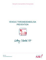
PREVENT VENTILATOR ASSOCIATED<br />
PNEUMONIA<br />
<strong>Getting</strong> <strong>Getting</strong> <strong>Getting</strong> <strong>Getting</strong> <strong>Started</strong> <strong>Started</strong> <strong>Started</strong> <strong>Started</strong> <strong>Kit</strong> <strong>Kit</strong><br />
<strong>Kit</strong> <strong>Kit</strong>
<strong>Safer</strong> <strong>Healthcare</strong> <strong>Now</strong>! Prevent Ventilator Associated Pneumonia <strong>Getting</strong> <strong>Started</strong> <strong>Kit</strong><br />
<strong>Safer</strong> <strong>Healthcare</strong> <strong>Now</strong>!<br />
We invite you to join <strong>Safer</strong> <strong>Healthcare</strong> <strong>Now</strong>! to help improve the safety of the Canadian<br />
healthcare system. <strong>Safer</strong> <strong>Healthcare</strong> <strong>Now</strong>! is a national program supporting Canadian<br />
healthcare organizations to improve safety through the use of quality improvement methods and<br />
the integration of evidence in practice.<br />
To learn more about this intervention, to find out how to join <strong>Safer</strong> <strong>Healthcare</strong> <strong>Now</strong>! and to<br />
gain access to additional resources, contacts, and tools, visit our website at<br />
www.saferhealthcarenow.ca<br />
This <strong>Getting</strong> <strong>Started</strong> <strong>Kit</strong> has been written to help engage your interprofessional/interdisciplinary<br />
teams in a dynamic approach for improving quality and safety while providing a basis for getting<br />
started. The <strong>Getting</strong> <strong>Started</strong> <strong>Kit</strong> represents the most current evidence, knowledge and practice,<br />
as of the date of publication and includes what has been learned since the first kits were<br />
released in 2005. We remain open to working consultatively on updating the content, as more<br />
evidence emerges, as together we make healthcare safer in Canada.<br />
Note:<br />
The <strong>Getting</strong> <strong>Started</strong> <strong>Kit</strong>s for all <strong>Safer</strong> <strong>Healthcare</strong> <strong>Now</strong>! interventions are the same and available<br />
in both French and English.<br />
This document is in the public domain and may be used and reprinted without permission<br />
provided appropriate reference is made to <strong>Safer</strong> <strong>Healthcare</strong> <strong>Now</strong>!<br />
June 2012 2
<strong>Safer</strong> <strong>Healthcare</strong> <strong>Now</strong>! Prevent Ventilator Associated Pneumonia <strong>Getting</strong> <strong>Started</strong> <strong>Kit</strong><br />
Acknowledgements<br />
The Canadian Patient Safety Institute (CPSI) is acknowledged<br />
for their financial and in-kind support of the <strong>Safer</strong> <strong>Healthcare</strong><br />
<strong>Now</strong>! <strong>Getting</strong> <strong>Started</strong> <strong>Kit</strong>s.<br />
We wish to thank and acknowledge the Canadian ICU<br />
Collaborative and faculty members who have contributed<br />
significantly to the work of the Ventilator acquired<br />
pneumonia teams and the revisions to this kit.<br />
In particular, we acknowledge the work of Ms. Paule Bernier,<br />
Dr. Paul Boiteau, Ms. Rosmin Esmail , Mr. Gordon Krahn, Dr.<br />
Denny Laporta, and Dr. John Muscedere.<br />
June 2012 3
<strong>Safer</strong> <strong>Healthcare</strong> <strong>Now</strong>! Prevent Ventilator Associated Pneumonia <strong>Getting</strong> <strong>Started</strong> <strong>Kit</strong><br />
Canadian ICU Collaborative Faculty<br />
December 2011<br />
Chaim Bell; MD, PhD, FRCPC<br />
Associate Professor of Medicine and Health Policy, Management & Evaluation<br />
CIHR/CPSI Chair in Patient Safety & Continuity of Care<br />
University of Toronto; St. Michael's Hospital<br />
Paule Bernier, P.Dt., MSc<br />
Nutritionist, Critical Care Team, Jewish General Hospital;<br />
Safety and Improvement Advisor, <strong>Safer</strong> <strong>Healthcare</strong> <strong>Now</strong>! (Québec)<br />
Soins de santé plus sécuritaires maintenant! SSPSM (Québec)<br />
Paul Boiteau, MD, FRCPC (Past-Chair and Financial Officer)<br />
Department Head, Critical Care Medicine, Calgary Health Region;<br />
Professor of Medicine, University of Calgary<br />
Mike Cass, BSc, RN, MScN<br />
Advanced Practice Nurse, Trillium Health Centre<br />
Leanne Couves<br />
Improvement Associates Ltd.<br />
Vanda DesRoches, RN, BN<br />
Clinical Development Nurse<br />
Prince County Hospital<br />
Greg Duchscherer, RRT, FCSRT<br />
Quality Improvement and Patient Safety Leader<br />
Department of Critical Care Medicine<br />
Alberta Health Services – Calgary Zone<br />
Bruce Harries, MBA<br />
Improvement Associates Ltd.<br />
Gordon Krahn, BSc, RRT<br />
Quality and Research Coordinator, BC Children’s Hospital<br />
Denny Laporta, MD, FRCPC<br />
Intensivist, Department of Adult Critical Care, Jewish General Hospital<br />
Faculty of Medicine, McGill University<br />
Anne MacLaurin, RN, BScN, MN<br />
Project Manager<br />
Canadian Patient Safety Institute<br />
June 2012 4
<strong>Safer</strong> <strong>Healthcare</strong> <strong>Now</strong>! Prevent Ventilator Associated Pneumonia <strong>Getting</strong> <strong>Started</strong> <strong>Kit</strong><br />
Claudio Martin, MD, FRCPC (Collaborative Chair)<br />
Intensivist, London Health Sciences Centre, Critical Care Trauma Centre<br />
Professor of Medicine and Physiology, University of Western Ontario<br />
Chair/Chief of Critical Care Western<br />
Cathy Mawdsley, RN, MScN, CNCC<br />
Clinical Nurse Specialist – Critical Care, London Health Sciences Centre;<br />
Adjunct Professor, School of Nursing, University of Western Ontario<br />
Sherissa Microys, MD, FRCPC, Major<br />
Assistant Professor, University of Ottawa; Intensivist, Ottawa Hospital;<br />
Major, Canadian Forces<br />
John Muscedere, MD, FRCPC<br />
Assistant Professor of Medicine, Queens University;<br />
Intensivist, Kingston General Hospital<br />
Tracie Northway, RN, MScN, CNCCP(C)<br />
Project Manager, Strategic Implementation<br />
BC Children's Hospital and Sunny Hill Health Center<br />
Yoanna Skrobik, MD, FRCPC<br />
Intensivist, Hôpital Maisonneuve Rosemont, Montréal<br />
Expert Panel for the new Pain, Sedation and Delirium Guidelines,<br />
Society of Critical Care Medline (SCCM)<br />
Jennifer Turple, BSc Pharm, ACPR<br />
Medication Safety Specialist<br />
ISMP Canada<br />
June 2012 5
<strong>Safer</strong> <strong>Healthcare</strong> <strong>Now</strong>! Prevent Ventilator Associated Pneumonia <strong>Getting</strong> <strong>Started</strong> <strong>Kit</strong><br />
Summary of Revisions from Previous Versions of<br />
the <strong>Getting</strong> <strong>Started</strong> <strong>Kit</strong><br />
1. The case for preventing ventilator associated pneumonia<br />
2. The definition of <strong>VAP</strong> was clarified<br />
3. Adult <strong>VAP</strong> Bundle: has gone from 4 to 5 elements<br />
Specific Revisions:<br />
a. The recommendation for HOB elevation has been reworded to “we recommend that<br />
the head of the bed be elevated to 45°. When this is not possible, attempts to raise<br />
the head of the bed at least > 30° should be considered,<br />
b. The recommendation for daily evaluation of readiness for extubation has been revised<br />
to reflect new evidence.<br />
c. The recommendation for endotracheal tubes with subglottic secretion drainage has<br />
been revised to reflect new evidence<br />
d. The recommendation for oral tubes has been removed from the bundle and replaced<br />
with “Initiate safe enteral nutrition within 24-48h of ICU admission”<br />
e. The recommendation for oral decontamination with Chlorhexidine has been upgraded<br />
as a <strong>VAP</strong> Bundle element and revised to include general recommendations for oral<br />
care.<br />
4. Additional revisions to reflect new evidence were made for the following.<br />
a. Hand hygiene<br />
b. VTE prophylaxis<br />
c. The promotion of patient mobility and autonomy.<br />
The Pediatric section was not revised in this version, as there was no notable evidence to<br />
modify current <strong>VAP</strong> prevention practices.<br />
June 2012 6
<strong>Safer</strong> <strong>Healthcare</strong> <strong>Now</strong>! Prevent Ventilator Associated Pneumonia <strong>Getting</strong> <strong>Started</strong> <strong>Kit</strong><br />
Table of Contents<br />
PREVENT VENTILATOR ASSOCIATED PNEUMONIA ........................................................ 1<br />
<strong>Safer</strong> <strong>Healthcare</strong> <strong>Now</strong>! ....................................................................................... 2<br />
Acknowledgements ........................................................................................... 3<br />
Canadian ICU Collaborative Faculty ....................................................................... 4<br />
Summary of Revisions from Previous Versions of the <strong>Getting</strong> <strong>Started</strong> <strong>Kit</strong> .......................... 6<br />
Table of Contents ............................................................................................. 7<br />
Background ..................................................................................................... 9<br />
Goal ........................................................................................................................ 9<br />
The Case for Preventing Ventilator-Associated Pneumonia in Adults and Children ............................ 9<br />
Preventing <strong>VAP</strong> in Adult Patients ........................................................................ 11<br />
Defining <strong>VAP</strong> in Adults ................................................................................................... 11<br />
The Adult <strong>VAP</strong> Bundle: Concept and Potential Impact ............................................................ 12<br />
Adult <strong>VAP</strong> Bundle: Five Components of Care ........................................................................ 12<br />
1. Elevation of the Head of the Bed to 45° when possible, otherwise attempt to maintain the head<br />
of the bed greater than 30° .............................................................................................................. 12<br />
2. Daily Evaluation of Readiness for Extubation .................................................................................. 15<br />
3. Subglottic Secretion Drainage .......................................................................................................... 19<br />
4. Oral care and Decontamination with Chlorhexidine ........................................................................ 22<br />
5. Initiation of safe enteral nutrition within 24-48h of ICU admission ................................................ 25<br />
Additional Evidence Based Components of Care ...................................................... 28<br />
1. Hand Hygiene .......................................................................................................... 28<br />
2. Practices That Promote Patient Mobility and Autonomy...................................................... 29<br />
Choice of Sedatives, Analgesics and Antipsychotics ............................................................................... 29<br />
Delirium screening and management .................................................................................................... 29<br />
Early Exercise .......................................................................................................................................... 30<br />
3. Venous Thromboembolism (VTE) Prophylaxis ................................................................... 32<br />
Preventing <strong>VAP</strong> in Children ............................................................................... 32<br />
1. Elevation of the Head of Bed (HOB) in infants and children ............................................................ 33<br />
2. Proper positioning of oral or nasal gastric tube in infant and children ........................................... 33<br />
3. Oral Care in children ......................................................................................................................... 33<br />
4. Eliminate the routine use of instil for suctioning for pediatric patients .......................................... 34<br />
5. Keep the ventilator tubing in a dependant position ........................................................................ 34<br />
June 2012 7
<strong>Safer</strong> <strong>Healthcare</strong> <strong>Now</strong>! Prevent Ventilator Associated Pneumonia <strong>Getting</strong> <strong>Started</strong> <strong>Kit</strong><br />
Additional Components for the Pediatric Population .............................................................. 34<br />
Components of the Adult bundle which Are Not Included ........................................................ 34<br />
Implementing the <strong>VAP</strong> Bundle in Adults and Children ............................................... 35<br />
1. Forming the Team .................................................................................................... 35<br />
2. Setting Aims ........................................................................................................... 36<br />
3. Using the Model for Improvement ................................................................................. 36<br />
4. <strong>Getting</strong> <strong>Started</strong> ........................................................................................................ 37<br />
5. First Test of Change .................................................................................................. 38<br />
6. Measurement .......................................................................................................... 38<br />
1. <strong>VAP</strong> Rate ........................................................................................................... 38<br />
2. <strong>VAP</strong> Bundle Compliance ...................................................................................................................... 39<br />
7. Track Measures over Time .......................................................................................... 40<br />
8. Barriers that may be Encountered ................................................................................. 41<br />
9. Work to Achieve a High Level of Compliance .................................................................... 41<br />
10. Tips for Gathering Data ............................................................................................ 41<br />
Frequently Asked Questions: <strong>VAP</strong> ........................................................................ 42<br />
APPENDIX A: Technical Descriptions..................................................................... 46<br />
1.0 <strong>VAP</strong> Rate per 1000 Ventilator Days – Worksheet .............................................................. 46<br />
1.0 <strong>VAP</strong> Rate per 1000 Ventilator Days – Technical Description ................................................. 47<br />
2.0 Adult <strong>VAP</strong> Bundle Compliance – Worksheet ..................................................................... 49<br />
3.0 Paediatric <strong>VAP</strong> Bundle Compliance – Worksheet ............................................................... 50<br />
2.0 ADULT <strong>VAP</strong> Bundle Compliance /3.0 PAEDIATRIC <strong>VAP</strong> Bundle Compliance – Technical Description . 51<br />
APPENDIX B: Sample Checklists and Daily Goals ....................................................... 53<br />
APPENDIX C: Sample Enteral feeding pre-printed orders ............................................ 62<br />
APPENDIX D: Criteria for Ventilator Associated Pneumonia (from 2009 PDF version) ........ 63<br />
Footnotes to Ventilator Associated Pneumonia Criteria ............................................. 68<br />
References.................................................................................................... 69<br />
June 2012 8
<strong>Safer</strong> <strong>Healthcare</strong> <strong>Now</strong>! Prevent Ventilator Associated Pneumonia <strong>Getting</strong> <strong>Started</strong> <strong>Kit</strong><br />
Background<br />
Goal<br />
The goal is to prevent ventilator-associated pneumonia (<strong>VAP</strong>) by implementing the five<br />
components of care called the “<strong>VAP</strong> Bundle.” The current <strong>VAP</strong> Bundle was modified to reflect<br />
the elements of practice that have the greatest evidence for their ability to decrease <strong>VAP</strong>.<br />
Teams are also strongly encouraged to implement the Additional evidence-based components of<br />
care described in this document.<br />
The Case for Preventing Ventilator-Associated Pneumonia in<br />
Adults and Children<br />
Nosocomially acquired infections in the intensive care unit (ICU) are common; higher rates are<br />
associated with increased severity of illness, utilization of invasive monitoring and treatment,<br />
morbidity, mortality and health care costs. 1,2,3,4 Invasively mechanically ventilated patients are<br />
particularly susceptible to nosocomial infections and pneumonia. Pneumonia that occurs in this<br />
context (i.e. mechanical ventilation with an endotracheal tube) is termed ventilator-associated<br />
pneumonia (<strong>VAP</strong>).<br />
The reason that <strong>VAP</strong> remains relevant is that the number of patients requiring mechanical<br />
ventilation increased in the past decade and is expected to increase further in the future. 5,6 In<br />
spite of intensive efforts to prevent <strong>VAP</strong>, it remains relatively common. The most recent data<br />
from the United States where surveillance data are available, reported that the incidence<br />
ranged from 2 to 10 cases per 1000 ventilator days. <strong>VAP</strong> is a cause of morbidity in mechanically<br />
ventilated patients resulting in prolongation of mechanical ventilation, ICU, and hospital days<br />
by an average of 7.6, 8.7 and 11.5 days, respectively. 7,8 Although the attributable mortality of<br />
<strong>VAP</strong> is controversial, it may be substantial if therapy is delayed or inappropriate. 9 Further, wide<br />
scale use of antibiotics for nosocomial infections such as <strong>VAP</strong> also exposes patients to antibioticrelated<br />
diarrhoea and colitis which carry their own burden and impact on outcome. 10,11<br />
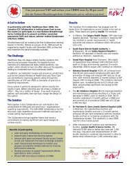
Estimates of the costs to the healthcare system from <strong>VAP</strong> have ranged from $10 000-16 000 US. 8,<br />
12,13 In Canada it is estimated that the prevention of one case of <strong>VAP</strong> could result in a cost saving<br />
of approximately $14,000 per patient. 14 It is estimated that the number of adult cases of <strong>VAP</strong> in<br />
Canada are around 4 000 per year, resulting in approximately 230 deaths consuming 17 000<br />
excess ICU days or 2% of all ICU days in Canada, at an estimated cost to the Canadian health care<br />
system of CAN $46 million per year.<br />
In the pediatric population, although <strong>VAP</strong> is an important clinical entity, there are fewer studies<br />
quantifying the problem. 15,16,17 The latest rates for pediatric <strong>VAP</strong> from the National Nosocomial<br />
Infections Surveillance (NNIS) group are 0 to 4.6 per 1,000 ventilator days with an average of<br />
1.8 per 1,000 ventilator days. 3 The presence of <strong>VAP</strong> in children leads to a longer duration of<br />
ventilation and increased length of stay and associated costs. 15,16,17 It is estimated that in the<br />
pediatric population, <strong>VAP</strong> prolongs hospital length of stay by 8.7days. <strong>VAP</strong> is also associated with<br />
increased mortality. In one study the difference in mortality rate was <strong>VAP</strong> 19.1% vs. non-<strong>VAP</strong> 7.2%. 16<br />
June 2012 9
<strong>Safer</strong> <strong>Healthcare</strong> <strong>Now</strong>! Prevent Ventilator Associated Pneumonia <strong>Getting</strong> <strong>Started</strong> <strong>Kit</strong><br />
Two points are worthy of mention when discussing <strong>VAP</strong> rates. Because multiple alternative<br />
diagnoses can mimic the clinical signs of <strong>VAP</strong>, the apparent rate of <strong>VAP</strong> in ICUs may vary<br />
significantly, depending on the prevalence of other ICU-acquired conditions; this has raised the<br />
concern that <strong>VAP</strong> rate may be an unreliable measure of quality of care. 18 Secondly, it is<br />
suspected that surveillance underestimates <strong>VAP</strong> occurrence and that true rates are likely much<br />
higher. 19,20 Despite these concerns regarding reported <strong>VAP</strong> rates, there appears to be no<br />
controversy that all efforts should be made to reduce <strong>VAP</strong>. 21 The Canadian ICU Collaborative<br />
Faculty and Canadian Patient Safety Institute’s <strong>Safer</strong> <strong>Healthcare</strong> <strong>Now</strong>! supports the reporting of<br />
<strong>VAP</strong> rates in combination with measurements of adherence to <strong>VAP</strong> prevention practices. 22 By<br />
reporting both, the incidence of <strong>VAP</strong> over time can be tracked and the adherence to best<br />
practices can similarly be followed. Thus the correlation between both measures can be<br />
observed for any particular institution allowing for insight into both practice and surveillance<br />
methods.<br />
For more information, the reader is referred to the topic “<strong>VAP</strong> Diagnosis” in the FAQ Section.<br />
In summary <strong>VAP</strong> is a common problem in Canadian ICUs, which is associated with poor outcomes,<br />
in vulnerable critically ill patient populations. There are evidence-based practices that can<br />
reduce the incidence of <strong>VAP</strong>; implementation of these practices have been effective in reducing<br />
<strong>VAP</strong> and its associated sequelae. 23,24,25,26,27,28,29 Although it is often argued that in the Canadian<br />
<strong>Healthcare</strong> system money is not saved by improving efficiency (because each discharged patient<br />
is replaced by a new patient with comparable overall costs), our incentive to reduce <strong>VAP</strong> should<br />
be directed towards liberating wasted ICU and hospital days, thus improving ICU access for other<br />
patients in need.<br />
June 2012 10
<strong>Safer</strong> <strong>Healthcare</strong> <strong>Now</strong>! Prevent Ventilator Associated Pneumonia <strong>Getting</strong> <strong>Started</strong> <strong>Kit</strong><br />
Preventing <strong>VAP</strong> in Adult Patients<br />
Defining <strong>VAP</strong> in Adults<br />
Ventilator-associated pneumonia (<strong>VAP</strong>) is defined as a pneumonia occurring in patients requiring<br />
a device intermittently or continuously to assist respiration through a tracheostomy or<br />
endotracheal tube. Further, the device must have been in place within the 48-hour period<br />
before onset of infection and for at least two consecutive days.<br />
Diagnostic criteria are as follows:<br />
a) Radiographic abnormalities:<br />
New or progressive, and persistent chest radiographic opacity(ies) compatible with<br />
Pneumonia, e.g. infiltrate, consolidation or cavitation<br />
b) And at least 1 of the following:<br />
• WBC ≥ 12,000 or < 4,000<br />
• Temperature > 38 0 C with no other cause<br />
c) And at least 2 of the following:<br />
• tracheal secretions: new onset of purulence, or change in character, or increase in<br />
volume<br />
• increase in suctioning requirements<br />
• inspiratory crackles (rales) or bronchial breath sounds on auscultation<br />
• Worsening gas exchange (e.g., O2 desaturations; PaO2/FiO2< 240, an increase in<br />
oxygenation or ventilatory requirements.<br />
If multiple episodes are suspected, one needs to look for resolution of the initial infection. The<br />
additional isolation of a new pathogen alone is not indicative of a new episode of pneumonia.<br />
The full spectrum of a combination of new signs, symptoms and radiographic evidence is<br />
required.<br />
The Faculty acknowledges that different opinions on timelines for inclusion of patients may<br />
arise. Most of the critical care literature refers to <strong>VAP</strong> in patients who have been intubated for<br />
at least 48h. The CDC recommends 30 including patients supported by a breathing device within<br />
the 48h before the onset of the infection. Canadian guidelines for the prevention of <strong>VAP</strong> were<br />
developed using a variety of definitions as reported by the original authors of the evidentiary<br />
base. 31 The primary purpose of our Collaborative and SHN is not research but aims at improving<br />
performance within each institution. The Faculty believes that adopting a congruent definition<br />
will not only allow intra-unit comparison over time but inter-unit comparisons, however it must<br />
be remembered that benchmarking and comparison between centres, although interesting, are<br />
not the aims of this effort. 32<br />
For more information, the reader is referred to the topic “<strong>VAP</strong> Diagnosis” in the FAQ Section<br />
June 2012 11
<strong>Safer</strong> <strong>Healthcare</strong> <strong>Now</strong>! Prevent Ventilator Associated Pneumonia <strong>Getting</strong> <strong>Started</strong> <strong>Kit</strong><br />
The Adult <strong>VAP</strong> Bundle: Concept and Potential Impact<br />
Care bundles, in general, are groupings of best practices with respect to a disease process that<br />
individually improve care, but when applied together may result in substantially greater<br />
improvement. The science supporting each bundle component is sufficiently established that the<br />
bundle is considered best practice. Bundles have been demonstrated to reduce <strong>VAP</strong> by the<br />
Canadian ICU Collaborative teams, examples of which are illustrated in this guide and by<br />
published data from pediatric and adult centres. 33,34<br />
<strong>Safer</strong> <strong>Healthcare</strong> <strong>Now</strong>! (SHN) has defined a “<strong>VAP</strong> bundle” as a group of evidence-based practices<br />
that, when implemented together, should result in reductions in the incidence of <strong>VAP</strong>. The<br />
Canadian Campaign has endorsed the inclusion of practices that are recommended by the<br />
published Clinical Practice Guideline Committee of the Canadian Critical Care Society and the<br />
Canadian Critical Care Trials Group. 31<br />
A recent ICU collaborative improvement project at IHI reported an average 45% reduction in the<br />
incidence of <strong>VAP</strong> using a “<strong>VAP</strong> bundle”. 35 Moreover, there is a trend toward greater success<br />
among teams that comply more fully with every element of the bundle.<br />
Compliance with the <strong>VAP</strong> bundle can be measured by simple assessment of the completion of<br />
each item. The approach has been most successful when all elements are executed together, an<br />
“all or none” strategy.<br />
The components of SHNs <strong>VAP</strong> Bundle are (not listed in order of importance):<br />
1. Elevation of the head of the bed to 45° when possible, otherwise attempt to maintain the<br />
head of the bed greater than 30° should be considered<br />
2. Daily evaluation of readiness for extubation.<br />
3. The utilization of endotracheal tubes with subglottic secretion drainage.<br />
4. Oral care and decontamination with Chlorhexidine.<br />
5. Initiation of safe enteral nutrition within 24-48h of ICU admission.<br />
Adult <strong>VAP</strong> Bundle: Five Components of Care<br />
1. Elevation of the Head of the Bed to 45° when possible, otherwise attempt to<br />
maintain the head of the bed greater than 30°<br />
Elevation of the head of the bed (HOB) is correlated with reductions in <strong>VAP</strong> rates and is an<br />
integral part of the <strong>VAP</strong> bundle. 36<br />
The rationale for this intervention is two-fold: 1) to decrease the risk of aspiration of<br />
aerodigestive (e.g. oropharyngeal and gastrointestinal) fluids. 37,38,39 and 2) to improve patients’<br />
lung volumes and ventilation. For example, patients in the supine position will have lower<br />
spontaneous tidal volumes on pressure support ventilation than those seated in an upright<br />
position. Although patients may be on mandatory modes of ventilation, the improvement in<br />
position may aid ventilatory efforts and minimize atelectasis. 40,41<br />
June 2012 12
<strong>Safer</strong> <strong>Healthcare</strong> <strong>Now</strong>! Prevent Ventilator Associated Pneumonia <strong>Getting</strong> <strong>Started</strong> <strong>Kit</strong><br />
In addition HOB elevation is congruent with other emerging concepts in the management of<br />
ventilated patients such as safe and timely enteral nutrition, patient interaction and orientation<br />
with environment, and liberation from immobility (eg. early ambulation) and from ventilatory<br />
support. 42<br />
In a recently performed meta-analysis of the three available RCTs studying the semi-recumbent<br />
position 36 , a total of 337 mechanically ventilated ICU patients were evaluated, The odds of<br />
developing clinically diagnosed <strong>VAP</strong> were significantly lower among patients randomized to the<br />
semi-recumbent 45° position compared to patients randomized to the supine position (OR =<br />
0.47; 95% CI, 0.27-0.82). A sub-analysis regarding the incidence of microbiologically documented<br />
<strong>VAP</strong>, ICU length of stay, and the duration of mechanical ventilation showed that patients<br />
randomized to the semi-recumbent 45° position had a trend toward better clinical outcomes.<br />
The authors concluded that 1) the usual practice of back-rest elevation of 15° to 30° is not<br />
sufficient to prevent <strong>VAP</strong> in mechanically ventilated patients, and 2) patients positioned semirecumbently<br />
45° have significantly lower incidence of clinically diagnosed <strong>VAP</strong> compared to<br />
patients positioned supinely.<br />
Is 45° the correct evidence-based angle of head of bed elevation? A more careful analysis of the<br />
three trials is informative in this regard. The first trial was a single-center study of 86<br />
mechanically ventilated patients assigned to semi-recumbent or supine body position. 43 The<br />
investigators demonstrated that suspected cases of ventilator-associated pneumonia in the<br />
supine position had an incidence of 34%, while in the semi-recumbent position suspected cases<br />
had an incidence of 8% (p=0.003). Similarly, confirmed cases were 23% and 5% respectively<br />
(p=0.018). Unfortunately there was no mention of how the head of bed angle was measured, nor<br />
of how and to what extent this was achieved and adhered to. Excluded from the study were<br />
patients have undergone recent abdominal surgery, neurosurgical intervention, previous<br />
endotracheal intubation, or in refractory shock. The second trial was multi-centered and<br />
compared the semi recumbent position targeted to 45 0 backrest elevation compared to a control<br />
position (10 0 backrest elevation). 44 The investigators observed that the targeted 45 0 backrest<br />
elevation was not reached, and the difference in the attained treatment position of 28 0 did not<br />
prevent the onset of <strong>VAP</strong> compared to the 10 0 control position. The authors did not clearly<br />
describe why the aimed position of 45 0 was not achieved. This study showed that 1) raising the<br />
HOB between 10 0 and 30 0 is not effective in preventing <strong>VAP</strong>, and 2) maintaining the HOB at 45 0 is<br />
a challenging task and underscores the need for concerted and continuous efforts by all team<br />
members to maintain this standard under routine conditions. Interestingly, the authors also<br />
found no difference in the development of pressure sores in both groups, suggesting that at least<br />
the intervention was not harmful. In both study groups, most patients had stage 1 or 2 pressure<br />
sores and in the majority of these cases, the pressure sores were present at the heel and/or<br />
sacral region. The third study is a prospective randomized trial comparing semi-recumbency with<br />
head of the bed elevation at 45 0 (intervention) to 25 0 (control). 45 The rate of <strong>VAP</strong> was 5/17<br />
(29.4%) in the patients whose HOB was elevated to 45 0 vs. 7/13 (53.8%) in the control group. The<br />
degree of compliance with 45 0 was not measured and co-interventions were not reported.<br />
Unfortunately the small sample size and methodological analysis limit any conclusions from this<br />
pilot study where 46% of randomized patients were withdrawn because of intubation/ventilation<br />
<strong>Safer</strong> <strong>Healthcare</strong> <strong>Now</strong>! Prevent Ventilator Associated Pneumonia <strong>Getting</strong> <strong>Started</strong> <strong>Kit</strong><br />
The potential for harm associated with HOB elevation has been addressed in the published<br />
literature. Pressure ulcers are more likely to occur at higher HOB elevation in critically ill<br />
patients 46 as well as in normal subjects. 47 Of note is that the Wound, Ostomy and Continence<br />
Nurses Society recommends maintaining the HOB at 30 0 elevation for supine positions. 48 Other<br />
factors similarly impede bedside practitioners’ adherence to higher degrees of HOB elevation 49<br />
decreased systolic blood pressure, SOFA score 50 , sliding down the bed, skin shearing, insomnia,<br />
inability to accurately estimate back rest elevation. 51 In order to address these issues, the<br />
American Association of Critical Care Nurses issued a Practice Alert for <strong>VAP</strong> prevention in 2004<br />
which was revised in 2007 to include evidence-based Practice Alert Statements and tools. 52<br />
A systematic review on the benefits and disadvantages of semi-upright position in ventilated<br />
patients was done by a European expert panel. 53 Based on their results, they recommended that<br />
ICU caregivers “elevate the head of the bed of mechanically ventilated patients to a 20 to 45°<br />
position and preferably in a ≥30° position as long as it is does not pose risks and conflicts with<br />
other nursing tasks, medical interventions or with patients’ wishes”.<br />
It thus appears that:<br />
• the optimal semi-recumbent HOB elevation position that reduces the development of<br />
aspiration and <strong>VAP</strong>, while deriving the least risk to patients is not known.<br />
• the level of evidence for the use of lower HOB elevation to prevent the most<br />
controversial complication - sacral pressure ulcers- is not as strong as that for HOB<br />
elevation to prevent aspiration and <strong>VAP</strong>. 54<br />
We conclude that until further evidence becomes available, patients without contraindications<br />
to HOB elevation should be kept at 45°, and when this is not possible, keeping the head of the<br />
bed above 30° should be considered. In all cases, the exact angle strategy is determined on a<br />
patient-to-patient basis according to individual patient needs.<br />
What changes can we make that will result in improvement?<br />
Hospital teams across Canada and the United States have developed and tested process and<br />
system changes that allowed them to improve performance on elevation of HOB. These<br />
measures support the implementation of the <strong>VAP</strong> bundle. Some of these changes are:<br />
• Identify local implementation challenges (e.g. insufficient awareness of the benefit of<br />
the 45 0 backrest elevation, disagreement about who is responsible for patients' bed<br />
positioning, difficulties in enabling and reinforcing such strategies) to tailor improvement<br />
strategies to the environment in your ICU. 55<br />
• Be aware of other practical challenges in maintaining head of bed elevation throughout<br />
the day and night such as: 1) patients sliding down the bed if overly elevated, 2) bed<br />
often needs to be lowered for procedures such as line placement, bathing, wound care,<br />
patient turns, etc., 3) caregivers forget to re-elevate the head of the bed following these<br />
procedures.<br />
• Determine what constitute valid contraindications for semi-recumbency in your ICU<br />
population (e.g. recent spine surgery or spinal cord trauma, abdominal wound, unstable<br />
pelvic fracture, mod-high-grade sacral ulcer, hemodynamic instability, increased<br />
June 2012 14
<strong>Safer</strong> <strong>Healthcare</strong> <strong>Now</strong>! Prevent Ventilator Associated Pneumonia <strong>Getting</strong> <strong>Started</strong> <strong>Kit</strong><br />
intracranial pressure (HOB >45 0 contraindicated), undergoing procedure, post-removal<br />
femoral arterial sheath, ECMO/VAD,etc.).<br />
• Implement mechanisms to ensure head-of-the-bed elevation, such as including<br />
documentation of this intervention on nursing flow sheets (electronic or paper) at regular<br />
intervals (e.g., every 4 hours), including HOB elevation on daily goal sheets or discussing<br />
it as a topic at daily multidisciplinary rounds.<br />
• Bring a protractor into the ICU to demonstrate exactly what 45 0 elevation looks like.<br />
Once you have measured 45 0 for that bed, place a piece of coloured tape on the wall<br />
behind the bed and verify compliance during ventilator checks.<br />
• When purchasing new beds include a specification about monitoring of HOB position (a QA<br />
project done at the JGH in Montreal identified that mechanical measuring devices are<br />
more accurate than electronic devices).<br />
• Educate all personnel and create an environment where all allied health care<br />
professionals, not only nurses and MDs, are encouraged to notify nursing if the head of<br />
the bed is not elevated; alternately, have these disciplines chart on the position of the<br />
HOB and empower them and others to carefully place the patient in this position with<br />
nursing assistance. Include other personnel such as orderlies and radiology technicians.<br />
• Educate patients and families to the importance of elevation of HOB and create an<br />
environment where family is encouraged to notify nursing if the head of the bed is not<br />
elevated.<br />
• Include this intervention on standard orders for the initiation and weaning of mechanical<br />
ventilation, delivery of tube feedings, and provision of oral care.<br />
• Use reminders within the patient care areas including the use of communication boards<br />
at every bedside which actually empower families to ensure that the HOB of their loved<br />
one is indeed elevated to 45 0 in the absence of contra-indications.<br />
• Provide educational material & posters for display in family waiting rooms.<br />
• Share and post compliance with the intervention in a prominent place in your ICU to<br />
encourage change and motivate staff.<br />
2. Daily Evaluation of Readiness for Extubation<br />
The timely liberation from mechanical ventilation is thought to help prevent <strong>VAP</strong> by minimizing<br />
“device exposure” , whereby the “device” is the ventilator-circuit-endotracheal tube (ETT)<br />
complex and “exposure” is the duration of mechanical ventilation (i.e. “device-days”). It should<br />
be noted that with current practice standards of ventilator management, the ETT component<br />
appears to carry the greatest burden of risk for pneumonia. 56 This is also supported by a recent<br />
review several small studies of non-invasive ventilation showing a marked reduction in<br />
pneumonia compared to invasive (e.g. with ETT) mechanical ventilation. 57<br />
In this context it thus appears sound for ICU teams to regularly re-evaluate the need for an<br />
endotracheal tube in their mechanically ventilated patients. This concept has been examined in<br />
June 2012 15
<strong>Safer</strong> <strong>Healthcare</strong> <strong>Now</strong>! Prevent Ventilator Associated Pneumonia <strong>Getting</strong> <strong>Started</strong> <strong>Kit</strong><br />
detail, and supporting evidence is presented in this section. The daily evaluation of readiness for<br />
extubation involves two central issues: minimization of unnecessary sedation, and testing the<br />
patient’s ability to assume unassisted breathing while still intubated.<br />
Minimization of unnecessary sedation<br />
Sedation has traditionally been prescribed in mechanically ventilated patients in order to<br />
maintain comfort, decrease pain and anxiety, improve patient-ventilator interaction, help<br />
maintain major organ homeostasis, facilitate nursing care by avoiding self-injury and to allow<br />
safe completion of daily activities and procedures. Unfortunately, over sedation may lead to<br />
unintended consequences, such as longer duration of mechanical ventilation and ICU stay,<br />
decreased communication with patient with consequent decreased ability to evaluate the<br />
patient for – among other items - delirium, weaning and readiness for extubation, as well as<br />
ventilator-related complications)such as neuromuscular weakness and pneumonia. 58<br />
In 2000, Kress reported the results of a randomized controlled trial in which 128 adult<br />
mechanically ventilated patients sedated by continuous IV infusion received either daily<br />
interruption of sedation (irrespective of clinical state) or sedation interruption at the clinician’s<br />
discretion. 59 Interruption was considered complete if the patient could perform 3 of 4 items on<br />
command: open eyes, squeeze hands, lift head and protrude tongue. Daily sedation interruption<br />
was associated with a marked and highly significant reduction in time on mechanical ventilation<br />
from 7.3 days to 4.9 days (p=0.004). Schweickert et al performed a post-hoc analysis of the<br />
Kress trial and found that patients undergoing spontaneous awakening trials via daily<br />
interruption of sedative infusions experienced significantly less complications associated with<br />
mechanical ventilation (<strong>VAP</strong>, upper gastrointestinal haemorrhage, bacteremia, barotrauma,<br />
venous thromboembolic disease, cholestasis or sinusitis requiring surgical intervention) than in<br />
those subjected to conventional sedation techniques (2.8% vs. 6.2%, p =.04). 60 In addition, these<br />
patients had a reduced ICU length of stay and were not at risk for worse psychological outcomes<br />
(anxiety, inability to cope with pain) after critical illness compared with conventional<br />
therapies. 61<br />
In an important proof-of-concept study by Strom et al showed that a no-sedation approach in<br />
mechanically ventilated ICU patients is associated with an increase in days without ventilation. 62<br />
In reality, as the intervention (no sedation) group was administered morphine as required, the<br />
true concept demonstrated was rather that a conservative approach of less sedation does not<br />
appear to cause harm in critically ill mechanically ventilated patients. Three caveats for this<br />
study are 1) the intervention group (“no” sedation) had a greater incidence of delirium, 2) the<br />
trial utilized more than usual resources, i.e. 1:1 patient: nurse ratios for all patients, 3) the trial<br />
was a single center study. A multicentre study is required to ascertain the reproducibility of<br />
these findings. In an observational study of 335 patients admitted to a mixed medical-surgical<br />
ICU, Salgado observed that minimal use of continuous sedation (42% of patients received some<br />
sedation, and only 10% of patients received sedation for >24 hours; 20% of ventilator hours were<br />
accompanied by a continuous sedative infusion) was feasible without apparent adverse effects<br />
(e.g. self-extubation requiring re-intubation). 63<br />
Interventional studies assessing the effect of implementing an ICU sedation protocol alone have<br />
provided inconsistent outcomes with respect to ventilator and ICU days, incidence of <strong>VAP</strong> and<br />
June 2012 16
<strong>Safer</strong> <strong>Healthcare</strong> <strong>Now</strong>! Prevent Ventilator Associated Pneumonia <strong>Getting</strong> <strong>Started</strong> <strong>Kit</strong><br />
extubation failure. 64,65 The benefits and risks of daily sedation interruption were also studied in a<br />
meta-analysis of five randomized controlled trials, comparing daily sedation interruption with no<br />
interruption in 699 critically ill patients. 66 Although daily sedation interruption was not<br />
associated with a significant reduction in duration of mechanical ventilation, length of intensive<br />
care unit or hospital stay, mortality, or self-extubation by the patients, it was however<br />
associated with a reduced risk of requiring tracheostomy (odds ratio 0.57, 95% confidence<br />
interval 0.35 to 0.92, P=0.02; I 2 =3%). The authors concluded that current evidence suggests that<br />
daily sedation interruption appears to be safe, but the significant heterogeneity and small<br />
sample sizes of the existing studies suggest that large randomised controlled studies with longterm<br />
survival follow-up are needed before daily sedation interruption can be recommended as a<br />
standard sedation practice for critically ill adult patients.<br />
However, the implementation and titration of ICU sedation is more than simply interrupting<br />
sedative infusions. It is rather a balancing act to minimize sedation-associated complications and<br />
improve patient comfort. Factors such as varying organizational models of medical and nursing<br />
care delivery and failure to link to other daily practices may render redundant any added<br />
advantage of a stand-alone sedation or weaning protocol.<br />
Testing the patient’s ability to assume unassisted breathing<br />
Two historic trials demonstrated the important value of daily spontaneous breathing trials in<br />
reducing the duration of mechanical ventilation. 67,68 The authors also noted during the process<br />
that weaning patients from ventilatory support was easier if patients were better able to cough<br />
and clear their secretions.<br />
We wish to acknowledge that specific weaning protocols are not proposed in this document. To<br />
this issue, a recent systematic review investigated the effect of weaning protocols on the<br />
duration of mechanical ventilation and other clinical outcomes. Despite a reduction in the<br />
duration of mechanical ventilation, weaning, and ICU stay when standardised weaning protocols<br />
are used, there was also significant heterogeneity among studies. 69 In another study reviewing<br />
international data, it was hypothesized that the observed large variability in organizational<br />
contexts and processes for weaning (e.g. regarding ICU structure, staffing, skill mix, education,<br />
roles, responsibilities, interdisciplinary organization, participation and collaboration) could<br />
account for some of the variability in weaning outcomes and perhaps in the added value of<br />
weaning protocols in ICUs. 70 ICU teams are urged to review the organizational context in which<br />
they wean their patients as well as the weaning process itself in order to optimize weaning<br />
outcomes. In passing, the contribution of non-invasive ventilation (NIV) to ventilator protocols<br />
may be one evidence-based method to facilitate liberation from mechanical ventilation in<br />
71, 72,73<br />
selected patients with respiratory failure.<br />
Linking the two<br />
The recent “Awakening and Breathing” trial linked the concepts of sedation interruption and<br />
regular reassessment of weaning and readiness for extubation. A “wake up and breathe”<br />
protocol that sequentially applies a daily spontaneous awakening trial (SAT) (interruption of<br />
sedation – whether constant infusion or p.r.n) and a daily spontaneous breathing trial (SBT)<br />
resulted in better outcomes for mechanically ventilated ICU patients than current standard<br />
approaches. 74 In this study, patients from four tertiary-care ICUs were randomized to<br />
June 2012 17
<strong>Safer</strong> <strong>Healthcare</strong> <strong>Now</strong>! Prevent Ventilator Associated Pneumonia <strong>Getting</strong> <strong>Started</strong> <strong>Kit</strong><br />
management with a daily SAT followed by an SBT (intervention group) or with sedation per usual<br />
care plus a daily SBT (control group). Patients in the intervention group spent more days<br />
breathing without assistance during the 28-day study period than did those in the control group<br />
(14.7 vs. 11.6 days; p=0.02) and were discharged from ICU (median time 9.1 days vs. 12.9 days;<br />
p=0.01) and the hospital earlier (median time 14.9 days vs. 19.2 days; p=0.04). Although more<br />
patients in the intervention group self-extubated than in the control group (p=0.03), the number<br />
of patients who required re-intubation after self-extubation was similar. Furthermore, during<br />
the year after enrolment, patients in the intervention group were less likely to die than were<br />
patients in the control group (Hazard Ratio 0.68; p=0.01) such that for every 7 patients treated<br />
with the intervention, one life was saved (number needed to treat was 7.4, 95% CI 4.2-35.5).<br />
The “wake up and breathe” flow sheets are readily available online. 75,76 Furthermore, in an a<br />
priori planned substudy conducted in one participating ICU during this trial, the authors found<br />
that the wake up and breathe protocol resulted in similar cognitive, psychological, and<br />
functional outcomes among patients tested 3 and 12 months post-ICU, ie the protocol benefits<br />
were not offset by adverse long-term outcomes. 77 It should be noted that the protocol was<br />
devised so that an SAT required holding even narcotics unless they were specifically prescribed<br />
for analgesia, underlining the importance of documenting the goals for medication use in these<br />
patients.<br />
What changes can we make that will result in improvement?<br />
Hospital teams across Canada and the United States have developed and tested process and<br />
system changes that allowed them to improve performance on daily sedation interruption<br />
and daily assessment of readiness to extubate. These measures, taken together, support the<br />
implementation of the <strong>VAP</strong> Bundle.<br />
Some of these changes are:<br />
• Implement a process to temporarily interrupt sedation (spontaneous awakening trial or<br />
SAT) daily at an appropriate time (e.g., before multidisciplinary rounds but after AM<br />
nursing change of shift) to reappraise the patients’ neurocognitive ability to assume a<br />
viable breathing pattern and his/her needs for sedation/analgesia. All patients receiving<br />
sedation administered either as continuous IV infusion or as PRN should be candidates for<br />
SAT.<br />
• Consider a SAT Safety Screen, with specific allowable contraindications (e.g. Patient<br />
receiving sedative infusion for active seizures or alcohol withdrawal, or escalating doses<br />
due to ongoing agitation, or the presence of neuromuscular blocking agents, has<br />
experienced myocardial ischemia within 24 hours or currently has increased intracranial<br />
pressure).<br />
• Spontaneous Awakening Trial (SAT). After having stopped all sedatives and analgesics<br />
used for sedation (continue analgesics for active pain), patient passes SAT if: opens eyes<br />
to verbal stimuli or tolerates sedation interruption for >4 hours. Patient fails if<br />
experiences: sustained anxiety, agitation, or pain, or has respiratory rate > 35 or Sp02 <<br />
88% for 5 or more minutes, or 2 or more signs of respiratory distress, or acute<br />
dysrhythmia. If SAT failed, medications are restarted at half dose and titrated.<br />
June 2012 18
<strong>Safer</strong> <strong>Healthcare</strong> <strong>Now</strong>! Prevent Ventilator Associated Pneumonia <strong>Getting</strong> <strong>Started</strong> <strong>Kit</strong><br />
• Include precautions to prevent self-extubation such as increased monitoring and vigilance<br />
during the trial. (see FAQ for further discussion)<br />
• Implement a process to standardize the performance of Spontaneous Breathing Trials<br />
(SBT).<br />
• If patient has passed a SAT screen, consider a SBT screen:<br />
Comfort: able to follow commands, adequate cough during suctioning<br />
Gas exchange: Pao2≥60 mmHg on Fio2 ≤ 0.4 and PEEP ≤ 10 cmH20<br />
Hemodynamics: acceptable MAP with no/minimal vasopressor/ionotropic infusions and<br />
no active cardiac ischemia.<br />
Breathing: respiratory rate ≤ 35 and minute ventilation ≤ 15 LPM.<br />
o If passes SBT screen, perform SBT on minimal ventilator support (current Fio2, Tpiece<br />
or tracheal collar, or CPAP 5cmH20 + PS ≤ 7 cmH20 for 1-2 hours.<br />
o SBT is failed if ≥ 1 of these signs occurs for ≥ 5 minutes: respiratory rate >35/min<br />
or 140/min or changed by >20% baseline, systolic<br />
BP >180 or
<strong>Safer</strong> <strong>Healthcare</strong> <strong>Now</strong>! Prevent Ventilator Associated Pneumonia <strong>Getting</strong> <strong>Started</strong> <strong>Kit</strong><br />
tract including the mouth and oropharynx become colonized with pathogenic organisms soon<br />
after ICU admission. 79 As previously discussed, measures to prevent <strong>VAP</strong> that aim to reduce the<br />
quantity of aspirated pathogenic bacteria, such as the reduction of bacterial loads in the<br />
oropharynx with antiseptic mouth care and elevation of the head of the bed, have also been<br />
shown to be effective in reducing <strong>VAP</strong>. 36,80<br />
There has been increasing research on endotracheal tube (ETT) design as a means of reducing<br />
the risk of <strong>VAP</strong>; these include changes to materials composing the tube, changes to cuff design<br />
and subglottic secretion drainage (SSD). Kollef et al. studied the effects of silver coated ETTs<br />
versus conventional ETTs and found that there was a 35.9% decrease in <strong>VAP</strong> in patients intubated<br />
longer than 24 hours receiving silver coated ETTs with a number needed to treat of 37 to prevent<br />
1 case of <strong>VAP</strong>. 81 Several mechanisms were postulated; silver has broad-spectrum antimicrobial<br />
activity, prevents bacterial adhesion to the ETT and may prevent biofilm formation. However,<br />
there were no differences in duration of MV, length of ICU stay and overall mortality. This study<br />
has several limitations including a low incidence of <strong>VAP</strong>, increased presence of patients with<br />
chronic obstructive lung disease and a high rate of pneumonia within 24 hours in the control<br />
group. Even though they are costlier than conventional ETTs, in an economic analysis of silver<br />
coated ETTs they were found to be associated with cost savings. 82 Nevertheless, silver coated<br />
ETTs have only been studied in a single RCT with significant limitations and further studies are<br />
required to determine the role of these novel ETTs, specifically in relation to other <strong>VAP</strong><br />
preventive strategies.<br />
The prevention of aspiration of pathogen laden secretions that accumulate above the<br />
inflated cuffs of ETTs has also been the research since there is increasing evidence that<br />
standard cuffs do not prevent micro-aspiration due to folds in the membrane when<br />
inflated and pressed against tracheal mucosa. 1 To reduce micro-aspiration around the<br />
cuff research has focused on cuff design improvements, the reduction of secretions<br />
accumulating above the cuff through sub-glottic secretion drainage (SSD) or both.<br />
Cuff design changes have focused on the geometry of the cuff or the cuff material. Changing the<br />
cylindrical shape of the cuff to a tapered one such that there is a better zone of apposition to<br />
the tracheal mucosa has been found to reduce aspiration in lab models by 90%. 83,84 However,<br />
clinical studies studying the significance of this have not been published and these are required.<br />
The replacement of standard polyvinyl chloride cuffs with an ultra thin polyurethane cuff (PUC)<br />
also decreases fluid leakage around the cuff. There are two clinical studies studying the effect<br />
of these type of cuffs. In one randomized clinical study of a PUC cuffed ETT combined with SSD,<br />
there was a significant reduction in the rate of <strong>VAP</strong>. 85 However, a standard ETT was utilized in<br />
the control group in this study and it is unclear if the reduction in <strong>VAP</strong> rate was the result of the<br />
PUC cuff or the SSD. The second study of a PUC cuffed ETT employed a time series analysis. 86<br />
With the replacement of standard ETTs with a PUC cuffed tube the rate of <strong>VAP</strong> fell and then rose<br />
slightly when the tubes were withdrawn. Given the large amount of clinical evidence for SSD,<br />
the role of PUC cuffed tubes remains to be determined.<br />
June 2012 20
<strong>Safer</strong> <strong>Healthcare</strong> <strong>Now</strong>! Prevent Ventilator Associated Pneumonia <strong>Getting</strong> <strong>Started</strong> <strong>Kit</strong><br />
A measure that reduces the amount of aspirated secretions into the lower respiratory tract is the<br />
evacuation of secretions that pool above the cuff of the endotracheal tube or sub-glottic<br />
secretion drainage. 87 Sub-glottic secretion drainage (SSD) through a specialized ETT<br />
incorporating a suction port above the cuff as a method to prevent <strong>VAP</strong> was first reported in<br />
1992. 88 This strategy has undergone a large amount of study to ascertain its effectiveness for the<br />
reduction of <strong>VAP</strong>. A meta-analysis of five randomized controlled studies published in 2005 by<br />
Dezfulian et al concluded that it was an effective intervention for the prevention of early onset<br />
<strong>VAP</strong> among patients expected to require >72 hours of MV. 89 Further in this meta-analysis, SSD<br />
was associated with reduced duration of MV and ICU length of stay although there was no effect<br />
on mortality.<br />
A repeat meta-analysis of 13 randomized controlled studies studying ETTs with SSD including a<br />
total of 2442 patients was published in 2011. 90 In this meta-analysis, it was found that SSD was<br />
associated with a highly significant reduction of <strong>VAP</strong> of approximately 50% (risk ratio 0.55 (95% CI<br />
0.46 – 0.66, p < 0.00001) with no heterogeneity (I 2 = 0%).Further the time to first <strong>VAP</strong> was<br />
significantly increased in the SSD group. The use of SSD was associated with reduced ICU length<br />
of stay (- 1.68 days, 95% CI -3.20 to -0.17, p = 0.03) and decreased duration of MV (- 1.18 days,<br />
95%CI - 2.19 to - 0.18, p = 0.02). There was no effect on mortality. It should be emphasized that<br />
the expected duration of mechanical ventilation for these ETTs to be placed was variable and in<br />
only 6 of the studies was the inclusion criteria greater than 72 hours. In 5 of the studies, it was<br />
greater than 24 hours and in 2 it was not specified. Adverse events such as re-intubation or post<br />
extubations stridor were not increased in the patients receiving SSD.<br />
Airway difficulties have been reported in animal models instrumented with ETTs with SSD but<br />
the significance of this in humans is not known. 91 Case reports in humans have reported stridor<br />
and stenosis of the airway. In spite of this, they have been adopted in wide scale clinical use<br />
without incident.<br />
In an older cost effectiveness analysis, the utilization of ETT with SSD was shown to be cost<br />
effective. 92 Given the morbidity and costs associated with <strong>VAP</strong>, the low numbers needed to treat<br />
with SSD to prevent 1 case of <strong>VAP</strong> (NNT of 11) 90 and the low acquisition cost of ETTs with SSD,<br />
they should be routinely used in all patients who are expected to be invasively mechanically<br />
ventilated long enough to put them at risk for <strong>VAP</strong>.<br />
What changes can we make that will result in improvement?<br />
All high risk for prolonged mechanical ventilation and <strong>VAP</strong> should receive ETTs with SSD.<br />
Identification of these patients at the time of intubation is difficult for clinicians and<br />
implementing population based methods may improve their utilization. Some of the<br />
processes that can be put in place to increase the utilization of ETTs with SSD are as follows:<br />
• Respiratory therapy (RT) will need to be intimately involved in the implementation of<br />
these ETTs.<br />
o Having an RT champion is crucial for this implementation.<br />
o Procedures required for maintaining ETTs with SSD will need to be put in place;<br />
either continuous or intermittent suction on the suction ports of the ETT.<br />
o All RTs working in the institution will need to inserviced on the importance of SSD.<br />
June 2012 21
<strong>Safer</strong> <strong>Healthcare</strong> <strong>Now</strong>! Prevent Ventilator Associated Pneumonia <strong>Getting</strong> <strong>Started</strong> <strong>Kit</strong><br />
• Utilizing ETTs with SSD in all patients intubated in the emergency department. These<br />
patients are likely to be intubated for longer periods of time. This can be facilitated as<br />
follows.<br />
o Discussion with emergency department physicians to emphasize the importance of<br />
these ETTs.<br />
o Stocking the emergency department with only ETTs with SSD.<br />
o Since ETTs with SSD are slightly larger than tubes without SSD, standard ETTs<br />
should be stocked in the difficult airway cart or tray.<br />
• Utilizing ETTs with SSD in all patients intubated on the medical wards or the in the<br />
Intensive Care Unit. This can be facilitated as follows.<br />
o Discussion with all the ICU attendings, physicians who respond to cardiac arrests,<br />
physicians who are likely to intubabe patients on general hospital wards and<br />
physicians who may be part of medical response teams.<br />
o Stocking airway trays, cardiac arrest carts or intubation kits with only ETTs with<br />
SSD<br />
o Stocking standard ETTs only in difficult airway carts.<br />
• Utilizing ETTs with SSD in high risk patients coming from the operating room. This can be<br />
facilitated as follows.<br />
o Discussion with anaesthetists, anaesthesia housestaff (if any) and respiratory<br />
therapists working in the operating room.<br />
o Make it standard operating procedure that any patient who may require ICU post<br />
operatively will be intubated with an ETT with SSD in the operating room.<br />
• All patients intubated in the ICU for more than 24 hours with standard ETTs should be<br />
reviewed. The review would examine the implementation process for SSD failed and<br />
feedback provided to the clinicians involved in the original intubation. It should be<br />
recognized that some patients may not be candidates for these tubes and the review<br />
should take this into consideration. Patients who may not be candidates for ETTs with<br />
SSD or who may not have received these tubes appropriately.<br />
o Patients who are nasally intubated.<br />
o Patients in whom there was difficulty with the intubation process.<br />
o Patients from OR who were not expected to require ICU care but due to<br />
difficulties intra-operatively come to require post-operative mechanical<br />
ventilation.<br />
• Post the rates of utilization of ETTs with SSD in patients who are mechanically ventilated.<br />
4. Oral care and Decontamination with Chlorhexidine<br />
Oropharyngeal colonization as well as colonization of dental plaque have been identified as risk<br />
factors for <strong>VAP</strong> as there is high concordance between the bacteria isolated from the<br />
oropharyngeal cavity or the dental plaque and those recovered from tracheal aspirates. 93,94<br />
June 2012 22
<strong>Safer</strong> <strong>Healthcare</strong> <strong>Now</strong>! Prevent Ventilator Associated Pneumonia <strong>Getting</strong> <strong>Started</strong> <strong>Kit</strong><br />
Oral care<br />
Garcia et al showed that implementing a comprehensive oral and dental care system and<br />
protocol (without Chlorhexidine) for critically ill medical patients compared to “standard oral<br />
care” was associated with a decrease in <strong>VAP</strong> rate (12 vs. 8 / 1000 ventilation-days, P = .06 ). 95<br />
Duration of mechanical ventilation and length of stay in the intensive care unit differed<br />
significantly between groups, as did mortality. Compliance with protocol components exceeded<br />
80%.<br />
The oral care policies and practices in intubated critically ill patients are varied and no gold<br />
standard exists. There are very few well designed studies exploring the different protocols thus<br />
the strength of the evidence supporting the practice is not strong. 96<br />
The American Association of Critical Care Nurses (AACN) and Association for Professionals in<br />
Infection Control and Epidemiology (APIC) recommend a comprehensive oral hygiene program for<br />
patients in critical care and acute care settings. 97,98,99 ACCN has published practice guidelines<br />
addressing tooth brushing, use of toothpaste or cleansing solution, use of peroxide, and<br />
suctioning of the oropharynx after brushing or cleaning. However, these guidelines are not<br />
widely known. 100<br />
No standard tools for the assessment of the oral cavity of intubated critically ill patients are<br />
accepted. The AACN Procedure Manual for Critical Care recommends assessment of the oral<br />
cavity every 8 hours. In a study of nurses practice, Feider et al observed that the most common<br />
frequency for oral assessment was every 4 hours however, 93% of participants reported not using<br />
a standardized oral assessment tool. Therefore it is unclear what was actually assessed. 100 APIC<br />
has produced a scoring system for evaluating the oral cavity to help decide on the frequency of<br />
oral hygiene to be performed but it is unclear if this tool has been validated. 99<br />
The recommendation for a given oral care protocol is currently not possible due to an<br />
insufficient number of well-designed studies, the heterogeneity of practices pre-intervention<br />
and the lack of information regarding compliance with other components of care to reduce <strong>VAP</strong>.<br />
Questions also arise as to the safety of oral care procedure in labile patients. Little information<br />
is available but the study by Prendergast suggests that execution of oral care does not seem to<br />
affect intracranial pressure adversely. 101 Further studies are necessary although it is clear that a<br />
comprehensive oral care protocol is required in daily practice.<br />
Oral decontamination<br />
The benefits of oral decontamination with antibiotic-containing regimens on the rate of <strong>VAP</strong><br />
have been reported. However, the benefits of these antibiotic-containing regimens (e.g.,<br />
gentamicin/colistin/vancomycin), must be weighted against the risk of increased selection of<br />
antibiotic-resistant pathogens. 102 Ideally, oropharyngeal decontamination should be achieved<br />
with either antiseptics or antibiotic classes that are not used for patient treatment. In addition,<br />
such agents should have a low potential for induction and selection of antibiotic resistance.<br />
Chlorhexidine (CHG) and povidone-iodine (PI) are reported to have excellent antibacterial<br />
effects, and resistance rates of nosocomial pathogens have remained exceptionally low, despite<br />
their long-term use. 103,104,105,106,107<br />
June 2012 23
<strong>Safer</strong> <strong>Healthcare</strong> <strong>Now</strong>! Prevent Ventilator Associated Pneumonia <strong>Getting</strong> <strong>Started</strong> <strong>Kit</strong><br />
A study comparing an oral rinse of 10% PI aqueous solution to normal saline and to standard care<br />
in patients with severe head injury showed a significant reduction in <strong>VAP</strong> rate in the PI Group<br />
(8%, 39% and 42% respectively). Use of this product in selected populations should be<br />
considered. 108<br />
Chlorhexidine is a broad spectrum antibacterial agent that has been used extensively in healthy<br />
populations as an oral rinse to control dental plaque and to prevent and treat gingivitis. It is the<br />
most extensively studied antiseptic for oral decontamination in intubated critically ill patients.<br />
Its use has been evaluated in both medical and surgical ICU populations, and in varying<br />
concentrations including 0.12%, 0.2%, and 2%.<br />
Originally, three studies using CHG as a gel or as a rinse either before or after admission to ICU<br />
and one study comparing CHG to Listerine showed a decrease in <strong>VAP</strong> rates in the CHG groups as<br />
compared to the control groups. 106,107,109,110,111 One study using CHG as a gel did not show a<br />
reduction in <strong>VAP</strong> rate . Although the patient populations, the concentrations (0.12%, 0.2% and<br />
2.0%) of CHG used, the combination of therapies (antiseptic alone or with Colistin), the timing of<br />
the intervention and the physical form of the CHG (oral rinse vs. gel applied to oral cavity and<br />
teeth) differed in all studies, the evidence indicates that CHG should be considered in the<br />
routine care of ventilated patients<br />
Meta analyses published since 2006 have shown that oral decontamination is associated with a<br />
reduction of <strong>VAP</strong>. Studies included medical, cardiac surgery and other surgical patients. Most<br />
studies used Chlorhexidine but in various form and concentration and for a duration after<br />
intubation varying from 0-28 days or until pneumonia/extubation/discharge from ICU or death.<br />
112,113,114,115,116,117,118<br />
Sona et al conducted a pre-intervention and post-intervention observational study in a twenty<br />
four bed surgical/trauma/burn intensive care units in an urban university hospital. 119 The new<br />
oral care protocol included tooth brushing with toothpaste, rinsing, suctioning and application of<br />
CHG 0.12% solution. The pre-intervention hospital policy offered no specific guidelines on what<br />
products to use or how to perform oral care and it was inconsistently done. This oral care<br />
protocol was added to the other elements of <strong>VAP</strong> prevention implemented and sustained for<br />
several years. The new protocol showed a 46% reduction in <strong>VAP</strong> rate (P < .04). This reduction in<br />
<strong>VAP</strong> occurred without a change to the gram-negative or gram-positive microorganism profile.<br />
Staff compliance with the oral care protocol during the 12-month period, monitored biweekly,<br />
averaged 81%. The implementation of this oral care protocol proved to be cost-effective.<br />
Moreover the use of the existing available products was estimated to be 16-19 times less<br />
expensive compared to a commercial oral care kit.<br />
There is no conclusion as to the effect of toothpaste prior to, in conjunction with, and after CHG<br />
solution on the reduction in oral plaque and antimicrobial benefits. 120,121,122,123,124 However, Munro<br />
et al in randomized controlled clinical trial with a 2 x 2 factorial design conclude that<br />
Chlorhexidine, but not tooth brushing, reduced early ventilator-associated pneumonia in patients<br />
without pneumonia at baseline. 125<br />
The most common cited side effect of CHG in healthy individuals using it after dental procedure<br />
or for the treatment of gingivitis is teeth staining. This side effect was not reported in any of the studies.<br />
June 2012 24
<strong>Safer</strong> <strong>Healthcare</strong> <strong>Now</strong>! Prevent Ventilator Associated Pneumonia <strong>Getting</strong> <strong>Started</strong> <strong>Kit</strong><br />
Currently in Canada, the only concentration available for chlorhexidine is 0.12%. 126<br />
Initial doses of CHG given the night before and on call for cardiac surgery have been shown to be<br />
beneficial. Future research would be helpful to determine the validity of giving a first dose of<br />
CHG the night before and on call for other surgeries and to determine the optimal duration of<br />
use of CHG including assessing continuation of use after extubation until discharge from ICU to<br />
prevent colonization in the event of re-intubation.<br />
Oral decontamination should be integrated into the care plan of all intubated patients. Of note,<br />
the ACCN guidelines does not recommend generalized use of oral decontamination but their<br />
guidelines are prior to the more recently published meta-analysis. 127 Although definite<br />
recommendations with regards to product selection and concentration cannot be made, a<br />
frequent approach has been to use 15mL CHG solution every 12 hours after performing oral care.<br />
Use of pre-printed orders for all patients admitted to ICU helps in optimizing compliance.<br />
Selected products should be stored appropriately, dispensed in small formats and infrequently<br />
manipulated to avoid contamination of the solutions.<br />
5. Initiation of safe enteral nutrition within 24-48h of ICU admission<br />
The impact of nutrition support in critically ill patients has been widely studied. The extensive<br />
literature base has been reviewed by multiple groups and professional societies and multiple<br />
guidelines exist. 128,129,130,131,132<br />
In summary some of the consensus recommendations are as follows. Please refer to the original<br />
publications for in depth discussions.<br />
• In patients who are critically ill, enteral nutrition (EN) is the preferred route of<br />
nutritional support. Enteral nutrition should be utilized in patients with a functional<br />
gastro-intestinal tract and who are hemodynamically stable and/or who are adequately<br />
fluid resuscitated 128,129,131<br />
• Enteral nutrition should be started within 24 to 48 hours 117,118 the feeding should be<br />
advanced to goal within the next 48 to 72 hours 118 . The specific reasons for providing<br />
early EN are to maintain gut integrity, modulate stress and the systemic immune<br />
response, and to attenuate disease severity. Feedings started within this time frame<br />
(compared to feedings started after 72 hours) are associated with less gut permeability,<br />
diminished activation, and release of inflammatory cytokines (i.e. tumour necrosis factor<br />
[TNF and reduced systemic endotoxemia)]. 131 Early EN is associated with a reduction in<br />
infectious complications, reduced LOS and reduced mortality when compared to delayed<br />
initiation. 128,130,131 Supplemental parenteral nutrition (PN) or routine initiation of PN<br />
within the first week of ICU admission is not recommended. 128,131,133<br />
• The type and the quantity of nutrients can influence outcome. However this topic is<br />
beyond the scope of this document and is best discussed in the various guidelines.<br />
128,130,131<br />
June 2012 25
<strong>Safer</strong> <strong>Healthcare</strong> <strong>Now</strong>! Prevent Ventilator Associated Pneumonia <strong>Getting</strong> <strong>Started</strong> <strong>Kit</strong><br />
What improvements can we do to minimize the risk of aspiration or pneumonia when<br />
administering enteral nutrition?<br />
• Oral vs. nasal? When possible large bore feeding tubes should be placed orally while it is<br />
acceptable to place small bore feeding tubes nasally.<br />
• Obtain confirmation of correct placement of a newly blindly-inserted tube before<br />
initiating feeding or administering medication. Place a mark on the exit site of the<br />
feeding tube and monitor patient for tube dislodgement and reconfirm placement if in<br />
doubt. 132 We strongly recommend that medical order authorizing initiation of enteral<br />
nutrition include a statement about the anatomical location of the tip of the tube. This<br />
is best achieved by the use of pre-printed orders (see ex JGH). Readers are invited to<br />
read the Enteral Nutrition Practice Guidelines for further discussion on techniques for<br />
insertion and monitoring of feeding tubes.<br />
• Position patients correctly all the time. Elevation of the head of the bed (HOB) to 45 0<br />
(and if not possible, attempts to keep > 30 0 ) should be considered as an integral part of<br />
the <strong>VAP</strong> bundle. Moreover, there is a consensus that critically ill patients receiving<br />
enteral nutrition have the head of the bed elevated to 45°. When this is not possible,<br />
attempts to raise the head of the bed at least > 30° should be considered. Where this is<br />
not possible, attempts to raise the head of the bed as much as possible should be<br />
considered. 128,130,131,132 Among the recognized contraindications to a semi-recumbent<br />
position are an unstable spine, hemodynamic instability, prone positioning, and certain<br />
medical procedures (such as a central venous catheter insertion). The reverse<br />
Trendelenberg position to elevate the HOB, unless contraindicated, when the patient<br />
cannot tolerate a backrest elevated position should be used. The position should be<br />
returned to 45° as soon as possible after a procedure. When this is not possible,<br />
attempts to raise the head of the bed at least > 30° should be considered. To increase<br />
compliance use written (pre-printed orders).<br />
• Understand the limitations of Gastric residuals. Measurement of gastric residual<br />
volumes (GRV) to monitor tolerance to enteral nutrition is controversial and there is<br />
confusion as to its impact on clinical outcome as it does not reflect the pathophysiology<br />
of critical illness and is influenced by numerous factors including the type of tube, the<br />
syringe, the position of the patient etc“ the elevated residual volumes by themselves<br />
have little clinical meaning and that only when combined with vomiting, sepsis, sedation,<br />
or the need for pressor agents does the correlation with worsening patient outcome<br />
emerge.” 134 Nevertheless it is recommended to measure gastric residuals every 4 hours in<br />
critically ill patients. 132 Gastric residual volumes in the range of 200-500 mL should raise<br />
concern and lead to the implementation of measures to reduce risk of aspiration, but<br />
automatic cessation of feeding should not occur for gastric residual volumes 500 mL should result in holding EN<br />
and reassessing patient tolerance by use of an established algorithm including physical<br />
assessment, GI assessment, evaluation of glycemic control, minimization of sedation, and<br />
consideration of promotility agent use, if not already prescribed. 132<br />
June 2012 26
<strong>Safer</strong> <strong>Healthcare</strong> <strong>Now</strong>! Prevent Ventilator Associated Pneumonia <strong>Getting</strong> <strong>Started</strong> <strong>Kit</strong><br />
• Use continuous administration, especially in patients at risk of aspiration or intolerant<br />
to intragastric feeding. 131<br />
• Use motility agents: The Canadian Guidelines recommend use of a promotility agent in<br />
critically ill patients who experience feed intolerance (high gastric residuals, emesis).<br />
Given the safety concerns associated with erythromycin, their recommendation is made<br />
for metoclopramide. And they note that there are insufficient data to make a<br />
recommendation about the combined use of metoclopramide and erythromycin. 130 Again<br />
because of the controversy surrounding measurement of GRV, it is difficult to recommend<br />
a cut-off point but a GRV >250 mL appears acceptable. 128,130,132 The American guidelines<br />
recommend to use a prokinetic agent after a second GRV of 250mL or more. 128,132<br />
• Consider feeding in the small bowel vs. in the stomach. According to ASPEN, either<br />
gastric or small bowel feeding is acceptable in the ICU setting. 131 However, small bowel<br />
feeding compared to gastric feeding may be associated with a reduction in pneumonia in<br />
critically ill patients Clinical trials with the largest number of subjects having pneumonia<br />
as a primary outcome suggest that post-pyloric enteral nutrition reduces aspiration<br />
pneumonia in critically ill adult patients. 128,130 The Canadian guidelines recommends the<br />
routine use of small bowel feeding where logistically feasible. At the minimum, small<br />
bowel feeding should be considered for patients at high risk for intolerance to EN (on<br />
inotropes, continuous infusion of sedatives, or paralytic agents, or patients with high<br />
nasogastric drainage) or at high risk for regurgitation and aspiration (nursed in supine<br />
position), specifically if patients repeatedly demonstrate high gastric residuals and are<br />
not tolerating adequate amounts of EN intragastrically 130,131 . High gastric residual volumes<br />
are defined as 250ml or 500ml. 130,132 Of note, the recommendation for placement of long<br />
term enteral device is for patients who will need enteral nutrition for four weeks or<br />
more. 132<br />
• Use of protocol that incorporate key safety strategies (see Appendix D), including<br />
positioning, confirmation of tube placement, measurement of GRV accepting a higher<br />
threshold (250mL), use of pro-kinetics, use of small post-pyloric tubes 130<br />
• Include a clinical dietitian as part of the interdisciplinary team<br />
• Consider reorganisation of workload or work organisation so to allow the presence of<br />
a dietitian at least every day of the year, including week-ends and statutory holiday<br />
June 2012 27
<strong>Safer</strong> <strong>Healthcare</strong> <strong>Now</strong>! Prevent Ventilator Associated Pneumonia <strong>Getting</strong> <strong>Started</strong> <strong>Kit</strong><br />
Additional Evidence Based Components of Care<br />
1. Hand Hygiene<br />
The required practice is to perform hand hygiene according to the four moments of hand hygiene<br />
which include before and after approaching a mechanically ventilated patient (as described in<br />
the four moments of hand at www.handhygiene.ca)<br />
The key role of healthcare workers washing their hands in the transmission of pathogens from<br />
patient to patient was demonstrated over 150 years ago by Ignaz Semmelweis. This Viennese<br />
obstetrician dramatically reduced the mortality related to puerperal fever by implementing<br />
systematic hand disinfection in chlorinated lime before examining patients. Since then, routine<br />
hand washing before and after patient contact has been espoused as the most important<br />
infection control measure in hospitals. The endemic transmission of exogenous staphylococci and<br />
other potential pathogens by the hands of healthcare workers has been well-documented. 135<br />
This phenomenon is of particular concern in the ICU where patient care necessitates frequent<br />
contact. In fact, one study has shown that on average each ICU patient experiences on average<br />
159 direct and 191 indirect contacts by healthcare workers in a 24 hour period. Much of the<br />
previous literature in this field has identified the very poor rates of hand washing by healthcare<br />
workers before and after patient contacts (21-66%). 136<br />
Hospital-wide programs to improve compliance with hand hygiene have generally shown<br />
improvement in practices over the short term but more recently they have also shown reductions<br />
in nosocomial infections. Rosenthal and colleagues found a 42 % decrease in overall nosocomial<br />
infections (47.55 to 27.93 infections per 1000 patient-days) with implementation of an<br />
education, training and performance feedback program in 2 Argentinean ICUs. This was<br />
attributed to the observed progressive increase in hand hygiene practices over 20 months,<br />
climbing from a compliance rate of 23.1% at baseline to 64.5 % at the end of the study. 137<br />
Similarly, Johnson et al implemented a multifaceted hand hygiene culture-change program in<br />
five clinical areas of a large Australian university teaching hospital that had high levels of MRSA.<br />
They found significant reductions in hospital-wide rates of total clinical MRSA isolates (40%<br />
decrease), patient episodes of MRSA bacteremia (57% reduction) and clinical isolates of ESBLproducing<br />
E. coli and Klebsiella sp. (90% reduction) over 36 months in association with a doubling<br />
in hand hygiene compliance (21 to 42%). 138<br />
Thus, attention to hand hygiene plays an important role in the prevention of nosocomial<br />
infections in the ICU and is likely to be more rewarding since the advent of alcohol-based hand<br />
rub solutions. 139<br />
There is an emerging consensus among experts that educational campaigns alone have not<br />
produced sustained improvement. 140 Rather, in order to succeed, strategies must be multimodal<br />
and include at least 5 components: staff education, monitoring of practices and performance<br />
feedback, reminders in the workplace, adoption of an institutional safety climate, and, last but<br />
not least, a system change—the preferential recourse to the use of alcohol-based hand rub as the<br />
new standard for patient care. 141 Moreover, in its testing of the WHO recommendations, Ontario<br />
June 2012 28
<strong>Safer</strong> <strong>Healthcare</strong> <strong>Now</strong>! Prevent Ventilator Associated Pneumonia <strong>Getting</strong> <strong>Started</strong> <strong>Kit</strong><br />
points to the importance of engaging senior management so that hand hygiene becomes an<br />
organizational priority and to the use of opinion leaders and champions in modeling behaviour. 142<br />
A summary of recommendations from the WHO, pertaining to hand hygiene can be found at:<br />
www.who.int/patientsafety/information_centre/ghhad_download_link/en/<br />
The Canadian Patient Safety Institute sponsors “Stop! Clean your hands” www.handhygiene.ca.<br />
Additional helpful information will be found in the resources links provided.<br />
2. Practices That Promote Patient Mobility and Autonomy<br />
The deleterious effects of ICU-acquired delirium and neuromuscular weakness on patient<br />
outcomes are well-known. These two complications of critical illness are highly prevalent in a<br />
mixed ICU population, but go more often unrecognized than other ICU-acquired organ system<br />
failures despite being associated with increased ventilator-, ICU- and hospital days and<br />
mortality. 143,144,145<br />
Recent data support practices that help mitigate the effects of these complications on ICU<br />
outcomes, and have been incorporated in a proposed “ABCDE” bundle as “an integrated and<br />
interdisciplinary approach to the management of mechanically ventilated patients”. 146,147 The<br />
first two letters “A” and “B” of this proposed bundle (Awakening and Breathing coordination)<br />
constitute one of our <strong>VAP</strong> Bundle elements (see DAILY EVALUATION OF READINESS FOR<br />
EXTUBATION on page 15).<br />
This section will outline practices represented by the letters “C” (choice of<br />
sedatives/analgesics), “D” (delirium screening and management) and “E” (early exercise)<br />
intended to better “liberate (from mechanical ventilation and sedation) and animate” (through<br />
early mobilization) our ventilated ICU patients. 148 A free interactive webcast on this topic is<br />
available online at www.aacn.org/CE-ABCDE-bundle (last accessed July 20 2011), and is<br />
accredited by the National Councils on continuing education for nurses (AACN), pharmacists<br />
(ACPE) and physicians (ACCME)).<br />
Choice of Sedatives, Analgesics and Antipsychotics<br />
For a variety of reasons, the critically ill ventilated ICU patient is at increased risk of adverse<br />
events related to sedative, analgesic, and antipsychotic therapy. These events are myriad, and<br />
require knowledge, vigilance and strategies to prevent or minimize them. 149 Conversely, improved<br />
outcomes are noted when ICU teams utilize a structured approach to sedation and analgesia<br />
administration and titration. 150 Despite the limited literature on the use of antipsychotics for ICU<br />
delirium, there is evidence that an approach that incorporates delirium to these modalities can<br />
positively affect clinical outcomes. 151,152<br />
Delirium screening and management<br />
Delirium is a syndrome characterized by a disturbance of consciousness and a change in cognition<br />
that develop over a short period of time. 153 When due to a general medical condition (DSM code<br />
293.0), the disturbance tends to fluctuate during the course of the day, and there is clinical<br />
evidence that the disturbance is caused by the direct physiological consequences of a general<br />
medical condition. Delirium affecting ICU patients is complex and still poorly understood. 154 A<br />
large proportion of ICU patients develop delirium, and this is associated with longer stays,<br />
June 2012 29
<strong>Safer</strong> <strong>Healthcare</strong> <strong>Now</strong>! Prevent Ventilator Associated Pneumonia <strong>Getting</strong> <strong>Started</strong> <strong>Kit</strong><br />
increased costs, mortality and morbidity in ICU survivors. 155,156<br />
The most important step in delirium management is early recognition. This can support ICU<br />
teams by alerting them to changes in physiological status. The converse is particularly relevant,<br />
e.g. delays in identifying delirium may delay identification of important changes in critical<br />
illness, with its known consequences on patient outcome. In addition, the decision to initiate or<br />
titrate medications (for example, analgesia, sedation) depends on accurate assessment of<br />
delirium. Without appropriate cognitive status information, treatment will not match the needs<br />
of the patient. Three major delirium screening tools have been utilized - the Nursing Delirium<br />
Screening Scale, Confusion Assessment Method for the Intensive Care Unit (CAM-ICU) and<br />
Intensive Care Delirium Screening Checklist (ICDSC). Although the CAM-ICU and ICDSC vary in<br />
their approach - the CAM-ICU provides a yes-no syndrome recognition, whereas the ICDSC grades<br />
the syndrome to include a range of “subsyndromal delirium”, they have been validated in ICU<br />
patients and been successfully used in delirium screening and management. 157,158,159<br />
If delirium is detected, efforts should focus on identifying the cause, risk factors, and<br />
comorbidities which should then be minimized/or eliminated. 160,161,162 This is aided by the<br />
combined application of the other interventions (“ABC” and “E”) described in this Section of the<br />
<strong>VAP</strong> Get <strong>Started</strong> <strong>Kit</strong>. When these efforts are unsuccessful patients are treated with psychoactive<br />
medications. Unfortunately the pharmacologic management of delirium is far from<br />
straightforward, and in need of much work to improve our understanding of this syndrome and<br />
its response to various medications.<br />
Early Exercise<br />
The first trial reporting the results of a progressive mobilization protocol in ventilated ICU<br />
patients proved the principle that it could be performed safely with successful outcomes. 163<br />
After major organ (neurological and cardio-respiratory) stability was established, the<br />
investigators assessed protocol readiness. With increasing level of consciousness and strength,<br />
the level of mobility is progressed thus: physiotherapy is introduced, the first target being the<br />
sitting position in bed. Sitting on the edge of the bed is then attempted once the patient can<br />
move his arm against gravity; active transfer to the chair (out of bed) is then attempted once<br />
the patient can move his leg against gravity. Ambulation is the last target. 164,165<br />
Two other studies followed which strengthened this notion and recognized the negative role of<br />
sedation on neuromuscular performance and mobility. Schweickert paired SATs and SBTs (see<br />
page 15 - <strong>VAP</strong> Bundle Section “DAILY EVALUATION OF READINESS FOR EXTUBATION”) with early<br />
exercise and mobilization(physical and occupational therapy) in mechanically ventilated<br />
patients. 60 Patients randomized to the early mobility group were three times more likely to<br />
return to independent functional status at hospital discharge (primary endpoint, 59% vs.35%,<br />
p=0·02), had a shorter duration of delirium (median 2 vs. 4 days p=0·02) and more days<br />
breathing without assistance (24 vs.21 ventilator-free days, p=0·05). Furthermore, intervention<br />
patients had higher functional scores (p=0·05) at hospital discharge and ambulated greater<br />
distances without assistance (33 vs.0 m, p=0·004) than did those in the control group. These<br />
observations indicate that early mobility is well tolerated and feasible, decreases both ICU and<br />
hospital length of stay and improves functional outcomes at hospital discharge.<br />
June 2012 30
<strong>Safer</strong> <strong>Healthcare</strong> <strong>Now</strong>! Prevent Ventilator Associated Pneumonia <strong>Getting</strong> <strong>Started</strong> <strong>Kit</strong><br />
The link between sedation, delirium, mobilization and ICU was further established by the study<br />
led by Needham. 166 This quality improvement project utilized a similar protocol, sought to<br />
improve sedation practices and increase mobilization in a medical ICU. At the completion of the<br />
study, there was a marked decrease in prescription for benzodiazepines as well as lower doses of<br />
narcotics were given. Accordingly patients were awake and alert on twice as many ICU days and<br />
the number of delirium-free days doubled. During the same time there was a 286% increase<br />
(P
<strong>Safer</strong> <strong>Healthcare</strong> <strong>Now</strong>! Prevent Ventilator Associated Pneumonia <strong>Getting</strong> <strong>Started</strong> <strong>Kit</strong><br />
3. Venous Thromboembolism (VTE) Prophylaxis<br />
Applying deep venous thrombosis prophylaxis is an appropriate intervention in all patients who<br />
are sedentary; however, the higher incidence of deep venous thrombosis in critical illness<br />
justifies greater vigilance. The risk of venous thromboembolism is reduced if prophylaxis is<br />
consistently applied. A clinical practice guideline issued as part of the Seventh American College<br />
of Chest Physicians Conference on Antithrombotic and Thrombolytic Therapy recommends<br />
prophylaxis for patients admitted to the intensive care unit. The level of cited evidence was that<br />
of several randomized control trials. 168<br />
In a recent multicenter randomized trial across ICUs, dalteparin was not superior to unfractionated<br />
heparin in decreasing the incidence of proximal deep-vein thrombosis. 169 However<br />
there were reduced pulmonary emboli and a trend towards reduced incidence of heparin<br />
induced thrombocytopenia. Formal economic analyses are pending. The usage of pharmacologic<br />
prophylaxis remains excellent practice in the general care of ventilated patients. An important<br />
consideration is in patients where pharmacologic prophylaxis is contra-indicated and these<br />
patients sequential compression devices (a.k.a. “venodynes” or “pneumoboots”) may be useful<br />
although the evidence for their utilization is not as good as that for pharmacologic prophylaxis.<br />
For more information, refer to the <strong>Getting</strong> <strong>Started</strong> <strong>Kit</strong> on Venous Thromboembolism (VTE)<br />
Prophylaxis from <strong>Safer</strong> <strong>Healthcare</strong> <strong>Now</strong>!<br />
www.saferhealthcarenow.ca/en/interventions/vte/pages/default.aspx<br />
Preventing <strong>VAP</strong> in Children<br />
The challenge faced when dealing with the pediatric population is the lack of evidence to<br />
support best practice. Most of the practices are extrapolated from the adult literature. This<br />
requires assessing each of the adult recommendations based on risk and potential benefit.<br />
Diagnosis in Children<br />
The diagnosis of <strong>VAP</strong> faces similar difficulties to that of the adult population with there being no<br />
gold standard. To complicate matters further the Centre for Disease Control (CDC) definition is<br />
separated into categories by age range resulting in three definitions as opposed to the one<br />
definition for adults. See APPENDIX D<br />
Surveillance<br />
Surveillance for <strong>VAP</strong> is the same in pediatric as in adults. The rate is calculated per 1000<br />
ventilator days. It is recommended that all suspected incidences of <strong>VAP</strong> are reviewed and that<br />
the definition is applied in a consistent manner.<br />
The Pediatric <strong>VAP</strong> Bundle<br />
Care bundles are supposed to be based on clinical evidence such that the components of the<br />
bundle are considered standard of care. Because of the lack of evidence in children we need to<br />
assess what parts of the established adult bundle can be applied to the pediatric population.<br />
This is done by using the limited research available and using the concept of “low risk”. In other<br />
words applying the adult components where the risk of doing so does not out way the possible<br />
June 2012 32
<strong>Safer</strong> <strong>Healthcare</strong> <strong>Now</strong>! Prevent Ventilator Associated Pneumonia <strong>Getting</strong> <strong>Started</strong> <strong>Kit</strong><br />
benefit. Based on this rational the pediatric bundle was developed. There are a few small<br />
studies showing a decrease in paediatric <strong>VAP</strong> when some form of bundle is applied. 12,15<br />
The concept of preventing <strong>VAP</strong> in children is the same as in adults. The risk factors are similar,<br />
micro aspiration of gastric and oral secretions. 85 Prevention of micro aspiration is more<br />
challenging in children due the use of un-cuffed endotracheal tubes and the lack of CASS-<br />
ETtubes in appropriate sizes for the paediatric population. Other risk factors include<br />
11, 13<br />
reintubation, transport out of the ICU, genetic syndrome, and brochcoscopy.<br />
1. Elevation of the Head of Bed (HOB) in infants and children<br />
Elevation of the Head of Bed has been shown to be of benefit in the adult population and<br />
positioning has been shown to be of benefit in neonates in preventing <strong>VAP</strong>. 86 Although no<br />
evidence is available to support this in children the concept seems applicable.<br />
Contraindications exist if the patient is unstable from a cardio-vascular point of view or they<br />
have had orthopaedic spinal procedures which require them to lay flat.<br />
What changes can we make that will result in improvement?<br />
• Using a measuring device to ensure the patient’s upper torso is at a 30 to 45 degree<br />
angle to demonstrate what 30 to 45 degrees is. (Many people underestimate the<br />
degree of elevation)<br />
• Document the measurement on a daily flow sheet every 4 hours<br />
• Include the discussion on morning rounds for the appropriateness of maintaining HOB<br />
elevation.<br />
2. Proper positioning of oral or nasal gastric tube in infant and children<br />
Having a gastric tube that is in the stomach decreases the chances of gastric contents being<br />
aspirated.<br />
What changes can we make that will result in improvement?<br />
• Reviewing Chest X-rays and documenting the proper position of the gastric tube on a<br />
daily goals sheet.<br />
• Inform radiology of the initiative and they can help monitor.<br />
3. Oral Care in children<br />
The research into the association of <strong>VAP</strong> and oral care has been done in the adult population.<br />
Recognizing there is no literature to support oral care in the prevention of <strong>VAP</strong> in children<br />
routine oral care is a low risk procedure and maintaining at least the recommendation of the<br />
American Association of Dentistry with regard to oral care in infants and children is prudent. 87<br />
This includes:<br />
• Wiping of the babies’ gums with a clean gauze pad after each feeding to remove<br />
plaque and residual food<br />
• For children with teeth, brush them gently with a child’s size toothbrush and water<br />
(toothpaste is used for children two and older).<br />
What changes can we make that will result in improvement?<br />
• Institute an oral care guideline for all patients.<br />
June 2012 33
<strong>Safer</strong> <strong>Healthcare</strong> <strong>Now</strong>! Prevent Ventilator Associated Pneumonia <strong>Getting</strong> <strong>Started</strong> <strong>Kit</strong><br />
• Document oral care on a daily flow sheet.<br />
• Provide the appropriate equipment for oral care, tooth brushes for patients with<br />
dentition and swabs for those without dentition.<br />
4. Eliminate the routine use of instil for suctioning for pediatric patients<br />
The use of instil for suctioning is common practice in children based on the belief that it<br />
prevents the endotracheal tube from becoming blocked with secretions. There is no evidence to<br />
support this practice. 88, 89 There is evidence that instil flushes the biofilm coating the inside of<br />
50, 90<br />
the endotracheal tube into the lungs and might contribute to AP.<br />
What changes can we make that will result in improvement?<br />
• Educate the staff in regards to the risks vs. benefits of instil for suctioning.<br />
• Document any instances of blocked endotracheal tubes to evaluate the practice of<br />
not using instil.<br />
5. Keep the ventilator tubing in a dependant position<br />
Condensate from the humidified ventilator circuit can build up in the ventilator tubing. If the<br />
ventilator tubing is not in a dependant position the condensate can drain down the endotracheal<br />
tube washing the biofilm into the patient’s lungs. 86<br />
What changes can we make that will result in improvement?<br />
• Move the ventilators to allow the tubing to hang in a dependant position.<br />
• Take pictures of the tubing properly positioned and have them at the bedside for<br />
reference.<br />
Additional Components for the Pediatric Population<br />
• Hand Hygiene: As per the adult bundle<br />
• Use of oral decontamination solutions in children: Although there is no evidence in the<br />
literature for children the theory and risk assessment support this practice.<br />
Components of the Adult bundle which Are Not Included<br />
• Sedation Vacation: Sedation Vacations are not recommended for young pediatric patients due<br />
to the inability of the patient to comprehend what is happening. This might put them at risk<br />
for an unplanned extubation and reintubation which is a contributing factor for <strong>VAP</strong>. 13<br />
However, an appropriate assessment of the patients need for mechanical ventilation should<br />
be done on a daily basis as extubation is the most important factor in preventing <strong>VAP</strong>.<br />
• CASS-ETT: CASS-ETT are not currently available in common pediatric sizes.<br />
• Oral vs. Nasal endotracheal tubes. The science behind using oral vs. nasal endotracheal tubes<br />
was conducted in adults. 38, 39 In Children the axillofacial sinuses are not fully developed until<br />
12 years of age, 93 which conceivable reduces the possibility of the sinus being a source of<br />
bacteria and subsequent cause of <strong>VAP</strong>.<br />
• Given that there is no literature to support the use of oral vs. nasal tubes in children for the<br />
prevention of <strong>VAP</strong> and the risks of unplanned extubations associated with fixing of the<br />
endotracheal tube no recommendations are made with regards to oral vs. nasal tubes.<br />
June 2012 34
<strong>Safer</strong> <strong>Healthcare</strong> <strong>Now</strong>! Prevent Ventilator Associated Pneumonia <strong>Getting</strong> <strong>Started</strong> <strong>Kit</strong><br />
Implementing the <strong>VAP</strong> Bundle in Adults and<br />
Children<br />
1. Forming the Team<br />
SHN recommends a multidisciplinary team approach to ventilator care. Improvement teams<br />
should be heterogeneous in make-up, but homogeneous in mindset. The value of bringing diverse<br />
personnel together is that all members of the care team are given a stake in the outcome and<br />
work to achieve the same goal. In ventilator care, the team must include an intensive care<br />
physician and should include:<br />
• Intensive Care Nurses<br />
• Respiratory Therapists<br />
• Physiotherapists<br />
• Nutritionists<br />
• Infection Control Practitioners<br />
• Pharmacists<br />
All the stakeholders in the process must be included, in order to gain the buy-in and cooperation<br />
of all parties. For example, teams without nurses are bound to fail. Teams led by nurses and<br />
allied health professionals may be successful, but often lack leverage; physicians must also be<br />
part of the team.<br />
Some suggestions to attract and retain excellent team members include:<br />
• use data to define and solve the problem;<br />
• work with those who want to work on the project, rather than trying to convince those<br />
who do not;<br />
• schedule meetings in advance with dates/times that are MD friendly;<br />
• ensure that meetings are structured (agenda and minutes);<br />
• ensure meetings are managed effectively (attention to time allocation);<br />
• ensure that there is clarity about task delegation and time lines;<br />
• engage them in the overall goal of the Campaign;<br />
• find champions within the hospital that are of sufficiently high profile to lend the effort<br />
immediate credibility.<br />
The team needs encouragement and commitment from an authority in the intensive care unit.<br />
Identifying a champion increases a team’s motivation to succeed. When measures are not<br />
improving fast enough, the champion readdresses the problems with staff and helps to keep<br />
everybody on track toward the aims and goals.<br />
Eventually, the changes that are introduced become established. At some point, however,<br />
changes in the field or other changes in the ICU will require revisiting the processes that have<br />
been developed. Identifying a “process owner,” a figure who is responsible for the functioning of<br />
the process now and in the future, helps to maintain the long-term integrity of the effort.<br />
June 2012 35
<strong>Safer</strong> <strong>Healthcare</strong> <strong>Now</strong>! Prevent Ventilator Associated Pneumonia <strong>Getting</strong> <strong>Started</strong> <strong>Kit</strong><br />
2. Setting Aims<br />
Improvement requires setting aims. An organization will not improve without a clear and firm<br />
intention to do so. The aim should be time-specific and measurable; it should also define the<br />
specific population of patients that will be affected. Agreeing on the aim is crucial, as is<br />
allocating the people and resources necessary to accomplish the aim.<br />
An example of an aim that would be appropriate for reducing <strong>VAP</strong> can be as simple as,<br />
“Decrease the rate of <strong>VAP</strong> by 50% within one year.” Teams are more successful when they have<br />
unambiguous, focused aims. Setting numerical goals clarifies the aim, helps to create tension for<br />
change, directs measurement, and focuses initial changes. Once the aim has been set, the team<br />
needs to be careful not to back away from it deliberately or "drift" away from it unconsciously.<br />
3. Using the Model for Improvement<br />
In order to move this work forward, SHN and IHI recommend using the Model for Improvement. 170<br />
Developed by Associates in Process Improvement, the Model for Improvement is a simple yet<br />
powerful tool for accelerating improvement that has been used successfully by hundreds of<br />
health care organizations to improve many different healthcare processes and outcomes.<br />
The model has two parts:<br />
• Three fundamental questions that guide improvement teams to<br />
1) set clear aims<br />
2) establish measures that will tell if changes are leading to improvement, and<br />
3) identify changes that are likely to lead to improvement.<br />
• The Plan-Do-Study-Act (PDSA) cycle to conduct small-scale tests of change in real work<br />
settings — by planning a test, trying it, observing the results, and acting on what is<br />
learned. This is the scientific method, used for action-oriented learning.<br />
Implementation: After testing a change on a small scale, learning from each test, and refining<br />
the change through several PDSA cycles, the team can implement the change on a broader scale<br />
— for example, for an entire pilot population or on an entire unit.<br />
Spread: After successful implementation of a change or package of changes for a pilot<br />
population or an entire unit, the team can spread the changes to other parts of the organization<br />
or to other organizations.<br />
You can learn more about the Model for Improvement on www.IHI.org. The Canadian<br />
Collaborative to Improve Patient Care and Safety in the ICU provides Teams with the knowledge<br />
and support to successfully implement the model.<br />
http://www.improvementassociates.com/dnn/CanadianICUCollaborative/tabid/190/Default.aspx<br />
June 2012 36
<strong>Safer</strong> <strong>Healthcare</strong> <strong>Now</strong>! Prevent Ventilator Associated Pneumonia <strong>Getting</strong> <strong>Started</strong> <strong>Kit</strong><br />
The Model for Improvement<br />
Langley, G., Moen, R., Nolan, K., Nolan, T.,<br />
Norman, C. & Provost, L. (2009). The<br />
Improvement Guide. A Practical Approach to<br />
Enhancing Organizational Performance. 2 nd<br />
Edition. San Francisco: John Wiley & Sons, Inc.<br />
This material is reproduced with permission of<br />
John Wiley & Sons, Inc.<br />
4. <strong>Getting</strong> <strong>Started</strong><br />
Setting Aims<br />
Improvement requires setting aims. The aim should be<br />
time-specific and measurable; it should also define the<br />
specific population of patients or other system that will<br />
be affected.<br />
Establishing Measures<br />
Teams use quantitative measures to determine if a<br />
specific change actually leads to an improvement.<br />
Selecting Changes<br />
Ideas for change may come from the insights of those<br />
who work in the system, from change concepts or other<br />
creative thinking techniques, or by borrowing from the<br />
experience of others who have successfully improved.<br />
Testing Changes<br />
The Plan-Do-Study-Act (PDSA) cycle is shorthand for<br />
testing a change in the real work setting — by planning<br />
it, trying it, observing the results, and acting on what is<br />
learned. This is the scientific method adapted for<br />
action-oriented learning.<br />
Hospitals will not successfully implement the <strong>VAP</strong> bundle overnight. If they do, chances are that<br />
they are doing something sub-optimally. A successful program involves careful planning, testing<br />
to determine if the process is successful, making modifications as needed, re-testing, and<br />
careful implementation.<br />
• Select the team and the venue. Many hospitals will have only one ICU, making the choice<br />
easier.<br />
• Assess where you stand presently. Does the respiratory therapy department have a<br />
process in place now for ventilator care to prevent pneumonia? If so, work with the<br />
department to begin preparing for changes.<br />
• Contact the infectious diseases or infection control department. Learn about your<br />
ventilator associated pneumonia rate and how frequently the hospital reports it to<br />
regulatory agencies.<br />
• Organize an educational program. Teaching the core principles to the respiratory therapy<br />
department as well as to the ICU staff (doctors, nurses, therapists, and others) will open<br />
many people’s minds to the process of change.<br />
• Introduce the <strong>VAP</strong> bundle to the key stakeholders in the process.<br />
June 2012 37
<strong>Safer</strong> <strong>Healthcare</strong> <strong>Now</strong>! Prevent Ventilator Associated Pneumonia <strong>Getting</strong> <strong>Started</strong> <strong>Kit</strong><br />
5. First Test of Change<br />
Once a team has prepared the way for change by studying the current process and educating the<br />
key stakeholders, the next step is to begin testing the bundle at your institution.<br />
Begin using the bundle with one patient from the time of initiation of mechanical ventilation.<br />
Teams that are just starting can begin by testing and implementing one component of the<br />
bundle element at a time working towards consistently implementing all components of the <strong>VAP</strong><br />
bundle.<br />
• Measurement can be reported as compliance with the individual bundle element and<br />
should be noted on the worksheet accordingly.<br />
• It is recommended that <strong>VAP</strong> bundle compliance be measured as compliance with all four<br />
elements of the bundle rather than a 'part' of the bundle.<br />
• Work with each nurse and respiratory therapist who cares for the patient to be sure they<br />
are able to follow the demands of the bundle.<br />
• Make sure that the approach is carried over from shift to shift, to eliminate gaps in<br />
teaching and utilization.<br />
• Process feedback and incorporate suggestions for improvement.<br />
• Once the bundle has been applied to one patient, increase utilization to the remainder of<br />
the ICU.<br />
• Use PDSA cycles to introduce elements of the bundle. Engage in subsequent PDSA cycles<br />
to refine the process and make it more reliable.<br />
6. Measurement<br />
There is only one way to know if a change represents an improvement: measurement.<br />
SHN recommends that teams implementing the <strong>VAP</strong> bundle collect data on two measures.<br />
1. <strong>VAP</strong> Rate<br />
The total number of cases of <strong>VAP</strong> for a particular time period.<br />
For example, if in February there were 6 cases of <strong>VAP</strong>, the number of cases would be six for<br />
that month. We want to be able to understand that number as a proportion of the total<br />
number of days that patients were on ventilators.<br />
The process of attributing a day of mechanical ventilation (MV) to a patient should be kept<br />
simple and the same from day to day. One such process is to count the number of MV patients<br />
in the ICU at approximately the same time every day and assign one day of MV to each of<br />
these patients. Some institutions have elected to perform such a count at midnight when<br />
planned extubations are unlikely to occur. For example, on Monday there are seven<br />
mechanically ventilated patients at the time of the count which equates to seven days of<br />
mechanical ventilation. Add the total number of mechanical ventilation days for the month<br />
based on your daily log. Thus, if there are 168 total days of MV during the month (sum of the<br />
June 2012 38
<strong>Safer</strong> <strong>Healthcare</strong> <strong>Now</strong>! Prevent Ventilator Associated Pneumonia <strong>Getting</strong> <strong>Started</strong> <strong>Kit</strong><br />
daily mechanical ventilation days during all of February), then the <strong>VAP</strong> rate per 1000<br />
ventilator days would be (6/168) x 1000 = 35.7.<br />
Total no. <strong>VAP</strong> cases<br />
-----------------------<br />
No. ventilator days<br />
X 1000 = <strong>VAP</strong> Rate<br />
2. <strong>VAP</strong> Bundle Compliance<br />
In our experience, teams begin to demonstrate improvement in outcomes when they provide<br />
all five components of the <strong>VAP</strong> bundle. Therefore, we encourage Teams to measure<br />
compliance with the entire <strong>VAP</strong> bundle. We recognize however that for new Teams there is a<br />
learning curve and that not all aspects of the bundle can be implemented on day one of their<br />
improvement journey.<br />
Therefore, Teams can report compliance with individual bundle components.<br />
On a given day, select all the ventilated patients and assess them for compliance with the<br />
<strong>VAP</strong> bundle or selected components of the bundle. For Teams that have implemented all five<br />
components of the bundle, even if one bundle component is missing, the case is not in<br />
compliance with the bundle.<br />
For example, if there are seven ventilated patients, and six have all five bundle elements<br />
present, then 6/7 (86%) is the compliance with the <strong>VAP</strong> bundle. If all seven had all five<br />
elements completed, compliance would be 100%. If all seven were missing even a single item,<br />
compliance would be 0%.<br />
No. receiving ALL five components of <strong>VAP</strong> bundle<br />
--------------------------------------------------------<br />
No. on ventilators for the day of the sample<br />
X 100 = Bundle compliance<br />
APPENDIX A: Technical Descriptions contains further details on the technical descriptions of<br />
these measures, including definitions of terms, numerators, denominators, exclusions, and<br />
collection strategies.<br />
Appendix A also contains pictures of input screens from the SHN Patient Safety Metrics<br />
System. These can be used at the baseline stage (before you have started to implement the<br />
bundle) or implementation stage. You may be able to collect some or all measures<br />
retrospectively, through chart review, but ideally your data will be collected concurrently.<br />
SHN recommends that before your facility, team or unit begins implementing the<br />
intervention, you obtain baseline data, using the worksheets provided. Baseline data will give<br />
you a sense of where you are starting from, and what some of the potential areas of focus are<br />
for your facility or unit. We suggest that you take a “snapshot” of three months or more, or<br />
whatever is feasible for your organization.<br />
June 2012 39
<strong>Safer</strong> <strong>Healthcare</strong> <strong>Now</strong>! Prevent Ventilator Associated Pneumonia <strong>Getting</strong> <strong>Started</strong> <strong>Kit</strong><br />
7. Track Measures over Time<br />
Improvement takes place over time. Determining if improvement has really occurred and if it is<br />
a lasting effect requires observing patterns over time. Run charts are graphs of data over time<br />
and are one of the single most important tools in performance improvement. Using run charts<br />
has a variety of benefits:<br />
• They help improvement teams formulate aims by depicting how well (or poorly) a process<br />
is performing.<br />
• They help in determining when changes are truly improvements by displaying a pattern of<br />
data that you can observe as you make changes.<br />
• They give direction as you work on improvement and information about the value of<br />
particular changes.<br />
Example:<br />
Percent Bundle Compliance<br />
100<br />
90<br />
80<br />
70<br />
60<br />
50<br />
40<br />
30<br />
20<br />
10<br />
0<br />
Brandon Regional Health Centre<br />
<strong>VAP</strong> Bundle Compliance<br />
June 2012 40
<strong>Safer</strong> <strong>Healthcare</strong> <strong>Now</strong>! Prevent Ventilator Associated Pneumonia <strong>Getting</strong> <strong>Started</strong> <strong>Kit</strong><br />
8. Barriers that may be Encountered<br />
• Fear of change<br />
All change is difficult. The antidote to fear is knowledge about the deficiencies of the<br />
present process and optimism about the potential benefits of a new process.<br />
• Communication breakdown<br />
Organizations have not been successful when they failed to communicate with staff about<br />
the importance of ventilator care, as well as when they failed to provide ongoing<br />
teaching as new staff become involved in the process.<br />
• Physician & staff “partial buy-in” (e.g., “Just another flavour of the week”). In order to<br />
enlist support and engage staff, it is important to share baseline data on <strong>VAP</strong> rates and to<br />
share the results of improvement efforts. If the run charts suggest a large decrease in<br />
<strong>VAP</strong> compared to baseline, issues surrounding “buyin” tend to fade.<br />
• Unplanned extubations<br />
Perhaps the most risky aspect of lightening the sedation that the patient is receiving daily<br />
is the chance that patients might self-extubate. This risk can be diminished by ensuring<br />
that the process is adequately supervised and that appropriate restraints are applied to<br />
the patient’s arms in a comfortable fashion.<br />
9. Work to Achieve a High Level of Compliance<br />
Evidence shows that the greater the level of compliance with all of the components in a bundle,<br />
the better the outcomes.<br />
Several hospitals in the USA have achieved greater than 95% compliance with the ventilator<br />
bundle. Those hospitals tend to have the fewest cases of <strong>VAP</strong>. For example, some unpublished<br />
data from the IHI initiatives shows the following:<br />
Level of Reliability<br />
(compliance with all elements)<br />
Reduction in <strong>VAP</strong> Rate:<br />
Unchanged 22 %<br />
95% compliance 61 %<br />
10. Tips for Gathering Data<br />
Use a data collection form, such as the worksheets in Appendix A, which allows you to track<br />
compliance with the bundle elements over time. Using a data collection form makes it easier to<br />
create run charts each month as well. A hospital may also wish to use a <strong>VAP</strong> bundle checklist<br />
to help track the process (Appendix B).<br />
Note that the checklist is particularly effective if used in conjunction with a Daily Goals<br />
assessment form that can be completed during daily rounds on the patient (Appendix B).<br />
June 2012 41
<strong>Safer</strong> <strong>Healthcare</strong> <strong>Now</strong>! Prevent Ventilator Associated Pneumonia <strong>Getting</strong> <strong>Started</strong> <strong>Kit</strong><br />
Frequently Asked Questions: <strong>VAP</strong><br />
How do I Diagnose <strong>VAP</strong>?<br />
There is currently much controversy on the accuracy and relevance of current criteria for<br />
diagnosing <strong>VAP</strong>. The major difficulty in establishing universally accepted diagnostic criteria for<br />
<strong>VAP</strong> is that there is no reference standard for <strong>VAP</strong>. In addition, as none of the available<br />
diagnostic tests when performed alone provide an accurate diagnosis of <strong>VAP</strong>, a diagnostic<br />
strategy incorporating a combination of several clinical criteria is required, which then describes<br />
a clinical syndrome called “<strong>VAP</strong>”. Unfortunately, this strategy is burdensome and may lead to<br />
variability in ascertaining <strong>VAP</strong>. 171 The considerable inter-observer subjectivity, inter-institutional<br />
heterogeneity in surveillance/assessment and patient case mix may lead to gaming of the<br />
surveillance process if benchmarking and public reporting add undue pressures to hospital<br />
reputation and compensation instead of being used for internal quality improvement.<br />
In order to address these issues the Center for Disease Control and Prevention Division of<br />
<strong>Healthcare</strong> Quality Promotion is currently piloting various diagnostic strategies in order to<br />
achieve a revised definition for <strong>VAP</strong> that will be objective, streamlined, reliable and potentially<br />
automatable. This will not be a clinical definition but one that will be clinically credible and will<br />
hopefully predict patient outcomes. The reader will be kept abreast of these events through<br />
available CPSI/SHN communication resources (e.g. <strong>VAP</strong> Community of Practice, Canadian ICU<br />
Collaborative Listserv discussion group).<br />
Can I implement most of the <strong>VAP</strong> bundle, but exclude some items?<br />
While this is possible, it is not recommended. In fact, the goal of bundling therapies together<br />
aims to create a linkage between practices that makes the overall process more effective.<br />
Certainly, in terms of monitoring compliance with the <strong>VAP</strong> bundle, “picking and choosing” items<br />
would be unwise however we recognize that Teams starting their journey may implement<br />
components of the bundle in a staged process. Compliance with specific components of the<br />
bundle can in the early stages assist teams in targeting areas for improvement. Hence reporting<br />
compliance with components of the bundle for Teams beginning their process improvement is<br />
acceptable remembering that the ultimate goal however is to implement all elements as early as<br />
possible.<br />
How can you compare <strong>VAP</strong> rates between institutions?<br />
The practice of comparing rates of disease entities or patterns of therapy across institutions is<br />
commonly known as “benchmarking.” Benchmarking may not be a valid method to compare<br />
performance between facilities because of differences in patient population, resource<br />
availability, or severity of illness. Fortunately, none of the work required to improve the care of<br />
ventilated patients requires a comparison of rates between institutions. As long as you establish<br />
methods in your institution to determine the patterns and methods of your regular data<br />
collection, your results will be consistent over time with respect to your own performance and<br />
your own improvement, which is our primary interest. Presumably, any improvements you make<br />
would be reflected in any benchmarking work that you do for other organizations.<br />
June 2012 42
<strong>Safer</strong> <strong>Healthcare</strong> <strong>Now</strong>! Prevent Ventilator Associated Pneumonia <strong>Getting</strong> <strong>Started</strong> <strong>Kit</strong><br />
What are the inclusion and exclusion criteria for the <strong>VAP</strong> bundle? For the individual<br />
bundle elements?<br />
No specific exclusion criteria exist, but good clinical judgment should be exercised in<br />
conjunction with a close reading of the evidence cited in this How-to Guide document. Likewise,<br />
no specific inclusion criteria are available. Instead, teams interested in improving their<br />
performance should develop these standards in conjunction with their clinical staff and apply<br />
them uniformly over time. In so doing, teams will have an accurate standard whereby they can<br />
measure their own progress in comparison to the only standard that is truly meaningful: their<br />
own data. As an example, some institutions have proposed criteria for excluding patients from<br />
various parts of the bundle.<br />
One institution excludes patients from interruption of sedation if any of the following criteria<br />
apply:<br />
• Open abdominal wound in which fascia is not closed, unless ordered by a physician<br />
• Documentation of intra-cranial hypertension (ICP > 20) in previous 24 hours, unless<br />
ordered by a physician<br />
• Severe gas exchange abnormalities (e.g., P/F
<strong>Safer</strong> <strong>Healthcare</strong> <strong>Now</strong>! Prevent Ventilator Associated Pneumonia <strong>Getting</strong> <strong>Started</strong> <strong>Kit</strong><br />
Some people use sedation scales to manage over sedation. Is this a reasonable<br />
substitute for the interruption of sedation in the bundle?<br />
The use of subjective and objective criteria may be helpful in maintaining the desired level of<br />
sedation, despite changes in medical personnel and sedation goals. Although no true reference<br />
measure or criterion exists for sedation assessment, several subjective patient assessment<br />
scoring systems have been developed, including the following:<br />
• Motor Activity Assessment Scale (MAAS) 172<br />
• The Sedation-Agitation Scale (SAS) 173<br />
• The Richmond Agitation-Sedation Scale (RASS) 174<br />
However, these scales are not substitutes for the standard of interruption of sedation. In the<br />
Kress trial, patients were in fact subjected to both a sedation scale and interruption of sedation.<br />
Should I include patients with tracheotomies in the ventilator bundle?<br />
The ventilator bundle has primarily been tested on intubated patients, rather than those with<br />
tracheotomies, so we do not have specific evidence to adequately tell you the effect of the<br />
current <strong>VAP</strong> bundle on this population. Some bundle components are not applicable such as the<br />
presence of a CASS-ETtube. These patients may still however benefit from the other <strong>VAP</strong> bundle<br />
components.<br />
If a patient is admitted to the ICU without a CASS-ETT, what do we do?<br />
The decision to change a regular ETT to a CASS-ETT must take into consideration the patient<br />
specific risks associated with the change of such a tube (loss of airway, regurgitation and<br />
aspiration, cardiopulmonary arrest, etc.). Specifically, one must balance the fact that we know<br />
that re-intubated patients have a higher risk of <strong>VAP</strong> 175 against the protective effects of an initial<br />
CASS-ETT intubation. We do not have specific evidence about the risk-benefit ratio of electively<br />
re-intubating an ICU patient with a CASS-ETT.<br />
I would like to implement the use of CASS-ETT, but I am concerned about reports of<br />
tracheal injury.<br />
It is the Faculty’s opinion that the weight of current evidence favours the use of the CASS-<br />
ETTube. In 2004, an in vivo study on sheep documented tracheal mucosal injury at the level of<br />
the subglottic suction orifice, along with heavy tracheal bacterial colonization when in the sheep<br />
that were maintained “head-up”. 91 During that study, the sheep were in the prone position with<br />
the head remaining midline and posterior neck flexed. This position alters the normal curvature<br />
of the ET tube and places the subglottic suction orifice in the upper subglottic region.<br />
Extrapolation of these findings to humans may be limited, in as much as only one small case<br />
series reported such injury in two of five patients with the Hi-Lo Evac, developing laryngeal<br />
edema immediately after extubation and requiring reintubation. 176 Whether the CASS-ETT<br />
contributed to laryngeal edema alone is not known. Standard ET tubes are known to be<br />
associated with tracheal trauma because they do not conform to the patient’s anatomy resulting<br />
in pressure on soft tissue. ET tube suctioning and suction catheters have been known to cause<br />
mucosal injury by denuding the tracheal mucosa at the site where the suction catheter lumen<br />
contacts the tracheal tissue during suction application. The potential for a CASSETT to cause<br />
similar mucosal injury is not known. However, Valles reported no increase in post-extubation<br />
June 2012 44
<strong>Safer</strong> <strong>Healthcare</strong> <strong>Now</strong>! Prevent Ventilator Associated Pneumonia <strong>Getting</strong> <strong>Started</strong> <strong>Kit</strong><br />
edema or reintubations in more than 3,000 patients over 10 years using CASS-ETT, and reported<br />
no more tracheal mucosal injury than that accounted by prolonged intubation. 177 In addition,<br />
Dragoumanis et al. identified an impaired ability of CASS-ET Tubes to reliably drain subglottic<br />
secretions because of intermittent occlusion of the suction channel. 178<br />
In response to this communication the manufacturer redesigned the tube by increasing the<br />
diameter of the subglottic aspiration channel and lowering its dorsal orifice to immediately<br />
above the superior (proximal) junction of the inflation cuff and ET Tube.<br />
June 2012 45
<strong>Safer</strong> <strong>Healthcare</strong> <strong>Now</strong>!<br />
APPENDIX A: Techni Technical Descriptions<br />
1.0 <strong>VAP</strong> Rate per 1000 Ventilator Days – Worksheet<br />
June 2012<br />
Prevent Ventilator Associated Pneumonia <strong>Getting</strong> <strong>Started</strong> <strong>Kit</strong><br />
46
<strong>Safer</strong> <strong>Healthcare</strong> <strong>Now</strong>! Prevent Ventilator Associated Pneumonia <strong>Getting</strong> <strong>Started</strong> <strong>Kit</strong><br />
1.0 <strong>VAP</strong> Rate per 1000 Ventilator Days – Technical<br />
Description<br />
Intervention(s): Prevention of Ventilator-Associated Pneumonia<br />
Ventilator-associated pneumonia (<strong>VAP</strong>) is defined as a pneumonia occurring in<br />
patients requiring a device intermittently or continuously to assist respiration through<br />
a tracheostomy or endotracheal tube. Further, the device must have been in place<br />
within the 48-hour period before onset of infection and for at least two consecutive<br />
days.<br />
Diagnostic criteria are as follows:<br />
a) Radiographic abnormalities:<br />
New or progressive, and persistent chest radiographic opacity(ies) compatible<br />
with<br />
Pneumonia, e.g. infiltrate, consolidation or cavitation<br />
b) And at least 1 of the following:<br />
• WBC ≥ 12,000 or < 4,000<br />
• Temperature > 38 0 C with no other cause<br />
c) And at least 2 of the following:<br />
• tracheal secretions: new onset of purulence, or change in character, or<br />
increase in volume<br />
• increase in suctioning requirements<br />
• inspiratory crackles (rales) or bronchial breath sounds on auscultation<br />
• Worsening gas exchange (e.g., O2 desaturations; PaO2/FiO2< 240, an increase<br />
in oxygenation or ventilatory requirements<br />
CALCULATION DETAILS:<br />
Numerator Definition: Total number of <strong>VAP</strong> cases in all ICUs in the organization during the<br />
set time interval<br />
Numerator Exclusions:<br />
• Exclude non invasive ventilation days<br />
• For adult population: Exclude patients less than 18 years of age at the date of ICU<br />
admission<br />
• For pediatric population: Exclude patients 18 years old and more<br />
Denominator Definition: Number of ventilator days in all ICUs in same time interval used in<br />
numerator (see definition below)<br />
Denominator Exclusions:<br />
• Same as the nominator<br />
Calculate as:<br />
[Number of Ventilator-Associated Pneumonias / Number of ventilator days]<br />
x 1,000 = <strong>VAP</strong> rate per 1000 ventilator days<br />
June 2012 47
<strong>Safer</strong> <strong>Healthcare</strong> <strong>Now</strong>! Prevent Ventilator Associated Pneumonia <strong>Getting</strong> <strong>Started</strong> <strong>Kit</strong><br />
Measurement Period Length: Measure monthly.<br />
Definition of Terms:<br />
• Ventilator-Associated Pneumonia: Pneumonia occurring in patients requiring a device<br />
intermittently or continuously to assist respiration through a tracheostomy or<br />
endotracheal tube. Further, the device must have been in place within the 48-hour<br />
period before onset of infection and for at least 2 consecutive days<br />
• Ventilator Day: Total number of days of exposure to ventilators by all patients in the<br />
selected population during the selected time period<br />
COLLECTION STRATEGY:<br />
Sampling Plan: Report the monthly <strong>VAP</strong> rate for the last several months (minimum three<br />
months). This will serve as your baseline. Continue to track the measure monthly. If<br />
possible, track the rate in an annotated run chart, with notes reflecting any interventions<br />
you made to improve. If your organization’s infection control practitioner reports data<br />
quarterly, we strongly encourage you to disaggregate this data and report monthly.<br />
Rate per 1000 Ventilator Days<br />
14<br />
12<br />
10<br />
8<br />
6<br />
4<br />
2<br />
0<br />
HSC General Hospital - Newfoundland and Labrador<br />
VTE Rate<br />
June 2012 48
<strong>Safer</strong> <strong>Healthcare</strong> <strong>Now</strong>!<br />
2.0 Adult <strong>VAP</strong> Bundle Compliance – Worksheet<br />
June 2012<br />
Prevent Ventilator Associated Pneumonia <strong>Getting</strong> <strong>Started</strong> <strong>Kit</strong><br />
49
<strong>Safer</strong> <strong>Healthcare</strong> <strong>Now</strong>!<br />
3.0 Paediatric <strong>VAP</strong> Bundle Compliance – Worksheet<br />
June 2012<br />
Prevent Ventilator Associated Pneumonia <strong>Getting</strong> <strong>Started</strong> <strong>Kit</strong><br />
50
<strong>Safer</strong> <strong>Healthcare</strong> <strong>Now</strong>! Prevent Ventilator Associated Pneumonia <strong>Getting</strong> <strong>Started</strong> <strong>Kit</strong><br />
2.0 ADULT <strong>VAP</strong> Bundle Compliance<br />
3.0 PAEDIATRIC <strong>VAP</strong> Bundle Compliance<br />
– Technical Description<br />
Intervention(s): Prevention of Ventilator-Associated Pneumonia<br />
Definition: The percentage of intensive care patients on mechanical ventilation for<br />
whom all elements of the <strong>VAP</strong> bundle are implemented unless contraindicated and<br />
documented on the daily goals sheet and/or elsewhere in the medical record through<br />
regular audit processes.<br />
Goal: 95% of all patients on mechanical ventilation in the intensive care unit(s) receive<br />
all five elements of the <strong>VAP</strong> bundle. Historically, this level of reliability has been<br />
achieved by building an infrastructure using multidisciplinary rounds and daily goals.<br />
CALCULATION DETAILS:<br />
Numerator Definition: Number of intensive care unit patients on mechanical ventilation at<br />
time of audit for which all elements of the <strong>VAP</strong> bundle are documented and in place.<br />
The five ADULT <strong>VAP</strong> bundle elements, unless contra-indicated, are:<br />
1. Elevation of the head of the bed to 45° when possible, otherwise attempt to maintain the<br />
head of the bed greater than 30° should be considered<br />
2. Daily evaluation of readiness for extubation.<br />
3. The utilization of endotracheal tubes with subglottic secretion drainage.<br />
4. Oral care and decontamination with Chlorhexidine.<br />
5. Initiation of safe enteral nutrition within 24-48h of ICU admission.<br />
The five Pediatric <strong>VAP</strong> bundle elements, unless contra-indicated, are:<br />
1. Elevation of the Head of Bed (HOB) in infants and children<br />
2. Proper positioning of oral or nasal gastric tube in infant and children<br />
3. Oral Care in pediatric patients<br />
4. Eliminate the routine use of instil for suctioning for paediatric patients<br />
5. Keep the ventilator tubing in a dependant position<br />
NOTE: This is an “all or nothing” indicator. If any of the elements are not documented or<br />
visualized at the time of audit, do not count the patient in the numerator. If a bundle element<br />
is contraindicated for a particular patient and this is documented appropriately in the medical<br />
record, then the bundle can still be considered compliant with regard to that element.<br />
Numerator Exclusions:<br />
• Exclude patients receiving non invasive ventilation<br />
• For adult population: Exclude patients less than 18 years of age at the date of ICU<br />
admission<br />
• For pediatric population: Exclude patients 18 years old and more<br />
June 2012 51
<strong>Safer</strong> <strong>Healthcare</strong> <strong>Now</strong>! Prevent Ventilator Associated Pneumonia <strong>Getting</strong> <strong>Started</strong> <strong>Kit</strong><br />
Denominator Definition: Total number of ICU mechanically ventilated patients<br />
Denominator Exclusions:<br />
• Same as numerator<br />
Measurement Period Length:<br />
Report compliance on a monthly basis. However you will need to conduct weekly sample of<br />
mechanically ventilated patients. The aim is to sample approximately 10% of the total<br />
ventilator days in a given month. For example, if a unit has 300 ventilator days per month,<br />
this means sampling 7-8 patients per week.<br />
Definition of Terms:<br />
• <strong>VAP</strong> Bundle - A group of interventions for all patients on mechanical ventilation (unless<br />
medically contraindicated) that, when implemented together, result in better outcomes<br />
than when implemented individually. When implemented with a higher level of reliability,<br />
basic structural changes are required on unit to maintain compliance.<br />
• Elements of the bundle: see previous descriptions of each element<br />
Calculate as: [Number of intensive care unit patients on mechanical ventilation for whom all<br />
elements of the ventilator bundle are documented and in place / Total number of intensive<br />
care unit patients on mechanical ventilation on day of week of sample] x 100<br />
Comments: Incorporating all elements of the <strong>VAP</strong> bundle into your daily goals form and<br />
reviewing them daily during multidisciplinary rounds allows for easy review of bundle<br />
compliance during weekly survey. This also serves as a reminder during rounds to increase<br />
compliance with the bundle elements.<br />
COLLECTION STRATEGY:<br />
Use a daily goal sheet and/or medical record as data source. Review for implementation of<br />
the <strong>VAP</strong> bundle. Visually confirm compliance with head-of-the-bed elevation, placement of<br />
oral tubes ,use of CASS-ETTUBE tubes (adults) and position of the ventilator tubing in a<br />
dependant position (in pediatric)<br />
Sampling Plan: The sample should include all patients on mechanical ventilation in the<br />
intensive care unit(s). Only patients with all aspects of <strong>VAP</strong> bundle in place are recorded as<br />
being in compliance with the <strong>VAP</strong> bundle. The recommended sample size should equal 10% of<br />
an ICU’s total ventilator days in a month Conduct the sample one day per week.. Rotate the<br />
days of the week and the shifts. On the day of the sample, examine the medical records of all<br />
patients on mechanical ventilation for evidence of bundle compliance that day and visually<br />
confirm compliance with elements of the bundle. Team may more easily sample 100% of<br />
patients if they have a rounding system in place and can collect information as part of<br />
rounds.<br />
June 2012 52
<strong>Safer</strong> <strong>Healthcare</strong> <strong>Now</strong>! Prevent Ventilator Associated Pneumonia <strong>Getting</strong> <strong>Started</strong> <strong>Kit</strong><br />
APPENDIX B: Sample Checklists and Daily Goals<br />
SAMPLE <strong>VAP</strong> BUNDLE CHECKLIST<br />
Calgary Health Region<br />
(For information purposes only)<br />
June 2012 53
<strong>Safer</strong> <strong>Healthcare</strong> <strong>Now</strong>! Prevent Ventilator Associated Pneumonia <strong>Getting</strong> <strong>Started</strong> <strong>Kit</strong><br />
Date of Audit:<br />
_________________<br />
Eastern Health, St. John’s, N.L.<br />
Cardiac/Critical Care Program<br />
<strong>VAP</strong> bundle Audit Tool<br />
(NOTE: this is an example based on the 2009 <strong>VAP</strong> Bundle)<br />
Time: _________<br />
# of Ventilated Patients in the Unit: _______<br />
Person Performing the Audit: _____________________________________________________________<br />
Patient<br />
Information:<br />
A. Head of the Bed Elevation<br />
Hospital (ID) Number: _____________________ Bed #: _______________<br />
Admitted From: __________________________________________________<br />
Number of Days on the Ventilator: _________________________<br />
1. On inspection was the HOB elevated to > 30 degrees?<br />
YES NO, appropriate for the following reason:<br />
Hemodynamic Instability CRRT<br />
Unstable Spines, Thoracic/Lumbar Undergoing Procedure<br />
Other: ______________________________________________<br />
NO No reason documented<br />
B. Use of an Evac Endotracheal Tube<br />
1. Is an Evac ETT insitu?<br />
YES NO, appropriate for the following reason:<br />
Patient from another region < 6.0 ETT<br />
Post-op patient (ICU adm, not predicted)<br />
Other: ______________________________________________<br />
NO No reason documented<br />
June 2012 54
<strong>Safer</strong> <strong>Healthcare</strong> <strong>Now</strong>! Prevent Ventilator Associated Pneumonia <strong>Getting</strong> <strong>Started</strong> <strong>Kit</strong><br />
C. Oral versus Nasal Gastric Tube<br />
1. Is there an Oral gastric tube in situ?<br />
YES NO, appropriate for the following reason:<br />
Oral trauma preventing placement<br />
Post oral, esophageal or upper GI surgery<br />
Tracheostomy in situ<br />
Sutured nasal tube<br />
Other: ______________________________________________<br />
NO No reason documented<br />
D. Ventilator Weaning Assessment<br />
1. Has the patient been assessed for weaning criteria? (Daily Screen)<br />
YES NO<br />
2. Did the patient pass the daily weaning screen?<br />
YES NO<br />
3. Has patient had SBT?<br />
YES NO<br />
4. If receiving continuous sedation/analgesia infusions has the patient had a sedation vacation.<br />
YES NO Not Appropriate Due to Clinical Conditions N/A<br />
E Sedation/Analgesia Scale Usage<br />
1. Is the patient’s sedation level being titrated and documented with the SAS?<br />
YES NO N/A<br />
2. Is the patients’ analgesia level being titrated and documented with the Pain Scale?<br />
YES NO N/A<br />
June 2012 55
<strong>Safer</strong> <strong>Healthcare</strong> <strong>Now</strong>! Prevent Ventilator Associated Pneumonia <strong>Getting</strong> <strong>Started</strong> <strong>Kit</strong><br />
F DVT Prophylaxis<br />
1. Is the patient on DVT Prophylaxis?<br />
YES Indicate what DVT prophylaxis in place:<br />
Heparin/Lovenox SCD TEDS<br />
NO No reason documented<br />
G Stress Ulcer Prophylaxis<br />
1. Is the patient on Stress Ulcer Prophylaxis?<br />
YES NO, appropriate for the following reason: ________________________________<br />
NO No reason documented<br />
H 1. Is the patient on enteral feeds?<br />
YES, at target YES, not at target<br />
Indicate why? ____________________________________________________________<br />
NO, appropriate for the following reason:<br />
GI rest post surgery/trauma<br />
High residuals<br />
Other ________________________________________________________________<br />
NO, No reason documented<br />
2. If unable to feed enterally with orogastric tube; is the patient being fed:<br />
With TPN<br />
Not fed, No Reason Given<br />
I 1. Is the patient on glucose monitoring?<br />
YES nomogram<br />
NO, Appropriate for the following reason: ________________________________________<br />
NO, No Reason<br />
June 2012 56
<strong>Safer</strong> <strong>Healthcare</strong> <strong>Now</strong>! Prevent Ventilator Associated Pneumonia <strong>Getting</strong> <strong>Started</strong> <strong>Kit</strong><br />
J Is the Evac Tube maintained as required?<br />
1. Is the Evac Tube suction line connected to 20 mmHg continuously?<br />
YES NO<br />
2. Is the suction line irrigated with air Q3h?<br />
YES NO<br />
3. Is the Evac suction line patent?<br />
YES NO<br />
4. Is cuff pressure documented Q3h?<br />
YES NO<br />
What is the cuff pressure? 22-24 cmH2O < 22 cmH2O<br />
If less than 22 cm H2O, what is it on average? _______________________________________<br />
(For information purposes only)<br />
June 2012 57
<strong>Safer</strong> <strong>Healthcare</strong> <strong>Now</strong>! Prevent Ventilator Associated Pneumonia <strong>Getting</strong> <strong>Started</strong> <strong>Kit</strong><br />
<strong>VAP</strong> Bundle<br />
CNS<br />
CVS<br />
Respiratory<br />
GI/GU<br />
Palliser Health Region- Daily Goals Sheet<br />
ICU Ventilator Checklist<br />
(To be used as a guide to facilitate discussions at morning rounds)<br />
Goal Notes<br />
ICU discharge planning: What needs to be<br />
done for the patient to prepare for<br />
transfer out of ICU<br />
Is the reason for ICU admission resolved?<br />
What is the patient’s greatest safety risk?<br />
Is the HOB ≥ 30 degrees?<br />
Was a SBT attempted?<br />
Does the patient have an orogastric tube?<br />
Can a sedation vacation be attempted?<br />
Does the patient have an EVAC tube?<br />
Does patient have adequate pain control?<br />
Is patient appropriately sedated?<br />
Is patient hemodynamically stable?<br />
What is patient’s volume status?<br />
DVT prophylaxis<br />
What are morning lab results?<br />
(cultures, drug levels, etc)<br />
What are x-ray results?<br />
Frequency of suctioning?<br />
Type of sputum - ? purulent<br />
ABG’s<br />
Ventilator setting changes?<br />
(ventilation/oxygenation)<br />
PUD prophylaxis?<br />
Nutritional support<br />
-Tube feed residuals<br />
Bowel regimen<br />
Can any catheters/tubes be D/C?<br />
June 2012 58
<strong>Safer</strong> <strong>Healthcare</strong> <strong>Now</strong>! Prevent Ventilator Associated Pneumonia <strong>Getting</strong> <strong>Started</strong> <strong>Kit</strong><br />
Integ/MSK<br />
Psycho- social<br />
Mobilization (?PT consult)<br />
Skin care/integrity<br />
Family updated?<br />
Social issues to address?<br />
Emotional/spiritual issues?<br />
Code status addressed?<br />
Personal directive in place?<br />
**Shaded areas not to be completed at this time.<br />
DO NOT PLACE THIS FORM ON PATIENT CHART<br />
(For information purposes only)<br />
June 2012 59
<strong>Safer</strong> <strong>Healthcare</strong> <strong>Now</strong>! Prevent Ventilator Associated Pneumonia <strong>Getting</strong> <strong>Started</strong> <strong>Kit</strong><br />
Daily Goals and <strong>VAP</strong> Prevention Checklist<br />
Jewish General Hospital<br />
June 2012 60
<strong>Safer</strong> <strong>Healthcare</strong> <strong>Now</strong>! Prevent Ventilator Associated Pneumonia <strong>Getting</strong> <strong>Started</strong> <strong>Kit</strong><br />
June 2012 61
<strong>Safer</strong> <strong>Healthcare</strong> <strong>Now</strong>! Prevent Ventilator Associated Pneumonia <strong>Getting</strong> <strong>Started</strong> <strong>Kit</strong><br />
APPENDIX C: Sample Enteral feeding pre-printed<br />
orders<br />
Jewish General Hospital, Montréal, Québec<br />
Dept of Adult Critical Care<br />
June 2012 62
<strong>Safer</strong> <strong>Healthcare</strong> <strong>Now</strong>! Prevent Ventilator Associated Pneumonia <strong>Getting</strong> <strong>Started</strong> <strong>Kit</strong><br />
APPENDIX D: Criteria for Ventilator Associated<br />
Pneumonia (from 2009 PDF version)<br />
June 2012 63
<strong>Safer</strong> <strong>Healthcare</strong> <strong>Now</strong>! Prevent Ventilator Associated Pneumonia <strong>Getting</strong> <strong>Started</strong> <strong>Kit</strong><br />
Criteria for Ventilator Associated Pneumonia Infants<br />
< 1 year of age<br />
Start tracking these criteria from the day the patient is intubated,<br />
Date intubated: / Week of ventilation: ___ Day of ventilation:<br />
Criteria checked today – nil radiology findings present.<br />
New or progressive and persistent infiltrate 2<br />
Consolidation Cavitation Pneumatoceles<br />
If any of the above findings are present on 2 consecutive days 1<br />
then consider the following criteria:<br />
Worsening gas exchange (O2 sat < 94%, ↑ FiO2<br />
requirement, ↑ mean airway pressure, or ↑ ventilation)<br />
� �<br />
If the above is present then consider the following:<br />
• Temperature instability with no other recognized<br />
cause<br />
• Leukopenia ( < 4 x10⁶ ) or leukocytosis ( > 15 x106<br />
WBC/L) and left shift (> 10% band forms)<br />
• New onset of purulent sputum, 3 or change in<br />
character of sputum, 4 or ↑ respiratory secretions, or<br />
↑ suctioning requirements<br />
• New apnea, tachypnea, 5 nasal flaring with retraction<br />
of chest wall or grunting<br />
• New wheezing, rales, 6 or rhonchi<br />
• New cough Bradycardia ( 170 beats/min )<br />
1 2 3 4 5 6 7<br />
� � � � � � �<br />
NB: Complete clinical criteria part only after x-ray criteria are met. If radiological<br />
findings, worsening gas exchange and 3 other clinical findings are present indicates<br />
a <strong>VAP</strong><br />
June 2012 64<br />
�<br />
�<br />
�<br />
�<br />
�<br />
�<br />
�<br />
�<br />
�<br />
�<br />
�<br />
�<br />
�<br />
�<br />
�<br />
�<br />
�<br />
�<br />
�<br />
�<br />
�<br />
�<br />
�<br />
�<br />
�<br />
�<br />
�<br />
�<br />
�<br />
�<br />
�<br />
�<br />
�<br />
�<br />
�<br />
�<br />
�<br />
�<br />
�<br />
�<br />
�<br />
�<br />
�<br />
�<br />
�<br />
�<br />
�<br />
�<br />
�<br />
�<br />
�<br />
�<br />
�<br />
�<br />
�<br />
�<br />
�<br />
�<br />
�<br />
�<br />
�<br />
�<br />
�<br />
�<br />
�<br />
�<br />
�<br />
�<br />
�<br />
�<br />
�<br />
�<br />
�<br />
�<br />
�<br />
�<br />
�<br />
�<br />
�<br />
�<br />
�<br />
�
<strong>Safer</strong> <strong>Healthcare</strong> <strong>Now</strong>! Prevent Ventilator Associated Pneumonia <strong>Getting</strong> <strong>Started</strong> <strong>Kit</strong><br />
Criteria for Ventilator Associated Pneumonia Children<br />
1 - 12 years of age<br />
Start tracking these criteria from the day the patient is intubated,<br />
Date intubated: / Week of ventilation: ___ Day of ventilation:<br />
Criteria checked – nil criteria met.<br />
• New or progressive and persistent infiltrate 2<br />
• Consolidation<br />
• Cavitation<br />
If any of the above findings are present on 2 consecutive days 1<br />
then consider the following criteria:<br />
• Fever (>38.4°C) or hypothermia (< 36.5°C) with no other<br />
•<br />
recognized cause<br />
Leukopenia ( < 4 x10⁶ ) or leukocytosis ( > 15 x106 WBC/L)<br />
• New onset of purulent sputum, 3 change in character of<br />
sputum, 4 ↑ respiratory secretions, or ↑ suctioning<br />
requirements<br />
• New onset or worsening cough, dyspnea, apnea, or<br />
tachypnea 5<br />
• New rales, 6 � �<br />
� �<br />
� �<br />
� �<br />
� �<br />
•<br />
or bronchial breath sounds<br />
Worsening gas exchange (O2 sat < 94%, ↑ FiO2 requirement,<br />
↑ mean airway pressure, or ↑ ventilation)<br />
� �<br />
1 2 3 4 5 6 7<br />
� � � � � � �<br />
NB: Complete clinical criteria part only after x-ray criteria are met. If radiological findings<br />
and 3 clinical findings are present indicates a <strong>VAP</strong><br />
June 2012 65<br />
�<br />
�<br />
�<br />
�<br />
�<br />
�<br />
�<br />
�<br />
�<br />
�<br />
�<br />
�<br />
�<br />
�<br />
�<br />
�<br />
�<br />
�<br />
�<br />
�<br />
�<br />
�<br />
�<br />
�<br />
�<br />
�<br />
�<br />
�<br />
�<br />
�<br />
�<br />
�<br />
�<br />
�<br />
�<br />
�<br />
�<br />
�<br />
�<br />
�<br />
�<br />
�<br />
�<br />
�<br />
�<br />
�<br />
�<br />
�<br />
�<br />
�<br />
�
<strong>Safer</strong> <strong>Healthcare</strong> <strong>Now</strong>! Prevent Ventilator Associated Pneumonia <strong>Getting</strong> <strong>Started</strong> <strong>Kit</strong><br />
Criteria for Ventilator Associated Pneumonia Adolescents<br />
> 12 years of age.<br />
Start tracking these criteria from the day the patient is intubated,<br />
Date intubated: / Week of ventilation: ___ Day of ventilation:<br />
Criteria checked – nil criteria met.<br />
• New or progressive and persistent infiltrate 2<br />
• Consolidation<br />
• Cavitation<br />
If any of the above findings are present on 2 consecutive days<br />
then consider the following criteria:<br />
Fever (>38.4°C) with no other recognized cause<br />
� �<br />
Leukopenia ( < 4 x10⁶ ) or leukocytosis ( > 12 x106<br />
WBC/L)<br />
If either of the above present then consider following:<br />
• New onset of purulent sputum,3 change in character<br />
of sputum,4 ↑ respiratory secretions, or ↑ suctioning<br />
requirements<br />
• New onset or worsening cough, dyspnea, apnea, or<br />
tachypnea5<br />
• New rales,6 or bronchial breath sounds<br />
• Worsening gas exchange (O2 sat < 94%, ↑ FiO2<br />
requirement,<br />
ventilation)<br />
↑ mean airway pressure, or ↑<br />
1 2 3 4 5 6 7<br />
� � � � � � �<br />
NB: Complete clinical criteria part only after x-ray criteria are met. If radiology<br />
findings plus 1 clinical criteria from each of the other sections are present indicates<br />
a <strong>VAP</strong><br />
June 2012 66<br />
�<br />
�<br />
�<br />
�<br />
�<br />
�<br />
�<br />
�<br />
�<br />
�<br />
�<br />
�<br />
�<br />
�<br />
�<br />
�<br />
�<br />
�<br />
�<br />
�<br />
�<br />
�<br />
�<br />
�<br />
�<br />
�<br />
�<br />
�<br />
�<br />
�<br />
�<br />
�<br />
�<br />
�<br />
�<br />
�<br />
�<br />
�<br />
�<br />
�<br />
�<br />
�<br />
�<br />
�<br />
�<br />
�<br />
�<br />
�<br />
�<br />
�<br />
�<br />
�<br />
�<br />
�<br />
�<br />
�<br />
�<br />
�<br />
�<br />
�<br />
�
<strong>Safer</strong> <strong>Healthcare</strong> <strong>Now</strong>! Prevent Ventilator Associated Pneumonia <strong>Getting</strong> <strong>Started</strong> <strong>Kit</strong><br />
June 2012 67
<strong>Safer</strong> <strong>Healthcare</strong> <strong>Now</strong>! Prevent Ventilator Associated Pneumonia <strong>Getting</strong> <strong>Started</strong> <strong>Kit</strong><br />
Footnotes to Ventilator Associated Pneumonia<br />
Criteria<br />
1. Occasionally, in non-ventilated patients, the diagnosis of nosocomial pneumonia may be<br />
quite clear on the basis of symptoms, signs, and a single definitive chest radiograph.<br />
However, in patients with other pulmonary or cardiac disease (for example, congestive heart<br />
failure, interstitial lung disease, respiratory distress syndrome, bronchopulmonary dysplasia,<br />
pulmonary edema, or chronic obstructive pulmonary disease) or smoke or inhalation<br />
pulmonary injury, the diagnosis of pneumonia may be particularly difficult. Other noninfectious<br />
conditions (for example, pulmonary edema from decompensated congestive heart<br />
failure) may simulate the presentation of pneumonia. In these more difficult cases, serial<br />
chest radiographs must be examined to help separate infectious from non-infectious<br />
pulmonary processes. To help confirm difficult cases, it may be useful to review radiographs<br />
on the day of diagnosis, 3 days prior to the diagnosis and on days 2 and 7 after the diagnosis.<br />
Pneumonia may have rapid onset and progression, but it does not resolve quickly.<br />
Radiographic changes of pneumonia persist for several weeks. As a result, rapid<br />
radiographic resolution suggests that the patient does not have pneumonia, but rather a noninfectious<br />
process such as atelectasis or congestive heart failure.<br />
2. Note that there are many ways of describing the radiographic appearance of pneumonia.<br />
Examples include, but are not limited to, "air-space disease," "focal opacification," and<br />
"patchy areas of increased density." Although perhaps not specifically delineated as<br />
"pneumonia" by the radiologist, in the appropriate clinical setting these alternative<br />
descriptive wordings should be seriously considered as potentially positive findings.<br />
3. Purulent sputum is defined as secretions from the lungs, bronchi, or trachea that contain >25<br />
neutrophils and 75 breaths per minute;<br />
• babies 60 breaths per minute;<br />
• infants 2-12 months old >50 breaths per minute;<br />
• children >1 year old >30 breaths per minute<br />
• children >12 years >25 breaths per minute<br />
6. Rales may be described as "crackles.”<br />
June 2012 68
<strong>Safer</strong> <strong>Healthcare</strong> <strong>Now</strong>! Prevent Ventilator Associated Pneumonia <strong>Getting</strong> <strong>Started</strong> <strong>Kit</strong><br />
References<br />
1 Vincent JL, Bihari D, Suter PM, Bruining HA, White J, et al. The prevalence of nosocomial<br />
infection in intensive care units in Europe.Results of the European prevalence of infection in<br />
intensive care (EPIC) study.JAMA 1995; 274: 639-644.<br />
2 Vincent JL. Sakr Y, Sprung C. et al. Sepsis in European intensive care units: Results of the<br />
SOAP study.Crit Care Med 2006; 34: 344-353.<br />
3 Edwards J, Peterson K, Mu Y. et al. National <strong>Healthcare</strong> Safety Network (NHSN) report: Data<br />
summary for 2006 through 2008, issued December 2009 Am J Infect Control 2009; 37: 783-<br />
805.<br />
4 Durand-Zaleski I, Chaix C, Brun-Buisson C. Costs of nosocomial infections in the ICU and<br />
impact of programs to reduce risks and costs. ClinPulm Med. 2002; 9: 33-38.<br />
5 Needham DM, Bronskill SE, Sibbald WJ, Pronovost PJ, Laupacis A.Mechanical ventilation in<br />
Ontario, 1992-2000: Incidence, survival, and hospital bed utilization of non-cardiac surgery<br />
adult patients. Crit Care Med 2004; 32: 1504-1509.<br />
6 Needham D, Bronskill S, Calinawan J, Sibbald W,Pronovost P, Laupacis A. Projected incidence<br />
of mechanical ventilation in Ontario to 2026: Preparing for the aging baby boomers.Crit Care<br />
Med 2005; 33: 574-579.<br />
7 Muscedere J., Day A., Heyland D.K. Mortality, attributable mortality and time to clinical<br />
event analysis as endpoints for trials of Ventilator Associated Pneumonia and Hospital<br />
Acquired Pneumonia. Clinical Infectious Diseases. 51 Suppl 1:S120-5, 2010<br />
8 Safdar N, Dezfulian C, Collard HR, Saint S. Clinical and economic consequences of ventilatorassociated<br />
pneumonia: a systematic review. Crit Care Med.2005; 33:2 184-93.<br />
9 Kuti E., Patel A., Coleman C. Impact of inappropriate antibiotic therapy on mortality in<br />
patients with ventilator-associated pneumonia and blood stream infection: A meta-analysis. J<br />
Crit Care.2008; 23: 91–100.<br />
10 Leclair MA. Allard C. Lesur O. Pepin J. Clostridium difficile infection in the intensive care<br />
unit. J Intensive Care Med. 2010; 25: 23-30.<br />
11 Riddle DJ. Dubberke ER. Clostridium difficile infection in the intensive care unit.Infec Dis<br />
Clin of North America.2009; 23: 727-43.<br />
12 Hugonnet, S., et al., Impact of ventilator-associated pneumonia on resource utilization and<br />
patient outcome.Infect Control HospEpidemiol, 2004; 25: 1090-1096.<br />
13 Warren DK, et al. Outcome and attributable cost of ventilator-associated pneumonia among<br />
intensive care unit patients in a suburban medical center.Crit Care Med 2003; 31: 1312-1313.<br />
14 Muscedere, J, Martin C, Heyland D., The impact of ventilator-associated pneumonia on the<br />
Canadian health care system.J Crit Care. 2008; 23: p. 5-10.<br />
15 Almuneef M., et al., Ventilator-associated pneumonia in a pediatric intensive care unit in<br />
Saudi Arabia: a 30-month prospective surveillance. Infect Control HospEpidemiol, 2004; 25:<br />
753-758.<br />
June 2012 69
<strong>Safer</strong> <strong>Healthcare</strong> <strong>Now</strong>! Prevent Ventilator Associated Pneumonia <strong>Getting</strong> <strong>Started</strong> <strong>Kit</strong><br />
16 Bigham M. et al., Ventilator-Associated Pneumonia in the Pediatric Intensive Care Unit:<br />
Characterizing the Problem and Implementing a Sustainable Solution. The Journal of<br />
PediatricsVolume 154, Issue 4, April 2009, Pages 582-587.e2.<br />
17 Elward A, Warren D, Fraser V. Ventilator-Associated Pneumonia in Pediatric Intensive Care<br />
Unit Patients: Risk Factors and Outcomes. Pediatrics, 2002; 109: 758-764.<br />
18 M Klompas, Kulldorff M, Platt R. Risk of Misleading Ventilator-Associated Pneumonia Rates<br />
with Use of Standard Clinical and Microbiological Criteria.Clin Infect Dis 2008; 46: 1443–<br />
1446.<br />
19 Morrow L, Malesker M, Farrington K. Diagnostic criteria and Intensity of surveillance affect<br />
reportable ventilator associated pneumonia rates. Chest 2006; 130: Supplement 101S.<br />
20 Drees M, Hausman S, Rogers A, Freeman L, Wroten K. Underestimating the impact of<br />
ventilator-associated pneumonia by use of surveillance data.Infect ContrHospEpidemiol.<br />
2010; 31: 650-652.<br />
21 Klompas M, Classen D, Arias K. et al. Strategies to Prevent Ventilator-Associated Pneumonia<br />
in Acute Care Hospitals. S H E A / I D S A P R A C T I C E RECOMMENDATION. Infect Control<br />
HospEpidemiol2008; 29: S31–S40.<br />
22 Klompas M, Platt R. Ventilator-Associated Pneumonia—The Wrong Quality Measure for<br />
Benchmarking.Ann Intern Med.2007; 147: 803-805.<br />
23 Lai KK, Baker Sp, FontecchioSa: Impact of a program of Intensive Surveillance and<br />
interventions targeting ventilator associated pneumonia and its cost effectiveness.<br />
InfContrHospEpidem 2003; 24: 859-863.<br />
24 Zack J., Garrison T., Trovillon E., Clinkscale D. et al. Effect of an education program aimed<br />
at reducing the occurrence of ventilator-associated pneumonia. Crit Care Med 2002; 30:<br />
2407-2412.<br />
25 Esmail R, G Duchscherer, J Giesbrecht, J King, P Ritchie, D Zuege. Prevention of Ventilator-<br />
Associated Pneumonia in the Calgary Health Region: A Canadian Success Story. <strong>Healthcare</strong><br />
Quarterly 2008; 11: 129-136.<br />
26 Babcock HM, Zack JE, Garrison T, TrovillionE, Jones M, Fraser VJ, Kollef MH: An educational<br />
intervention to reduce ventilator-associated pneumonia in an integrated health system: a<br />
comparison of effects. Chest2004; 125: 2224–2231.<br />
27 Berenholtz SM, JC Pham, DA Thompson, DM. Needham, LH Lubomski, RC Hyzy, R Welsh, SE<br />
Cosgrove, JB Sexton, E Colantuoni, SR. Watson, CA Goeschel, PJ Pronovost. Collaborative<br />
Cohort Study of an Intervention to Reduce Ventilator-Associated Pneumonia in the Intensive<br />
Care Unit. Infect Control HospEpidemiol 2011; 32: 305-314.<br />
28 L Bouadma, B Mourvillier, V Deiler, B Le Corre, I Lolom, B Régnier, M Wolff, J-C Lucet.A<br />
multifaceted program to prevent ventilator-associated pneumonia: Impact on compliance<br />
with preventive measures. Crit Care Med 2010; 38:789 –796.<br />
29 L Bouadma, E Deslandes, I Lolom, B Le Corre, B Mourvillier, B Regnier, R Porcher, M Wolff, J-<br />
C Lucet Long-Term Impact of a Multifaceted Prevention Program on Ventilator-Associated<br />
Pneumonia in a Medical Intensive Care UnitClin Infect Dis 2010; 51: 1115–1122.<br />
June 2012 70
<strong>Safer</strong> <strong>Healthcare</strong> <strong>Now</strong>! Prevent Ventilator Associated Pneumonia <strong>Getting</strong> <strong>Started</strong> <strong>Kit</strong><br />
30 Horan, T.C., M. Andrus, and M.A. Dudeck, CDC/NHSN surveillance definition of health careassociated<br />
infection and criteria for specific types of infections in the acute care setting.Am<br />
J Infect Control, 2008; 36: 309-32.<br />
31 Muscedere J, et al., Comprehensive evidence-based clinical practice guidelines for<br />
ventilator-associated pneumonia: prevention. J Crit Care, 2008; 23: 126-137.<br />
32 Tablan, O.C., et al., Guidelines for preventing health-care--associated pneumonia, 2003:<br />
recommendations of CDC and the <strong>Healthcare</strong> Infection Control Practices Advisory Committee.<br />
MMWR Recomm Rep, 2004; 53: 1-36.<br />
33 Esmail, R., et al., Prevention of ventilator-associated pneumonia in the Calgary health<br />
region: a Canadian success story! Healthc Q. 2008; 11: p. 129-136.<br />
34 Couves, L, Harries B. Improving patient care and safety in the ICU: Improvement Guide.<br />
2007: Edmonton, Alberta.<br />
35 Website:<br />
www.ihi.org/IHI/Topics/CriticalCare/IntensiveCare/ImprovementStories/ReducingVentilat<br />
orAssociatedPneumoniaChangingCulture.htm. Accessed March 25, 2009.<br />
36 VG Alexiou, Ierodiakonou V, Dimopoulos G, Falagas ME. Impact of patient position on the<br />
incidence of ventilator-associated pneumonia: A meta-analysis of randomized controlled<br />
trials. J Crit Care 2009; 24: 515–522.<br />
37 Ibanez J, Penafiel A, Marse P, Jorda R, et al. Incidence of gastroesophageal reflux and<br />
aspiration in mechanically ventilated patients using small-bore nasogastric tubes. J Parenter<br />
Enteral Nutr, 2000;24:103-106.<br />
38 Ibanez J, Penafiel A, Raurich JM, Marse P, et al. Gastroesophageal reflux in intubated<br />
patients receiving enteral nutrition: Effect of supine and semirecumbent positions. J<br />
Parenter Enteral Nutr, 1992;16:419422<br />
39 Torres A, Serra-Batlles J, Ros E, Piera C, et al. Pulmonary aspiration of gastric contents in<br />
patients receiving mechanical ventilation: The effect of body position. Ann Int Med,<br />
1992;116:540-543<br />
40 Klingstedt C, Hedenstierna G, Lundquist H, et al. The influence of body position and<br />
differential ventilation on lung dimensions and atelectasis formation in anaesthetized man.<br />
Acta Anaesthesiol Scand. 1990;34:315-322;<br />
41 Richard J-C, Maggiore SM, Mancebo J, Lemaire F, Jonson B, Brochard L. effects of vertical<br />
positioning on gas exchange and lung volumes in acute respiratory distress syndrome.<br />
Intensive Care Med. 2006;32:1623-1626).<br />
42 Cook D, Meade M, Hand L, McMullin J. Toward understanding evidence uptake:<br />
semirecumbency for pneumonia prevention.Crit Care Med. 2002;30:1472-1477<br />
43 Drakulovic MB, et al., Supine body position as a risk factor for nosocomial pneumonia in<br />
mechanically ventilated patients: a randomised trial. Lancet, 1999; 354: 1851-1858.<br />
44 vanNieuwenhoven C, et al., Feasibility and effects of the semirecumbent position to prevent<br />
ventilator-associated pneumonia: a randomized study. Crit Care Med 2006; 34: 396-402.<br />
June 2012 71
<strong>Safer</strong> <strong>Healthcare</strong> <strong>Now</strong>! Prevent Ventilator Associated Pneumonia <strong>Getting</strong> <strong>Started</strong> <strong>Kit</strong><br />
45 Keeley L. Reducing the risk of ventilator-acquired pneumonia through head of bed elevation.<br />
NursCrit Care 2007; 12: 287-94.<br />
46 Sideranko S, Quinn A, Burns K, Froman RD. Effects of position and mattress overlay on sacral<br />
and heel pressures in a clinical population. Res Nurs Health. 1992; 15:245–251<br />
47 Defloor T. The effect of position and mattress on interface pressure. Appl Nurs Res.<br />
2000;13:2–11<br />
48 Ratliff CR, Bryant DE. Guidelines for the Prevention and Management of Pressure Ulcers.<br />
Glenview, IL: Wound, Ostomy, and Continence Nursing Society; 2003. WOCN Clinical Practice<br />
Guideline<br />
49 Grap MJ, Cantley M, Munro M, Corley M. Use of backrest elevation in critical care: a pilot<br />
study. Am J Crit Care. 1999;8:475–480; Grap MJ, Munro CL, Bryant S, Ashtiani B. Predictors<br />
of backrest elevation in critical care. Intensive Crit Care Nurs. 2003;19:68–74<br />
50 Rose L, Baldwin I, Crawford T, Parke R. Semirecumbent Positioning in Ventilator-dependent<br />
Patients: a multicenter, Observational study. Am J Crit Care 2010;19:e100-e108<br />
51 Grap MJ, Munro CL. Quality improvement in backrest elevation: improving outcomes in<br />
critical care. AACN Clin Issues. 2005;16:133–139<br />
52 <strong>VAP</strong> Educational Presentation, Directions for Conducting Audits of HOB Elevation and Data<br />
Collection Tool for HOB Elevation. http://www.aacn.org/wd/practice/content/vappractice-alert.pcms?menu=practice<br />
53 BS Niël-Weise, P Gastmeier, A Kola, RP Vonberg, JC Wille, PJ van den Broek, for the Bed<br />
Head Elevation Study Group. An evidence-based recommendation on bed head elevation for<br />
mechanically ventilated patients. Critical Care 2011, 15:R111<br />
54 Grap MJ, Munro CL. Quality improvement in backrest elevation: improving outcomes in<br />
critical care. AACN Clin Issues. 2005;16:133–139<br />
55 Cook D, Meade M, Hand L, McMullin J. Toward understanding evidence uptake:<br />
semirecumbency for pneumonia prevention. Crit Care Med 2002; 30: 1472-1427.<br />
56 Pneumatikos I, Dragoumanis C, Bouros D. Ventilator-associated Pneumonia or Endotracheal<br />
Tube-associated Pneumonia?An Approach to the Pathogenesis and Preventive Strategies<br />
Emphasizing the Importance of Endotracheal Tube. Anesthesiology 2009; 110: 673–80.<br />
57 Burns K, Adhikari N, Keenan S, Meade M. Noninvasive positive pressure ventilation as a<br />
weaning strategy for intubated adults with respiratory failure.Cochrane Database of<br />
Systematic Reviews 2010; (8): CD004127.<br />
58 Kollef MH, Levy NT, Ahrens TS, Schaiff R, Prentice D, Sherman G: The use of continuous i.v.<br />
sedation is associated with prolongation of mechanical ventilation. Chest 1998; 114: 541–548.<br />
59 Kress J, et al., Daily interruption of sedative infusions in critically ill patients undergoing<br />
mechanical ventilation. N Engl J Med, 2000. 342: 1471-7.<br />
60 Schweickert W, et al., Daily interruption of sedative infusions and complications of critical<br />
illness in mechanically ventilated patients. Crit Care Med, 2004; 32: 1272-1276.<br />
61 Kress J, et al., The long-term psychological effects of daily sedative interruption on critically<br />
ill patients. Am J RespirCrit Care Med, 2003; 168: 1457-1461.<br />
June 2012 72
<strong>Safer</strong> <strong>Healthcare</strong> <strong>Now</strong>! Prevent Ventilator Associated Pneumonia <strong>Getting</strong> <strong>Started</strong> <strong>Kit</strong><br />
62 Strom T, Martinussen T, Toft P. A protocol of no sedation for critically ill patients receiving<br />
mechanical ventilation: a randomized trial. Lancet 2010; 375: 475-480.<br />
63 Salgado D. Favory R, Goulart M, Brimioulle S, Vincent JL. Towards less sedation in the<br />
intensive care unit: a prospective observational study. J Crit Care.2011; 26: 113-121.<br />
64 Bucknall T, Manias E, Presneill J. A randomized trial of protocol-directed sedation<br />
management for mechanical ventilation in an Australian intensive care unit.Crit Care Med,<br />
2008; 36: 1444-1450.<br />
65 Quenot J, et al., Effect of a nurse-implemented sedation protocol on the incidence of<br />
ventilator-associated pneumonia. Crit Care Med, 2007; 35: 2031-2036.<br />
66 Augustes R, Ho K. Meta-analysis of randomised controlled trials on daily sedation interruption<br />
for critically ill adult patients.AnaesthIntens Care 2011; 39:401-409.<br />
67 Esteban A, et al. A comparison of four methods of weaning patients from mechanical<br />
ventilation.Spanish Lung Failure Collaborative Group.N Engl J Med, 1995; 332: 345-350.<br />
68 Ely E, et al. Effect on the duration of mechanical ventilation of identifying patients capable<br />
of breathing spontaneously. N Engl J Med, 1996; 335: 1864-1869.<br />
69 Blackwood B, Alderdice F, Burns K, Cardwell C, Lavery G, O'Halloran P. Use of weaning<br />
protocols for reducing duration of mechanical ventilation in critically ill adult patients:<br />
Cochrane systematic review and meta-analysis. BMJ. 2011; 342: c7237.<br />
70 Rose L, Blackwood B, Burns S, Frazier S, Egerod I. International perspectives on the<br />
influence of structure and process of weaning from mechanical ventilation. Am J Crit<br />
Care. 2011; 20:: e10-18.<br />
71 Nava S, et al. Noninvasive mechanical ventilation in the weaning of patients with respiratory<br />
failure due to chronic obstructive pulmonary disease. A randomized, controlled trial.Ann<br />
Intern Med, 1998; 128: p. 721-728.<br />
72 Ferrer M, et al. Noninvasive ventilation during persistent weaning failure: a randomized<br />
controlled trial. Am J RespirCrit Care Med, 2003; 168: p. 70-76.<br />
73 Burns, K.E., N.K. Adhikari, and M.O. Meade, A meta-analysis of noninvasive weaning to<br />
facilitate liberation from mechanical ventilation.Can J Anaesth, 2006; 53: 305-315.<br />
74 Girard T, et al. Efficacy and safety of a paired sedation and ventilator weaning protocol for<br />
mechanically ventilated patients in intensive care (Awakening and Breathing Controlled trial):<br />
a randomised controlled trial. Lancet, 2008; 371: 126-134.<br />
75 www.icudelirium.org/delirium/WakeUPandBreathe.html<br />
76 http://www.mc.vanderbilt.edu/icudelirium/sedation.html<br />
77 Jackson J, Girard T, Gordon S. et al. Long-term cognitive and psychological outcomes in the<br />
awakening and breathing controlled trial. Am J RespirCrit Care Med. 2010; 182: 183-191.<br />
78 Safdar N., Crnich C.J., Maki D.G. The pathogenesis of ventilator-associated pneumonia: its<br />
relevance to developing effective strategies for prevention. Respiratory Care 2005; 50:<br />
725-739.<br />
June 2012 73
<strong>Safer</strong> <strong>Healthcare</strong> <strong>Now</strong>! Prevent Ventilator Associated Pneumonia <strong>Getting</strong> <strong>Started</strong> <strong>Kit</strong><br />
79 Kallet R.H., Quinn T.E. The gastrointestinal tract and ventilator-associated<br />
pneumonia.Respiratory Care 2005; 50: 910-921.<br />
80 Drakulovic MB, Torres A, Bauer TT, et al: Supine body position as a risk factor for nosocomial<br />
pneumonia in mechanically ventilated patients: A randomised trial. Lancet 1999;354: 1851-<br />
1858.<br />
81 Kollef MH,Afessa B, Anzueto A et al and the NASCENT Investigation Group. Silver-coated<br />
endotracheal tubes and incidence of ventilator-associated pneumonia: the NASCENT<br />
randomized trial.JAMA 2008; 300: 805-13.<br />
82 Shorr A, Zilberberg M, Kollef M: Coast-effectiveness analysis of silver-coated endotracheal<br />
tubes to reduce the incidence of ventilator-associated pneumonia, Infect Control<br />
HospEpidemiol 2009, 30:759–763.<br />
83 Dave MH, Frotzler A, Spielmann N, Madjdpour C, Weiss M: Effect of tracheal tube cuff shape<br />
on fluid leakage across the cuff: an in vitro study, Brit J Anaes 2010; 105: 538–43.<br />
84 TaperGuard Endotracheal Tube FDA 510(k) Premarket Notification. Available at<br />
http://www.accessdata.fda.gov/cdrh_docs/pdf9/K090352.pdf. Accessed August 2011.<br />
85 Lorente L, Lecuona M, Jimenez A, Mora ML, Sierra A. Influence of an endotracheal tube with<br />
polyurethane cuff and subglottic secretion drainage on pneumonia. Am J RespirCrit Care<br />
Med. 2007; 176: 1079–1083.<br />
86 Miller, M.A., A polyurethane cuffed endotracheal tube is associated with decreased rates of<br />
ventilator-associated pneumoniaJournal of Critical Care (2011) 26, 280–286<br />
87 Greene R, Thompson S, Jantsch HS, et al:Detection of pooled secretions above endotrachealtube<br />
cuffs: value of plain radiographs in sheep cadavers and patients.Am J Roentgenol 1994;<br />
163: 1333-7.<br />
88 Mahul P, Auboyer C, Jospe R, et al: Prevention of nosocomial pneumonia in intubated<br />
patients: respective role of mechanical subglottic secretions drainage and stress ulcer<br />
prophylaxis. Intensive Care Med 1992;18: 20-5.<br />
89 Dezfulian, C, Shojania K, et al: Subglottic secretion drainage for preventing ventilatorassociated<br />
pneumonia: a meta-analysis. Am J Med 2005; 118: 11-8.<br />
90 Muscedere J, Rewa O, MaKechnie K, Laporta D, Jiang X. Heyland D.K. Sub-Glottic Secretion<br />
for the prevention of ventilator associated pneumonia; A systematic review and metaanalysis.<br />
Crit Care Med, 2011; 39: 1985-1991<br />
91 Berra L, De Marchi L, et al :Evaluation of continuous aspiration of subglottic secretion in an<br />
in vivo study. Crit Care Med 2004; 32: 2071-2078.<br />
92 Shorr A, O’Malley PG. Continuous Subglottic Suctioning for the Prevention of Ventilator<br />
Associated Pneumonia: Potential Economic Implications. Chest 2001; 119: 228 – 235.<br />
93 Garrouste-Orgeas M, Chevret S, Arlet G et al. Oropharyngeal or gastric colonization and<br />
nosocomial pneumonia in adult intensive care unit patients. A prospective study based on<br />
genomic DNA analysis. Am J RespirCrit Care Med, 1997;156: 1647-1655.<br />
June 2012 74
<strong>Safer</strong> <strong>Healthcare</strong> <strong>Now</strong>! Prevent Ventilator Associated Pneumonia <strong>Getting</strong> <strong>Started</strong> <strong>Kit</strong><br />
94 Scannapieco F, Bush R, Paju S. Associations between periodontal disease and risk for<br />
nosocomial bacterial pneumonia and chronic obstructive pulmonary disease.A systematic<br />
review. Ann Periodontol, 2003; 8: 54-69.<br />
95 Garcia R, Jendresky L, Colbert L, et al. Reducing ventilator associated pneumonia through<br />
advanced oral-dental care: a 48-month study. Am J Crit Care.2009; 18: 523-534.<br />
96 Feider L, Mitchell P. Validity and reliability of an oral care practice survey for the orally<br />
intubated adult critically ill patient. Nurs Res 2009; 58: 374-377.<br />
97 Vollerman K, Sole M. Endotracheal tube and oral care , in AACN procedure manual for critical<br />
care , Debra Lynn-McHale Wiegand, ed. St Louis,MO: Elsevier; 2010; 31-35.<br />
98 AACN practice alert : Oral care for patients at risk for Ventilator-associated pneumonia.<br />
www.aacn.org. Last accessed July 31 2011.<br />
99 An APIC guide .Guide to the Elimination of Ventilator-Associated Pneumonia 2009<br />
www.apic.org.Last accessed July 31 2011<br />
100 Feider L, Mitchell P, Bridges E. Oral care practices for orally intubated critically ill adults.<br />
American Journal ofCritical Care, 2010;19: 175-183.<br />
101 Prendergast V, Hallberg I, Jahnke H, et al. Intracranial pressure in intubated patients in a<br />
neuroscience intensive care unit. Am J Crit Care, 2009; 18: 368-376.<br />
102 Bergmans, DCJJ, Bonten MJM, Gaillard CA, et al., Prevention of ventilator-associated<br />
pneumonia by oral decontamination - a prospective, randomized, double-blind, placebocontrolled<br />
study. Am J RespirCrit Care Med 2001; 164: 382-388.<br />
103 Hammond SA., Morgan JR, Russell AC. Comparative susceptibility of hospital isolates of gramnegative<br />
bacteria to antiseptics and disinfectants. J Hosp Infect,1987;9: 255-264.<br />
104 Shiraishi T, Nakagawa Y. Evaluation of the bactericidal activity of povidone-iodine and<br />
commercially available gargle preparations. Dermatology, 2002; 204 Suppl 1: 37-41.<br />
105 Houston, S, Hougland P, Anderson JJ, et al., Effectiveness of 0.12% chlorhexidinegluconate<br />
oral rinse in reducing prevalence of nosocomial pneumonia in patients undergoing heart<br />
surgery. Am J Crit Care, 2002; 11: 567-570.<br />
106 Koeman, M, van der Ven AJ, Hak E, et al., Oral decontamination with chlorhexidine reduces<br />
the incidence of ventilator-associated pneumonia. Am J RespirCrit Care Med, 2006;173: 1348-<br />
1355.<br />
107 Fourrier F, Dubois D, Pronnier P, et al., Effect of gingival and dental plaque antiseptic<br />
decontamination on nosocomial infections acquired in the intensive care unit: a double-blind<br />
placebo-controlled multicenter study. Crit Care Med, 2005; 33: 1728-1735.<br />
108 Seguin P, Tanguy M, Laviolle B, et al. Effect of oropharyngeal decontamination by povidoneiodine<br />
on ventilator-associated pneumonia in patients with head trauma. Crit Care Med,<br />
2006; 34: 1514-1519.<br />
109 Fourrier F, Cau-Pottier E, Boutigny H, et al., Effects of dental plaque antiseptic<br />
decontamination on bacterial colonization and nosocomial infections in critically ill patients.<br />
Intensive Care Med, 2000; 26: 1239-1247.<br />
June 2012 75
<strong>Safer</strong> <strong>Healthcare</strong> <strong>Now</strong>! Prevent Ventilator Associated Pneumonia <strong>Getting</strong> <strong>Started</strong> <strong>Kit</strong><br />
110 DeRiso AJ 2nd, Ladowski JS, Dillo TA et al., Chlorhexidinegluconate 0.12% oral rinse reduces<br />
the incidence of total nosocomial respiratory infection and nonprophylactic systemic<br />
antibiotic use in patients undergoing heart surgery. Chest, 1996; 109: 1556-1561<br />
111 Houston, S, Hougland P, Anderson JJ, et al., Effectiveness of 0.12% chlorhexidinegluconate<br />
oral rinse in reducing prevalence of nosocomial pneumonia in patients undergoing heart<br />
surgery. Am J Crit Care, 2002; 11: 567-570.<br />
112 Pineda LA, Saliba RG, El Solh AA. Effect of oral decontamination with chlorhexidine on the<br />
incidence of nosocomial pneumonia: a meta-analysis. Crit Care, 2006;10: R35.<br />
113 Chan EY, Ruest A, O Meade M, Cook D.Oral decontamination for prevention of pneumonia in<br />
mechanically ventilated adults: systematic review and meta-analysis. BMJ, 2007; 334: 889.<br />
114 Kola A, Gastmeier P. Efficacy of oral chlorhexidine in preventing lower respiratory tract<br />
infections.Meta-analysis of randomized controlled trials.J Hosp Infect, 2007; 66: 207-216.<br />
115 Tantipong H, ,Morkchareonpong C,Jaiyindee S. Randomized controlled trial and metaanalysis<br />
of oral decontamination with 2% chlorhexidine solution for the prevention of<br />
ventilator-associated pneumonia. Infect Control HospEpidemiol, 2008; 29: 131-136.<br />
116 Siempos II, Falagas ME.Oral decontamination with chlorhexidine reduces the incidence of<br />
nosocomial pneumonia (Letter).Critical Care, 2007; 11: 402.<br />
117 Chlebicki M, Safdar N .Topical chlorhexidine for prevention of ventilator-associated<br />
pneumonia: A meta-analysis, Crit Care Med 2007; 35: 595–602.<br />
118 Gastmeier P, Geffers C. Prevention of ventilator-associated pneumonia: analysis of studies<br />
published since 2004.J Hosp Infect 2007; 67: 1-8.<br />
119 Sona CS, Zack JE, Schallom ME, et al. The impact of a simple, low-cost oral care protocol on<br />
ventilator-associated pneumonia rates in a surgical intensive care unit. J Intensive Care Med,<br />
2009; 24: 54-62.<br />
120 Moghaddas O. The effect of different methods of using chlorhexidine mouth rinse on<br />
microbial plaque (Abstract). International association for dental research, Iranian section,<br />
annual meeting december 2009. Last accessed August 2, 2011:<br />
http://iadr.confex.com/iadr/iran09/preliminaryprogram/abstract_140952.htm<br />
121 Sheen S, Owens J, Addy M. The effect of toothpaste on the propensity of chlorhexidine and<br />
cetylpyridinium chloride to produce staining in vitro: a possible predictor of inactivation. J<br />
ClinPeriodontol.2001; 28: 46-51.<br />
122 Barkvoll P, Rølla G, Svendsen A. Interaction between chlorhexidinedigluconate and sodium<br />
lauryl sulfate in vivo. J ClinPeriodontol.1989; 16: 593-595.<br />
123 Van Strydonck DA, TimmermanMF, Van DerWeijden GA. Chlorhexidinemouthrinse in<br />
combination with an SLS containing sodium lauryl sulphatedentrifice and a dentrifice slurry. J<br />
Clin Periodontol. 2006; 33: 340-344.<br />
124 Pobo A, Lisboa T, Rodriguez A, et al. A randomized trial of dental brushing for preventing<br />
ventilator-associated pneumonia Chest, 2009; 136: 433–439.<br />
125 Munro C, Grap M, Jones D, et al. Chlorhexidine,tooth brushing, and preventing ventilator<br />
associated pneumonia in critically ill adults. Am J Crit Care 2009; 18: 428-437.<br />
June 2012 76
<strong>Safer</strong> <strong>Healthcare</strong> <strong>Now</strong>! Prevent Ventilator Associated Pneumonia <strong>Getting</strong> <strong>Started</strong> <strong>Kit</strong><br />
126 Santé et bien-être Canada: http://webprod3.hc-sc.gc.ca/dpd-bdpp/dispatchrepartition.do?lang=fra.<br />
Last accessed July 31 2011<br />
127 Vollerman K, Sole ML. Endotracheal tube and oral care , in AACN procedure manual for<br />
critical care , Debra Lynn-McHale Wiegand, ed. St Louis, MO: Elsevier; 2010; 31-35.<br />
128 Critical Illness Evidence-Based Nutrition Practice Guideline . The American Dietetic Evidence<br />
Library 2006 http://www.adaevidencelibrary.com/topic.cfm?cat=2799 last accessed July<br />
17 2011<br />
129 Kreymanna KG, MM Bergerb, NE.P Deutzc, et al.. ESPEN Guidelines on Enteral Nutrition:<br />
Intensive care Clinical Nutrition,2006; 25: 210–223.<br />
130<br />
Canadian Clinical Practice Guidelines for nutrition support in adult critically ill patients,<br />
January 2009<br />
http://www.criticalcarenutrition.com/index.php?option=com_content&view=article&id=1<br />
8&Itemid=10 last accessed July 16 2011<br />
131 McClave SA, RG Martindale, VW Vanek, et al the A.S.P.E.N. Board of Directors; and the<br />
American College of Critical Care Medicine. Guidelines for the Provision and Assessment of<br />
Nutrition Support Therapyin the Adult Critically Ill Patient:Society of Critical Care Medicine<br />
(SCCM) and American Society for Parenteral and Enteral Nutrition (A.S.P.E.N.).<br />
JPEN,2009;33:277-316<br />
132 Bankhead R, J Boullata,S Brantley, et al and the A.S.P.E.N. Board of Directors. Enteral<br />
Nutrition Practice Recommendations JPEN,2009;33:122-167.<br />
133 Casaer MP, D Mesotten, G Hermans, et al. Early versus Late Parenteral Nutrition in Critically<br />
Ill Adults. published on June 29, 2011, at NEJM.org. Last accessed July 15, 2011<br />
http://www.nejm.org/doi/full/10.1056/NEJMoa1102662?query=featured_home&&<br />
134 McClave SA, JELukan , JAStefater, et al. Poor validity of residual volume as a marker for risk<br />
of aspiration in critically ill patients. Crit Care Med. 2005;33:324-330.<br />
135 Eggimann P, Pittet D. Infection control in the ICU. Chest, 2001; 120: 2059-2093.<br />
136 McArdle F, et al. How much time is needed for hand hygiene in intensive care? A prospective<br />
trained observer study of rates of contact between healthcare workers and intensive care<br />
patients.J Hosp Infect. 2006; 62: 304-310.<br />
137 Rosenthal V, Guzman S, Safdar N. Reduction in nosocomial infection with improved hand<br />
hygiene in intensive care units of a tertiary care hospital in Argentina.Am J Infect Control,<br />
2005; 33: 392-397.<br />
138 Johnson P, et al. Efficacy of an alcohol/chlorhexidine hand hygiene program in a hospital<br />
with high rates of nosocomial methicillin-resistant Staphylococcus aureus (MRSA) infection.<br />
Med J Aust, 2005; 183: 509-514.<br />
139 Hugonnet S, Perneger T, Pittet D. Alcohol-based handrub improves compliance with hand<br />
hygiene in intensive care units. Arch Intern Med, 2002; 162: 1037-1043.<br />
140 Mah M, Tam Y, Deshpande S. Social marketing analysis of 20 [corrected] years of hand<br />
hygiene promotion. Infect Control HospEpidemiol, 2008; 29: 262-270.<br />
June 2012 77
<strong>Safer</strong> <strong>Healthcare</strong> <strong>Now</strong>! Prevent Ventilator Associated Pneumonia <strong>Getting</strong> <strong>Started</strong> <strong>Kit</strong><br />
141 Pittet D, Commentary: hand hygiene: it's all about when and how. Infect Control<br />
HospEpidemiol, 2008; 29: 957-959.<br />
142 Care O, Just Clean Your Hands. Ontario’s step-by-step guide to implementing a multifaceted<br />
hand hygiene program in your hospital]. Accessed March 16, 2009. Available from:<br />
http://www.oahpp.ca/services/documents/jcyh/jcyh-for-hosptials/tools-forimplementation/a-quick-guide-to-just-clean-your-hands.pdf<br />
143 Morandi A, Brummel N, Ely E. Sedation, delirium and mechanical ventilation: the ‘ABCDE’<br />
approach. Curr Opinion Crit Care 2011; 17: 43–49.<br />
144 Bannerjee A, Girard T, Pandharipande P. The complex interplay between delirium, sedation,<br />
and early mobility during critical illness: applications in the trauma unit .Curr Opinion in<br />
Anaesth.2011; 24:195-201.<br />
145 Needham D. Mobilizing Patients in the Intensive Care Unit.Improving Neuromuscular<br />
Weakness and Physical Function JAMA.2008; 300: 1685-1690.<br />
146 Vasilevskis E, Ely E, Speroff T, Pun B, Boehm L, Dittus R. Reducing Iatrogenic Risks ICU-<br />
Acquired Delirium and Weakness—Crossing the Quality Chasm.Chest 2010; 138: 1224-1233.<br />
147 Morandi A, Brummel N, Ely E. Sedation, delirium and mechanical ventilation: the ‘ABCDE’<br />
approach. Curr Opinion Crit Care 2011; 17:43–1749.<br />
148 Pandharipande P, Banerjee A, McGrane M. Ely E. Liberation and animation for ventilated ICU<br />
patients: the ABCDE bundle for the back-end of critical care. Critical Care 2010; 14:157-159.<br />
149 Devlin J, Mallow-Corbett S, Riker R. Adverse drug events associated with the use of<br />
analgesics, sedatives, and antipsychotics in the intensive care unit. Crit Care Med. 2010;<br />
38(Suppl): S231-S243.<br />
150 Sessler C, Pedram S. Protocolized and target-based sedation and analgesia in the ICU. Crit<br />
Care Clin 2009; 25: 489-513.<br />
151 Devlin J, Skrobik Y. Antipsychotics for the Prevention and Treatment of Delirium in the<br />
Intensive Care Unit: What Is Their Role? Harv Rev Psych 2011; 19: 59–67.<br />
152 Skrobik Y, Ahern S, Leblanc M, Marquis F, Awissi D, Kavanagh B. Protocolized intensive care<br />
unit management of analgesia, sedation, and delirium improves analgesia and subsyndromal<br />
delirium rates. Anesthesia & Analgesia.2010; 111: 451-63.<br />
153 Diagnostic and Statistical Manual of Mental Disorders, Fourth Edition Text Revision,<br />
http://www.psychiatryonline.com/ accessed July 23 2011.<br />
154 Maldonado J. Pathoetiological model of delirium: a comprehensive understanding of the<br />
neurobiology of delirium and an evidence-based approach to prevention and treatment. Crit<br />
Care Clin 2008; 24: 789-856.<br />
155 TD Girard, Jackson JC, Pandharipande PP, et al. Duration of delirium as a predictor of longterm<br />
cognitive impairment in survivors of critical illness.Crit Care Med 2010; 38:1513–1520.<br />
156 Inouye S, Rushing J, Foreman M. et al. Does delirium contribute to poor hospital outcomes? A<br />
three-site epidemiologic study.J Gen Intern Med 1998; 13: 234–242.<br />
157 Luetz A, Heymann A, Radtke F, et al. Different assessment tools for intensive care unit<br />
delirium: Which score to use? Crit Care Med 2010; 38: 409–418.<br />
June 2012 78
<strong>Safer</strong> <strong>Healthcare</strong> <strong>Now</strong>! Prevent Ventilator Associated Pneumonia <strong>Getting</strong> <strong>Started</strong> <strong>Kit</strong><br />
158 Ely E, Margolin R, Francis J, et al. Evaluation of delirium in critically ill patients: Validation<br />
of the Confusion Assessment Method for the Intensive Care Unit (CAM-ICU). Crit Care Med<br />
2001; 29: 1370–1379.<br />
159 Bergeron N, Dubois M, Dumont M, Dial S, Skrobik Y. Intensive Care Delirium Screening<br />
Checklist: evaluation of a new screening tool.Intensive Care Med. 2001; 27: 859-864.<br />
160 King M, Render M, Ely E. et al. Liberation and animation: strategies to minimize brain<br />
dysfunction in critically ill patients. SeminRespirCrit Care Med 2010; 31: 87–96.<br />
161 Van Rompaey B, Elseviers M, Schuurmans M, Shortridge-Baggett L, Truijen S, Bossaert L. Risk<br />
factors for delirium in intensive care patients: a prospective cohort study. Crit Care. 2009,<br />
13: R77.<br />
162 Ouimet S, Kavanagh B, Gottfried S, Skrobik Y. Incidence, risk factors and consequences of<br />
ICU delirium. Intensive Care Med2007; 33: 66-73.<br />
163 Bailey P, Thomsen G, Spuhler V, et al. Early activity is feasible and safe in respiratory failure<br />
patients. Crit Care Med2007; 35:139–145.<br />
164 Ross A, Morris P. Crit Care Nurse. 2010; 30(2 Suppl): S11-13.<br />
165 Kress J. Clinical trials of early mobilization of critically ill patients.Crit Care Med 2009;<br />
37(Suppl): S442-447.<br />
166 Needham D, Korupolu R, Zanni JM, et al. Early physical medicine and rehabilitation for<br />
patients with acute respiratory failure: a quality improvement project. Arch Phys Med<br />
Rehabil 2010; 91: 536–542.<br />
167 Gosselink R, Bott J, Johnson M, et al. Physiotherapy for adult patients with critical illness:<br />
recommendations of the European Respiratory Society and European Society of Intensive<br />
Care Medicine Task Force on Physiotherapy for Critically Ill Patients. Intensive Care Med<br />
2008; 34:1188–1199.<br />
168 Geerts, W.H., et al., Prevention of venous thromboembolism: the Seventh ACCP Conference<br />
on Antithrombotic and Thrombolytic Therapy. Chest, 2004.126(3 Suppl): p. 338S-400S.<br />
169 The PROTECT Investigators for the Canadian Critical Care Trials Group and the Australian and<br />
New Zealand Intensive Care Society Clinical Trials Group. Dalteparin versus Unfractionated<br />
Heparin in Critically Ill Patients.N Engl J Med 2011; 364: 1305-1314.<br />
170 Langley, G., Moen, R., Nolan, K., Nolan, T., Norman, C. & Provost, L. (2009). The<br />
Improvement Guide. A Practical Approach to Enhancing Organizational Performance. 2 nd<br />
Edition. San Francisco: John Wiley & Sons, Inc. This material is reproduced with permission<br />
of John Wiley & Sons, Inc.<br />
171 Klompas M. Interobserver variability in ventilator associated pneumonia surveillance. Am J<br />
Infect Control 2010;38:237-9<br />
172 Devlin JW, Boleski G, Mlynarek M, et al: Motor Activity Assessment Scale: A valid and reliable<br />
sedation scale for use with mechanically ventilated patients in an adult surgical intensive<br />
care unit. Crit Care Med 1999; 27: 1271–1275<br />
173 Riker, RR, Picard J, Fraser GL.Prospective evaluation of the Sedation-Agitation Scale for<br />
adult critically ill patients. Critical Care Medicine. 27(7):1325-1329, July 1999.<br />
June 2012 79
<strong>Safer</strong> <strong>Healthcare</strong> <strong>Now</strong>! Prevent Ventilator Associated Pneumonia <strong>Getting</strong> <strong>Started</strong> <strong>Kit</strong><br />
174 Gosnell MS, Grap MJ, et al: The Richmond Agitation–Sedation Scale: Validity and reliability in<br />
adult intensive care unit patients. Am J Respir Crit Care Med 2002; 166:1338–1344<br />
175 Pawar M, Mehta Y, Khurana P, Chaudhary A, Kulkarni V, Trehan N.J Ventilator-associated<br />
pneumonia: Incidence, risk factors, outcome, and microbiology. Cardiothorac Vasc Anesth.<br />
2003 Feb;17(1):22-8.<br />
176 Girou E, Buu-Hoi A, Stephan F, et al: Airway colonisation in long-term mechanically<br />
ventilated patients. Effect of semi-recumbent position and continuous subglottic suctioning.<br />
Intensive Care Med 2004; 30:225–233<br />
177 Valles J: Monitoring intra-cuff pressure in subglottic aspiration. Crit Care Med 2005; 33:1469–<br />
1470<br />
178 Dragoumanis C, Vretzakis G, Papaioannou, V, Didilis V, Vogiatzaki T, Pneumatikos I.<br />
Investigating the failure to aspirate subglottic secretions with the Evac endotracheal tube.<br />
Anesthesia & Analgesia 2007; 105 (4): 1083-1085.<br />
June 2012 80