A New Dry Blotting System for Rapid Protein Transfer ... - Invitrogen
A New Dry Blotting System for Rapid Protein Transfer ... - Invitrogen
A New Dry Blotting System for Rapid Protein Transfer ... - Invitrogen

You also want an ePaper? Increase the reach of your titles
YUMPU automatically turns print PDFs into web optimized ePapers that Google loves.
A <strong>New</strong> <strong>Dry</strong> <strong>Blotting</strong> <strong>System</strong> <strong>for</strong> <strong>Rapid</strong><br />
<strong>Protein</strong> <strong>Transfer</strong> from Polyacrylamide<br />
Gels to Membranes<br />
Tom Diller 1 , Jennifer Miller 1 , Tim Updyke 1 , Galia Feldman 2 ,<br />
Inbal Simcha 2 , and Cory Szafranski 1<br />
1 <strong>Invitrogen</strong> Corporation, Carlsbad, CA, USA<br />
2 <strong>Invitrogen</strong> Corporation, Ness Ziona, Israel<br />
Introduction<br />
Western blotting is a standard method <strong>for</strong> the immunologic<br />
visualization of proteins that have been separated by gel<br />
electrophoresis. Many techniques are available <strong>for</strong> transferring<br />
(blotting) proteins from gels to membranes prior to<br />
immunodetection, including electroblotting gels onto capture<br />
membranes via “wet transfer” techniques. These wet<br />
techniques are typically per<strong>for</strong>med in a tank with a large<br />
volume of buffer and require one to two hours or more <strong>for</strong> setup<br />
and transfer of proteins. Semi-dry protein transfer<br />
alleviates some of these drawbacks but requires significant<br />
ef<strong>for</strong>t to “sandwich” together the layers of gel, blotting paper,<br />
and membranes, and semi-dry blotting still requires about an<br />
hour to transfer the proteins. In this study, we evaluate a new<br />
“dry blotting” device, called the iBlot <strong>Dry</strong> <strong>Blotting</strong> <strong>System</strong>, <strong>for</strong><br />
protein transfer after gel electrophoresis. This buffer-free<br />
system is equipped with its own integrated power supply,<br />
utilizes preassembled transfer stacks with an integrated<br />
nitrocellulose membrane, and transfers proteins in a very short<br />
period of time (six to seven minutes). We compare dry<br />
blotting to conventional wet and semi-dry blotting <strong>for</strong> different<br />
protein standards and E. coli or Human colon cancer cell<br />
lysates at various load levels.<br />
Experimental Section<br />
Sodium dodecyl sulfate polyacrylamide gel electrophoresis<br />
(SDS-PAGE) was per<strong>for</strong>med with samples using four distinct<br />
types of gradient mini gels:<br />
NuPAGE ® Novex ® 4-12% Bis-Tris Gels with MES running<br />
buffer (<strong>Invitrogen</strong>)<br />
Novex ® 4-20% Tris-Glycine Gels (<strong>Invitrogen</strong>)<br />
5-20% Tris-Glycine Gels (Brand X)<br />
4-20% Tris-Glycine Gels (Brand Y)<br />
4-12% Bis-Tris Gels (Brand Z)<br />
To compare dry blotting to wet blotting, gels were loaded with<br />
three samples of varying type and protein mass amounts,<br />
listed by lane number below (<strong>for</strong> Figures 1-4*):<br />
1. 3 µl SeeBlue ® Plus2 Pre-Stained <strong>Protein</strong> Standard<br />
(<strong>Invitrogen</strong>)<br />
2. 0.5 µl MagicMark XP Western <strong>Protein</strong> Standard<br />
(<strong>Invitrogen</strong>)<br />
3. 1.0 µl MagicMark XP Western <strong>Protein</strong> Standard<br />
4. 2.0 µl MagicMark XP Western <strong>Protein</strong> Standard<br />
5. 3.0 µl MagicMark XP Western <strong>Protein</strong> Standard<br />
6. 10 ng E. coli lysate<br />
7. 25 ng E. coli lysate<br />
8. 50 ng E. coli lysate<br />
9. 100 ng E. coli lysate<br />
10. 200 ng E. coli lysate<br />
11. 300 ng E. coli lysate<br />
12. 6 µl SeeBlue ® Plus2 Pre-Stained <strong>Protein</strong> Standard<br />
(*Note: Figure 4 contains an extra lane of 3 µl SeeBlue ® Plus2<br />
Pre-Stained <strong>Protein</strong> Standard between lanes 5 and 6).<br />
The following samples were loaded onto a 4-12% Bis-Tris gel<br />
(Brand Z) to compare dry blotting to semi-dry blotting (Figure<br />
5), by lane number:<br />
1. 0.0625 µg SW480 Human colon cancer cell lysate<br />
(Alexis) (control)<br />
2. 0.125 µg SW480 lysate<br />
3. 0.25 µg SW480 lysate<br />
4. 0.5 µg SW480 lysate<br />
5. 1.0 µg SW480 lysate<br />
6. 2.0 µg SW480 lysate<br />
7. 5 µl SeeBlue ® Plus2 Pre-Stained <strong>Protein</strong> Standard<br />
8. 0.5 µl MagicMark XP Western <strong>Protein</strong> Standard<br />
9. 1.0 µl MagicMark XP Western <strong>Protein</strong> Standard<br />
10. 2.0 µl MagicMark XP Western <strong>Protein</strong> Standard<br />
11. 4.0 µl MagicMark XP Western <strong>Protein</strong> Standard<br />
12. 5 µl SeeBlue ® Plus2 Pre-Stained <strong>Protein</strong> Standard<br />
SDS-PAGE protocols and protein transfers to nitrocellulose<br />
membranes were per<strong>for</strong>med according to manufacturers’<br />
instructions. <strong>Dry</strong> versus wet transfers were compared using<br />
the iBlot <strong>Dry</strong> <strong>Blotting</strong> device and an <strong>Invitrogen</strong> XCell II<br />
(Wet) Blot Module (Figures 1-4). Immunodetection of proteins<br />
on the resulting membranes in Figures 1-4 was per<strong>for</strong>med<br />
using a WesternBreeze ® Chromogenic Immunodetection Kit<br />
(<strong>Invitrogen</strong>) <strong>for</strong> detection of rabbit primary antibodies. An<br />
anti-E. coli primary antibody (DAKO) was used (1:2000) with a<br />
chromogen development time of 15 minutes.<br />
A semi-dry transfer apparatus (Trans-Blot SD, Bio-Rad) was<br />
used to compare iBlot dry transfer to a semi-dry transfer in<br />
Figure 5. Immunodetection <strong>for</strong> Figure 5 was per<strong>for</strong>med with a<br />
WesternBreeze ® Chromogenic Immunodetection kit <strong>for</strong><br />
detection of mouse primary antibodies. Anti-Actin (1:5,000)<br />
and anti-Tubulin (1:10,000, both Sigma) and a chromogen<br />
development time of 15 minutes were used <strong>for</strong> detection in<br />
this case.<br />
Results<br />
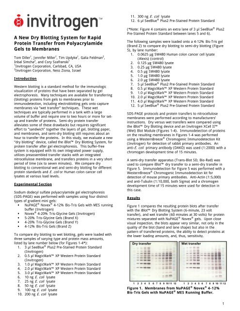
Figure 1 compares the resulting protein blots after transfer<br />
with the iBlot <strong>Dry</strong> <strong>Blotting</strong> <strong>System</strong> (6-minute, 23 volt<br />
transfer), and wet transfer (60 minutes at 30 volts) <strong>for</strong> protein<br />
mixtures separated with NuPAGE ® Novex ® gels. Upon close<br />
visual inspection, the blots appear very similar, not only in the<br />
quality of the blot (band and lane shape) but also in the<br />
pattern of transferred proteins, the ability to detect proteins at<br />
the lower loading amounts, and, thus, sensitivity.<br />
<strong>Dry</strong> transfer Wet transfer<br />
1 2 3 4 5 6 7 8 9 10 11 12 1 2 3 4 5 6 7 8 9 10 11 12<br />
Figure 1. Membranes from NuPAGE ® Novex ® 4-12%<br />
Bis-Tris Gels with NuPAGE ® MES Running Buffer.<br />
1
Figure 2 compares data <strong>for</strong> dry blotting the same samples from<br />
Novex ® Tris-Glycine Gels <strong>for</strong> 6 minutes at 23 volts with data<br />
obtained <strong>for</strong> wet blotting with the protocol-recommended 60<br />
minutes at 25 volts. Again, the dry transfer membrane data is<br />
visually comparable to the wet transfer data. Note that protein<br />
bands <strong>for</strong> the pre-stained SeeBlue ® Plus2 Standard (lanes 1<br />
and 12) are more evident on the dry blotted membrane than<br />
with wet blot, particularly in the higher molecular weight<br />
region. This may indicate that the iBlot TM transfer system<br />
possesses greater sensitivity as compared to wet transfer of<br />
some protein samples and gel types.<br />
<strong>Dry</strong> transfer Wet transfer<br />
1 2 3 4 5 6 7 8 9 10 11 12 1 2 3 4 5 6 7 8 9 10 11 12<br />
Figure 2. Membranes from Novex ® 4-20% Tris-Glycine<br />
Gels.<br />
Figures 3 and 4 are similar comparisons of the same samples<br />
transferred from other brands of Tris-Glycine gels to<br />
nitrocellulose membranes (6 minutes/23 volts with dry<br />
transfer; 60 minutes/25 volts <strong>for</strong> Brand X and Brand Y gels via<br />
wet transfer). The data indicate that dry transfer of proteins<br />
using the iBlot <strong>Dry</strong> <strong>Blotting</strong> <strong>System</strong> yields results that are<br />
very comparable to results obtained using standard wet<br />
transfer methods that have been used historically <strong>for</strong> protein<br />
transfer and which take significantly longer to per<strong>for</strong>m. The<br />
quality of the data and the sensitivity of the western detection<br />
technique seem reduced in the data obtained with Brand X and<br />
Y gels as compared to that obtained from NuPAGE ® and<br />
Novex ® gels, while the blots from NuPAGE ® Novex ® Bis-Tris<br />
gels appear to outper<strong>for</strong>m the other gel types with respect to<br />
band quality and sensitivity.<br />
<strong>Dry</strong> transfer Wet transfer<br />
1 2 3 4 5 6 7 8 9 10 11 12 1 2 3 4 5 6 7 8 9 10 11 12<br />
Figure 3. Membranes from 5-20% Tris-Glycine Gels<br />
(Brand X).<br />
<strong>Dry</strong> transfer Wet transfer<br />
1 2 3 4 5 6 7 8 9 10 11 12 1 2 3 4 5 6 7 8 9 10 11 12<br />
Figure 4. Membranes from 4-20% Tris-Glycine Gels<br />
(Brand Y).<br />
<strong>Dry</strong> transfer of proteins (6 minutes/23 volts) from a 4-12%<br />
Bis-Tris gel to nitrocellulose is compared with semi-dry (60<br />
minutes/20 volts) protein transfer in Figure 5. Data <strong>for</strong> these<br />
gels, as well as NuPAGE ® Novex ® gels and other competitive<br />
gels we compared (data not shown), indicate that dry transfer<br />
of proteins using the iBlot dry blotting system is superior to<br />
semi-dry blotting <strong>for</strong> detection of proteins. The ability to<br />
detect tubulin and actin at lower protein masses in lanes 1-6<br />
and the more pronounced bands evident in the MagicMark<br />
XP Standard lanes (8-11) of Figure 5 demonstrate this higher<br />
sensitivity.<br />
<strong>Dry</strong> transfer Semi-dry<br />
transfer<br />
1 2 3 4 5 6 7 8 9 10 11 12 1 2 3 4 5 6 7 8 9 10 11 12<br />
Figure 5. Membranes from 4-12% Bis-Tris Gels (Brand<br />
Z).<br />
Summary<br />
For the conditions and samples tested, the iBlot <strong>Dry</strong> <strong>Blotting</strong><br />
<strong>System</strong> provides overall visual immunodetection results that<br />
are comparable to results obtained using a standard wet<br />
blotting system but in a fraction of the time and with both less<br />
ef<strong>for</strong>t and less volume of reagents. When used with the<br />
iBlot system, visual immunodetection results of Brand X and<br />
Y Tris-Glycine gels are similar to results obtained using Novex ®<br />
Tris-Glycine gels, indicating that the dry blotting system can be<br />
used with gels from a variety of sources. In addition, dry<br />
protein transfer enables significantly more sensitive detection<br />
of proteins than semi-dry protein transfer and requires much<br />
less time.<br />
Finally, <strong>for</strong> dry versus wet protein transfer, the quality and<br />
sensitivity of the resulting protein blots are comparable among<br />
all the gels included within this study, yet the best results<br />
appear to be obtained from NuPAGE ® Novex ® gels. These<br />
results indicate that western blot protein transfers can be<br />
optimized by choosing the blotting procedure and gel type that<br />
is best suited to your sample and that satisfactory results can<br />
now be obtained with dry blotting in just minutes as opposed<br />
to waiting <strong>for</strong> hours <strong>for</strong> data from the laboratory.<br />
For more in<strong>for</strong>mation, visit www.invitrogen.com/iBlot.<br />
<strong>Invitrogen</strong> Corporation<br />
1600 Faraday Avenue, Carlsbad, CA 92008 USA<br />
Contact: protein.analysis@invitrogen.com<br />
2