Magnetic Chromatography brochure - Invitrogen
Magnetic Chromatography brochure - Invitrogen
Magnetic Chromatography brochure - Invitrogen

Create successful ePaper yourself
Turn your PDF publications into a flip-book with our unique Google optimized e-Paper software.
A<br />
C<br />
A<br />
pH 3<br />
Lysate (unfractionated)<br />
655 spots<br />
pH 3<br />
400 mM fraction<br />
Reduced Sample Complexity<br />
802 spots, 68 % unique to this fraction 817 spots, 84 % unique to this fraction<br />
DYNAL<br />
invitrogen bead separations<br />
pH 10<br />
pH 10<br />
B<br />
D<br />
pH 3<br />
200 mM fraction<br />
867 spots, 84 % unique to this fraction<br />
pH 3 pH 10<br />
600 mM fraction<br />
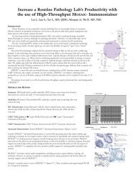
Fig. 2. Reduced sample complexity using Dynabeads® WCX<br />
A cell lysate from a human cancer cell line (SW480) was fractionated using Dynabeads® WCX. Proteins were adsorbed to Dynabeads® WCX using low salt conditions<br />
(50 mM NaP buffer pH 7, 50 mM NaCl) and desorbed step-wise with increasing salt concentrations (50 mM NaP buffer pH 7 including 200 mM, 400 mM or 600 mM<br />
NaCl). The figure shows unfractionated lysate (A) and the three fractions (B,C,D) as analysed by 2-D gel electrophoresis. Fraction B, C and D enabled the detection of<br />
2,211 unique resolved spots and of these only 38 spots were found to be present in all three fractions (2%). Analysing the same amount of protein from unfractionated<br />
lysate enabled the detection of only 655 spots.<br />
Fractions were prepared for gel analysis by desalting using a commercially available 2-D gel clean up kit. Samples were then focused in pH 3 - 10 (11 cm) IPG strips<br />
and resolved in 8 - 16 % SDS-PAGE gels. Approximately 100 µg protein was applied to each strip. Gels were silver stained and analysed using a commercially available<br />
2-D gel analysis software.<br />
Fig. 3. Reduced sample complexity using Dynabeads® RPC Protein<br />
A. Fractionation of a protein mixture containing diamine oxidase and alcohol dehydrogenase using Dynabeads® RPC Protein. The protein mixture (containing 10 μg<br />
total protein) was adsorbed to Dynabeads® RPC Protein in 200 mM NaCl, 0.1 % TFA. Salts were subsequently washed away and proteins desorbed step-wise using<br />
increasing concentrations of acetonitrile (20 %, 30 % and 40 %). One quarter of each fraction was applied to a 10-20 % SDS-PAGE gel. The gel was silver stained.<br />
Diamine oxidase and alcohol dehydrogenase are found in distinct fractions.<br />
B. A mixture containing Insulin (1), Ubiquitin I (2), Cytochrome C (3) and Myoglobin (4) (Bruker Daltonics’ Protein Calibration Standard I) was fractionated using<br />
Dynabeads® RPC Protein. The mixture was adsorbed to Dynabeads® RPC Protein in 200 mM NaCl, 0.1 % TFA. Salts were subsequently washed away and proteins/<br />
peptides desorbed step-wise using increasing concentrations of acetonitrile (15 %, 30 % and 40 %). An aliquot of protein/peptide mixture or eluate (1 μl) was mixed<br />
with an equal amount of MALDI matrix (1 μl sinapinic acid) and 0.5 μl of the sample/matrix mixture spotted onto a MALDI target plate. Data was obtained using a<br />
commercially available MALDI-TOF/TOF MS instrument.<br />
B<br />
Intens. (a.u.)<br />
Intens. [a.u.]<br />
Intens. [a.u.]<br />
Intens. [a.u.]<br />
Intens. [a.u.]<br />
4<br />
x10<br />
3<br />
2<br />
1<br />
0<br />
x104<br />
2.5<br />
2.0<br />
1.5<br />
1.0<br />
0.5<br />
0 0<br />
x104<br />
2.0<br />
1.5<br />
1.0<br />
0.5<br />
0 0<br />
4000<br />
3000<br />
2000<br />
1000<br />
0<br />
1<br />
pH 10<br />
Unfractionated mixture<br />
Pro1_g7_160305\0_G7\1\1SLin Raw<br />
2 3 4<br />
Pro2_g8_160305\0_G8\1\1SLin Raw<br />
15 % acetonitrile fraction<br />
Pro3_h6_160305\0_H6\1\1SLin Raw<br />
30 % acetonitrile fraction<br />
Pro4_h8_160305\0_H8\1\1SLin Raw<br />
40 % acetonitrile fraction<br />
6000 8000 10000 12000 14000 16000 18000<br />
m/z<br />
3