96-well phosphoELISA™ Sample Preparation for ... - Invitrogen
96-well phosphoELISA™ Sample Preparation for ... - Invitrogen
96-well phosphoELISA™ Sample Preparation for ... - Invitrogen

You also want an ePaper? Increase the reach of your titles
YUMPU automatically turns print PDFs into web optimized ePapers that Google loves.
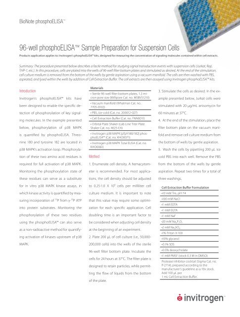
BioNote phosphoELISA <br />
<strong>96</strong>-<strong>well</strong> phosphoELISA <strong>Sample</strong> <strong>Preparation</strong> <strong>for</strong> Suspension Cells<br />
Products application applies to: <strong>Invitrogen</strong>’s phosphoELISA kits, designed <strong>for</strong> measuring the concentration of signaling molecules contained within cell extracts.<br />
Summary: The procedure presented below describes a facile method <strong>for</strong> studying signal transduction events with suspension cells (Jurkat, Raji,<br />
THP-1, etc.). In this procedure, cells are plated into the <strong>well</strong>s of <strong>96</strong>-<strong>well</strong> filter bottom plates and stimulated as desired. At the end of the stimulation,<br />
cell culture medium is removed from the bottom of the <strong>well</strong>s by gentle aspiration using a vacuum manifold. The cells are then washed with PBS,<br />
aspirated, and lysed within the <strong>well</strong>s by addition of Cell Extraction Buffer. The cell extracts are then assayed using <strong>Invitrogen</strong> phosphoELISA kits.<br />
Introduction<br />
<strong>Invitrogen</strong>’s phosphoELISA kits have<br />
been designed to enable the specifi c de-<br />
tection of phosphorylation of key signal-<br />
ing molecules. In the example presented<br />
below, phosphorylation of p38 MAPK<br />
is quantifi ed by phosphoELISA. Threo-<br />
nine 180 and tyrosine 182 are located in<br />
p38 MAPK’s activation loop. Phosphoryla-<br />
tion of these two amino acid residues is<br />
required <strong>for</strong> full activation of p38 MAPK.<br />
Monitoring the phosphorylation state of<br />
these residues can serve as a substitute<br />
<strong>for</strong> in vitro p38 MAPK kinase assays, in<br />
which kinase activity is quantifi ed by mea-<br />
suring incorporation of 32 P from γ- 32 P ATP<br />
into protein substrates. Monitoring the<br />
phosphorylation of these two residues<br />
using the phosphoELISA can also serve<br />
as a non-radioactive method <strong>for</strong> quantify-<br />
ing activation of kinases upstream of p38<br />
MAPK.<br />
Materials<br />
• Sterile <strong>96</strong>-<strong>well</strong> fi lter bottom plates, 1.2 micron<br />
pore size (Millipore Cat. no. MSBVS1210)<br />
• Vacuum manifold (Whatman Cat. no.<br />
7705-0102)<br />
• PBS, ice-cold (Cat. no. 200012-027)<br />
• Cell Extraction Buff er (Cat. no. FNN0011)<br />
• Orbital Plate Shaker (Lab Line Titer Plate<br />
Shaker Cat. no. 4625-EA)<br />
• <strong>Invitrogen</strong> p38 MAPK [pTpY180/182] phosphoELISA<br />
(Cat. no. KHO0071)<br />
• <strong>Invitrogen</strong> p38 MAPK Total ELISA (Cat. no.<br />
KHO0061)<br />
Method<br />
1. Enumerate cell density. A hemacytom-<br />
eter is recommended. For most applica-<br />
tions, the cell density should be adjusted<br />
to 0.25-1.0 X 10 6 cells per milliliter cell<br />
culture medium. It is important to note<br />
that this value may require some optimi-<br />
zation <strong>for</strong> each specifi c application. Cell<br />
doubling time is an important factor to<br />
be considered when adjusting cell density<br />
at the beginning of an experiment.<br />
2. Plate 200 μL of cell culture (i.e., 50,000-<br />
200,000 cells) into the <strong>well</strong>s of the sterile<br />
<strong>96</strong>-<strong>well</strong> fi lter bottom plate. Incubate the<br />
cells <strong>for</strong> 24 hours at 37°C. The fi lter plate is<br />
designed to retain particles, while permit-<br />
ting the fl ow of liquids from the bottom<br />
of the plate.<br />
3. Stimulate the cells as desired. In the ex-<br />
ample presented below, Jurkat cells were<br />
stimulated with 20 μg/mL anisomycin <strong>for</strong><br />
60 minutes at 37°C.<br />
4. At the end of the stimulation, place the<br />
fi lter bottom plate on the vacuum mani-<br />
fold and remove cell culture medium from<br />
the bottom of <strong>well</strong>s by gentle aspiration.<br />
5. Wash the cells by pipetting 200 μL ice<br />
cold PBS into each <strong>well</strong>. Remove the PBS<br />
from the bottom of the <strong>well</strong>s by gentle<br />
aspiration. Repeat two times <strong>for</strong> a total of<br />
three washings.<br />
Cell Extraction Buff er Formulation<br />
•10 mM Tris, pH 7.4<br />
•100 mM NaCl<br />
•1 mM EDTA<br />
•1 mM EGTA<br />
•1 mM NaF<br />
•20 mM Na 4P2O7 •2 mM Na3VO4 •1% Triton X-100<br />
•10% glycerol<br />
•0.1% SDS<br />
•0.5% deoxycholate<br />
•1 mM PMSF (stock 0.3 M in DMSO)<br />
Protease inhibitor cocktail (Sigma Cat. no.<br />
P-2714), prepared according to the<br />
manufacturer’s guideline as a 10x stock.<br />
Add 100 μL per<br />
1 mL Cell Extraction Buff er.
BioNote phosphoELISA<br />
6. Pipette 300 μL protease-inhibitor-<br />
supplemented Cell Extraction Buff er into<br />
each <strong>well</strong>. Incubate the plate on ice <strong>for</strong> 30<br />
minutes.<br />
7. Thoroughly mix the contents of each<br />
<strong>well</strong> by pipetting up and down 5-6 times.<br />
A multi-channel pipette is desirable <strong>for</strong><br />
this application. At this point in the proce-<br />
dure, the extracts are ready <strong>for</strong> analysis. Al-<br />
ternatively, the extracts may be stored in<br />
the fi lter bottom plate at -20°C <strong>for</strong> future<br />
analysis. Frozen plates should be thawed<br />
on ice in preparation of completing the<br />
assays.<br />
8. Place the plate on an orbital shaker and<br />
mix <strong>for</strong> 1 minute.<br />
9. Prepare the <strong>96</strong>-<strong>well</strong> plate <strong>for</strong> ELISA as<br />
follows:<br />
For ELISA <strong>Sample</strong> Wells: Pipette 95 μL Stan-<br />
dard Diluent Buff er (included in the ELISA<br />
kits) into the <strong>well</strong>s of the ELISA plates des-<br />
ignated <strong>for</strong> samples. Transfer 5 μL cell ex-<br />
tract from the fi lter plate into the sample<br />
<strong>well</strong>s of the ELISA plates. Place the ELISA<br />
p38 [pTpY180/182] Levels in Anisomycin-Treated<br />
and Non-Treated Jurkat Cells<br />
Cells/ Well<br />
plates on an orbital shaker to thoroughly<br />
mix the contents of the <strong>well</strong>s.<br />
For ELISA Standard Wells: Prepare stan-<br />
dards as indicated in the assay protocol<br />
and pipette into designated <strong>well</strong>s.<br />
10. Complete the ELISAs as directed by the<br />
assay protocols.<br />
Results and Discussion<br />
Jurkat cells were seeded into the <strong>well</strong>s<br />
of the fi lter bottom plate at densities of<br />
50,000, 100,000, and 200,000 cells per 200<br />
μg/mL. The seeded cells were incubated<br />
<strong>for</strong> 24 hours at 37°C, then treated with<br />
anisomycin (20 μg/mL) <strong>for</strong> 60 minutes, or<br />
left untreated. At the end of the incuba-<br />
tion period, the cells were lysed by the<br />
method described above, and assayed in<br />
parallel with the p38 MAPK [pTpY180/182]<br />
phophosELISA and the p38 MAPK Total<br />
ELISA.<br />
Figure 1—Anisomycin increases phosphorylation of p38 MAPK at<br />
threonine 180 and 182.<br />
The results presented in Figure 1 were ob-<br />
tained with the p38 MAPK [pTpY180/182]<br />
phosphoELISA (Cat. no. KHO0071). The<br />
data presented show that anisomycin<br />
©2006 <strong>Invitrogen</strong> Corporation. All rights reserved. These products may be covered by one or more Limited Use Label Licenses (see <strong>Invitrogen</strong> catalog or www.invitrogen.com). By use of these products you accept the<br />
terms and conditions of all applicable Limited Use Label Licenses. For research use only. Not intended <strong>for</strong> any animal or human therapeutic or diagnostic use, unless otherwise stated. F-1028-BN-pELISA US 1006<br />
p38 (pg/mL)<br />
250<br />
200<br />
150<br />
100<br />
50<br />
0<br />
treatment increases the level of phos-<br />
phorylation of p38 MAPK at threonine 180<br />
and 182. The data presented in Figure 1<br />
also show that the quantity of p38 MAPK<br />
[pTpY180/182] measured with this kit is<br />
directly proportional to the number of<br />
cells seeded into the <strong>well</strong>s of the fi lter bot-<br />
tom plate.<br />
The results presented in Figure 2 were ob-<br />
tained with the p38 MAPK Total ELISA (Cat.<br />
no. KHO0061). This kit was designed to<br />
permit normalization of p38 MAPK phos-<br />
phorylation state. The data presented<br />
show that anisomycin treatment has<br />
no impact on the amount to total p38<br />
MAPK measured in cell lysates, indicating<br />
anisomycin increases p38 MAPK<br />
[pTpY180/182] by enhancing phosphory-<br />
lation state (Figure 1), rather than al-<br />
tering total p38 MAPK level. The data<br />
presented in Figure 2 also show that the<br />
quantity of total p38 MAPK measured with<br />
this kit is directly proportional to the num-<br />
ber of cells seeded into the <strong>well</strong>s of the<br />
fi l t e r b o t t o m p l a t e .<br />
p38 (Total) Levels in anisomycin-treated<br />
and non-treated Jurkat cells<br />
50,000 100,000 200,000<br />
Cells/Well<br />
Figure 2—Aninomycin treatment does not change the total<br />
p38 MAPK level in cell lysates.<br />
www.invitrogen.com