Crystallography and Lectin Structure Database - CNRS
Crystallography and Lectin Structure Database - CNRS
Crystallography and Lectin Structure Database - CNRS

You also want an ePaper? Increase the reach of your titles
YUMPU automatically turns print PDFs into web optimized ePapers that Google loves.
16 U. Krengel <strong>and</strong> A. Imberty<br />
the first Nobel Prize in physics for his discovery in 1901. A few years later, it was<br />
proposed that the wavelength of X-rays were of the same magnitude as interatomic<br />
distances, <strong>and</strong> in 1912, a crystal diffraction experiment confirmed this<br />
hypothesis. The first crystal structure was solved in the same year. It then took a<br />
few decades until this method could be successfully applied to large <strong>and</strong> fragile<br />
macromolecules such as proteins <strong>and</strong> DNA. Dorothy Crowfoot Hodgkin played a<br />
leading role in this work. The year 1962 marked a period of particular recognition<br />
of macromolecular X-ray structure analysis, as both the Nobel Prize in physiology<br />
<strong>and</strong> that in chemistry went to l<strong>and</strong>mark X-ray structures: Watson, Crick,<br />
<strong>and</strong> Wilkins received honors for their famous 3D-structural model of DNA <strong>and</strong><br />
Kendrew <strong>and</strong> Perutz for the first protein structures solved, those of myoglobin<br />
<strong>and</strong> hemoglobin.<br />
As the name “X-ray crystallography” suggests, protein crystals are a necessary<br />
precondition for the study of proteins or protein lig<strong>and</strong> complexes by this method.<br />
Crystals are needed in order to enhance the scattering power of the subjects under<br />
investigation. Normally, molecules scatter X-rays only weakly, but if they are regularly<br />
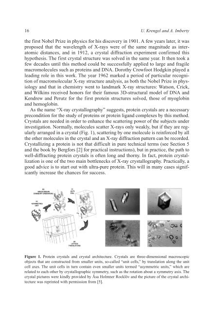
arranged in a crystal (Fig. 1), scattering by one molecule is reinforced by all<br />
the other molecules in the crystal <strong>and</strong> an X-ray diffraction pattern can be recorded.<br />
Crystallizing a protein is not that difficult in pure technical terms (see Section 5<br />
<strong>and</strong> the book by Bergfors [2] for practical instructions), but in practice, the path to<br />
well-diffracting protein crystals is often long <strong>and</strong> thorny. In fact, protein crystallization<br />
is one of the two main bottlenecks of X-ray crystallography. Practically, a<br />
good advice is to start out with ultra-pure protein. This will in many cases significantly<br />
increase the chances for success.<br />
Figure 1. Protein crystals <strong>and</strong> crystal architecture. Crystals are three-dimensional macroscopic<br />
objects that are constructed from smaller units, so-called “unit cells,” by translation along the unit<br />
cell axes. The unit cells in turn contain even smaller units termed “asymmetric units,” which are<br />
related to each other by crystallographic symmetry, such as the rotation about a symmetry axis. The<br />
crystal pictures were kindly provided by Åsa Holmner Rocklöv <strong>and</strong> the picture of the crystal architecture<br />
was reprinted with permission from [5].