Traitement des infections à germes intracellulaires - Infectiologie
Traitement des infections à germes intracellulaires - Infectiologie
Traitement des infections à germes intracellulaires - Infectiologie

Create successful ePaper yourself
Turn your PDF publications into a flip-book with our unique Google optimized e-Paper software.
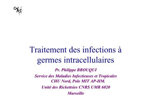
<strong>Traitement</strong> <strong>des</strong> <strong>infections</strong> <strong>à</strong><br />
<strong>germes</strong> <strong>intracellulaires</strong><br />
Pr. Philippe BROUQUI<br />
Service <strong>des</strong> Maladies Infectieuses et Tropicales<br />
CHU Nord, Pole MIT AP-HM. AP HM.<br />
Unité Unit <strong>des</strong> Rickettsies CNRS UMR 6020<br />
Marseille
• Concept différent<br />
• Pharmacocinétique<br />
• Antibiogramme<br />
• Identification<br />
moléculaire <strong>des</strong><br />
résistances aux<br />
antibiotiques<br />
Introduction<br />
• Femme enceinte et<br />
enfant<br />
• Nouvelles molécules<br />
• <strong>Traitement</strong>s courts<br />
• <strong>Traitement</strong>s alternatifs
• Bactérie intracellulaire<br />
– Notion de sanctuaire<br />
– Pas d ’antibiogramme<br />
– Intérêt de la biologie<br />
moléculaire<br />
Définition
Blood<br />
Pharmacocinetic<br />
Cell target<br />
Intracellular<br />
compartment pH
Extracellular medium<br />
Active drug<br />
diffusion<br />
pinocytosis<br />
metabolism and<br />
inactivation<br />
subcellular distribution<br />
cytosolic activity<br />
subcellular activity<br />
Antibiotic uptake<br />
Inactive drug<br />
cytosol<br />
pH 4.5<br />
Phagolysosome<br />
bacteria
Destruction of bacteria in phagocytic cells<br />
Strategy for survival of intracellular bacteria in cells<br />
nucleus<br />
cytosol<br />
Phagolysosome<br />
(complete fusion)<br />
Killing of the bacteria<br />
pH 4,5<br />
<br />
Bacteria<br />
Early phagosome<br />
<br />
<br />
Lysosomes<br />
pH 6,5<br />
Acidification<br />
Late phagosome<br />
(incomplete fusion)
Survival and Intracellular location of<br />
Escape phagosome live in<br />
the cytosol<br />
Listeria spp<br />
Shigella spp<br />
Rickettsia spp<br />
bacteria<br />
Adapt in acidic environment<br />
and live in the<br />
phagolysosome<br />
C burnetii<br />
Y pestis<br />
S aureus<br />
Y pseudotuberculosis<br />
Inhibits fusion and live<br />
in the phagosome<br />
Chlamydia spp<br />
Ehrlichia<br />
Legionella spp<br />
Y enterocolitica<br />
Brucella spp
Facteurs influençant l’activité<br />
intracellulaire d’un antibiotique<br />
• Pénétration intracellulaire de l’antibiotique<br />
• Localisation sub-cellulaire de l’antibiotique<br />
/microorganisme<br />
• Sensibilité du microorganisme <strong>à</strong> l’antibiotique<br />
• Influence du milieu intracellulaire sur le couple<br />
antibiotique/microorganisme<br />
– pH, enzymes, fixation protéique...
Métho<strong>des</strong> d’étude de la pénétration<br />
intracellulaire <strong>des</strong> antibiotiques<br />
• Choix de la cellule<br />
• Temps de contact avec l’antibiotique<br />
• Élimination de l’antibiotique extracellulaire<br />
• Dosage de l’antibiotique intracellulaire<br />
– Autoradiographie<br />
– Fluorescence (tétracyclines, fluoroquinolones..)<br />
– Dosage chimique et biologique après lyse cellulaire
Pénétration intracellulaire <strong>des</strong><br />
antibiotiques<br />
• Macrophages #Polynucléaires<br />
– Pénètrent bien : (C/E>4)<br />
• Azythromycine,Roxithromycine,Erythromycine<br />
• Clindamycine<br />
• Quinolones<br />
– Pénètrent : (C/E 1-4)<br />
• Rifampicine, Doxycycline, Chloramphénicol, Bactrim<br />
• Aminoglycosi<strong>des</strong> ++<br />
– Pénètrent pas :( C/E
Métho<strong>des</strong> d’étude de la localisation sub-<br />
cellulaire <strong>des</strong> antibiotiques<br />
• Fractionnement cellulaire en gradient de<br />
saccharose<br />
– Dosage biochimique ou biologique <strong>des</strong> fractions<br />
• Utilisation <strong>des</strong> traceurs cellulaires (double<br />
marquage)<br />
– Autoradiographie, microscope confocal<br />
• Différenciation lysosome/cytosol
?<br />
?<br />
Localisation subcellulaire <strong>des</strong> ATB<br />
• Cytosol<br />
– Cyclines<br />
– Chloramphenicol<br />
– Rifampicine<br />
– Quinolones<br />
• Lysosomes<br />
– Aminosi<strong>des</strong><br />
– Erythromycine<br />
– Clindamycine<br />
– Quinolones<br />
– Rifampicine<br />
– Cyclines et chloramphénicol<br />
???
Moyens pour la mesure du pH<br />
• Marqueur chimique<br />
intracellulaire<br />
– Dextran ( base faible) ,<br />
traceurs fluorescents..<br />
• Microscopie confocale
Rôle du pH dans l ’activité <strong>des</strong><br />
antibiotiques<br />
Antibiotique PH optimal<br />
Quinolones<br />
Erythromycine<br />
Betalactamines<br />
Aminosi<strong>des</strong><br />
Ethambutol<br />
Cyclines<br />
Rifampicine<br />
Pyrazinamide<br />
8<br />
7.8<br />
7<br />
7.5<br />
7<br />
6.6<br />
Antibiotic uptake, subcellular localisation and<br />
pH of optimum activity of antibiotics<br />
Antibiotic Mode of entry Cytosol Lysosomes PH of optimum activity<br />
Aminoglycosi<strong>des</strong><br />
Betalactams<br />
Chloramphenicol<br />
Erythromycin<br />
Fluoroquinolones<br />
Rifampin<br />
Tetracyclines<br />
Pinocytosis<br />
Diffusion<br />
Diffusion<br />
Transport<br />
Unknow<br />
Diffusion<br />
Diffusion<br />
+<br />
++<br />
+<br />
++<br />
++<br />
++<br />
+++<br />
Unknow<br />
Symbols : + = low concentration ; ++ = medium concentration ; +++ = high concentration<br />
+++<br />
++<br />
++<br />
++<br />
7<br />
7<br />
7<br />
7.8<br />
8<br />
Co-localisation ATB-Bactéries<br />
• Lysosomes et P-L<br />
– Antibiotiques<br />
• Aminoglycosi<strong>des</strong>,erythromycine,quinolones,rifampicine<br />
,clindamycine, cyclines ?<br />
– Bactéries<br />
• C burnetii, Y pestis, S aureus, Y pseudotuberculosis<br />
• Cytosol<br />
– Antibiotiques<br />
• Cyclines, rifampicine,chloramphénicol, quinolones<br />
– Bactéries<br />
• Rickettsia spp, Shigella spp, Listeria spp
Co-localisation ATB-Bactéries<br />
• Phagosome<br />
–ATB ?<br />
– Bactéries<br />
• Chlamydia spp, Ehrlichia spp, Legionella spp, Y<br />
enterocolitica, Brucella spp.
In vitro antibiotic susceptibility testing<br />
guinea pig model<br />
embryonated eggs<br />
cell systems<br />
– plaque assay (McDade J., Appl.Microbiol. 1969)<br />
– dye uptake assay (Raoult D. et al J. Infect. Dis. 1987)<br />
plaque assay dye uptake assay<br />
– IF assay (Ives T.J. et al Antimicrob. Agents Chemother. 1997)<br />
– flow cytometry (Kelly D.J. et al Am.J.Trop.Med.Hyg. 1995)
• Methods<br />
A/<br />
B/<br />
Tissue-Cell culture system<br />
[BACTERIA]<br />
[ANTIBIOTICS]<br />
[BACTERIA]<br />
Media without<br />
ATB<br />
Incubation at 37°C for several days<br />
P3 laboratory
Dye uptake assay<br />
•Distribution of cells in 96-well microplate<br />
•Incubation at 37°C with 5% CO 2 for 24-48<br />
hours<br />
to obtain confluent monolayers<br />
. After incubation, cell monolayers are<br />
stained with neutral red dye (vital dye) dye)<br />
for<br />
1h at 37°C<br />
37
Dye uptake assay<br />
Uninfected cells OD = 1<br />
Infected cells 2000 PFU OD = 0<br />
Infected cells 200 PFU OD = 0.150<br />
Infected cells 20 PFU OD = 0.700<br />
•The The optical density at 492 nm of each well<br />
is determined using a spectrophotometer<br />
The MIC corresponds to the lowest antibiotic concentration<br />
for which the mean OD at 492 nm is higher than that of the<br />
20 PFU controls<br />
Doxycycline 0.015 µg/ml OD =0<br />
Doxycycline 0.03 µg/ml OD = 0<br />
Doxycycline 0.06 µg/ml OD = 0<br />
Doxycycline 0.125 µg/ml OD =0.89 MIC<br />
Doxycycline 0.25 µg/ml OD = 0.95<br />
Doxycycline 0.5 µg/ml OD =1<br />
Doxycycline 1 µg/ml OD = 1
•Cells are prepared in Petri dishes<br />
•Supernantant is discarded by<br />
aspiration of the medium<br />
Plaque assay<br />
The plaque assay system is currently the recommended technique allowing<br />
enumeration of the inoculum (Plaque Forming Unit PFU) and evaluation of<br />
the bacteriostatic activity of antibiotics. antibiotics<br />
•Cells are infected for 1 hour<br />
with a bacterial inoculum.
Plaque assay<br />
• Antibiotics are added at different concentrations at the same time, whereas no antibiotics are<br />
added in drug-free controls<br />
• Infected cells are then overlaid with Eagle MEM with 2% fetal calf serum and 0.5 % agar.<br />
Petri dishes are incubated 7 to 10 days at 37°C in a 5% CO2 atmosphere.
Plaque assay: assay:<br />
results<br />
Doxycycline 0.015 µg/ml Doxycycline 0.03 µg/ml Doxycycline 0.06 µg/ml<br />
Doxycycline 0.125 µg/ml Doxycycline 0.25 µg/ml Doxycycline 0.5 µg/ml<br />
The MICs are defined as the lowest antibiotic concentration allowing complete inhibition of<br />
plaque formation, as compared to a drug-free drug free control
Diff-quick Diff quick assay<br />
Evaluation of the growth of bacteria (as determined by counting the percentage of infected cells<br />
after Diff Quick staining) staining)<br />
with or without antibiotics as compared to a primary inoculum<br />
•Grow Grow bacteria in cell culture in 96-<br />
well plates until > 50% of cells are<br />
infected<br />
•Add Add antibiotics (three wells for each<br />
antibiotic concentration tested)<br />
•Incubation Incubation of the plates at 37°C 37 C for 10<br />
days<br />
•Harvest Harvest cell cultures each day during<br />
10 days<br />
•Preparation Preparation of sli<strong>des</strong> by cytospin<br />
centrifugation
•Diff Diff Quick staining of the cytospin smears<br />
Infected cells (%)<br />
120%<br />
100%<br />
80%<br />
60%<br />
40%<br />
20%<br />
0%<br />
0 1 2 3 4 5 6 7 8 9 Days<br />
Diff-quick Diff quick assay<br />
Control Telithromycin (8 µg/ml) Doxycycline (1µg/ml)<br />
•Determination<br />
Determination of the percentage of infected cells<br />
Morulae<br />
The MIC is the lowest antibiotic<br />
concentration allowing reduction<br />
of the percentage of infected cells<br />
to < 10% after 10 days<br />
incubation of cultures with the<br />
antibiotic
150 cm 2 flasks<br />
C. burnetiiinfected P388D1 cells<br />
Titer C. burnetii<br />
10 -1 to 10 -9<br />
Shell vials<br />
Incubate6 days<br />
immunofluorescence<br />
Bactericidal assay<br />
•Antibiotic exposure of infected cells for 24 hours<br />
•Cell monolayers are harvested and lysed by thermal shock<br />
•Titration of each bacterial suspension onto uninfected cells and incubation for 6 days.<br />
•Cells are then stained by immunofluorescence<br />
Wash<br />
lyse cells<br />
25 cm 2 flasks<br />
1 - cells adhere<br />
2 - add new media<br />
3 - add drug<br />
4 - incubate 24 hours
Negative control<br />
Bactericidal assay: assay:<br />
results<br />
Bacteriocidal activity was deduced from the reduction of the Vacuole Forming Unit (VFU)<br />
in cultures receiving antibiotics as compared with drug-free drug free controls<br />
Pure 10-1 VFU 10-2 VFU<br />
10-3 VFU<br />
Doxycycline + hydroxychloroquine<br />
10-4 VFU 10-5 VFU
Assay in 24-well microplates<br />
Cells are harvested<br />
Extraction of DNA<br />
PCR mix<br />
Light-Cycler<br />
Light Cycler assay<br />
Evaluation of the growth of bacteria (as determined by the quantification of DNA copies)<br />
with or without antibiotics as compared to a primary inoculum<br />
Disposition of samples in the carrousel<br />
The Lightcycler machine<br />
Software analysis of results
Light-Cycler<br />
Light Cycler assay<br />
MIC was defined as the first antibiotic<br />
concentration allowing the inhibition of growth<br />
of bacteria as compared to the number of DNA<br />
copies at day 0.
L ’évaluation moléculaire de l ’activité<br />
• Quinolones<br />
<strong>des</strong> antibiotiques<br />
– (DNA gyrase de C burnetii, et de M tuberculosis)<br />
• Rifampicine<br />
– (RpoB de M tuberculosis et <strong>des</strong> rickettsies )<br />
• Izionazide<br />
– ( katG de M tuberculosis : Rinder H: Mol Diagn 1999)<br />
• Tetracyclines<br />
– ( Aminov RI Appl Environ Microbiol 2000)
Le génome bactérien :<br />
evaluation <strong>des</strong> résitances in silico<br />
• Permet de comprendre les mécanismes de la<br />
résistance aux antibiotiques<br />
– Anomalie dans les voies métaboliques<br />
• T. whiplei<br />
– Mutations sur les génes cibles<br />
• 23 s pour les macroli<strong>des</strong><br />
• Prévoir la résistance<br />
– F tularensis et macroli<strong>des</strong>
L ’évaluation in vivo de l ’activité <strong>des</strong><br />
• Modèles animaux<br />
antibiotiques<br />
– Endocardites <strong>à</strong> S aureus (Maurin AAC 97)<br />
– Endocardites <strong>à</strong> C burnetii (LaScola JID 98)<br />
• Essai cliniques<br />
– Irremplaçable<br />
– Discordance<br />
• Brucellose, FBM
Fièvre Q<br />
• Coxiella burnetii<br />
– Forme aiguë : pneumonie +/- hépatite +/- fièvre<br />
– Forme chronique : endocardites ou <strong>infections</strong> vasculaires<br />
Transaminases<br />
élevées<br />
90<br />
Fièvre<br />
90<br />
Pneumopathies<br />
45
• In vitro sensible <strong>à</strong> :<br />
Fièvre Q : traitement<br />
– Doxycycline<br />
– Bactrim<br />
– Sparfloxacine<br />
– Ofloxacine<br />
– Moxifloxacine<br />
– Roxithyromycine<br />
– Clarithromycine<br />
– Telithromycine<br />
– Rifampicine<br />
– Ciprofloxacine/Erythromyci<br />
ne variable<br />
• <strong>Traitement</strong><br />
– Forme aiguë<br />
• Doxycycline 200 mg/J 15<br />
jours (A)<br />
• Bactrim ? ( E et FE )<br />
– Endocardites<br />
• Doxycycline 200 mg /J et<br />
Plaquenil® 600mg/J<br />
pendant 18mois ( A)<br />
Chloroquinémie Chloroquin mie # 1-2 1 2 micro litre/ml
• Ehrlichioses<br />
monocytiques<br />
Ehrlichioses humaines<br />
– Amblyomma americanum<br />
– USA<br />
– E chaffeensis<br />
• Ehrlichioses<br />
granulocytique<br />
– Ixo<strong>des</strong> spp<br />
– USA et Europe<br />
– E phagocytophila<br />
Syndrome grippal après piqûre de tique
• In vitro sensible <strong>à</strong> :<br />
Ehrlichioses traitement<br />
– Doxycycline<br />
– Rifampicine<br />
– Quinolones<br />
• E phagocytophila<br />
seulement<br />
• In vitro résistant <strong>à</strong> :<br />
– Chloramphénicol, Bactrim<br />
pénicilline et érythromycine<br />
– Modérément sensible <strong>à</strong><br />
Aminoglycosi<strong>des</strong><br />
• <strong>Traitement</strong><br />
– Doxycycline 7-14 jours<br />
(A et E )<br />
– Rifampicine ?
Rickettsioses boutonneuses<br />
• Fièvre boutonneuse méditerranéenne<br />
– R conorii<br />
– Ubiquitaire<br />
• TIBOLA<br />
– R slovaca<br />
– Dermacentor marginatus<br />
– Europe<br />
• Fièvre Africaine <strong>à</strong> tique<br />
– R africae<br />
– Afrique du Sud<br />
Amblyomma<br />
R sanguineus
Rickettsioses : traitement<br />
• In vitro sensible <strong>à</strong> :<br />
– Doxycycline<br />
– Quinolones<br />
– Josamycine<br />
– Roxithromycine<br />
– Clarithromycine<br />
– Telithromycine<br />
– Rifampicine ?<br />
• <strong>Traitement</strong><br />
–Forme simple<br />
• Doxycycline 200mgune<br />
fois (A,E)<br />
• Josamycine 50 mg/kg 10<br />
jours<br />
– Forme Malignes<br />
• Doxycycline 200 mg / J<br />
pendant 10 j<br />
Souches résistantes r sistantes <strong>à</strong> la Rifampicine
• Agent infectieux<br />
– B.burgdorferi strico sensu<br />
– B garinii<br />
– B afzelii<br />
• Localisation intracellulaire<br />
Maladie de Lyme<br />
• In vitro sensible <strong>à</strong>:<br />
– Pénicilline<br />
– Erythromycine<br />
– Doxycycline<br />
– Ceftriaxone<br />
• In cellulo sensible <strong>à</strong><br />
– Erythromicine<br />
– Doxycycline
Bartonelloses<br />
• Bartonella bacilliformis<br />
– Fièvre d ’Oroya ou maladie<br />
de Carrion<br />
– Verruga peruana
• Bartonella henselae<br />
– Maladie <strong>des</strong> griffes du chat<br />
Bartonelloses<br />
• Bartonella quintana<br />
– Fièvre <strong>des</strong> tranchés et bactériémie<br />
chronique chez les SDF<br />
• B henselae et B quintana<br />
– Angiomatose Bacillaire ( ID)<br />
– Péliose hépatique et splénique (ID)<br />
– Endocardites
Bartonelloses: traitement<br />
• In cellulo sensible <strong>à</strong>:<br />
– Aminoglycosi<strong>des</strong> ( bactéricide)<br />
– Doxycycline<br />
– Quinolones<br />
– Rifampicine<br />
– Erythromycine<br />
Bartonella henselae<br />
Bartonella quintana<br />
Bartonella elizabethae<br />
Brucella abortus<br />
Brucella melitensis<br />
Ochrobactrum anthropi<br />
Bartonella bacilliformis
Regimen for<br />
Disease Adults Children Strength of<br />
Typical CSD<br />
Complicated CSD<br />
Trench fever OR<br />
Chronic bacteremia<br />
with B. quintana<br />
BA*<br />
PH*<br />
<strong>Traitement</strong> <strong>des</strong> bartonelloses<br />
No recommendation<br />
OR for patients with extensive<br />
lymphadenopathy, consider:<br />
Azithromycin 500 mg PO the first day and<br />
250 mg PO on days 2 to 5 as a single daily<br />
dose<br />
Doxycycline 100 mg PO BID for 4 wks<br />
AND rifampin 300 mg PO BID for 3 wks<br />
Doxycycline 200 mg PO QD for 4 wks<br />
AND Gentamicin 3 mg/kg IV QD for 2<br />
wks<br />
Erythromycin 500 mg PO QID for 3<br />
months<br />
OR doxycycline 100 mg PO BID for 3<br />
months<br />
Erythromycin 500 mg PO QID for 4<br />
months<br />
OR doxycycline 100 mg PO BID for 4<br />
months<br />
No recommendation<br />
OR for patients with extensive<br />
lymphadenopathy, consider:<br />
Azithromycin 10 mg/kg PO on day 1, and<br />
5 mg/kg PO on days 2 to 5 as a single<br />
daily dose<br />
Unknown<br />
Unknown<br />
Erythromycin ethylsuccinate 40 mg/kg PO<br />
total/day, in four divided doses (maximum<br />
total daily dose of 2 g/d) for 3 months<br />
Erythromycin ethylsuccinate 40 mg/kg PO<br />
total/day, in four divided doses (maximum<br />
total daily dose of 2 g/d) for 4 months<br />
recommendation<br />
BI<br />
CIII<br />
BII<br />
AI<br />
AII<br />
AII<br />
AII<br />
AII<br />
Reference<br />
(56)<br />
(8; 86)<br />
(56)<br />
(36)<br />
(58)<br />
(58)<br />
(58)<br />
(58)
Endocarditis<br />
Endocarditis<br />
Carrion’s disease<br />
Oroya fever<br />
Verruga peruana<br />
<strong>Traitement</strong> <strong>des</strong> bartonelloses<br />
Suspected Bartonella, culture-negative:<br />
Gentamicin 3 mg/kg/d IV for 14 days<br />
AND<br />
ceftriaxone 2 g IV or IM QD for 6 weeks<br />
+/- doxycycline 100 mg PO or IV BID for<br />
6 weeks<br />
Documented Bartonella, culture-positive:<br />
Gentamicin 3 mg/kg/d IV for 14 days<br />
AND<br />
doxycycline 100 mg PO BID for 6 wks<br />
Chloramphenicol 500 mg PO or IV QID<br />
for 14 days AND another antibiotic (beta<br />
lactam preferred)<br />
OR ciprofloxacin 500 mg PO BID for 10d<br />
Rifampin 10 mg/kg/d PO for 14d<br />
OR Streptomycin 15-20 mg/kg/d IM for<br />
10d<br />
Unknown<br />
Chloramphenicol 50-75mg/kg/d PO or IV<br />
divided into four doses for 14 days AND<br />
another antibiotic (beta lactam preferred)<br />
OR ciprofloxacin in children 7-12 years<br />
250 mg PO BID for 10d<br />
Rifampin 10 mg/kg/d PO for 14d<br />
(maximum total daily dose of 600<br />
mg/day)<br />
AII<br />
BII<br />
BII<br />
BII<br />
BII<br />
AII<br />
BIII<br />
AII<br />
AII<br />
(85)<br />
(85)<br />
(85)<br />
(85)<br />
(85)<br />
(85)<br />
**<br />
(68)<br />
(68)
Legionelloses (Legionella pneumophila)<br />
• In vitro sur cellules<br />
– Doxycycline<br />
– Bactrim<br />
– ofloxacine,ciprofloxacine,sparfloxacine,<br />
– rifampicine<br />
– érythromycine,roxithromycine,azithromycin<br />
e,clarithromycine<br />
• <strong>Traitement</strong> de référence ( Durée 14-21 jours )<br />
– Azithromycine IV ( non disponible en France)<br />
– Levofoxacine IV ( Tavanic® 250-750 mg / jour )<br />
– ERYTHROMYCINE 2g per os /j ou 4 g IV par J pd 10-14 J<br />
– et Rifampicine<br />
– Autres traitements possibles mais non référencés<br />
• bactrim, doxycycline,ciprofloxacine,azithromycine,clarithromycine
• In vitro sur cellules<br />
• <strong>Traitement</strong><br />
Brucellose<br />
– Aminoglycosi<strong>des</strong> +++, Bactrim,<br />
doxycycline,quinolones, rifampicine<br />
– Doxycycline et / rifampicine ou<br />
streptomycine 6 semaines (A)<br />
– ( Enfant , Femme enceinte) ???
Mycobactéries atypiques<br />
• In vitro sur cellules<br />
– Clarythromycine,azythromycine, sparfloxacine<br />
avec<br />
• éthambutol et/ou amikacine et /ou rifampicine<br />
• Hétérogeneité <strong>des</strong> souches ++++<br />
• <strong>Traitement</strong> <strong>des</strong> formes disséminées ++<br />
– Clarythromycine ou azythromycine et éthambutol<br />
et au moins 1 parmi<br />
• Clofazimine,rifampicine, rifabutine,ciprofloxacine,<br />
amikacine
• In vitro<br />
• <strong>Traitement</strong><br />
Chlamydia spp.( hors urétrites)<br />
– Doxycycline,ciprofloxacine, ofloxacine, sparfloxacine,<br />
rifampicine, érythromycine, azithromycine, clarithromycine,<br />
roxithromicine<br />
– Pneumonies adultes ( C pneumoniae et C psittaci)<br />
• Doxycycline 10-21 j ours<br />
• érythromycine ou azythromycine<br />
– Pneumonie de l’enfant<br />
• Erythromycine ou azythromycine<br />
– LGV ou maladie de Nicolas Favre ( C trachomatis)<br />
• Doxycycline 21 jours ou erythromycine
• Urétrite <strong>à</strong>C trachomatis<br />
<strong>Traitement</strong> courts<br />
– Azythromycine 1g en une prise unique ou<br />
• Doxycycline 200 mg/j pd 7 jours ou érythromycine 2g/j pd 7 j ou<br />
1g/j pd 14 J<br />
• Fièvre typhoïde<br />
– In vitro sur cellules :<br />
chloramphénicol,quinolones,(ceftriaxone et ampicilline ?)<br />
– <strong>Traitement</strong> : Ceftriaxone 3g/j 5 jours ou Quinolones 5 jours<br />
• Rickettsioses<br />
– FBM, typhus
Merci pour votre attention