Multi-Bracket Appliance in Management of Mandibular ...
Multi-Bracket Appliance in Management of Mandibular ...
Multi-Bracket Appliance in Management of Mandibular ...

Create successful ePaper yourself
Turn your PDF publications into a flip-book with our unique Google optimized e-Paper software.
120 <strong>Multi</strong>-bracket appliance <strong>in</strong> management <strong>of</strong>mandibular reconstruction<br />
b<br />
a<br />
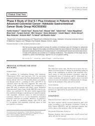
Figure 1. (a) <strong>Bracket</strong> with a rectangular groove is shown. Arch-wire is <strong>in</strong>serted<br />
through this groove. (b) Rectangular arch-wire has a proper elasticity to<br />
ma<strong>in</strong>ta<strong>in</strong> the occlusal situation.<br />
<strong>Multi</strong>-bracket appliances are frequently used <strong>in</strong> orthodontic<br />
treatment (Figs Ia and 1b). <strong>Bracket</strong>s are bonded to the teeth<br />
directly and firmly, and rectangular arch wires control the position<br />
<strong>of</strong> each <strong>in</strong>dividual tooth three-dimensionally. This appliance has<br />
the ability to move the teeth <strong>in</strong> a predeterm<strong>in</strong>ed manner, and<br />
therefore has become widely used <strong>in</strong> the orthodontic field.<br />
The present paper describes three cases <strong>in</strong> which the multibracket<br />
appliance was applied <strong>in</strong> mandibular reconstruction us<strong>in</strong>g<br />
a vascularized bone graft.<br />
MATERIALS AND METHODS<br />
PREPARATION OF MULTI-BRACKET ApPLIANCE<br />
First, the extent <strong>of</strong> the mandibular excision was estimated.<br />
Standard edgewise brackets (0.018 x 0.025 <strong>in</strong>ch slot) were<br />
bonded to the teeth <strong>in</strong> the maxilla and <strong>in</strong> the rema<strong>in</strong><strong>in</strong>g mandible,<br />
and dental impressions were taken . Rectangular wires (0.017 x<br />
0.022 <strong>in</strong>ch) passively fitted with the brackets were made on a<br />
dental model and hooks for maxillo-mandibular fixation were<br />
soldered. An acrylic bite plate to determ<strong>in</strong>e the maxillo-mandibular<br />
relationship was also fabricated on the same dental model <strong>in</strong><br />
two <strong>of</strong> the three cases. Prior to surgery, the fabricated arch-wire<br />
was ligated to the brackets after some adjustment <strong>in</strong> the outpatient<br />
room. Orthodontists performed these procedures.<br />
INTRAOPERATION<br />
After mandibular resection, the occlusion <strong>of</strong> the rema<strong>in</strong><strong>in</strong>g teeth<br />
was replicated precisely. Wires for maxillo-mandibular fixation<br />
were then applied and the bite plate was placed between the<br />
maxillary and mandibular dentition <strong>in</strong> two cases. The harvested<br />
bone was then contoured to replicate the bone defect and then<br />
carefully fixed with m<strong>in</strong>i plates to ma<strong>in</strong>ta<strong>in</strong> the occlusion.<br />
POSTSURGICAL TREATMENTS<br />
Wires for maxillo-mandibular fixation were removed 4 weeks<br />
after surgery when bone union was recognized by roentgenography.<br />
The multi-bracket appliance was worn for a further 3<br />
months when the wound contraction became mild. A plate<br />
type-reta<strong>in</strong>er was used after remov<strong>in</strong>g the multi-bracket appliance.<br />
RESULTS<br />
CASE I<br />
A 45-year-old woman presented with recurrent adenoid cystic<br />
carc<strong>in</strong>oma <strong>of</strong> the right sub-mandibular gland (Figs 2a and 2b).<br />
The planned mandibular resection approach was from above the<br />
right angle to the right lateral <strong>in</strong>cisor. She had lost her maxillary<br />
<strong>in</strong>cisors and she used removable partial dentures. <strong>Bracket</strong>s and<br />
arch-wires were applied to the proposed residual teeth accord<strong>in</strong>g<br />
to the protocol and an orthodontic plate with four artificial<br />
anterior teeth was also fashioned to ma<strong>in</strong>ta<strong>in</strong> the occlusion <strong>of</strong>the<br />
rema<strong>in</strong><strong>in</strong>g dentition (Fig. 2c). Resection <strong>of</strong> the mandible from<br />
above the right angle to the right can<strong>in</strong>e <strong>in</strong>clud<strong>in</strong>g the sub-mandibular<br />
s<strong>of</strong>t tissue and sk<strong>in</strong>, and right radical neck dissection were<br />
carried out. The facial sk<strong>in</strong> and mandibular defects were<br />
reconstructed us<strong>in</strong>g a fibula osteoseptocutaneous flap from the<br />
left lower leg (Fig. 2d) . The mucosal defect was directly sutured.<br />
As the rema<strong>in</strong><strong>in</strong>g mandible was rigidly fixed to the maxilla, the<br />
fibula was fixed accurately without any shift <strong>of</strong>the mandible. The<br />
wires for maxillo-mandibular fixation were removed 4 weeks<br />
after the operation, and the multi-bracket appliance was worn for<br />
a further 3 months. One year later, she was satisfied with her facial<br />
appearance and demonstrated stable occlusion <strong>of</strong> the residual<br />
teeth (Figs 2e and 2f). Removable partial dentures were worn <strong>in</strong><br />
the reconstructed mandible and maxilla (Fig. 2g). She was able<br />
to eat all foods without difficulty.<br />
CASE 2<br />
A 44-year-old man presented with left mand ibular malignant<br />
schwannoma (Figs 3a and 3b) . A segmental mandibulectomy<br />
Downloaded from<br />
http://jjco.oxfordjournals.org/<br />
by guest on March 2, 2013