Multi-Bracket Appliance in Management of Mandibular ...
Multi-Bracket Appliance in Management of Mandibular ...
Multi-Bracket Appliance in Management of Mandibular ...

You also want an ePaper? Increase the reach of your titles
YUMPU automatically turns print PDFs into web optimized ePapers that Google loves.
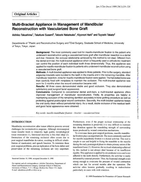
I Orig<strong>in</strong>al Articles I<br />
<strong>Multi</strong>-<strong>Bracket</strong> <strong>Appliance</strong> <strong>in</strong> <strong>Management</strong> <strong>of</strong> <strong>Mandibular</strong><br />
Reconstruction with Vascularized Bone Graft<br />
Jpn J Cl<strong>in</strong> Oncol1999;29(3)1l9-126<br />
Akihiko Takushima 1 , Takafumi Susami 2 , Takashi Nakatsuka 1 , Kiyonori Harii 1 and Tuyoshi Takato 2<br />
Departments <strong>of</strong> 1Plastic and Reconstructive Surgery and 20 ral Surgery, Graduate School <strong>of</strong> Medic<strong>in</strong>e, University<br />
<strong>of</strong> Tokyo, Tokyo, Japan<br />
INTRODUCTION<br />
Background: The most commonly used tool for maxillo-mandibular fixation to the patient who<br />
underwent reconstruction us<strong>in</strong>g a vascularized bone graft after mandibular resection is a dental<br />
arch-bar. However, the occlusal relationship achieved by this method is not ideal. Different from<br />
the dental arch-bar, the multi-bracket appliance which is frequently used <strong>in</strong> orthodontic treatment<br />
can control the position <strong>of</strong> each <strong>in</strong>dividual tooth three dimensionally. Thus, this appliance was<br />
applied for maxillo-mandibular fixation to patients who underwent mandibular reconstruction us<strong>in</strong>g<br />
a vascularized bone graft.<br />
Methods: A multi-bracket appliance was applied to three patients. Prior to the surgery, standard<br />
edgewise brackets were bonded to the teeth <strong>in</strong> the maxilla and <strong>in</strong> the rema<strong>in</strong><strong>in</strong>g mandible. After<br />
mandibular resection, wires for maxillo-mandibularfixation were applied. The harvested bone was<br />
then carefully fixed with m<strong>in</strong>iplates to ma<strong>in</strong>ta<strong>in</strong> the occlusion. The multi-bracket appliance was<br />
worn for 3 months when the wound contraction became mild.<br />
Results: All three cases demonstrated stable and good occlusion. They also demonstrated<br />
satisfactory post-surgical facial appearance.<br />
Conclusions: Compared to conventional dental arch-bars, a multi-bracket appliance <strong>of</strong>fers<br />
improved management <strong>of</strong> mandibular reconstruction. Firstly, its properties are helpful <strong>in</strong><br />
ma<strong>in</strong>ta<strong>in</strong><strong>in</strong>g occlusion <strong>of</strong> the rema<strong>in</strong><strong>in</strong>g dentition accurately <strong>in</strong> bone graft<strong>in</strong>g procedure as well as<br />
protect<strong>in</strong>g aga<strong>in</strong>st postsurgical wound contraction. Secondly, the multi-bracket appliance keeps<br />
the oral cavity clean without periodontal <strong>in</strong>jury. As a result, stable occlusion <strong>of</strong> the residual teeth<br />
and good facial appearance were obta<strong>in</strong>ed.<br />
Key words: maxillo-mandibularfixation - bracket - vascularized bone.<br />
<strong>Mandibular</strong> reconstruction after tumor ablation presents several<br />
challenges for reconstructive surgeons. Although microsurgical<br />
tissue transfer leads to relatively high quality morphological<br />
tissue restoration (1), functional recovery is less than ideal (2).<br />
Destruction <strong>of</strong> the rema<strong>in</strong><strong>in</strong>g occlusion <strong>of</strong>ten occurs due to<br />
post-surgical mandibular deviation, result<strong>in</strong>g <strong>in</strong> difficult rehabilitation<br />
<strong>of</strong> masticatory and speech function. To m<strong>in</strong>imize these<br />
post-surgical problems, precise replication <strong>of</strong> the bone defect and<br />
preservation <strong>of</strong> the rema<strong>in</strong><strong>in</strong>g occlusion are very important.<br />
Received October 19,1998; accepted December 9,1998<br />
For repr<strong>in</strong>ts and all correspondence: Akihiko Takushima, Department <strong>of</strong><br />
Plastic and Reconstructive Surgery, Graduate School <strong>of</strong> Medic<strong>in</strong>e,<br />
University <strong>of</strong> Tokyo, 7-3-1, Hongo, Bunkyo-ku, Tokyo 113, Japan.<br />
E-mail: takushima-pla@h.u-tokyo.ac.jp<br />
Furthermore, even if the proper occlusal relationship <strong>of</strong> the<br />
rema<strong>in</strong><strong>in</strong>g dentition is preserved, it is very difficult to ma<strong>in</strong>ta<strong>in</strong><br />
this dentition aga<strong>in</strong>st the remodel<strong>in</strong>g strength <strong>of</strong> surround<strong>in</strong>g<br />
tissue produced by wound contraction mechanisms.<br />
To overcome these post-surgical problems, maxillo-mandibular<br />
fixation plays an important role. The most commonly used tool<br />
for maxillo-rnandibular fixation is a dental arch-bar. This tool is<br />
simple to use and helpful <strong>in</strong> fix<strong>in</strong>g the mandible to the maxilla<br />
dur<strong>in</strong>g the early postsurgical phase to obta<strong>in</strong> primary union <strong>of</strong> the<br />
transferred bone (3). However, the occlusal relationship achieved<br />
by this method is not always ideal because <strong>of</strong> its <strong>in</strong>sufficient<br />
rigidity. This appliance is applied by circumdental wires and is not<br />
adhered to the teeth directly. Furthermore, the arch-bar is easily<br />
deformed by external pressure. Thus, the fixational strength is not<br />
strong enough to overcome the pressure <strong>of</strong> wound contraction<br />
which can last for several months after the operation. The<br />
arch-bar <strong>of</strong>ten loosens gradually, result<strong>in</strong>g <strong>in</strong> malocclusion<br />
particularly <strong>in</strong> cases with wide resection.<br />
© 1999 Foundation for Promotion <strong>of</strong> Cancer Research<br />
Downloaded from<br />
http://jjco.oxfordjournals.org/<br />
by guest on March 2, 2013
120 <strong>Multi</strong>-bracket appliance <strong>in</strong> management <strong>of</strong>mandibular reconstruction<br />
b<br />
a<br />
Figure 1. (a) <strong>Bracket</strong> with a rectangular groove is shown. Arch-wire is <strong>in</strong>serted<br />
through this groove. (b) Rectangular arch-wire has a proper elasticity to<br />
ma<strong>in</strong>ta<strong>in</strong> the occlusal situation.<br />
<strong>Multi</strong>-bracket appliances are frequently used <strong>in</strong> orthodontic<br />
treatment (Figs Ia and 1b). <strong>Bracket</strong>s are bonded to the teeth<br />
directly and firmly, and rectangular arch wires control the position<br />
<strong>of</strong> each <strong>in</strong>dividual tooth three-dimensionally. This appliance has<br />
the ability to move the teeth <strong>in</strong> a predeterm<strong>in</strong>ed manner, and<br />
therefore has become widely used <strong>in</strong> the orthodontic field.<br />
The present paper describes three cases <strong>in</strong> which the multibracket<br />
appliance was applied <strong>in</strong> mandibular reconstruction us<strong>in</strong>g<br />
a vascularized bone graft.<br />
MATERIALS AND METHODS<br />
PREPARATION OF MULTI-BRACKET ApPLIANCE<br />
First, the extent <strong>of</strong> the mandibular excision was estimated.<br />
Standard edgewise brackets (0.018 x 0.025 <strong>in</strong>ch slot) were<br />
bonded to the teeth <strong>in</strong> the maxilla and <strong>in</strong> the rema<strong>in</strong><strong>in</strong>g mandible,<br />
and dental impressions were taken . Rectangular wires (0.017 x<br />
0.022 <strong>in</strong>ch) passively fitted with the brackets were made on a<br />
dental model and hooks for maxillo-mandibular fixation were<br />
soldered. An acrylic bite plate to determ<strong>in</strong>e the maxillo-mandibular<br />
relationship was also fabricated on the same dental model <strong>in</strong><br />
two <strong>of</strong> the three cases. Prior to surgery, the fabricated arch-wire<br />
was ligated to the brackets after some adjustment <strong>in</strong> the outpatient<br />
room. Orthodontists performed these procedures.<br />
INTRAOPERATION<br />
After mandibular resection, the occlusion <strong>of</strong> the rema<strong>in</strong><strong>in</strong>g teeth<br />
was replicated precisely. Wires for maxillo-mandibular fixation<br />
were then applied and the bite plate was placed between the<br />
maxillary and mandibular dentition <strong>in</strong> two cases. The harvested<br />
bone was then contoured to replicate the bone defect and then<br />
carefully fixed with m<strong>in</strong>i plates to ma<strong>in</strong>ta<strong>in</strong> the occlusion.<br />
POSTSURGICAL TREATMENTS<br />
Wires for maxillo-mandibular fixation were removed 4 weeks<br />
after surgery when bone union was recognized by roentgenography.<br />
The multi-bracket appliance was worn for a further 3<br />
months when the wound contraction became mild. A plate<br />
type-reta<strong>in</strong>er was used after remov<strong>in</strong>g the multi-bracket appliance.<br />
RESULTS<br />
CASE I<br />
A 45-year-old woman presented with recurrent adenoid cystic<br />
carc<strong>in</strong>oma <strong>of</strong> the right sub-mandibular gland (Figs 2a and 2b).<br />
The planned mandibular resection approach was from above the<br />
right angle to the right lateral <strong>in</strong>cisor. She had lost her maxillary<br />
<strong>in</strong>cisors and she used removable partial dentures. <strong>Bracket</strong>s and<br />
arch-wires were applied to the proposed residual teeth accord<strong>in</strong>g<br />
to the protocol and an orthodontic plate with four artificial<br />
anterior teeth was also fashioned to ma<strong>in</strong>ta<strong>in</strong> the occlusion <strong>of</strong>the<br />
rema<strong>in</strong><strong>in</strong>g dentition (Fig. 2c). Resection <strong>of</strong> the mandible from<br />
above the right angle to the right can<strong>in</strong>e <strong>in</strong>clud<strong>in</strong>g the sub-mandibular<br />
s<strong>of</strong>t tissue and sk<strong>in</strong>, and right radical neck dissection were<br />
carried out. The facial sk<strong>in</strong> and mandibular defects were<br />
reconstructed us<strong>in</strong>g a fibula osteoseptocutaneous flap from the<br />
left lower leg (Fig. 2d) . The mucosal defect was directly sutured.<br />
As the rema<strong>in</strong><strong>in</strong>g mandible was rigidly fixed to the maxilla, the<br />
fibula was fixed accurately without any shift <strong>of</strong>the mandible. The<br />
wires for maxillo-mandibular fixation were removed 4 weeks<br />
after the operation, and the multi-bracket appliance was worn for<br />
a further 3 months. One year later, she was satisfied with her facial<br />
appearance and demonstrated stable occlusion <strong>of</strong> the residual<br />
teeth (Figs 2e and 2f). Removable partial dentures were worn <strong>in</strong><br />
the reconstructed mandible and maxilla (Fig. 2g). She was able<br />
to eat all foods without difficulty.<br />
CASE 2<br />
A 44-year-old man presented with left mand ibular malignant<br />
schwannoma (Figs 3a and 3b) . A segmental mandibulectomy<br />
Downloaded from<br />
http://jjco.oxfordjournals.org/<br />
by guest on March 2, 2013
a<br />
Jpn J Cl<strong>in</strong> Oneal 1999;29(3) 121<br />
Figure2. (a) Preoperative frontal view. (b) Preoperative coronal MRI show<strong>in</strong>g<br />
adenoid cystic carc<strong>in</strong>oma <strong>in</strong> the submandibular gland (arrows). (c) Preoperat ive<br />
occlusal situation. The patient lost her maxillary <strong>in</strong>cisors and used removable<br />
partial dentures. <strong>Bracket</strong>s and arch-wires were applied to the proposed residual<br />
teeth and an orthodontic plate with four artificial anterior teeth was also<br />
fashioned. (d) After the maxillo-mandibul ar fixation, the fibula was fixed to the<br />
mandible with m<strong>in</strong>iplates. Then, the peroneal vessels were anastomosed.<br />
Downloaded from<br />
http://jjco.oxfordjournals.org/<br />
by guest on March 2, 2013
122 <strong>Multi</strong>-bracketappliance <strong>in</strong> management <strong>of</strong>mandibular reconstruction<br />
from above the left angle to the midl<strong>in</strong>e was planned. <strong>Bracket</strong>s<br />
and arch-wires were applied to the proposed residual teeth.<br />
Resection <strong>of</strong> the mandible from above the right angle to the right<br />
lateral <strong>in</strong>cisor and right radical neck dissection were carried out.<br />
The defects <strong>of</strong> the mandible and oral mucosa were reconstructed<br />
us<strong>in</strong>g a fibula osteoseptocutaneous flap from the right lower leg<br />
(Fig. 3c). One year after the operation, he demonstrated good<br />
Figure 2. (e) Postoperative frontal viewone year after surgery. (f) The patients<br />
shows a 30mmopen<strong>in</strong>g. (g)Postoperative occlusalsituation.Removablepartial<br />
dentureswere worn <strong>in</strong> the maxilla.<br />
mastication and was able to consume a completely normal diet<br />
(Figs 3d and 3e).<br />
CASE 3<br />
A 13-year-old boy presented with left mandibular myx<strong>of</strong>ibroma<br />
(Figs 4a and 4b). Similar to the other two cases, brackets and<br />
Downloaded from<br />
http://jjco.oxfordjournals.org/<br />
by guest on March 2, 2013
Jpn J Cl<strong>in</strong> OncoI1999;29(3) 123<br />
Figure 3. (a) Preoperativefrontalview. (b) PreoperativeCT show<strong>in</strong>gmalignant<br />
schwannoma (arrows). (c) The mandible was fixed to the maxilla by wires<br />
through arch-wires. Then, the fibula was trimmedto matchto the defect <strong>of</strong> the<br />
mandibleand transferred.<br />
Downloaded from<br />
http://jjco.oxfordjournals.org/<br />
by guest on March 2, 2013
124 <strong>Multi</strong>-bracket appliance <strong>in</strong> management <strong>of</strong>mandibular reconstruction<br />
Figure 3. (d) Postoperative frontal view one year after surgery. (e) Postoperative<br />
panoramic radiograph one year after surgery.<br />
arch-wires were applied to the proposed residual teeth (Fig. 4c).<br />
Segmental mandibulectomy from above the left angle to the left<br />
can<strong>in</strong>e was performed. Bone defect was reconstructed with a<br />
fibula flap from the left lowerleg. There wereno sk<strong>in</strong> defectsand<br />
a small mucosal defect was directly sutured. One year after the<br />
operation, he was very satisfied with his facial appearance. He<br />
alsodemonstratedstableand goodocclusion. (Figs4d,4e and4f).<br />
DISCUSSION<br />
Despite the smallsamplesize,the presentresults from mandibular<br />
reconstruction us<strong>in</strong>g a multi-bracket appliance are very<br />
encourag<strong>in</strong>g. This technique <strong>of</strong>fers two major advantages over<br />
the conventional dental arch-bar.<br />
First, themulti-bracket appliance keepstheorig<strong>in</strong>aldentalarch<br />
form firmly. <strong>Bracket</strong>s are bonded to the <strong>in</strong>dividual teeth directly<br />
without any loosen<strong>in</strong>g. Rectangular arch-wires control the<br />
<strong>in</strong>dividualtoothpositionthree-dimensionally and have considerable<br />
stiffness to counter the external pressure. These properties<br />
are helpful <strong>in</strong> ma<strong>in</strong>ta<strong>in</strong><strong>in</strong>g occlusion <strong>of</strong> the rema<strong>in</strong>ig dentition<br />
accurately<strong>in</strong> bonegraft<strong>in</strong>gprocedureas wellas protect<strong>in</strong>gaga<strong>in</strong>st<br />
postsurgical wound contraction. On the other hand, the dental<br />
arch-bar is attached to teeth by ligature wires and <strong>of</strong>ten loosens<br />
<strong>in</strong> maxillo-mandibular fixation . Furthermore, this appliancedoes<br />
not demonstrate sufficient mechanical strength. In contrast, the<br />
dental arch-bar, when used for mandibular fracture, enables<br />
accuratefixation <strong>of</strong> the mandible. This is due to the lack <strong>of</strong> bony<br />
ands<strong>of</strong>t tissuedefects. Thus,thearchedshape <strong>of</strong>the mandiblecan<br />
be perfectlyrestoredwhichpreservesthe rigid maxilla-mandibular<br />
fixation. Baurmash reports that there is little difference<strong>in</strong> the<br />
strength<strong>of</strong> attachment if one is deal<strong>in</strong>gwitha fulldentalarch, but<br />
the difference<strong>in</strong> strength is evidentwhen only a small number<strong>of</strong><br />
anchor<strong>in</strong>gsare available (4).<br />
In the case <strong>of</strong> mandibular resection follow<strong>in</strong>g tumor ablation,<br />
the tissue defect is generally large and the number <strong>of</strong> rema<strong>in</strong><strong>in</strong>g<br />
teeth is small. Initially horseshoe-shaped mandibles are divided<br />
<strong>in</strong>to two relatively straightsticks. A small number<strong>of</strong> circumdental<br />
wires is considered to be <strong>in</strong>sufficient to hold the arch-bars<br />
rigidly whichresults <strong>in</strong>considerable movement<strong>of</strong> the mandibles,<br />
even under maxillo-mandibular fixation. Therefore, hold<strong>in</strong>g the<br />
mandibles<strong>in</strong> positionpreciselydur<strong>in</strong>gbonegraft<strong>in</strong>gprocedureis<br />
difficult. Furthermore, as the teethelongatedur<strong>in</strong>g maxilla-mandibular<br />
fixation, circumdental wires tend to loosen a few weeks<br />
after surgery and the support<strong>in</strong>g teeth move toward the reconstructed<br />
direction. Re-tighten<strong>in</strong>g <strong>of</strong> the wires is <strong>of</strong>ten required<br />
which sometimes leads to periodontal <strong>in</strong>jury (5). We have<br />
occasionally experienced deterioration <strong>of</strong> occlusion after the<br />
removal <strong>of</strong> the arch-bars due to woundcontractionand resultant<br />
boneremodel<strong>in</strong>gwhichcont<strong>in</strong>uefor several monthsaftersurgery.<br />
The multi-bracketapplianceappearsto solvethese problems, but<br />
the contribution<strong>of</strong> the acrylicplate should also be considered.<br />
The multi-bracket appliance also keeps the oral cavity clean.<br />
This appliance is designed for long-term use <strong>in</strong> orthodontic<br />
treatmentwhileavoid<strong>in</strong>gperiodontal <strong>in</strong>jury. Thus, long-term use<br />
<strong>of</strong> this appliance ma<strong>in</strong>ta<strong>in</strong>s the occlusal relationship aga<strong>in</strong>st<br />
wound contraction. In contrast, arch-bars occasionally cause<br />
g<strong>in</strong>gival <strong>in</strong>fection around the circumdental wires and early<br />
removal <strong>of</strong> theseappliances is unavoidable. The wound contracture<br />
progresses after removal and deviation <strong>of</strong> the rema<strong>in</strong><strong>in</strong>g<br />
mandible occurs, result<strong>in</strong>g <strong>in</strong> destruction <strong>of</strong> occlusion. In the<br />
present series, multi-brackets were appliedfor 3 months without<br />
any complications.<br />
The goals <strong>of</strong> mandibularreconstruction are the restoration <strong>of</strong><br />
aestheticcontourandoral function <strong>in</strong>clud<strong>in</strong>gmastication, speech,<br />
Downloaded from<br />
http://jjco.oxfordjournals.org/<br />
by guest on March 2, 2013
Jpn J Cl<strong>in</strong> OncoI1999;29(3) 125<br />
Figure 4. (a) Preoperative frontal view. (b) Preoperative CT show<strong>in</strong>g<br />
mandibular myx<strong>of</strong>ibroma (arrows). (c) Preoperative occlusal situation.<br />
(d) ostoperative frontal view one year after surgery.<br />
Downloaded from<br />
http://jjco.oxfordjournals.org/<br />
by guest on March 2, 2013
126 <strong>Multi</strong>-bracket appliance <strong>in</strong> management <strong>of</strong>mandibular reconstruction<br />
Figure 4. (e) Postoperative panoramic radiograph one year after surgery.<br />
(f) Postoperative occlusal situation.<br />
and swallow<strong>in</strong>g. Although multi-brackets require the cooperation<br />
<strong>of</strong> orthodontists, and it is time-consum<strong>in</strong>g to articulate the dental<br />
models and apply the multi-brack et appliance, this appliance<br />
provide s improved functional dental rehabilitation.<br />
References<br />
I. Nakatsuka T, Harii K, Yamada A, Veda K, Ebihara S. Dual free flap transfer<br />
us<strong>in</strong>g forearm flap for mandibular reconstruction. Head Neck Surg<br />
1992;14:452-8.<br />
2. Wei FC,SeahC5.Tsai YC, Liu51,Tsai MS. Fibula osteoseptocutaneous flap<br />
for reconstruction <strong>of</strong> composite mandibular defects. Plast Reconstr Surg<br />
1994;93:294-304.<br />
3. Hidalgo DA.Fibula freeflap:a newmethod<strong>of</strong> mandiblereconstruction.Plast<br />
Reconstr Surg 1989;84:71-9.<br />
4. Baurrnash H. Bondedarchbars <strong>in</strong> oral andmaxill<strong>of</strong>acial surgery. An update.<br />
Oral Surg, OralMed, Oral PathoI1993;76:553- 6.<br />
5. Baurrnash H, Farr D, Baurmash M. Direct bond<strong>in</strong>g <strong>of</strong> arch bars <strong>in</strong> the<br />
management <strong>of</strong> maxillomandibular <strong>in</strong>juries. J Oral Maxill<strong>of</strong>ac Surg<br />
1988;46:813-5.<br />
Downloaded from<br />
http://jjco.oxfordjournals.org/<br />
by guest on March 2, 2013