pigmented lesions of oral mucosa.pdf
pigmented lesions of oral mucosa.pdf
pigmented lesions of oral mucosa.pdf

Create successful ePaper yourself
Turn your PDF publications into a flip-book with our unique Google optimized e-Paper software.
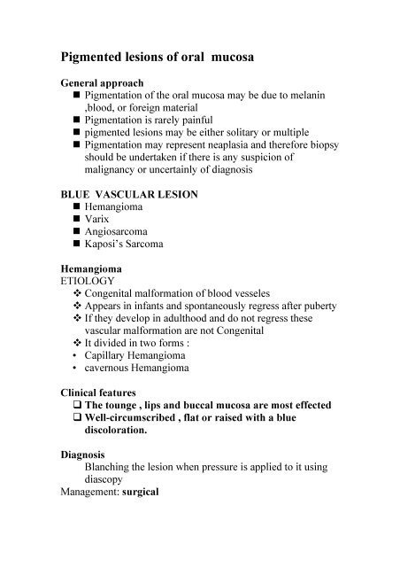
Pigmented <strong>lesions</strong> <strong>of</strong> <strong>oral</strong> <strong>mucosa</strong><br />
General approach<br />
Pigmentation <strong>of</strong> the <strong>oral</strong> <strong>mucosa</strong> may be due to melanin<br />
,blood, or foreign material<br />
Pigmentation is rarely painful<br />
<strong>pigmented</strong> <strong>lesions</strong> may be either solitary or multiple<br />
Pigmentation may represent neaplasia and therefore biopsy<br />
should be undertaken if there is any suspicion <strong>of</strong><br />
malignancy or uncertainly <strong>of</strong> diagnosis<br />
BLUE VASCULAR LESION<br />
Hemangioma<br />
Varix<br />
Angiosarcoma<br />
Kaposi’s Sarcoma<br />
Hemangioma<br />
ETIOLOGY<br />
Congenital malformation <strong>of</strong> blood vesseles<br />
Appears in infants and spontaneously regress after puberty<br />
If they develop in adulthood and do not regress these<br />
vascular malformation are not Congenital<br />
It divided in two forms :<br />
• Capillary Hemangioma<br />
• cavernous Hemangioma<br />
Clinical features<br />
The tounge , lips and buccal <strong>mucosa</strong> are most effected<br />
Well-circumscribed , flat or raised with a blue<br />
discoloration.<br />
Diagnosis<br />
Blanching the lesion when pressure is applied to it using<br />
diascopy<br />
Management: surgical
varix<br />
ETIOLOGY<br />
Pathologic dilatations <strong>of</strong> veins or venules<br />
represent a degenerative change in the adventitia <strong>of</strong> the<br />
venous wall<br />
Clinical features<br />
the chief site is the ventral tongue<br />
Lingual varicosities appear as tortuous serpentine blue,<br />
red, and purple elevations that course over the<br />
ventrolateral surface <strong>of</strong> the tongue, with extension<br />
anteriorly.<br />
on the lower lip, appearing as a focal raised<br />
pigmentation<br />
no clinical consequence. They are painless and are not<br />
subject to rupture and hemorrhage<br />
Diagnosis<br />
The varix resembles the hemangioma both clinically<br />
and histologically, yet it is distinguished by two<br />
features :<br />
(1) the patient’s age<br />
(2) its etiology<br />
Angiosarcoma<br />
not related to human immunodeficiency virus (HIV)<br />
can arise anywhere in the body.<br />
the <strong>oral</strong> cavity is an extremely rare site for such tumors<br />
Kaposi’s Sarcoma<br />
ETIOLOGY<br />
A proliferation <strong>of</strong> endothelial cells producing a mass.<br />
The factors have been proposed in the development <strong>of</strong><br />
this tumer:<br />
Genetic predispostion<br />
Environmental factors<br />
Infection
Immune dysregulation<br />
(HHV8)<br />
Clinical features<br />
The <strong>oral</strong> sites most involved the entire palate,<br />
Gingiva ,tounge.<br />
Diagnosis: biopsy<br />
Management: Excision, Radiotherapy, chemotherapy<br />
BROWN MELANOTIC LESIONS<br />
• Ephelis and Oral Melanotic Macule<br />
• Nevocellular Nevus and Blue Nevus<br />
• Malignant Melanoma<br />
• Drug-Induced Melanosis<br />
• Physiologic Pigmentation<br />
• Smoker’s Melanosis<br />
• Endocrinopathic Pigmentation<br />
• HIV Oral Melanosis<br />
• Peutz-Jeghers Syndrome<br />
Ephelis and Oral Melanotic Macule<br />
ETIOLOGY<br />
Accumulation <strong>of</strong> melanin in the epithelium and<br />
superficial connective tissues<br />
The cause is unknown<br />
Clinical features<br />
Uniformly flat, brown macules with distinct borders<br />
Less than 0.5 CM in size<br />
The most common sites : lips, buccal <strong>mucosa</strong>, gingiva<br />
Diagnosis: Biopsy
Management:<br />
No treatment is requied<br />
Nevocellular Nevus and Blue Nevus<br />
ETIOLOGY<br />
nevi are due to benign proliferations <strong>of</strong> melanocytes<br />
There are two major types, based on histology, and these<br />
two types tend to show differences clinically<br />
Nevocellular nevi<br />
arise from basal-layermelanocytes<br />
Since proliferation is minimal, these nevi are macular and<br />
are classified as (junctional nevi)<br />
With time, the melanocytes form clusters<br />
at the epitheliomesenchymal junction and begin to<br />
proliferate (compound nevi)<br />
The second type <strong>of</strong> nevus, not derived from basal-layer<br />
melanocytes, is the blue nevus<br />
neither the ordinary nor the cellular form has the potential<br />
to become a melanoma<br />
Clinical features<br />
both nevocellular and blue nevi tend<br />
to be brown and may be macular or nodular,sharply-defined<br />
border .<br />
Less than 0.5 CM in size<br />
The most common sites : the palate and gingiva but may<br />
also be encountered in the buccal <strong>mucosa</strong> and on the lips.<br />
Diagnosis: Biopsy<br />
Management: Excisional biopsy<br />
Malignant Melanoma<br />
ETIOLOGY<br />
There are no known predisposing factors for intra<strong>oral</strong> melanoma<br />
Mucosal melanomas are extremely rare.
Clinical features<br />
melanomas may appear flat or nodular<br />
the coloration can be quite varied, ranging from brown<br />
to black to blue<br />
show jagged irregular margins and ulceration<br />
Less than 0.5 CM in size<br />
Melanomas arising in the <strong>oral</strong> <strong>mucosa</strong> tend to occur on<br />
the anterior labial gingiva and the<br />
anterior aspect <strong>of</strong> the hard palate<br />
melanomas become more diffuse, nodular, and<br />
tumefactive, with foci <strong>of</strong> hyper- and hypopigmentation<br />
Diagnosis: Biopsy<br />
Management<br />
Excision with wide margins is the treatment <strong>of</strong> choice;<br />
once nodularity has envolved<br />
MRI and CT studies should be undertaken to explore<br />
regional metastases to the submandibular and cervical<br />
lymph nodes.<br />
Drug-Induced Melanosis:<br />
Drugs associated with <strong>oral</strong> <strong>mucosa</strong>l pigmentation<br />
Antimalarials: quinacrine, chloroquine,<br />
hydroxychloroquine<br />
Quinidine<br />
Zidovudine (AZT)<br />
Tetracycline<br />
Minocycline<br />
Chlorpromazine<br />
ETIOLOGY<br />
The pathogenesis <strong>of</strong> drug-induced<br />
pigmentation varies, depending on the causative drug.<br />
involve accumulation <strong>of</strong> melanin, deposits <strong>of</strong> the drug or<br />
one <strong>of</strong> its metabolites, synthesis <strong>of</strong> pigments under the
influence <strong>of</strong> the drug or deposition <strong>of</strong> iron after damage to<br />
the dermal vessels.<br />
Quinidine<br />
Mucosal discolouration is described as blue–grey or blue–<br />
black<br />
in most cases only the hard palate is involved<br />
Physiologic Pigmentation<br />
ETIOLOGY<br />
due to greater melanocyte activity rather than a greater<br />
number <strong>of</strong> melanocytes<br />
Clinical features<br />
The colour ranges from light to dark<br />
brown.<br />
The attached gingiva is the most common intra<strong>oral</strong> site <strong>of</strong><br />
such pigmentation<br />
bilateral, well-demarcated, ribbon-like, dark brown band<br />
that usually spares the marginal gingiva<br />
Pigmentation <strong>of</strong> the buccal <strong>mucosa</strong>, hard palate, lips and<br />
tongue may also be seen as brown patches with less welldefined<br />
borders.<br />
Smoker’s Melanosis<br />
ETIOLOGY<br />
increased production <strong>of</strong> melanin, which may provide a biologic<br />
defence against the noxious agents present in tobacco smoke.<br />
Clinical features<br />
The brown–black <strong>lesions</strong> most <strong>of</strong>ten involve the<br />
anteriorlabial gingiva followed by the buccal <strong>mucosa</strong>.<br />
Smoker’s melanosis usually disappears within 3 years <strong>of</strong><br />
smoking cessation
Endocrinopathic Pigmentation<br />
ETIOLOGY<br />
In both <strong>of</strong> Addison’s disease and pituitary-based Cushing’s<br />
syndrome these endocrine disorders, the cause <strong>of</strong><br />
hyperpigmentation is oversecretion <strong>of</strong> ACTH, a hormone with<br />
melanocyte-stimulating properties.<br />
Clinical features<br />
• Bronzing <strong>of</strong> the skin and patchy melanosis <strong>of</strong> the <strong>oral</strong><br />
<strong>mucosa</strong><br />
• The skin may appear tanned, and the gingiva ,palate, and<br />
buccal <strong>mucosa</strong> may be blotchy<br />
HIV Oral Melanosis<br />
ETIOLOGY The etiology remains undetermined.<br />
Clinical features The pigmentation resembles most <strong>of</strong> the<br />
other diffuse macular pigmentations; the buccal <strong>mucosa</strong> is<br />
the most frequently affected site, but the gingiva, palate,<br />
and tongue may also be involved.<br />
Peutz-Jeghers Syndrome<br />
Multiple focal melanotic brown macules are concentrated<br />
about the lips while the remaining facial skin is less<br />
strikingly involved.The macules appear as freckles or<br />
ephelides<br />
usually measuring < 0.5 cm in diameter<br />
Similar <strong>lesions</strong> may occur on the anterior tongue, buccal<br />
<strong>mucosa</strong>, and <strong>mucosa</strong>l surface <strong>of</strong> the lips. Ephelides are<br />
also seen on the fingers and hands<br />
GRAY /BLACK PIGMENTATIONS<br />
Amalgam Tattoo ,Graphite Tattoo<br />
Pigmentation Related to Heavy-Metal Ingestion
Amalgam Tattoo<br />
the most common source <strong>of</strong> solitary or focal pigmentation<br />
in the <strong>oral</strong> <strong>mucosa</strong> is the amalgam tattoo<br />
<strong>lesions</strong> are macular and gray or even black and are usually<br />
seen in the buccal <strong>mucosa</strong>, gingiva, or palate<br />
Graphite Tattoo<br />
Graphite tattoos tend to occur on the palate and represent<br />
traumatic implantation from a lead pencil.<br />
The <strong>lesions</strong> are usually macular, focal, and gray or black.<br />
Pigmentation Related to Heavy-Metal Ingestion<br />
Ingestion <strong>of</strong> heavy metals or metal salts can be an<br />
occupational hazard since many metals are used in<br />
industry and in paints.<br />
Lead, mercury, and bismuth have all been shown to be<br />
deposited in <strong>oral</strong> tissue if ingested in sufficient quantities<br />
or over a long course <strong>of</strong> time.<br />
in the <strong>oral</strong> cavity, the pigmentation is usually found along<br />
the free marginal gingiva, where it dramatically outlines<br />
the gingival cuff, resembling eyeliner.<br />
This metallic line has a gray to black appearance.<br />
The heavy metals may be associated with systemic<br />
symptoms <strong>of</strong> toxicity, including behavi<strong>oral</strong><br />
changes,neurologic disorders, and intestinal pain