The Protein Truncation Test - Promega
The Protein Truncation Test - Promega
The Protein Truncation Test - Promega

Create successful ePaper yourself
Turn your PDF publications into a flip-book with our unique Google optimized e-Paper software.
THE PROTEIN<br />
TRUNCATION TEST<br />
4C H A P T E R<br />
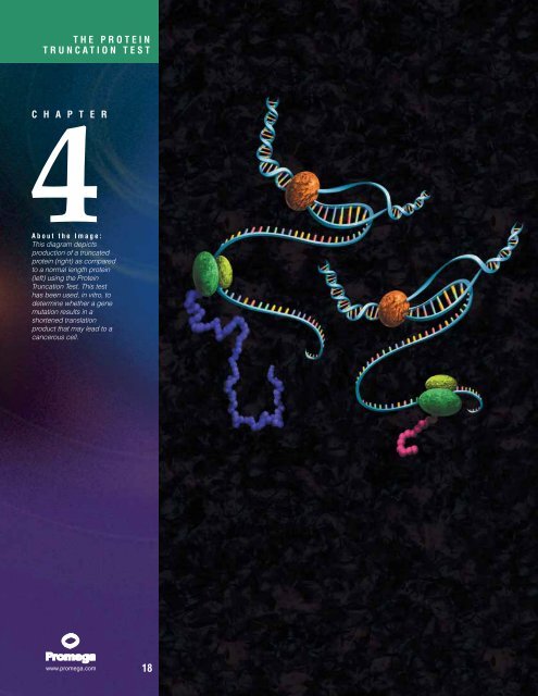
About the Image:<br />
This diagram depicts<br />
production of a truncated<br />
protein (right) as compared<br />
to a normal length protein<br />
(left) using the <strong>Protein</strong><br />
<strong>Truncation</strong> <strong>Test</strong>. This test<br />
has been used, in vitro, to<br />
determine whether a gene<br />
mutation results in a<br />
shortened translation<br />
product that may lead to a<br />
cancerous cell.<br />
18
Chapter Four: <strong>The</strong> <strong>Protein</strong> <strong>Truncation</strong> <strong>Test</strong><br />
Contents Page<br />
Introduction ............................................................................................................ 19<br />
PTT Principle .......................................................................................................... 20<br />
Source Considerations................................................................................................ 21<br />
Detection and Primer Design ........................................................................................ 22<br />
Introduction<br />
Mutations in a gene can range from large deletions to single point mutations. Many of the large<br />
deletions or translocations can be readily detected. For example, 95% of the cases of chronic myelogenous<br />
leukemia contain the Philadelphia chromosome, which is a translocation of part of<br />
chromosome 22 to chromosome 9. <strong>The</strong> abnormality can be detected by Southern blotting as aberrant<br />
or additional reactive bands when compared to normal samples (1). In this translocation, the<br />
abl proto-oncogene is translocated into the bcr gene resulting in the expression of a bcr-abl fusion<br />
protein. <strong>The</strong> chimeric transcript can be readily detected by RT-PCR (f) (2). Point mutations or small<br />
deletions, however, are much more difficult to detect. In Duchenne muscular dystrophy (DMD), for<br />
example, one third of the reported mutations in the gene DMD are not detectable as intragenic deletions<br />
or duplications (3–5). Techniques such as single strand confirmation polymorphism (6) can<br />
detect sequence differences but cannot distinguish between a polymorphism that may result in no<br />
phenotype (e.g., conservative amino acid change) and a polymorphism with a definite effect on the<br />
protein produced (e.g., premature termination of sequence).<br />
A rapid solution to these problems can be achieved through a procedure known as PTT (protein<br />
truncation test).<br />
C H A P T E R F O U R<br />
THE PROTEIN<br />
TRUNCATION TEST<br />
19<br />
References<br />
1. Lesieur, A. et al. (1994) Diag.<br />
Mol. Pathol. 3, 75.<br />
2. Lee, K-O. et al. (1996) J.<br />
Biochem. Mol. Biol. 29, 241.<br />
3. den Dunnen, J.T. et al. (1989)<br />
Am. J. Hum. Genet. 45, 835.<br />
4. Chamberlain, J.S. et al. (1988)<br />
Nucl. Acids Res. 16, 11141.<br />
5. Loenig, M. et al. (1987) Cell 50,<br />
509.<br />
6. Orita, M. et al. (1989) Genomics<br />
5, 874.<br />
TO ORDER<br />
Phone<br />
1-800-356-9526<br />
Fax<br />
1-800-356-1970<br />
Online<br />
www.promega.com
PROMEGA IN VITRO RESOURCE<br />
References (continued)<br />
7. Roest, P.A.M. et al. (1993) Hum.<br />
Mol. Genet. 2, 1719.<br />
8. Baklanov, M.M. et al. (1996)<br />
Nucl. Acids Res. 24, 3659.<br />
9. TNT ® Quick Coupled<br />
Transcription/Translation<br />
Systems Technical Manual<br />
#TM045, <strong>Promega</strong> Corporation.<br />
10. Kozak, M. (1986) Cell 44, 283.<br />
11. pGEM ®-T and pGEM ®-T Easy<br />
Vector Systems Technical<br />
Manual #TM042, <strong>Promega</strong><br />
Corporation.<br />
*For Laboratory Use.<br />
TO ORDER<br />
Phone<br />
1-800-356-9526<br />
Fax<br />
1-800-356-1970<br />
Online<br />
www.promega.com<br />
20<br />
PTT Principle<br />
A simple way to judge whether a mutation<br />
results in a truncation or not, is to translate the<br />
protein in vitro. Roest et al. (7) developed the<br />
protein truncation test (PTT) to rapidly screen for<br />
these mutations. PTT is composed of four steps:<br />
i) isolation of nucleic acid, either genomic DNA,<br />
total RNA or poly (A)+ RNA; ii) amplification of a<br />
specific region of the gene of interest; iii) in vitro<br />
transcription and translation of the product of the<br />
amplification reaction; and iv) detection of the<br />
translation products. <strong>The</strong> shorter protein<br />
products of the mutant alleles are easily distinguished<br />
from the full-length protein product of<br />
the normal allele (Figure 1). PTT has been used<br />
to analyze many genes in addition to DMD<br />
(Table 1).<br />
Amplified sequences for PTT can be generated<br />
across the entire protein coding sequence or<br />
they can be generated to specific exons. <strong>The</strong><br />
key feature of PTT is a specifically designed<br />
PCR primer to allow coupled in vitro transcription/translation<br />
of the amplified sequence. <strong>The</strong><br />
primer contains a T7 bacteriophage promoter<br />
Genomic DNA<br />
1 2<br />
Exons<br />
3 4 5<br />
Forward Primer<br />
T7<br />
ATG<br />
ATG<br />
AUG<br />
RNA<br />
Figure 1. Schematic diagram of the <strong>Protein</strong> <strong>Truncation</strong> <strong>Test</strong>.<br />
T7<br />
in vitro<br />
Transcription/<br />
Translation<br />
+<br />
sequence at the 5′-end that directs transcription.<br />
Usually, additional nucleotides are present<br />
upstream of the T7 promoter. Even the addition<br />
of a single G nucleotide upstream of the promoter<br />
increases the transcriptional efficiency<br />
(8). While T7 is the most commonly used promoter,<br />
T3 RNA polymerase promoter can be<br />
used as well. SP6 promoters are not well-suited<br />
for coupled transcription/translation of linear<br />
DNA (9). <strong>Promega</strong> offers a system specifically<br />
for the expression of PCR (f) products, the TNT ®<br />
T7 Quick for PCR DNA (c,d,e) (Cat.# L5540)*. A<br />
3–6bp spacer separates the promoter<br />
sequence from an optimal eukaryotic translation<br />
initiation sequence, which includes the initiation<br />
codon ATG. <strong>The</strong> optimal eukaryotic translation<br />
initiation sequence is referred to as a Kozak<br />
consensus sequence (10). <strong>The</strong> bacteriophage<br />
promoter, spacer and Kozak sequence are followed<br />
by sequences specific to the target<br />
(Table 2). At the 3´-end of the target, the primer<br />
can include a stop codon if the amplified<br />
sequence does not contain the native stop<br />
codon (9). Restriction enzyme recognition sites<br />
can also be engineered into both primers to aid<br />
PCR<br />
<strong>Protein</strong><br />
Cells from blood<br />
or tissue sample<br />
RNA<br />
Exons<br />
1 2 3 4 5<br />
Reverse Primer<br />
dsDNA<br />
Reverse<br />
Transcription<br />
cDNA<br />
PCR<br />
dsDNA<br />
Agarose gel electrophoresis<br />
of PCR products<br />
SDS-PAGE plus autoradiography<br />
– Full-length protein<br />
– Truncated protein<br />
mRNA<br />
1770MA04_7B
in subcloning of the PCR product if verification<br />
of a mutation is needed. <strong>The</strong> advent of PCR<br />
product cloning vectors has abrogated the<br />
need for inclusion of the restriction sites into<br />
PCR primers (11).<br />
Source Considerations<br />
<strong>The</strong> PTT test can be applied to individual exons<br />
of a gene via amplification of genomic DNA.<br />
Hogervorst et al. (12) analyzed genomic DNA of<br />
stored heparinized blood for mutations in the<br />
breast and ovarian cancer gene, BRCA1.<br />
Greater than 75% of the reported mutations in<br />
BRCA1 result in truncated proteins. Primers<br />
were designed to amplify exon 11, which<br />
encodes 61% of the BRCA1 gene product.<br />
Members of 35 families were analyzed, and all<br />
produced the correct size of PCR (f) product from<br />
the exon. <strong>The</strong> PCR product was transcribed<br />
and translated in vitro with [ 35S]methionine and<br />
analyzed by SDS-PAGE and autoradiography.<br />
Six mutations resulting in truncated proteins<br />
were identified. <strong>The</strong> mutant PCR products were<br />
directly sequenced and were found to be the<br />
result of either insertions or deletions yielding<br />
frameshift mutations and premature stop<br />
codons. Genomic DNA has also been used to<br />
analyze the genes BRCA2 (13), APC (14,15)<br />
and PLEC1 (16) by PTT.<br />
Use of genomic DNA as the source of nucleic<br />
acid for PTT has some drawbacks in that indi-<br />
Table 1. Genes Analyzed with the <strong>Protein</strong> <strong>Truncation</strong> <strong>Test</strong> a .<br />
vidual exons must be analyzed. To analyze the<br />
entire coding sequence of a gene like BRCA1,<br />
24 individual exons would need to be amplified<br />
and analyzed. Besides requiring a large number<br />
of amplifications, assuming all the exons are<br />
large enough to translate, analysis of the individual<br />
exons could miss truncation mutations<br />
that could result in aberrant exon splicing. In the<br />
same study that amplified exon 11 of the<br />
BRCA1 gene from genomic DNA for PTT analysis,<br />
Hogervorst et al. (12) isolated total RNA<br />
from freshly isolated peripheral blood lymphocytes.<br />
<strong>The</strong> sequences corresponding to exons<br />
2–10 were amplified by RT-PCR (f) and analyzed<br />
by PTT. One subject had a mutation in one<br />
allele that resulted, first, in a smaller RT-PCR<br />
product and, second, in a truncated protein by<br />
PTT. <strong>The</strong> mutation was directly sequenced and<br />
resulted from aberrant splicing of exons 9 and<br />
10. Thus, using RT-PCR and PTT, larger portions<br />
of a gene can be amplified and analyzed, picking<br />
up aberrant splicing mutations not identified<br />
by analysis of the exons via amplification of<br />
genomic DNA. In most cases, when RT-PCR is<br />
used as the method to generate targets, the<br />
entire coding region is broken into several<br />
smaller fragments. For example, three amplifications<br />
were used to test the entire coding region<br />
of the TSC2 gene by PTT (17). When using multiple<br />
targets to span an entire coding region, the<br />
amplimers should overlap so that a mutation at<br />
the 3′-end of one target (that does not cause a<br />
Condition Gene Ref.<br />
Familial Adenomatous Polyposis APC 14,15<br />
Hereditary Desmoid Disease APC 22<br />
Ataxia Telangiectasia ATM 23<br />
Hereditary Breast and Ovarian Cancer BRCA1 12<br />
BRCA2 13<br />
Familial Hypocalciuric Hypercalcemia CASR 24<br />
Cystic Fibrosis CFTR 25<br />
Chorioderemia CHM 26<br />
Duchenne Muscular Dystrophy DMD 7,27<br />
Fanconi Anaemia FAA 28<br />
Congenital Muscular Dystrophy laminin-α2 29<br />
Hereditary Non-Polyposis Colorectal Cancer hMSH2 30<br />
hMLH1 31<br />
Neurofibromatosis Type 1 NF1 32<br />
Neurofibromatosis Type 2 NF2 33<br />
Aniridia PAX6 34<br />
Paroxysmal Nocturnal Haemoglobinuria PIG-A 35<br />
Polycystic Kidney Disease PKD1 36,37<br />
Epidermolysis Bullosa with Muscular Dystrophy PLEC1 16,44–46<br />
Dystrophic Epidermolysis Bullosa COL7A1 43<br />
Breast Cancers, Gliomas, Melanomas PTEN/MMAC1 38,39,40<br />
Rubenstein-Taybi Syndrome RTS 41<br />
Familial Tuberous Sclerosis TSC2 17<br />
aMore references available in <strong>The</strong> <strong>Protein</strong> <strong>Truncation</strong> <strong>Test</strong> Bibliography (BL002) and Mutation Detection (BR043) also available on the Internet at www.promega.com<br />
C H A P T E R F O U R<br />
THE PROTEIN<br />
TRUNCATION TEST<br />
21<br />
References (continued)<br />
12. Hogervorst, F.B.L. et al. (1995)<br />
Nat. Genet. 10, 208.<br />
13. Lancaster, J.M. et al. (1996) Nat.<br />
Genet. 13, 238.<br />
14. Powell, S.M. et al. (1993) New<br />
Eng. J. Med. 329, 1982.<br />
15. van der Luijt, R. et al. (1994)<br />
Genomics 20, 1.<br />
16. Dang, M. et al. (1998) Lab.<br />
Invest. 78, 195.<br />
17. van Bakel, I. et al. (1997) Hum.<br />
Mol. Genet. 6, 1409.<br />
18. Hogervorst, F.B.L. (1997)<br />
<strong>Promega</strong> Notes 62, 7.<br />
19. Transcend Non-Radioactive<br />
Translation Detection Systems<br />
Technical Bulletin #TB182,<br />
<strong>Promega</strong> Corporation.<br />
20. Kirchgesser, M. et al. (1998)<br />
Clin. Chem. Lab. Med. 36, 567.<br />
21. Rowan, A.J. and Bodmer, W.F.<br />
(1997) Hum. Mutat. 9, 172.<br />
22. Eccles, D.M. et al. (1996) Am. J.<br />
Hum. Genet. 59, 1193.<br />
23. Wright, J. et al. (1996) Am. J.<br />
Hum. Genet. 59, 839.<br />
24. Bai, M. et al. (1997) J. Clin.<br />
Invest. 99, 1917.<br />
25. Romey, M. et al. (1996) Hum.<br />
Genet. 98, 328.<br />
26. Beaufrère, L. et al. (1997) Exp.<br />
Eye Res. 65, 849.<br />
27. Gardner, R.J. et al. (1995) Am.<br />
J. Hum. Genet. 57, 311.<br />
28. Lo Ten Foe, J.R. et al. (1996)<br />
Nat. Genet. 14, 320.<br />
29. Pegoraro, E. et al. (1998)<br />
Neurology 51, 101.<br />
30. Liu, B. et al. (1994) Cancer Res.<br />
54, 4590.<br />
31. Papadopoulos, N. et al. (1994)<br />
Science 263, 1625.<br />
32. Heim, R.A. et al. (1994) Nat.<br />
Genet. 8, 218.<br />
33. MacCollin, M. et al. (1994) Am.<br />
J. Hum. Genet. 55, 314.<br />
34. Axton, R. et al. (1997) J. Med.<br />
Genet. 34, 279.<br />
35. Maugard, C. et al. (1997) Br. J.<br />
Haematol. 98, 21.<br />
36. Roelfsema, J.H. et al. (1996)<br />
Nephrol. Dial. Transplant.<br />
11(suppl. 6), 5.<br />
TO ORDER<br />
Phone<br />
1-800-356-9526<br />
Fax<br />
1-800-356-1970<br />
Online<br />
www.promega.com
PROMEGA IN VITRO RESOURCE<br />
References (continued)<br />
37. Roelfsema, J.H. et al. (1997)<br />
Am. J. Hum. Genet. 61, 1044.<br />
38. Li, J. et al. (1997) Science 275,<br />
1943.<br />
39. Furnari, F.B. et al. (1997) Proc.<br />
Natl. Acad. Sci. USA 94, 12479.<br />
40. Robertson, G.P. et al. (1998)<br />
Proc. Natl. Acad. Sci. USA 95,<br />
9418.<br />
41. Petrij, F. et al. (1995) Nature 376,<br />
348.<br />
42. Sarkar, G. and Sommer, S.S.<br />
(1989) Science 244, 331.<br />
43. Whittock, N.V. et al. (1999) J.<br />
Invest. Dermatol. 113, 673.<br />
44. Takizawa, Y. et al. (1999) J.<br />
Invest. Dermatol. 112, 109.<br />
45. Kunz, M. et al. (2000) J. Invest.<br />
Dermatol. 114, 376.<br />
46. Rouan F. et al. (1989) J. Invest.<br />
Dermatol. 114, 381.<br />
TO ORDER<br />
Phone<br />
1-800-356-9526<br />
Fax<br />
1-800-356-1970<br />
Online<br />
www.promega.com<br />
22<br />
significant change in molecular weight) will be<br />
detected in another target having the same<br />
codons near the 5′-end.<br />
Detection and Primer Design<br />
<strong>The</strong> detection method for PTT products must be<br />
considered when designing primers for amplification<br />
(18). Typically, [ 35 S]methionine is the<br />
label of choice but other labels such as<br />
[ 35 S]cysteine and [ 3 H]leucine could be used as<br />
well. Thus, the amplified segments should contain<br />
one or more of these amino acids. <strong>The</strong><br />
reactions are resolved on an SDS-PAGE gel and<br />
either directly dried or fluorographically<br />
enhanced and exposed to X-ray film (9). <strong>The</strong><br />
dried gels can also be analyzed by phosphorimaging.<br />
When radioactive incorporation is not<br />
an option, non-radioactive techniques are available.<br />
<strong>Protein</strong>s can be tagged with biotin by<br />
inclusion of biotinylated lysine tRNA in the translation<br />
reaction (9,19). <strong>The</strong> biotin moiety is then<br />
detected with a streptavidin-enzyme conjugate<br />
and developed via either a colorimetric or<br />
chemiluminescent reaction (19). For example,<br />
PTT has been applied to the APC gene using<br />
translation with a biotinylated lysine tRNA (20).<br />
Other methods for non-radioactive detection<br />
include the inclusion of an epitope tag in the 5′primer<br />
so that the translation products can be<br />
analyzed by Western blotting with an antibody<br />
that binds the epitope (21). When dealing with a<br />
heterozygous condition, both the normal and<br />
mutant targets will be amplified and both the<br />
truncated and full-length protein will be detected,<br />
unless the allelle is on the X or Y<br />
chromosome of male subjects, no matter which<br />
detection method is chosen.<br />
PTT offers a quick and easy method for analyzing<br />
a protein coding sequence for truncation<br />
mutations. However, the method has some limitations.<br />
If the truncated sequence does not<br />
translate well or does not contain the appropriate<br />
amino acid for labeling, the mutation could<br />
be overlooked. Also, if the truncation is very<br />
near the 3′-end of the target, truncation could<br />
be missed due to the inability of SDS-PAGE to<br />
resolve such differences. If the mutations are<br />
very near the 5′-end of the coding sequence,<br />
the mutation could be missed as well.<br />
Refinements of PTT detection, such as the<br />
incorporation of an epitope tag into the 5′ PCR<br />
primer (21), could allow detection of these<br />
mutations, since incorporation of a specific<br />
amino acid is not needed for detection. Finally,<br />
incorporation of fluorescence-tagged amino<br />
acids may simplify the detection of proteins by<br />
PTT and can possibly be used for quantitation<br />
of the mutant protein (18).<br />
Table 2. Sequences of Different T7-Modified Oligonucleotide Primers for In Vitro Transcription<br />
and Translation a .<br />
Eukaryotic<br />
Translation<br />
Restriction T7 Bacteriophage Initiation<br />
Site Sequence Sequence Spacer Sequence Ref.<br />
GGATCC TAATACGACTCACTATAGGG AG CCACC ATG 13,42<br />
GGATCC TAATACGACTCACTATAGGG AG CCACC ATG G 30,31<br />
GGATCC TAATACGACTCACTATAGG AACAG CCACC ATG 7,15<br />
nnn b TAATACGACTCACTATAGG AACAG CCACC ATG G 12,28<br />
aSequences provided are for only the upstream portion of the 5 ′ primer that is not gene specific. For gene-specific use, the eukaryotic translation<br />
initiation sequence would be followed by 17–20 bases exactly complementary to the sequence of interest.<br />
bn = any nucleotide