Puss Caterpillar (Larva), Southern Flannel Moth (Adult ...
Puss Caterpillar (Larva), Southern Flannel Moth (Adult ...
Puss Caterpillar (Larva), Southern Flannel Moth (Adult ...

Create successful ePaper yourself
Turn your PDF publications into a flip-book with our unique Google optimized e-Paper software.
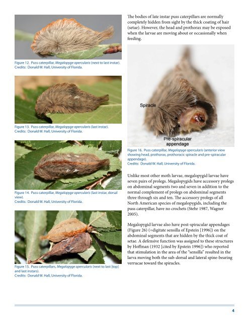
Figure 12. <strong>Puss</strong> caterpillar, Megalopyge opercularis (next to last instar).<br />
Credits: Donald W. Hall, University of Florida.<br />
Figure 13. <strong>Puss</strong> caterpillar, Megalopyge opercularis (last instar).<br />
Credits: Donald W. Hall, University of Florida.<br />
Figure 14. <strong>Puss</strong> caterpillar, Megalopyge opercularis (last instar, dorsal<br />
view).<br />
Credits: Donald W. Hall, University of Florida.<br />
Figure 15. <strong>Puss</strong> caterpillars, Megalopyge opercularis (next to last [top]<br />
and last instars).<br />
Credits: Donald W. Hall, University of Florida.<br />
The bodies of late instar puss caterpillars are normally<br />
completely hidden from sight by the thick coating of hair<br />
(setae). However, the head and prothorax may be exposed<br />
when the larvae are moving about or occasionally when<br />
feeding.<br />
Figure 16. <strong>Puss</strong> caterpillar, Megalopyge opercularis (anterior view<br />
showing head, prothorax, prothoracic spiracle and pre-spiracular<br />
appendage).<br />
Credits: Donald W. Hall, University of Florida.<br />
Unlike most other moth larvae, megalopygid larvae have<br />
seven pairs of prolegs. Megalopygids have accessory prolegs<br />
on abdominal segments two and seven in addition to the<br />
normal complement of prolegs on abdominal segments<br />
three through six and ten. The accessory prolegs of all<br />
North American species of megalopygids, including the<br />
puss caterpillar, have no crochets (Stehr 1987, Wagner<br />
2005).<br />
Megalopygid larvae also have post-spiracular appendages<br />
(Figure 26) (=digitate sensilla of Epstein [1996]) on the<br />
abdominal segments that are hidden by the thick coat of<br />
setae. A defensive function was assigned to these structures<br />
by Hoffman (1932 [cited by Epstein 1996]) who reported<br />
that stimulation in the area of the “sensilla” resulted in the<br />
larva moving both the sub-dorsal and lateral spine-bearing<br />
verrucae toward the spiracles.<br />
4