Ch05: Red and White Lesions of the Oral Mucosa
Ch05: Red and White Lesions of the Oral Mucosa
Ch05: Red and White Lesions of the Oral Mucosa

Create successful ePaper yourself
Turn your PDF publications into a flip-book with our unique Google optimized e-Paper software.
5<br />
▼<br />
RED AND WHITE LESIONS OF<br />
THE ORAL MUCOSA<br />
INDRANEEL BHATTACHARYYA, DDS, MSD<br />
DONALD M. COHEN, DMD, MS, MBA<br />
SOL SILVERMAN JR., DDS, MS<br />
▼ HEREDITARY WHITE LESIONS<br />
Leukoedema<br />
<strong>White</strong> Sponge Nevus<br />
Hereditary Benign Intraepi<strong>the</strong>lial Dyskeratosis<br />
Dyskeratosis Congenita<br />
▼ REACTIVE/INFLAMMATORY WHITE LESIONS<br />
Linea Alba (<strong>White</strong> Line)<br />
Frictional (Traumatic) Keratosis<br />
Cheek Chewing<br />
Chemical Injuries <strong>of</strong> <strong>the</strong> <strong>Oral</strong> <strong>Mucosa</strong><br />
Actinic Keratosis (Cheilitis)<br />
Smokeless Tobacco–Induced Keratosis<br />
Nicotine Stomatitis<br />
Sanguinaria-Induced Leukoplakia<br />
▼ INFECTIOUS WHITE LESIONS AND WHITE AND<br />
RED LESIONS<br />
<strong>Oral</strong> Hairy Leukoplakia<br />
C<strong>and</strong>idiasis<br />
Mucous Patches<br />
Parulis<br />
▼ IDIOPATHIC “TRUE” LEUKOPLAKIA<br />
Etiology<br />
Clinical Features<br />
Varieties<br />
Histopathologic Features<br />
Diagnosis <strong>and</strong> Management<br />
Prognosis<br />
▼ BOWEN’S DISEASE<br />
▼ ERYTHROPLAKIA<br />
Clinical Features<br />
Histopathologic Features<br />
Differential Diagnosis<br />
Treatment <strong>and</strong> Prognosis<br />
85<br />
▼ ORAL LICHEN PLANUS<br />
Etiology <strong>and</strong> Diagnosis<br />
Clinical Features<br />
Histopathologic Features<br />
Immun<strong>of</strong>luorescent Studies<br />
Differential Diagnosis<br />
Clinical Course <strong>and</strong> Prognosis<br />
Treatment<br />
▼ LICHENOID REACTIONS<br />
Drug-Induced Lichenoid Reactions<br />
Graft-versus-Host Disease<br />
▼ LUPUS ERYTHEMATOSUS (SYSTEMIC AND<br />
DISCOID)<br />
Clinical Features, Diagnosis, <strong>and</strong> Treatment<br />
Histopathologic Features<br />
Malignant Potential, Importance, <strong>and</strong> Scope <strong>of</strong> <strong>Oral</strong> <strong>Lesions</strong><br />
▼ DEVELOPMENTAL WHITE LESIONS: ECTOPIC<br />
LYMPHOID TISSUE<br />
▼ FORDYCE’S GRANULES<br />
Features<br />
Treatment<br />
▼ GINGIVAL AND PALATAL CYSTS OF THE<br />
NEWBORN AND ADULT<br />
Features in <strong>the</strong> Newborn<br />
Features in <strong>the</strong> Adult<br />
▼ MISCELLANEOUS LESIONS<br />
Geographic Tongue<br />
Hairy Tongue (Black Hairy Tongue)<br />
<strong>Oral</strong> Submucous Fibrosis
86 Diagnosis <strong>and</strong> Management <strong>of</strong> <strong>Oral</strong> <strong>and</strong> Salivary Gl<strong>and</strong> Diseases<br />
Any condition that increases <strong>the</strong> thickness <strong>of</strong> <strong>the</strong> epi<strong>the</strong>lium<br />
causes it to appear white by increasing <strong>the</strong> distance to <strong>the</strong> vascular<br />
bed. <strong>Lesions</strong> most <strong>of</strong>ten appear white because <strong>of</strong> a thickening<br />
<strong>of</strong> <strong>the</strong> keratin layer, or hyperkeratosis. O<strong>the</strong>r common<br />
causes <strong>of</strong> a white appearance include acanthosis (a thickening<br />
<strong>of</strong> <strong>the</strong> spinous cell layer), an increase in <strong>the</strong> amount <strong>of</strong> edema<br />
fluid in <strong>the</strong> epi<strong>the</strong>lium (ie, leukoedema), <strong>and</strong> reduced vascularity<br />
in <strong>the</strong> underlying lamina propria. Surface ulcerations<br />
covered by a fibrin cap can also appear white, as would collapsed<br />
bullae. (These latter two types <strong>of</strong> lesions are covered in<br />
Chapter 4, “Ulcerative, Vesicular, <strong>and</strong> Bullous <strong>Lesions</strong>.”)<br />
▼ HEREDITARY WHITE LESIONS<br />
Leukoedema<br />
Leukoedema is a common mucosal alteration that represents a<br />
variation <strong>of</strong> <strong>the</strong> normal condition ra<strong>the</strong>r than a true pathologic<br />
change. 1,2 It has been reported in up to 90% <strong>of</strong> black adults <strong>and</strong><br />
up to 50% <strong>of</strong> black teenagers. 3,4 The incidence in white persons<br />
in different studies is highly variable (10 to 90%). 3 This difference<br />
can be attributed to <strong>the</strong> darker coloration <strong>of</strong> <strong>the</strong> mucosa<br />
in black persons, rendering <strong>the</strong> alteration more visible. 5,6<br />
Similar edematous changes have been reported in o<strong>the</strong>r<br />
mucosal surfaces, such as <strong>the</strong> vagina <strong>and</strong> larynx. 5<br />
FEATURES<br />
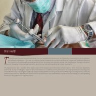
The most frequent site <strong>of</strong> leukoedema is <strong>the</strong> buccal mucosa bilaterally,<br />
<strong>and</strong> it may be seen rarely on <strong>the</strong> labial mucosa, s<strong>of</strong>t palate,<br />
<strong>and</strong> floor <strong>of</strong> <strong>the</strong> mouth. It usually has a faint, white, diffuse, <strong>and</strong><br />
filmy appearance, with numerous surface folds resulting in wrinkling<br />
<strong>of</strong> <strong>the</strong> mucosa (Figure 5-1). It cannot be scraped <strong>of</strong>f, <strong>and</strong> it<br />
disappears or fades upon stretching <strong>the</strong> mucosa. Microscopic<br />
examination reveals thickening <strong>of</strong> <strong>the</strong> epi<strong>the</strong>lium, with significant<br />
intracellular edema <strong>of</strong> <strong>the</strong> stratum spinosum. The surface <strong>of</strong><br />
<strong>the</strong> epi<strong>the</strong>lium may demonstrate a thickened layer <strong>of</strong> parakeratin.<br />
TREATMENT<br />
No treatment is indicated for leukoedema since it is a variation<br />
<strong>of</strong> <strong>the</strong> normal condition. No malignant change has been<br />
reported. 2<br />
<strong>White</strong> Sponge Nevus<br />
<strong>White</strong> sponge nevus (WSN) is a rare autosomal dominant disorder<br />
with a high degree <strong>of</strong> penetrance <strong>and</strong> variable expressivity;<br />
it predominantly affects noncornified stratified squamous<br />
epi<strong>the</strong>lium. 7 The disease usually involves <strong>the</strong> oral<br />
mucosa <strong>and</strong> (less frequently) <strong>the</strong> mucous membranes <strong>of</strong> <strong>the</strong><br />
nose, esophagus, genitalia, <strong>and</strong> rectum. 7,8 The lesions <strong>of</strong> WSN<br />
may be present at birth or may first manifest or become more<br />
intense at puberty. Genetic analyses <strong>of</strong> families with WSN have<br />
identified a missense mutation in one allele <strong>of</strong> keratin 13 that<br />
leads to proline substitution for leucine within <strong>the</strong> keratin<br />
gene cluster on chromosome 17. 9,10 A new study, using<br />
sequence analysis, has reported a glutamine insertion localized<br />
in <strong>the</strong> helix initiation motif <strong>of</strong> <strong>the</strong> 1A alpha helical domain <strong>of</strong><br />
Keratin 4 gene. 9<br />
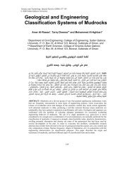
FIGURE 5-1 Leukoedema with a faint white diffuse filmy appearance<br />
<strong>and</strong> mild wrinkling <strong>of</strong> <strong>the</strong> mucosa.<br />
FEATURES<br />
<strong>White</strong> sponge nevus presents as bilateral symmetric white,<br />
s<strong>of</strong>t, “spongy,” or velvety thick plaques <strong>of</strong> <strong>the</strong> buccal mucosa.<br />
However, o<strong>the</strong>r sites in <strong>the</strong> oral cavity may be involved, including<br />
<strong>the</strong> ventral tongue, floor <strong>of</strong> <strong>the</strong> mouth, labial mucosa, s<strong>of</strong>t<br />
palate, <strong>and</strong> alveolar mucosa. The condition is usually asymptomatic<br />
<strong>and</strong> does not exhibit tendencies toward malignant<br />
change. The characteristic histopathologic features are epi<strong>the</strong>lial<br />
thickening, parakeratosis, a peculiar perinuclear condensation<br />
<strong>of</strong> <strong>the</strong> cytoplasm, <strong>and</strong> vacuolization <strong>of</strong> <strong>the</strong> suprabasal<br />
layer <strong>of</strong> keratinocytes. Electron microscopy <strong>of</strong> exfoliated cells<br />
shows numerous cellular granules composed <strong>of</strong> disordered<br />
aggregates <strong>of</strong> ton<strong>of</strong>ilaments. 7,8<br />
DIFFERENTIAL DIAGNOSIS<br />
The lesions <strong>of</strong> WSN may be grossly similar to those <strong>of</strong> o<strong>the</strong>r<br />
hereditary mucosal syndromes such as hereditary benign<br />
intraepi<strong>the</strong>lial dyskeratosis or pachyonychia congenita, infections<br />
such as c<strong>and</strong>idiasis, traumatic lesions seen in cheek chewing,<br />
<strong>and</strong> chemical burns or preneoplastic/neoplastic<br />
processes. 7 This differential diagnosis is best resolved in many<br />
cases by incisional biopsy specimens interpreted in <strong>the</strong> context<br />
<strong>of</strong> <strong>the</strong> clinical history <strong>and</strong> physical findings. 8<br />
TREATMENT<br />
No treatment is indicated for this benign <strong>and</strong> asymptomatic<br />
condition. Patients may require palliative treatment if <strong>the</strong> condition<br />
is symptomatic. One study has reported some relief <strong>of</strong><br />
symptoms with a tetracycline rinse. 11<br />
Hereditary Benign Intraepi<strong>the</strong>lial Dyskeratosis<br />
Hereditary benign intraepi<strong>the</strong>lial dyskeratosis (HBID), also<br />
known as Witkop’s disease, is a rare autosomal dominant disorder<br />
characterized by oral lesions <strong>and</strong> bilateral limbal conjunctival<br />
plaques. 12,13 This condition is noted specifically in a<br />
triracial isolate <strong>of</strong> white, Native American, <strong>and</strong> African<br />
American people <strong>and</strong> <strong>the</strong>ir descendants in Halifax county,<br />
North Carolina. It exhibits a high degree <strong>of</strong> penetrance. 12
<strong>Red</strong> <strong>and</strong> <strong>White</strong> <strong>Lesions</strong> <strong>of</strong> <strong>the</strong> <strong>Oral</strong> <strong>Mucosa</strong> 87<br />
FEATURES<br />
The oral lesions are similar to those <strong>of</strong> WSN, with thick, corrugated,<br />
asymptomatic, white “spongy” plaques involving <strong>the</strong><br />
buccal <strong>and</strong> labial mucosa. O<strong>the</strong>r intraoral sites include <strong>the</strong> floor<br />
<strong>of</strong> <strong>the</strong> mouth, <strong>the</strong> lateral tongue, <strong>the</strong> gingiva, <strong>and</strong> <strong>the</strong> palate. The<br />
oral lesions are generally detected in <strong>the</strong> first year <strong>of</strong> life <strong>and</strong><br />
gradually increase in intensity until <strong>the</strong> teens. The most significant<br />
aspect <strong>of</strong> HBID involves <strong>the</strong> bulbar conjunctiva, where<br />
thick, gelatinous, foamy, <strong>and</strong> opaque plaques form adjacent to<br />
<strong>the</strong> cornea. The ocular lesions manifest very early in life (usually<br />
within <strong>the</strong> first year). Some patients exhibit chronic relapsing<br />
ocular irritation <strong>and</strong> photophobia. The plaques may exhibit<br />
seasonal prominence, with many patients reporting more-pronounced<br />
lesions in <strong>the</strong> spring <strong>and</strong> regression during <strong>the</strong> summer<br />
months. A few cases <strong>of</strong> blindness due to corneal vascularization<br />
following HBID have been reported. 12 The<br />
histopathologic features <strong>of</strong> HBID are characteristic, <strong>and</strong> <strong>the</strong><br />
epi<strong>the</strong>lium exhibits marked parakeratin production with thickening<br />
<strong>of</strong> <strong>the</strong> stratum spinosum <strong>and</strong> <strong>the</strong> presence <strong>of</strong> numerous<br />
dyskeratotic cells. Ultrastructural findings in patients with<br />
HBID reveal <strong>the</strong> <strong>the</strong> presence <strong>of</strong> numerous vesicular bodies in<br />
immature dyskeratotic cells, densely packed ton<strong>of</strong>ilaments<br />
within <strong>the</strong> cytoplasm <strong>of</strong> <strong>the</strong>se cells, <strong>and</strong> <strong>the</strong> disappearance <strong>of</strong><br />
cellular bridging in mature dyskeratotic cells. 14<br />
TREATMENT<br />
Since HBID is a benign condition, no treatment is required for<br />
<strong>the</strong> oral lesions. For evaluation <strong>and</strong> treatment <strong>of</strong> <strong>the</strong> ocular<br />
lesions, <strong>the</strong> patient should be referred to an ophthalmologist. 12<br />
Dyskeratosis Congenita<br />
Dyskeratosis congenita, a recessively inherited genodermatosis, is<br />
unusual due to <strong>the</strong> high incidence <strong>of</strong> oral cancer in young affected<br />
adults. 15 It is a rare X-linked disorder characterized by a series <strong>of</strong><br />
oral changes that lead eventually to an atrophic leukoplakic oral<br />
mucosa, with <strong>the</strong> tongue <strong>and</strong> cheek most severely affected. 15 The<br />
oral changes occur in association with severely dystrophic nails<br />
<strong>and</strong> a prominent reticulated hyperpigmentation <strong>of</strong> <strong>the</strong> skin <strong>of</strong> <strong>the</strong><br />
face, neck, <strong>and</strong> chest. 15 Many cases also exhibit hematologic<br />
changes including pancytopenia, hypersplenism, <strong>and</strong> an aplastic<br />
or Fanconi’s anemia (ie, an anemia associated with an inherited<br />
inability to repair deoxyribonucleic acid [DNA] defects, leading<br />
to a high frequency <strong>of</strong> leukemia <strong>and</strong> lymphoma). 16,17<br />
The oral lesions commence before <strong>the</strong> age <strong>of</strong> 10 years as<br />
crops <strong>of</strong> vesicles with associated patches <strong>of</strong> white ulcerated<br />
necrotic mucosa <strong>of</strong>ten infected with C<strong>and</strong>ida. Erythroplakic<br />
changes <strong>and</strong> nail dystrophy follow, with leukoplakic lesions <strong>and</strong><br />
carcinoma supervening on <strong>the</strong> oral lesions in early adulthood. 15<br />
▼ REACTIVE AND INFLAMMATORY<br />
WHITE LESIONS<br />
Linea Alba (<strong>White</strong> Line)<br />
As <strong>the</strong> name implies, linea alba is a horizontal streak on <strong>the</strong><br />
buccal mucosa at <strong>the</strong> level <strong>of</strong> <strong>the</strong> occlusal plane extending from<br />
<strong>the</strong> commissure to <strong>the</strong> posterior teeth. It is a very common<br />
finding <strong>and</strong> is most likely associated with pressure, frictional<br />
irritation, or sucking trauma from <strong>the</strong> facial surfaces <strong>of</strong> <strong>the</strong><br />
teeth. This alteration was present in about 13% <strong>of</strong> <strong>the</strong> population<br />
in one study. 18–20<br />
CLINICAL FEATURES<br />
Linea alba is usually present bilaterally <strong>and</strong> may be pronounced<br />
in some individuals. It is more prominent in individuals<br />
with reduced overjet <strong>of</strong> <strong>the</strong> posterior teeth. It is <strong>of</strong>ten<br />
scalloped <strong>and</strong> restricted to dentulous areas. 18<br />
TREATMENT<br />
No treatment is indicated for patients with linea alba. The<br />
white streak may disappear spontaneously in some people. 18,19<br />
Frictional (Traumatic) Keratosis<br />
CLINICAL FEATURES<br />
Frictional (traumatic) keratosis is defined as a white plaque<br />
with a rough <strong>and</strong> frayed surface that is clearly related to an<br />
identifiable source <strong>of</strong> mechanical irritation <strong>and</strong> that will usually<br />
resolve on elimination <strong>of</strong> <strong>the</strong> irritant. These lesions may<br />
occasionally mimic dysplastic leukoplakia; <strong>the</strong>refore, careful<br />
examination <strong>and</strong> sometimes a biopsy are required to rule out<br />
any atypical changes. 20,21 Histologically, such lesions show<br />
varying degrees <strong>of</strong> hyperkeratosis <strong>and</strong> acanthosis. 22 Prevalence<br />
rates as high as 5.5% have been reported. 21 Such lesions are<br />
similar to calluses on <strong>the</strong> skin. Traumatic keratosis has never<br />
been shown to undergo malignant transformation. 23 <strong>Lesions</strong><br />
belonging to this category <strong>of</strong> keratosis include linea alba <strong>and</strong><br />
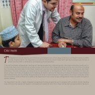
cheek, lip, <strong>and</strong> tongue chewing. Frictional keratosis is frequently<br />
associated with rough or maladjusted dentures (Figure<br />
5-2) <strong>and</strong> with sharp cusps <strong>and</strong> edges <strong>of</strong> broken teeth. 20<br />
TREATMENT<br />
Upon removal <strong>of</strong> <strong>the</strong> <strong>of</strong>fending agent, <strong>the</strong> lesion should resolve<br />
within 2 weeks. Biopsies should be performed on lesions that<br />
do not heal to rule out a dysplastic lesion.<br />
FIGURE 5-2 Leukoplakic-appearing area <strong>of</strong> frictional keratosis from an<br />
ill-fitting denture.
88 Diagnosis <strong>and</strong> Management <strong>of</strong> <strong>Oral</strong> <strong>and</strong> Salivary Gl<strong>and</strong> Diseases<br />
Cheek Chewing<br />
<strong>White</strong> lesions <strong>of</strong> <strong>the</strong> oral tissues may result from chronic irritation<br />
due to repeated sucking, nibbling, or chewing. 24–29<br />
These insults result in <strong>the</strong> traumatized area becoming thickened,<br />
scarred, <strong>and</strong> paler than <strong>the</strong> surrounding tissues. Cheek<br />
chewing is most commonly seen in people who are under<br />
stress or in psychological situations in which cheek <strong>and</strong> lip<br />
biting become habitual. Most patients with this condition are<br />
somewhat aware <strong>of</strong> <strong>the</strong>ir habit but do not associate it with<br />
<strong>the</strong>ir lesions. The white lesions <strong>of</strong> cheek chewing may sometimes<br />
be confused with o<strong>the</strong>r dermatologic disorders involving<br />
<strong>the</strong> oral mucosa, which can lead to misdiagnosis. 30 Chronic<br />
chewing <strong>of</strong> <strong>the</strong> labial mucosa (morsicatio labiorum) <strong>and</strong> <strong>the</strong><br />
lateral border <strong>of</strong> <strong>the</strong> tongue (morsicatio linguarum) may be<br />
seen with cheek chewing or may cause isolated lesions.<br />
Prevalence rates ranging from 0.12 to 0.5% have been reported<br />
in Sc<strong>and</strong>inavian populations, compared with a prevalence rate<br />
<strong>of</strong> 4.6% for South African school children in mental health<br />
treatment facilities; <strong>the</strong>se rates support <strong>the</strong> role <strong>of</strong> stress <strong>and</strong><br />
anxiety in <strong>the</strong> etiology <strong>of</strong> this condition. 26–28<br />
TYPICAL FEATURES<br />
The lesions are most frequently found bilaterally on <strong>the</strong> posterior<br />
buccal mucosa along <strong>the</strong> plane <strong>of</strong> occlusion. They may<br />
be seen in combination with traumatic lesions on <strong>the</strong> lips or<br />
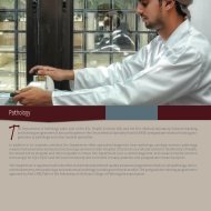
tongue. Patients <strong>of</strong>ten complain <strong>of</strong> roughness or small tags <strong>of</strong><br />
tissue that <strong>the</strong>y actually tear free from <strong>the</strong> surface. This produces<br />
a distinctive frayed clinical presentation (Figure 5-3).<br />
The lesions are poorly outlined whitish patches that may be<br />
intermixed with areas <strong>of</strong> ery<strong>the</strong>ma or ulceration. The occurrence<br />
is twice as prevalent in females <strong>and</strong> three times more<br />
common after <strong>the</strong> age <strong>of</strong> 35 years.<br />
The histopathologic picture is distinctive <strong>and</strong> includes<br />
hyperparakeratosis <strong>and</strong> acanthosis. The keratin surface is usually<br />
shaggy <strong>and</strong> ragged with numerous projections <strong>of</strong> keratin<br />
that demonstrate adherent bacterial colonies. When <strong>the</strong> lesion<br />
is seen on <strong>the</strong> lateral tongue, <strong>the</strong> clinical <strong>and</strong> histomorphologic<br />
features mimic those <strong>of</strong> oral hairy leukoplakia. 29<br />
FIGURE 5-3 Morsicatio buccarum represented by a frayed macerated<br />
irregular leukoplakic area in <strong>the</strong> cheek.<br />
TREATMENT AND PROGNOSIS<br />
Since <strong>the</strong> lesions result from an unconscious <strong>and</strong>/or nervous<br />
habit, no treatment is indicated. However, for those desiring<br />
treatment <strong>and</strong> unable to stop <strong>the</strong> chewing habit, a plastic<br />
occlusal night guard may be fabricated. Isolated tongue<br />
involvement requires fur<strong>the</strong>r investigation to rule out oral<br />
hairy leukoplakia especially when appropriate risk factors for<br />
infection with human immunodeficiency virus (HIV) are present.<br />
Differential diagnosis also includes WSN, chemical burns,<br />
<strong>and</strong> c<strong>and</strong>idiasis.<br />
Chemical Injuries <strong>of</strong> <strong>the</strong> <strong>Oral</strong> <strong>Mucosa</strong><br />
Transient nonkeratotic white lesions <strong>of</strong> <strong>the</strong> oral mucosa are<br />
<strong>of</strong>ten a result <strong>of</strong> chemical injuries caused by a variety <strong>of</strong> agents<br />
that are caustic when retained in <strong>the</strong> mouth for long periods<br />
<strong>of</strong> time, such as aspirin, silver nitrate, formocresol, sodium<br />
hypochlorite, paraformaldehyde, dental cavity varnishes, acidetching<br />
materials, <strong>and</strong> hydrogen peroxide. 31–42 The white<br />
lesions are attributable to <strong>the</strong> formation <strong>of</strong> a superficial<br />
pseudomembrane composed <strong>of</strong> a necrotic surface tissue <strong>and</strong><br />
an inflammatory exudate.<br />
SPECIFIC CAUSATIVE AGENTS<br />
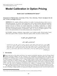
Aspirin Burn. Acetylsalicylic acid (aspirin) is a common<br />
source <strong>of</strong> burns <strong>of</strong> <strong>the</strong> oral cavity. 43–45 Usually, <strong>the</strong> tissue is<br />
damaged when aspirin is held in <strong>the</strong> mucobuccal fold area for<br />
prolonged periods <strong>of</strong> time for <strong>the</strong> relief <strong>of</strong> common dental<br />
pain (Figure 5-4).<br />
Silver Nitrate. Silver nitrate is commonly used by health care<br />
practitioners as a chemical cautery agent for <strong>the</strong> treatment <strong>of</strong><br />
aphthous ulcers. 46 It brings about almost instantaneous relief<br />
<strong>of</strong> symptoms by burning <strong>the</strong> nerve endings at <strong>the</strong> site <strong>of</strong> <strong>the</strong><br />
ulcer. However, silver nitrate <strong>of</strong>ten destroys tissue around <strong>the</strong><br />
immediate area <strong>of</strong> application <strong>and</strong> may result in delayed healing<br />
or (rarely) severe necrosis at <strong>the</strong> application site (Figure<br />
5-5). 46 Its use should be discouraged.<br />
FIGURE 5-4 Aspirin burn, creating a pseudomembranous necrotic<br />
white area.
<strong>Red</strong> <strong>and</strong> <strong>White</strong> <strong>Lesions</strong> <strong>of</strong> <strong>the</strong> <strong>Oral</strong> <strong>Mucosa</strong> 89<br />
FIGURE 5-5 Extensive tissue necrosis caused by injudicious use <strong>of</strong> silver<br />
nitrate.<br />
Hydrogen Peroxide. Hydrogen peroxide is <strong>of</strong>ten used as an<br />
intraoral rinse for <strong>the</strong> prevention <strong>of</strong> periodontal disease. At<br />
concentrations <strong>of</strong> ≥ 3%, hydrogen peroxide is associated with<br />
epi<strong>the</strong>lial necrosis. 40<br />
Sodium Hypochlorite. Sodium hypochlorite, or dental<br />
bleach, is commonly used as a root canal irrigant <strong>and</strong> may<br />
cause serious ulcerations due to accidental contact with oral<br />
s<strong>of</strong>t tissues. 32<br />
Dentifrices <strong>and</strong> Mouthwashes. Several cases <strong>of</strong> oral injuries<br />
<strong>and</strong> ulcerations due to <strong>the</strong> misuse <strong>of</strong> commercially available<br />
mouthwashes <strong>and</strong> dentifrices have been reported (Figure 5-<br />
6). 31,33,42,47 An unusual sensitivity reaction with severe ulcerations<br />
<strong>and</strong> sloughing <strong>of</strong> <strong>the</strong> mucosa has been reported to have<br />
been caused by a cinnamon-flavored dentifrice (Figure 5-7).<br />
However, <strong>the</strong>se lesions probably represent a sensitivity or allergic<br />
reaction to <strong>the</strong> cinnamon aldehyde in <strong>the</strong> toothpaste. 47<br />
This reaction can appear to be very similar to <strong>the</strong> reactions<br />
caused by o<strong>the</strong>r chemical agents such as aspirin <strong>and</strong> hydrogen<br />
FIGURE 5-6 Diffuse slough <strong>of</strong> marginal gingivae due to misuse <strong>of</strong> commercial<br />
mouthwash.<br />
FIGURE 5-7 Severe ulceration <strong>and</strong> sloughing <strong>of</strong> mucosa, caused by use<br />
<strong>of</strong> a cinnamon-containing dentifrice.<br />
peroxide. Caustic burns <strong>of</strong> <strong>the</strong> lips, mouth, <strong>and</strong> tongue have<br />
been seen in patients who use mouthwashes containing alcohol<br />
<strong>and</strong> chlorhexidine. 33,42 A case <strong>of</strong> an unusual chemical<br />
burn, confined to <strong>the</strong> masticatory mucosa <strong>and</strong> produced by<br />
abusive ingestion <strong>of</strong> fresh fruit <strong>and</strong> by <strong>the</strong> concomitant excessive<br />
use <strong>of</strong> mouthwash, has also been reported. 42<br />
TYPICAL FEATURES<br />
The lesions are usually located on <strong>the</strong> mucobuccal fold area<br />
<strong>and</strong> gingiva. The injured area is irregular in shape, white, covered<br />
with a pseudomembrane, <strong>and</strong> very painful. The area <strong>of</strong><br />
involvement may be extensive. When contact with <strong>the</strong> tissue is<br />
brief, a superficial white <strong>and</strong> wrinkled appearance without<br />
resultant necrosis is usually seen. Long-term contact (usually<br />
with aspirin, sodium hypochlorite, phenol, paraformaldehyde,<br />
etc) can cause severer damage <strong>and</strong> sloughing <strong>of</strong> <strong>the</strong> necrotic<br />
mucosa. The unattached nonkeratinized tissue is more commonly<br />
affected than <strong>the</strong> attached mucosa.<br />
TREATMENT AND PROGNOSIS<br />
The best treatment <strong>of</strong> chemical burns <strong>of</strong> <strong>the</strong> oral cavity is prevention.<br />
Children especially should be supervised while taking<br />
aspirin tablets, to prevent prolonged retention <strong>of</strong> <strong>the</strong> agent in<br />
<strong>the</strong> oral cavity. 31,41,45 The proper use <strong>of</strong> a rubber dam during<br />
endodontic procedures reduces <strong>the</strong> risk <strong>of</strong> iatrogenic chemical<br />
burns. Most superficial burns heal within 1 or 2 weeks. A<br />
protective emollient agent such as a film <strong>of</strong> methyl cellulose<br />
may provide relief. 37,45 However, deep-tissue burns <strong>and</strong> necrosis<br />
may require careful débridement <strong>of</strong> <strong>the</strong> surface, followed by<br />
antibiotic coverage. In case <strong>of</strong> ingestion <strong>of</strong> caustic chemicals or<br />
accidental exposure to severely corrosive agents, extensive scarring<br />
that may require surgery <strong>and</strong>/or pros<strong>the</strong>tic rehabilitation<br />
may occur. 48<br />
Actinic Keratosis (Cheilitis)<br />
Actinic (or solar) keratosis is a premalignant epi<strong>the</strong>lial lesion<br />
that is directly related to long-term sun exposure. 49 These<br />
lesions are classically found on <strong>the</strong> vermilion border <strong>of</strong> <strong>the</strong>
90 Diagnosis <strong>and</strong> Management <strong>of</strong> <strong>Oral</strong> <strong>and</strong> Salivary Gl<strong>and</strong> Diseases<br />
lower lip as well as on o<strong>the</strong>r sun-exposed areas <strong>of</strong> <strong>the</strong> skin. A<br />
small percentage <strong>of</strong> <strong>the</strong>se lesions will transform into squamous<br />
cell carcinoma. 50 Biopsies should be performed on<br />
lesions that repeatedly ulcerate, crust over, or show a thickened<br />
white area. These lesions are commonly found in individuals<br />
with extensive sun exposure, such as those with outdoor occupations<br />
<strong>and</strong>/or fair complexions. 49<br />
TYPICAL FEATURES<br />
Actinic keratosis may be seen on <strong>the</strong> skin <strong>of</strong> <strong>the</strong> forehead, cheeks,<br />
ears, <strong>and</strong> forearms. On <strong>the</strong> lip, it appears as a white plaque, oval<br />
to linear in shape, usually measuring < 1 cm in size (Figure 5-<br />
8). The surface may be crusted <strong>and</strong> rough to <strong>the</strong> touch.<br />
Histopathologically, <strong>the</strong> surface epi<strong>the</strong>lium appears atrophic,<br />
with a basophilic homogenous amorphous alteration <strong>of</strong> <strong>the</strong> collagen<br />
(solar elastosis) in <strong>the</strong> lamina propria. Varying degrees <strong>of</strong><br />
atypical features such as increased nucleocytoplasmic ratios,<br />
loss <strong>of</strong> cellular polarity <strong>and</strong> orientation, <strong>and</strong> nuclear <strong>and</strong> cellular<br />
atypia are found within <strong>the</strong> epi<strong>the</strong>lium. A mild lymphocytic<br />
infiltrate may also be noted in <strong>the</strong> lamina propria. 49<br />
TREATMENT AND PROGNOSIS<br />
The mainstay <strong>of</strong> treatment <strong>of</strong> actinic keratosis is surgery.<br />
Chemo<strong>the</strong>rapeutic agents such as topical 5-fluorouracil have<br />
been used with some success. 50 However, follow-up biopsies in<br />
individuals who were treated with 5-fluorouracil showed that<br />
<strong>the</strong> dysplastic changes persist in clinically healthy-appearing<br />
epi<strong>the</strong>lium. Patients treated with nonsurgical methods <strong>the</strong>refore<br />
require long-term follow-up. About 10% <strong>of</strong> <strong>the</strong>se lesions<br />
will undergo malignant transformation. 51<br />
Smokeless Tobacco–Induced Keratosis<br />
Chewing tobacco is an important established risk factor for <strong>the</strong><br />
development <strong>of</strong> oral carcinoma in <strong>the</strong> United States. 52<br />
Habitually chewing tobacco leaves or dipping snuff results in<br />
<strong>the</strong> development <strong>of</strong> a well-recognized white mucosal lesion in<br />
<strong>the</strong> area <strong>of</strong> tobacco contact, called smokeless tobacco keratosis,<br />
snuff dipper’s keratosis, or tobacco pouch keratosis. 53<br />
FIGURE 5-8 Distinctive raised white plaque, representing actinic<br />
cheilitis.<br />
While <strong>the</strong>se lesions are accepted as precancerous, <strong>the</strong>y are significantly<br />
different from true leukoplakia <strong>and</strong> have a much<br />
lower risk <strong>of</strong> malignant transformation. 54<br />
This habit was once almost universal in <strong>the</strong> United States<br />
<strong>and</strong> is very common among certain o<strong>the</strong>r populations, most<br />
notably in Sweden, India, <strong>and</strong> Sou<strong>the</strong>ast Asia. 52,55–60<br />
Smokeless tobacco use among white males in <strong>the</strong> United<br />
States has shown a recent resurgence. 60–62 The estimated proportion<br />
<strong>of</strong> adult men in <strong>the</strong> United States who regularly use<br />
“spit” tobacco ranges from 6 to 20%. 58,63 This range is attributed<br />
to significant geographic, cultural, <strong>and</strong> gender variations<br />
in chewing habits. 54 The cumulative incidence for<br />
smokeless tobacco use was highest for non-Hispanic white<br />
males. 54 Unfortunately, <strong>the</strong> habit starts relatively early in life,<br />
usually between <strong>the</strong> ages <strong>of</strong> 9 <strong>and</strong> 15 years, <strong>and</strong> is rarely begun<br />
after 20 years <strong>of</strong> age. Recent epidemiologic data indicate that<br />
over five million Americans use smokeless tobacco <strong>and</strong> that<br />
more than 750,000 <strong>of</strong> <strong>the</strong>se users are adolescents. It is estimated<br />
that each year in <strong>the</strong> United States, approximately<br />
800,000 young people between <strong>the</strong> ages <strong>of</strong> 11 <strong>and</strong> 19 years<br />
experiment with smokeless tobacco <strong>and</strong> that about 300,000<br />
become regular users. 55–59,63<br />
Smokeless tobacco contains several known carcinogens,<br />
including N-nitrosonornicotine (NNN), <strong>and</strong> <strong>the</strong>se have been<br />
proven to cause mucosal alterations. 64 In addition to its established<br />
role as a carcinogen, chewing tobacco may be a risk factor<br />
in <strong>the</strong> development <strong>of</strong> root surface caries <strong>and</strong>, to a lesser<br />
extent, coronal caries. This may be due to its high sugar content<br />
<strong>and</strong> its association with increased amounts <strong>of</strong> gingival<br />
recession. 58 The duration <strong>of</strong> exposure is very important in <strong>the</strong><br />
production <strong>of</strong> mucosal damage. Leukoplakia has been<br />
reported to develop with <strong>the</strong> habitual use <strong>of</strong> as little as three<br />
cans <strong>of</strong> snuff per week for longer than 3 years. 53 Although all<br />
forms <strong>of</strong> smokeless tobacco may result in mucosal alterations,<br />
snuff (a finely powdered tobacco) appears to be much more<br />
likely to cause such changes than is chewing tobacco. 53,59<br />
Smokeless tobacco is not consistently associated with<br />
increased rates <strong>of</strong> oral cancer. 54 Approximately 20% <strong>of</strong> all adult<br />
Swedish males use moist snuff, yet it has not been possible to<br />
detect any significant increase in <strong>the</strong> incidence <strong>of</strong> cancer <strong>of</strong> <strong>the</strong><br />
oral cavity or pharynx in Sweden. By international st<strong>and</strong>ards,<br />
<strong>the</strong> prevalence <strong>of</strong> oral cancer is low in Sweden. 52 This has been<br />
attributed to variations in <strong>the</strong> composition <strong>of</strong> snuff, in particular,<br />
<strong>the</strong> amount <strong>of</strong> fermented or cured tobacco in <strong>the</strong> mixture.<br />
The carcinogen NNN is present in much lower concentrations<br />
in Swedish snuff, probably because <strong>of</strong> a lack <strong>of</strong> fermentation<br />
<strong>of</strong> <strong>the</strong> tobacco. Also, <strong>the</strong> high level <strong>of</strong> snuff use might decrease<br />
<strong>the</strong> amount <strong>of</strong> cigarette smoking <strong>and</strong> <strong>the</strong>refore lead to a lesser<br />
prevalence <strong>of</strong> oral cancer. 52,54<br />
TYPICAL FEATURES<br />
Numerous alterations are found in habitual users <strong>of</strong> smokeless<br />
tobacco. Most changes associated with <strong>the</strong> use <strong>of</strong> smokeless<br />
tobacco are seen in <strong>the</strong> area contacting <strong>the</strong> tobacco. The most<br />
common area <strong>of</strong> involvement is <strong>the</strong> anterior m<strong>and</strong>ibular vestibule,<br />
followed by <strong>the</strong> posterior vestibule. 53 The surface <strong>of</strong> <strong>the</strong> mucosa
<strong>Red</strong> <strong>and</strong> <strong>White</strong> <strong>Lesions</strong> <strong>of</strong> <strong>the</strong> <strong>Oral</strong> <strong>Mucosa</strong> 91<br />
FIGURE 5-9 Snuff pouch with a white wrinkled mucosal surface.<br />
appears white <strong>and</strong> is granular or wrinkled (Figure 5-9); in some<br />
cases, a folded character may be seen (tobacco pouch keratosis).<br />
Commonly noted is a characteristic area <strong>of</strong> gingival recession<br />
with periodontal-tissue destruction in <strong>the</strong> immediate area <strong>of</strong> contact<br />
(Figure 5-10). This recession involves <strong>the</strong> facial aspect <strong>of</strong> <strong>the</strong><br />
tooth or teeth <strong>and</strong> is related to <strong>the</strong> amount <strong>and</strong> duration <strong>of</strong><br />
tobacco use. The mucosa appears gray or gray-white <strong>and</strong> almost<br />
translucent. Since <strong>the</strong> tobacco is not in <strong>the</strong> mouth during examination,<br />
<strong>the</strong> usually stretched mucosa appears fissured or rippled,<br />
<strong>and</strong> a “pouch” is usually present. This white tobacco pouch may<br />
become lea<strong>the</strong>ry or nodular in long-term heavy users (Figure 5-<br />
11). Rarely, an erythroplakic component may be seen. The lesion<br />
is usually asymptomatic <strong>and</strong> is discovered on routine examination.<br />
Microscopically, <strong>the</strong> epi<strong>the</strong>lium is hyperkeratotic <strong>and</strong> thickened.<br />
A characteristic vacuolization or edema may be seen in <strong>the</strong><br />
keratin layer <strong>and</strong> in <strong>the</strong> superficial epi<strong>the</strong>lium. Frank dysplasia is<br />
uncommon in tobacco pouch keratosis. 62<br />
FIGURE 5-10 Snuff pouch showing extensive periodontal tissue<br />
destruction <strong>and</strong> a thickened area <strong>of</strong> leukoplakia. (Courtesy <strong>of</strong> Dr. Robert<br />
Howell, West Virginia University, School <strong>of</strong> Dentistry)<br />
FIGURE 5-11 <strong>White</strong> lea<strong>the</strong>ry nodular tobacco pouch. These thickened<br />
areas are more worrisome for malignant transformation.<br />
TREATMENT AND PROGNOSIS<br />
Cessation <strong>of</strong> use almost always leads to a normal mucosal<br />
appearance within 1 to 2 weeks. 54,64,65 Biopsy specimens<br />
should be obtained from lesions that remain after 1 month.<br />
Biopsy is particularly indicated for those lesions that appear<br />
clinically atypical <strong>and</strong> that include such features as surface<br />
ulceration, erythroplakia, intense whiteness, or a verrucoid or<br />
papillary surface. 53, 62, 64 The risk <strong>of</strong> malignant transformation<br />
is increased fourfold for chronic smokeless tobacco users. 66<br />
Nicotine Stomatitis<br />
Nicotine stomatitis (stomatitis nicotina palati, smoker’s palate)<br />
refers to a specific white lesion that develops on <strong>the</strong> hard <strong>and</strong> s<strong>of</strong>t<br />
palate in heavy cigarette, pipe, <strong>and</strong> cigar smokers. The lesions are<br />
restricted to areas that are exposed to a relatively concentrated<br />
amount <strong>of</strong> hot smoke during inhalation.Areas covered by a denture<br />
are usually not involved. The lesion has become less common<br />
since pipe smoking has lost popularity. Although it is associated<br />
closely with tobacco smoking, <strong>the</strong> lesion is not considered<br />
to be premalignant. 67–69 Interestingly, nicotine stomatitis also<br />
develops in individuals with a long history <strong>of</strong> drinking extremely<br />
hot beverages. 70 This suggests that heat, ra<strong>the</strong>r than toxic chemicals<br />
in tobacco smoke, is <strong>the</strong> primary cause. Prevalence rates as<br />
high as 1.0 to 2.5% have been reported in populations <strong>of</strong> different<br />
cultures. 69–72 “Reverse smoking”(ie, placing <strong>the</strong> burning end<br />
<strong>of</strong> <strong>the</strong> cigarette in <strong>the</strong> oral cavity), seen in South American <strong>and</strong><br />
Asian populations, produces significantly more pronounced<br />
palatal alterations that may be erythroleukoplakic <strong>and</strong> that are<br />
definitely considered premalignant. 73<br />
TYPICAL FEATURES<br />
This condition is most <strong>of</strong>ten found in older males with a history<br />
<strong>of</strong> heavy long-term cigar, pipe, or cigarette smoking. Due<br />
to <strong>the</strong> chronic insult, <strong>the</strong> palatal mucosa becomes diffusely gray<br />
or white (Figure 5-12, A). Numerous slightly elevated papules<br />
with punctate red centers that represent inflamed <strong>and</strong> metaplastically<br />
altered minor salivary gl<strong>and</strong> ducts are noted.
92 Diagnosis <strong>and</strong> Management <strong>of</strong> <strong>Oral</strong> <strong>and</strong> Salivary Gl<strong>and</strong> Diseases<br />
A<br />
B<br />
FIGURE 5-12 A, Nicotine stomatitis with diffuse white change in <strong>the</strong><br />
palatal mucosa, along with elevated papules with red centers. B, Histologic<br />
appearance <strong>of</strong> nicotine stomatitis, showing hyperkeratosis <strong>and</strong> acanthosis<br />
with squamous metaplasia <strong>of</strong> <strong>the</strong> dilated salivary duct. (Hematoxylin <strong>and</strong><br />
eosin, ×40 original magnification)<br />
Microscopically, <strong>the</strong> surface epi<strong>the</strong>lium exhibits hyperkeratosis<br />
<strong>and</strong> acanthosis with squamous metaplasia <strong>and</strong> hyperplasia <strong>of</strong><br />
<strong>the</strong> salivary ducts as <strong>the</strong>y approach <strong>the</strong> surface (see Figure 5-12,<br />
B). The subjacent connective tissue <strong>and</strong> minor salivary gl<strong>and</strong>s<br />
exhibit a mild to moderate scattered chronic inflammation. No<br />
atypical or dysplastic changes are usually identified. 72<br />
TREATMENT AND PROGNOSIS<br />
Nicotine stomatitis is completely reversible once <strong>the</strong> habit is<br />
discontinued. The severity <strong>of</strong> inflammation is proportional to<br />
<strong>the</strong> duration <strong>and</strong> amount <strong>of</strong> smoking. 72 The lesions usually<br />
resolve within 2 weeks <strong>of</strong> cessation <strong>of</strong> smoking. Biopsy <strong>of</strong> nicotine<br />
stomatitis is rarely indicated except to reassure <strong>the</strong> patient.<br />
However, a biopsy should be performed on any white lesion <strong>of</strong><br />
<strong>the</strong> palatal mucosa that persists after 1 month <strong>of</strong> discontinuation<br />
<strong>of</strong> smoking habit.<br />
Sanguinaria-Induced Leukoplakia<br />
Sanguinaria extract, a mixture <strong>of</strong> benzophenanthridine alkaloids<br />
derived from <strong>the</strong> common bloodroot plant (Sanguinaria<br />
canadensis), has been used in oral rinses <strong>and</strong> toothpaste products<br />
since 1982. The most widely used product with<br />
Sanguinaria, Viadent, has been shown, through extensive clinical<br />
trials, to be effective against plaque buildup <strong>and</strong> gingivitis.<br />
74,75 Importantly, sanguinaria extract has also been shown to<br />
be carcinogenic in many studies. 76,77 In 1999, Damm <strong>and</strong> associates<br />
78 reported an increased prevalence <strong>of</strong> leukoplakia <strong>of</strong> <strong>the</strong><br />
maxillary vestibule in patients who used sanguinaria-based<br />
products on a routine basis. They conducted a retrospective<br />
review <strong>of</strong> 88 patients with leukoplakia <strong>of</strong> <strong>the</strong> maxillary vestibule<br />
<strong>and</strong> found that 84.1% <strong>of</strong> <strong>the</strong> patients reported having used<br />
Viadent. The prevalence <strong>of</strong> Viadent use was only 3% among<br />
r<strong>and</strong>omly selected adults in <strong>the</strong>ir study. Eversole <strong>and</strong> colleagues<br />
79 compared <strong>and</strong> contrasted biomarkers <strong>and</strong> ploidy data<br />
from maxillary gingival leukoplakias associated with dentifrices<br />
<strong>and</strong> mouth rinses containing sanguinaria with those from o<strong>the</strong>r<br />
forms <strong>of</strong> benign <strong>and</strong> premalignant mucosal keratosis. They<br />
used computerized image analysis <strong>and</strong> biomarker immunohistochemical<br />
assays to assess ploidy, DNA content, <strong>and</strong> p53<br />
<strong>and</strong> proliferating cell nuclear antigen immunoreactivity <strong>of</strong><br />
nuclei in tissue from <strong>the</strong>se groups. A significantly higher (fourfold)<br />
DNA content <strong>and</strong> higher numbers <strong>of</strong> cells with hyperploid<br />
nuclei were found in <strong>the</strong> group with sanguinaria-associated<br />
keratoses. Although this group did not harbor significant<br />
numbers <strong>of</strong> p53-expressing nuclei, a significant elevation in<br />
nuclei labeled with proliferating cell nuclear antigen was noted.<br />
The authors concluded that sanguinaria-associated keratoses<br />
show marker <strong>and</strong> image analysis pr<strong>of</strong>iles similar to those <strong>of</strong><br />
non-sanguinaria-induced dysplastic lesions <strong>of</strong> <strong>the</strong> lip <strong>and</strong><br />
mucosa. 79 Hence, preparations containing sanguinaria should<br />
be avoided until <strong>the</strong> risk for malignant transformation is determined.<br />
80 This recommendation is fur<strong>the</strong>r supported by <strong>the</strong><br />
lack <strong>of</strong> regression in some Viadent-induced leukoplakias<br />
months after <strong>the</strong> cessation <strong>of</strong> Viadent use.<br />
TYPICAL FEATURES<br />
Most patients are adults in <strong>the</strong> fourth to ninth decades <strong>of</strong> life.<br />
In <strong>the</strong> study by Damm <strong>and</strong> colleagues, <strong>the</strong> range <strong>of</strong> Viadent use<br />
before <strong>the</strong> development <strong>of</strong> lesions was 6 months to 12 years,<br />
with a mean <strong>of</strong> 4.4 years. 78 Typically, patients present with a<br />
white, velvety, wrinkled or corrugated patch <strong>of</strong> leukoplakia in<br />
<strong>the</strong> maxillary vestibule, involving both <strong>the</strong> attached gingiva <strong>and</strong><br />
vestibular mucosa (Figure 5-13, A). The lesions may also be<br />
seen in <strong>the</strong> anterior m<strong>and</strong>ibular vestibule (see Figure 5-13, B).<br />
The area is usually very distinct <strong>and</strong> sharply demarcated from<br />
<strong>the</strong> surrounding tissue. The lesions are localized to <strong>the</strong>se areas<br />
since <strong>the</strong> anterior portions <strong>of</strong> <strong>the</strong> maxillary <strong>and</strong> m<strong>and</strong>ibular
<strong>Red</strong> <strong>and</strong> <strong>White</strong> <strong>Lesions</strong> <strong>of</strong> <strong>the</strong> <strong>Oral</strong> <strong>Mucosa</strong> 93<br />
A B<br />
FIGURE 5-13 A, Typical white corrugated leukoplakia in <strong>the</strong> maxillary vestibule, associated with sanguinaria use. B, M<strong>and</strong>ibular vestibular lesion in<br />
<strong>the</strong> same patient.<br />
vestibule exhibit prolonged retention <strong>of</strong> <strong>the</strong> product due to <strong>the</strong><br />
greater distance from <strong>the</strong> major salivary ducts. Histopathologically,<br />
all biopsy specimens demonstrate significant<br />
surface keratosis with a verrucoid pattern. Minimal atypical<br />
changes (including basilar hyperplasia, nuclear hyperchromatism,<br />
<strong>and</strong> increased nucleocytoplasmic ratios) limited to <strong>the</strong><br />
lower one-third <strong>of</strong> <strong>the</strong> epi<strong>the</strong>lium are noted in most specimens.<br />
More significant atypical changes have also been reported. 78<br />
TREATMENT<br />
No appropriate treatment has been established for sanguinariainduced<br />
leukoplakia. However, an initial biopsy is m<strong>and</strong>atory. If<br />
a histopathologic diagnosis <strong>of</strong> dysplasia is rendered, <strong>the</strong> condition<br />
should be treated in a fashion similar to <strong>the</strong> treatment <strong>of</strong><br />
o<strong>the</strong>r potentially premalignant processes. The less severe changes<br />
should be managed according to clinical judgment, depending on<br />
<strong>the</strong> extent <strong>and</strong> duration <strong>of</strong> <strong>the</strong> lesion. In all cases, complete discontinuation<br />
<strong>of</strong> Sanguinaria-containing products <strong>and</strong> cessation<br />
<strong>of</strong> any o<strong>the</strong>r harmful habits such as tobacco or alcohol use is<br />
m<strong>and</strong>atory. All patients should be given careful clinical followup,<br />
with a biopsy <strong>of</strong> any recurrent or worsening lesion(s).<br />
▼ INFECTIOUS WHITE LESIONS AND<br />
WHITE AND RED LESIONS<br />
<strong>Oral</strong> Hairy Leukoplakia<br />
<strong>Oral</strong> hairy leukoplakia is a corrugated white lesion that usually<br />
occurs on <strong>the</strong> lateral or ventral surfaces <strong>of</strong> <strong>the</strong> tongue in<br />
patients with severe immunodeficiency. 81,82 The most common<br />
disease associated with oral hairy leukoplakia is HIV<br />
infection. 82 <strong>Oral</strong> hairy leukoplakia is reported in about 25% <strong>of</strong><br />
adults with HIV infection but is not as common in HIVinfected<br />
children. Its prevalence reaches as high as 80% in<br />
patients with acquired immunodeficiency syndrome (AIDS). 83<br />
Epstein-Barr virus (EBV) is implicated as <strong>the</strong> causative agent<br />
in oral hairy leukoplakia. 84–86 A positive correlation with<br />
decreasing cluster designation 4 (CD4) cell counts has been<br />
established in HIV-positive patients. The presence <strong>of</strong> this<br />
lesion has been associated with <strong>the</strong> subsequent development<br />
<strong>of</strong> AIDS in a large percentage <strong>of</strong> HIV positive patients. 83 Hairy<br />
leukoplakia has also occasionally been reported in patients<br />
with o<strong>the</strong>r immunosuppressive conditions, such as patients<br />
undergoing organ transplantation <strong>and</strong> patients undergoing<br />
prolonged steroid <strong>the</strong>rapy. 87–89 Rare cases may occur in<br />
immunocompetent persons after topical steroid <strong>the</strong>rapy. 90,91<br />
TYPICAL FEATURES<br />
<strong>Oral</strong> hairy leukoplakia most commonly involves <strong>the</strong> lateral border<br />
<strong>of</strong> <strong>the</strong> tongue but may extend to <strong>the</strong> ventral or dorsal surfaces.<br />
<strong>Lesions</strong> on <strong>the</strong> tongue are usually corrugated <strong>and</strong> may have<br />
a shaggy or frayed appearance, mimicking lesions caused by<br />
tongue chewing (Figure 5-14). <strong>Oral</strong> hairy leukoplakia may also<br />
present as a plaquelike lesion <strong>and</strong> is <strong>of</strong>ten bilateral.<br />
Histopathologic examination <strong>of</strong> <strong>the</strong> epi<strong>the</strong>lium reveals severe<br />
hyperparakeratosis with an irregular surface, acanthosis with<br />
superficial edema, <strong>and</strong> numerous koilocytic cells (virally affected<br />
FIGURE 5-14 Bilateral linear leukoplakic lesions on <strong>the</strong> dorsolateral<br />
tongue, suggestive <strong>of</strong> oral hairy leukoplakia. (Courtesy <strong>of</strong> Dr. Parnell Taylor,<br />
Riverton, Wyoming)
94 Diagnosis <strong>and</strong> Management <strong>of</strong> <strong>Oral</strong> <strong>and</strong> Salivary Gl<strong>and</strong> Diseases<br />
“balloon” cells) in <strong>the</strong> spinous layer. The characteristic microscopic<br />
feature is <strong>the</strong> presence <strong>of</strong> homogeneous viral nuclear<br />
inclusions with a residual rim <strong>of</strong> normal chromatin. 92–94 The<br />
definitive diagnosis can be established by demonstrating <strong>the</strong><br />
presence <strong>of</strong> EBV through in situ hybridization, electron<br />
microscopy, or polymerase chain reaction (PCR). 85,86<br />
DIFFERENTIAL DIAGNOSIS<br />
It is important to differentiate this lesion from o<strong>the</strong>r clinically<br />
similar entities such as hyperplastic c<strong>and</strong>idiasis, idiopathic<br />
leukoplakia, leukoplakia induced by tongue chewing, tobaccoassociated<br />
leukoplakia, lichen planus, lupus ery<strong>the</strong>matosus,<br />
WSN, <strong>and</strong> verrucous leukoplakia. Since oral hairy leukoplakia<br />
is considered to be highly predictive <strong>of</strong> <strong>the</strong> development <strong>of</strong><br />
AIDS, differentiation from o<strong>the</strong>r lesions is critical. 95<br />
TREATMENT AND PROGNOSIS<br />
No treatment is indicated. The condition usually disappears<br />
when antiviral medications such as zidovudine, acyclovir, or<br />
gancyclovir are used in <strong>the</strong> treatment <strong>of</strong> <strong>the</strong> HIV infection<br />
<strong>and</strong> its complicating viral infections. 81 Topical application <strong>of</strong><br />
podophyllin resin or tretinoin has led to short-term resolution<br />
<strong>of</strong> <strong>the</strong> lesions, but relapse is <strong>of</strong>ten seen. 81 The probability <strong>of</strong><br />
patients developing AIDS was found to be 48% at 16 months<br />
<strong>and</strong> as high as 83% at 31 months after <strong>the</strong> initial diagnosis <strong>of</strong><br />
oral hairy leukoplakia. 83<br />
TABLE 5-1 Classification <strong>of</strong> <strong>Oral</strong> C<strong>and</strong>idiasis<br />
Acute<br />
Pseudomembranous<br />
Atrophic (ery<strong>the</strong>matous)<br />
Antibiotic stomatitis<br />
Chronic<br />
Atrophic<br />
Denture sore mouth<br />
Angular cheilitis<br />
Median rhomboid glossitis<br />
Hypertrophic/hyperplastic<br />
C<strong>and</strong>idal leukoplakia<br />
Papillary hyperplasia <strong>of</strong> <strong>the</strong> palate (see denture sore mouth)<br />
Median rhomboid glossitis (nodular)<br />
Multifocal<br />
Mucocutaneous<br />
Syndrome associated<br />
Familial +/– endocrine c<strong>and</strong>idiasis syndrome<br />
Myositis (thymoma associated)<br />
Localized<br />
Generalized (diffuse)<br />
Immunocompromise (HIV) associated<br />
HIV = human immunodeficiency virus.<br />
C<strong>and</strong>idiasis<br />
“C<strong>and</strong>idiasis” refers to a multiplicity <strong>of</strong> diseases caused by a<br />
yeastlike fungus, C<strong>and</strong>ida, <strong>and</strong> is <strong>the</strong> most common oral fungal<br />
infection in humans. The various diseases are classified in<br />
Table 5-1 according to onset <strong>and</strong> duration (acute or chronic);<br />
clinical features, including color (ery<strong>the</strong>matous/atrophic);<br />
location (median rhomboid glossitis, denture stomatitis, multifocal<br />
c<strong>and</strong>idiasis, <strong>and</strong> angular cheilitis); <strong>the</strong> presence <strong>of</strong> skin<br />
lesions as well as oral lesions (mucocutaneous); <strong>and</strong> association<br />
with an immunocompromised host (HIV associated).<br />
O<strong>the</strong>r clinical features include a hyperplastic or hypertrophic<br />
appearance (papillary hyperplasia <strong>of</strong> <strong>the</strong> palate, c<strong>and</strong>idal<br />
leukoplakia, <strong>and</strong> hyperplastic median rhomboid glossitis).<br />
C<strong>and</strong>ida is predominantly an opportunistic infectious agent<br />
that is poorly equipped to invade <strong>and</strong> destroy tissue. The role<br />
<strong>of</strong> C<strong>and</strong>ida as opportunistic invader versus etiologic agent in<br />
patients with oral white lesions has not been clearly established.<br />
However, <strong>the</strong> demonstration <strong>of</strong> <strong>the</strong> catalytic role <strong>of</strong><br />
some C<strong>and</strong>ida strains in endogenous cellular nitrosamine production,<br />
96 <strong>the</strong> statistically significant association <strong>of</strong> certain<br />
strains with dysplastic red <strong>and</strong> white lesions (speckled leukoplakia),<br />
97,98 <strong>and</strong> <strong>the</strong> hyperplastic effects on epi<strong>the</strong>lium <strong>of</strong><br />
C<strong>and</strong>ida in vitro, 99 indicate that C<strong>and</strong>ida may be a carcinogen<br />
or promoting agent, ra<strong>the</strong>r than only an innocuous opportunistic<br />
infectious entity.<br />
ACUTE PSEUDOMEMBRANOUS CANDIDIASIS (THRUSH)<br />
Clinical Features. Thrush is <strong>the</strong> prototype <strong>of</strong> <strong>the</strong> oral infections<br />
caused by C<strong>and</strong>ida. It is a superficial infection <strong>of</strong> <strong>the</strong><br />
outer layers <strong>of</strong> <strong>the</strong> epi<strong>the</strong>lium, <strong>and</strong> it results in <strong>the</strong> formation<br />
<strong>of</strong> patchy white plaques or flecks on <strong>the</strong> mucosal surface<br />
(Figure 5-15, A). Removal <strong>of</strong> <strong>the</strong> plaques by gentle rubbing or<br />
scraping usually reveals an area <strong>of</strong> ery<strong>the</strong>ma or even shallow<br />
ulceration. Because <strong>of</strong> <strong>the</strong>ir prevalence, characteristic appearance,<br />
<strong>and</strong> ease <strong>of</strong> removal, <strong>the</strong> lesions <strong>of</strong> thrush are easily recognized,<br />
<strong>and</strong> a diagnosis <strong>of</strong> thrush is frequently made on <strong>the</strong><br />
basis <strong>of</strong> <strong>the</strong> appearance <strong>of</strong> <strong>the</strong> lesion. A smear demonstrating<br />
a yeast or myelin is helpful when <strong>the</strong> diagnosis is uncertain.<br />
Thrush is seen in children <strong>and</strong> in adults <strong>of</strong> all ages whenever<br />
<strong>the</strong> number <strong>of</strong> C<strong>and</strong>ida organisms in <strong>the</strong> oral cavity<br />
increases significantly. When C<strong>and</strong>ida is reduced or eliminated<br />
by <strong>the</strong> administration <strong>of</strong> antifungal agents, <strong>the</strong> lesions <strong>of</strong><br />
thrush rapidly disappear. Transient episodes <strong>of</strong> thrush may<br />
occur as isolated phenomena,with lesions that disappear spontaneously<br />
with minimal or no treatment. These episodes are<br />
usually unrelated to any recognized predisposing factor <strong>and</strong> are<br />
common in neonates <strong>and</strong> young children. Alternatively, <strong>the</strong><br />
lesions may promptly recur following treatment, suggesting<br />
<strong>the</strong> persistence <strong>of</strong> a predisposing factor, as is <strong>of</strong>ten seen in<br />
adult patients with c<strong>and</strong>idiasis.<br />
The typical lesions in infants are described as s<strong>of</strong>t white<br />
adherent patches on <strong>the</strong> oral mucosa. The intraoral lesions are<br />
generally painless <strong>and</strong> can be removed with little difficulty. In<br />
<strong>the</strong> adult, inflammation, ery<strong>the</strong>ma, <strong>and</strong> painful eroded areas<br />
are more <strong>of</strong>ten associated with this disease, <strong>and</strong> <strong>the</strong> typical
<strong>Red</strong> <strong>and</strong> <strong>White</strong> <strong>Lesions</strong> <strong>of</strong> <strong>the</strong> <strong>Oral</strong> <strong>Mucosa</strong> 95<br />
A B<br />
FIGURE 5-15 A, Patchy white plaque <strong>and</strong> flecks that rubbed <strong>of</strong>f represent thrush. The patient complained <strong>of</strong> a burning mouth. B, More-extensive<br />
pseudomembranous lesions associated with an ery<strong>the</strong>matous base in an adult with severe thrush. (Courtesy <strong>of</strong> Dr. T.J. Johnson, University <strong>of</strong> Nebraska Medical<br />
Center, College <strong>of</strong> Dentistry)<br />
pearly white plaquelike lesions are relatively inconspicuous at<br />
times. Any mucosal surface may be involved, <strong>and</strong> ery<strong>the</strong>matous<br />
or white areas <strong>of</strong>ten develop beneath partial or complete<br />
dentures. The lesions may involve <strong>the</strong> entire oral mucosa (see<br />
Figure 5-15, B) or may involve relatively localized areas where<br />
normal cleansing mechanisms are poor.<br />
A prodromal symptom <strong>of</strong> a rapid onset <strong>of</strong> a bad taste <strong>and</strong><br />
<strong>the</strong> loss <strong>of</strong> taste discrimination is described by some adults. A<br />
burning sensation <strong>of</strong> <strong>the</strong> mouth <strong>and</strong> throat may also precede<br />
<strong>the</strong> appearance <strong>of</strong> <strong>the</strong> white pseudomembranous lesions.<br />
Symptoms <strong>of</strong> this type in a patient receiving broad-spectrum<br />
antibiotics are strongly suggestive <strong>of</strong> thrush or o<strong>the</strong>r forms <strong>of</strong><br />
oral c<strong>and</strong>idiasis. Patients with immunodeficiencies, such as<br />
those suffering from AIDS or hematologic malignancies, are<br />
also especially susceptible to this form <strong>of</strong> c<strong>and</strong>idiasis.<br />
The differential diagnosis <strong>of</strong> thrush includes food debris,<br />
habitual cheek biting, <strong>and</strong> rarely, a genetically determined<br />
epi<strong>the</strong>lial abnormality such as white sponge nevus.<br />
Causative Organism <strong>and</strong> Frequency. The yeastlike fungus<br />
that causes thrush <strong>and</strong> o<strong>the</strong>r manifestations <strong>of</strong> c<strong>and</strong>idiasis<br />
occurs in both yeast <strong>and</strong> mycelial forms in <strong>the</strong> oral cavity. The<br />
organism grows by a budding <strong>of</strong> <strong>the</strong> yeast cells to form germ<br />
tubes <strong>and</strong> individual hyphal elements, which undergo limited<br />
peripheral branching to form a pseudomycelium. These phenomena<br />
can be demonstrated in smears <strong>and</strong> tissue sections<br />
<strong>and</strong> form <strong>the</strong> basis for confirmatory laboratory diagnostic tests<br />
for c<strong>and</strong>idiasis.<br />
C<strong>and</strong>ida species are normal inhabitants <strong>of</strong> <strong>the</strong> oral flora <strong>of</strong><br />
many individuals, but are present in <strong>the</strong> mouth <strong>of</strong> <strong>the</strong> healthy<br />
carrier in a low concentration <strong>of</strong> 200 to 500 cells per milliliter<br />
<strong>of</strong> saliva. 100,101 At this concentration, <strong>the</strong> organism cannot<br />
usually be identified by direct microscopic examination <strong>of</strong><br />
smears from <strong>the</strong> oral mucosa, <strong>and</strong> its presence can be demonstrated<br />
only by inoculation onto a selective medium such as<br />
Sabouraud agar. Saliva samples give a carrier rate <strong>of</strong> 20 to 30%<br />
for healthy young adults whereas imprint cultures, which sample<br />
colonized sites ra<strong>the</strong>r than detached cells <strong>and</strong> organisms in<br />
<strong>the</strong> mixed saliva, give a figure as high as 44%. 102 Imprint cultures<br />
suggest that <strong>the</strong> papillae <strong>of</strong> <strong>the</strong> posterior oral surface <strong>of</strong><br />
<strong>the</strong> tongue are <strong>the</strong> primary colonization site in <strong>the</strong> oral cavity<br />
<strong>of</strong> healthy dentate carriers <strong>and</strong> that o<strong>the</strong>r areas are contaminated<br />
or secondarily colonized from this site.<br />
The asymptomatic carrier state is affected by a number <strong>of</strong><br />
known factors, including <strong>the</strong> immune status <strong>of</strong> <strong>the</strong> host, <strong>the</strong><br />
strain <strong>of</strong> C<strong>and</strong>ida, <strong>the</strong> local oral environment, smoking, prior<br />
use <strong>of</strong> antibiotics, <strong>and</strong> <strong>the</strong> general health <strong>of</strong> <strong>the</strong> host. The carrier<br />
state is more prevalent in diabetic individuals, <strong>and</strong> <strong>the</strong><br />
density <strong>of</strong> C<strong>and</strong>ida at various oral sites is also increased in<br />
persons with diabetes. As reported by Guggenheimer <strong>and</strong> colleagues,<br />
diabetic patients with clinical features <strong>of</strong> c<strong>and</strong>idiasis<br />
were more likely to have a longer duration <strong>of</strong> insulin-dependent<br />
diabetes mellitus (IDDM), to have poorer glycemic control,<br />
<strong>and</strong> to experience <strong>the</strong> complications <strong>of</strong> nephropathy <strong>and</strong><br />
retinopathy. 103 The wearing <strong>of</strong> removable pros<strong>the</strong>tic appliances<br />
is also associated with higher asymptomatic carrier<br />
prevalence rates. Importantly, simple measures to improve <strong>the</strong><br />
oral health <strong>of</strong> <strong>the</strong> patient will reduce <strong>the</strong> rate <strong>of</strong> C<strong>and</strong>ida colonization<br />
<strong>of</strong> <strong>the</strong> oral mucosa <strong>and</strong> denture. 104<br />
Because C<strong>and</strong>ida spp are normal oral inhabitants, thrush<br />
<strong>and</strong> o<strong>the</strong>r forms <strong>of</strong> oral c<strong>and</strong>idiasis may be classified as specific<br />
endogenous infections. A variety <strong>of</strong> species <strong>of</strong> C<strong>and</strong>ida have<br />
been isolated from carriers <strong>and</strong> from patients with c<strong>and</strong>idiasis.<br />
C<strong>and</strong>ida albicans, C<strong>and</strong>ida tropicalis, <strong>and</strong> C<strong>and</strong>ida glabrata<br />
account for over 80% <strong>of</strong> medical isolates; C<strong>and</strong>ida parapsilopsis,<br />
C<strong>and</strong>ida guilliermondii, C<strong>and</strong>ida krusei, <strong>and</strong> C<strong>and</strong>ida<br />
pseudotropicalis are also recognized as pathogens. C<strong>and</strong>idiasis<br />
in HIV-positive patients is <strong>of</strong>ten associated with a shift in<br />
species, from C<strong>and</strong>ida albicans to C<strong>and</strong>ida glabrata <strong>and</strong><br />
C<strong>and</strong>ida krusei. 105 The particular species involved with a given<br />
oral infection is generally not thought to be <strong>of</strong> any significance,<br />
but C<strong>and</strong>ida albicans is most commonly found in<br />
thrush, <strong>and</strong> several subtypes <strong>of</strong> this species have been implicated<br />
as cocarcinogens in speckled leukoplakia. 96,97 Severity<br />
<strong>and</strong> refractoriness <strong>of</strong> C<strong>and</strong>ida infection to treatment possibly<br />
depend more on <strong>the</strong> site <strong>of</strong> involvement <strong>and</strong> on predisposing
96 Diagnosis <strong>and</strong> Management <strong>of</strong> <strong>Oral</strong> <strong>and</strong> Salivary Gl<strong>and</strong> Diseases<br />
factors than on properties <strong>of</strong> <strong>the</strong> infecting species. While certain<br />
phenotypic characteristics such as tissue invasion may<br />
give strains <strong>of</strong> C<strong>and</strong>ida a competitive advantage in <strong>the</strong> oral cavity,<br />
it is <strong>the</strong> host’s immunocompetence that ultimately determines<br />
whe<strong>the</strong>r clearance, colonization, or c<strong>and</strong>idiasis<br />
occurs. 106 Like o<strong>the</strong>r microorganisms involved in endogenous<br />
infections, C<strong>and</strong>ida spp are <strong>of</strong> low virulence, are not usually<br />
considered contagious, <strong>and</strong> are involved in mucosal infection<br />
only where <strong>the</strong>re is a definite local or systemic predisposition<br />
to <strong>the</strong>ir enhanced reproduction <strong>and</strong> invasion.<br />
Predisposing Factors. The following predisposing factors for<br />
oral c<strong>and</strong>idiasis have been defined by clinical observation:<br />
1. Marked changes in oral microbial flora (due to <strong>the</strong> use <strong>of</strong><br />
antibiotics [especially broad-spectrum antibiotics], excessive<br />
use <strong>of</strong> antibacterial mouth rinses, or xerostomia).<br />
2. Chronic local irritants (dentures <strong>and</strong> orthodontic<br />
appliances)<br />
3. Administration <strong>of</strong> corticosteroids (aerosolized inhalant<br />
<strong>and</strong> topical agents are more likely to cause c<strong>and</strong>idiasis<br />
than systemic administration)<br />
4. Poor oral hygiene<br />
5. Pregnancy<br />
6. Immunologic deficiency<br />
—congenital or childhood (chronic familial mucocutaneous<br />
c<strong>and</strong>idiasis ± endocrine c<strong>and</strong>idiasis syndrome<br />
[hypoparathyroidism, hypoadrenocorticism],<br />
<strong>and</strong> immunologic immaturity <strong>of</strong> infancy)<br />
—acquired or adult (diabetes, leukemia, lymphomas,<br />
<strong>and</strong> AIDS)<br />
— iatrogenic (from cancer chemo<strong>the</strong>rapy, bone marrow<br />
transplantation, <strong>and</strong> head <strong>and</strong> neck radiation)<br />
7. Malabsorption <strong>and</strong> malnutrition<br />
So important are <strong>the</strong>se predisposing factors in <strong>the</strong> etiology<br />
<strong>of</strong> this infection that it is extremely rare to find a case <strong>of</strong> oral<br />
c<strong>and</strong>idiasis in which one or more <strong>of</strong> <strong>the</strong>se factors cannot be<br />
identified. A diagnosis <strong>of</strong> thrush should always be followed by<br />
a search for a possible undiagnosed medical disorder, a review<br />
<strong>of</strong> <strong>the</strong> patient’s medications, <strong>and</strong> a search for some locally acting<br />
predisposing factor such as a denture.<br />
Xerostomia <strong>and</strong> chronic local irritants may alter <strong>the</strong> oral<br />
mucous membranes, predisposing <strong>the</strong>m to colonization <strong>and</strong><br />
invasion. Shifts in <strong>the</strong> bacterial flora <strong>of</strong>ten accompany <strong>the</strong>se situations<br />
<strong>and</strong> provide an opportunity for C<strong>and</strong>ida spp to<br />
increase. Radiation to <strong>the</strong> head <strong>and</strong> neck also affects <strong>the</strong> oral<br />
mucous membranes <strong>and</strong> produces xerostomia. In Sjögren’s<br />
syndrome, sarcoidosis, <strong>and</strong> o<strong>the</strong>r diseases <strong>of</strong> <strong>the</strong> salivary<br />
gl<strong>and</strong>s, xerostomia <strong>of</strong>ten develops gradually <strong>and</strong> is tolerated by<br />
<strong>the</strong> patient until superinfection with C<strong>and</strong>ida develops. The<br />
mucosal lesions, pain, <strong>and</strong> associated symptoms <strong>of</strong> thrush <strong>the</strong>n<br />
cause <strong>the</strong> patient to seek medical or dental care.<br />
Knowledge <strong>of</strong> <strong>the</strong> ecology <strong>and</strong> epidemiology <strong>of</strong> C<strong>and</strong>ida in<br />
<strong>the</strong> human mouth has increased substantially in recent years,<br />
particularly in regard to <strong>the</strong> attachment <strong>of</strong> <strong>the</strong> organism to oral<br />
mucosal surfaces. 107,108 C<strong>and</strong>ida colonization <strong>and</strong> infection<br />
depend on <strong>the</strong> initial ability <strong>of</strong> <strong>the</strong> organism to adhere to host<br />
surfaces. In immunocompromised hosts, adhesion varies significantly<br />
among species <strong>of</strong> C<strong>and</strong>ida. 109 Also, <strong>the</strong> quality <strong>of</strong> <strong>the</strong><br />
epi<strong>the</strong>lial cells in immunocompromised (HIV-positive)<br />
patients, including <strong>the</strong> cells’ receptivity to C<strong>and</strong>ida,may play<br />
a role in increasing <strong>the</strong> oral concentration <strong>of</strong> yeast. 110<br />
Histologic Features. Microscopic examination <strong>of</strong> <strong>the</strong> lesions<br />
<strong>of</strong> thrush reveals a localized superficial inflammatory reaction,<br />
with hyperparakeratosis <strong>and</strong> ulceration <strong>of</strong> <strong>the</strong> surface.<br />
The ulcer is covered with a fibrinoid exudate, in which are<br />
found large numbers <strong>of</strong> yeast <strong>and</strong> pseudohyphae. The fungi<br />
rarely penetrate below this superficial layer. This pseudomembrane<br />
imparts <strong>the</strong> characteristic white-flecked appearance to<br />
<strong>the</strong> mucosal lesions. Thrush is correctly described as an acute<br />
pseudomembranous c<strong>and</strong>idiasis.<br />
ACUTE ATROPHIC CANDIDIASIS<br />
Acute atrophic c<strong>and</strong>idiasis presents as a red patch <strong>of</strong> atrophic<br />
or ery<strong>the</strong>matous raw <strong>and</strong> painful mucosa, with minimal evidence<br />
<strong>of</strong> <strong>the</strong> white pseudomembranous lesions observed in<br />
thrush. Antibiotic sore mouth, a common form <strong>of</strong> atrophic<br />
c<strong>and</strong>idiasis, should be suspected in a patient who develops<br />
symptoms <strong>of</strong> oral burning, bad taste, or sore throat during or<br />
after <strong>the</strong>rapy with broad-spectrum antibiotics. Patients with<br />
chronic iron deficiency anemia may also develop atrophic c<strong>and</strong>idiasis<br />
(Figure 5-16).<br />
CHRONIC ATROPHIC CANDIDIASIS<br />
Chronic atrophic c<strong>and</strong>idiasis includes denture stomatitis (denture<br />
sore mouth), angular cheilitis, <strong>and</strong> median rhomboid glossitis.<br />
Denture Stomatitis (Denture Sore Mouth). Denture stomatitis<br />
is a common form <strong>of</strong> oral c<strong>and</strong>idiasis 111 that manifests<br />
as a diffuse inflammation <strong>of</strong> <strong>the</strong> maxillary denture-bearing<br />
areas <strong>and</strong> that is <strong>of</strong>ten (15 to 65% <strong>of</strong> cases) associated with<br />
angular cheilitis.<br />
FIGURE 5-16 A patient with a history <strong>of</strong> chronic iron deficiency anemia<br />
developed red, raw, <strong>and</strong> painful areas <strong>of</strong> <strong>the</strong> mucosa, diagnosed as acute<br />
atrophic c<strong>and</strong>idiasis.
<strong>Red</strong> <strong>and</strong> <strong>White</strong> <strong>Lesions</strong> <strong>of</strong> <strong>the</strong> <strong>Oral</strong> <strong>Mucosa</strong> 97<br />
At least 70% <strong>of</strong> individuals with clinical signs <strong>of</strong> denture stomatitis<br />
exhibit fungal growth, <strong>and</strong> this condition most likely results<br />
from yeast colonization <strong>of</strong> <strong>the</strong> oral mucosa, combined with bacterial<br />
colonization. 112 C<strong>and</strong>ida spp act as an endogenous infecting<br />
agent on tissue predisposed by chronic trauma to microbial<br />
invasion. 113 <strong>Lesions</strong> <strong>of</strong> chronic atrophic c<strong>and</strong>idiasis have also been<br />
frequently reported in HIV-positive <strong>and</strong> AIDS patients. 113<br />
Three progressive clinical stages <strong>of</strong> denture sore mouth have<br />
been described. 114,115 The first stage consists <strong>of</strong> numerous<br />
palatal petechiae (Figure 5-17, A). The second stage displays a<br />
more diffuse ery<strong>the</strong>ma involving most (if not all) <strong>of</strong> <strong>the</strong> denture-covered<br />
mucosa (see Figure 5-17, B <strong>and</strong> C). The third stage<br />
includes <strong>the</strong> development <strong>of</strong> tissue granulation or nodularity<br />
(papillary hyperplasia) (see Figure 5-17, D), commonly involving<br />
<strong>the</strong> central areas <strong>of</strong> <strong>the</strong> hard palate <strong>and</strong> alveolar ridges.<br />
Antifungal treatment will modify <strong>the</strong> bright red appearance<br />
<strong>of</strong> denture sore mouth <strong>and</strong> papillary hyperplasia specifically but<br />
will not resolve <strong>the</strong> basic papillomatous lesion, especially if <strong>the</strong><br />
lesions have been present for more than 1 year. Antifungal <strong>the</strong>rapy<br />
<strong>and</strong> cessation <strong>of</strong> denture wearing usually is advisable before<br />
surgical excision since elimination <strong>of</strong> <strong>the</strong> mucosal inflammation<br />
<strong>of</strong>ten reduces <strong>the</strong> amount <strong>of</strong> tissue that needs to be excised.<br />
Yeast attached to <strong>the</strong> denture plays an important etiologic<br />
role in chronic atrophic c<strong>and</strong>idiasis. 116 The attachment <strong>of</strong> yeast<br />
A B<br />
C<br />
to <strong>the</strong> patient’s appliances is increased by mucus <strong>and</strong> serum<br />
<strong>and</strong> decreased by <strong>the</strong> presence <strong>of</strong> salivary pellicle, suggesting<br />
an explanation for <strong>the</strong> severity <strong>of</strong> c<strong>and</strong>idiasis in xerostomic<br />
patients. Rinsing <strong>the</strong> appliance with a dilute (10%) solution <strong>of</strong><br />
household bleach, soaking it in boric acid, or applying nystatin<br />
cream before inserting <strong>the</strong> denture will eliminate <strong>the</strong> yeast.<br />
Disinfection <strong>of</strong> <strong>the</strong> appliance is an important part <strong>of</strong> <strong>the</strong> treatment<br />
<strong>of</strong> denture sore mouth. S<strong>of</strong>t liners in dentures provide a<br />
porous surface <strong>and</strong> an opportunity for additional mechanical<br />
locking <strong>of</strong> plaque <strong>and</strong> yeast to <strong>the</strong> appliance. In general, s<strong>of</strong>t<br />
liners are considered to be an additional hazard for patients<br />
who are susceptible to oral c<strong>and</strong>idiasis.<br />
Denture sore mouth is rarely found under a m<strong>and</strong>ibular<br />
denture. One possible explanation for this is that <strong>the</strong> negative<br />
pressure that forms under <strong>the</strong> maxillary denture excludes salivary<br />
antibody from this region, <strong>and</strong> yeast may reproduce,<br />
undisturbed, in <strong>the</strong> space between <strong>the</strong> denture <strong>and</strong> mucosa.<br />
The closer adaptation <strong>of</strong> <strong>the</strong> maxillary denture <strong>and</strong> palate may<br />
also bring <strong>the</strong> large number <strong>of</strong> yeasts adhering to <strong>the</strong> denture<br />
surface into contact with <strong>the</strong> mucosa.<br />
Angular Cheilitis. Angular cheilitis is <strong>the</strong> term used for an<br />
infection involving <strong>the</strong> lip commissures (Figure 5-18). The<br />
majority <strong>of</strong> cases are C<strong>and</strong>ida associated <strong>and</strong> respond<br />
FIGURE 5-17 Three progressive stages <strong>of</strong> denture sore mouth. A, Numerous palatal petechiae in a patient with an ill-fitting denture. B <strong>and</strong> C, More<br />
diffuse ery<strong>the</strong>ma is seen under a partial denture (B) <strong>and</strong> a full upper denture (C). D, This patient has developed a granular nodular overgrowth <strong>of</strong> <strong>the</strong> palate<br />
(papillary hyperplasia) secondary to a c<strong>and</strong>idal infection <strong>and</strong> an ill-fitting denture. (Courtesy <strong>of</strong> Dr. Robert Howell, West Virginia University, School <strong>of</strong> Dentistry)<br />
D
98 Diagnosis <strong>and</strong> Management <strong>of</strong> <strong>Oral</strong> <strong>and</strong> Salivary Gl<strong>and</strong> Diseases<br />
FIGURE 5-18 C<strong>and</strong>idal infection <strong>of</strong> <strong>the</strong> lip commissures (angular cheilitis).<br />
promptly to antifungal <strong>the</strong>rapy. There is frequently a coexistent<br />
denture stomatitis, <strong>and</strong> angular cheilitis is uncommon<br />
in patients with a natural dentition. O<strong>the</strong>r possible etiologic<br />
c<strong>of</strong>actors include reduced vertical dimension; a<br />
nutritional deficiency (iron deficiency anemia <strong>and</strong> vitamin<br />
B or folic acid deficiency) sometimes referred to as perlèche;<br />
<strong>and</strong> (more rarely) diabetes, neutropenia, <strong>and</strong> AIDS, as well<br />
as co-infection with Staphylococcus <strong>and</strong> beta-hemolytic<br />
Streptococcus. More-extensive desquamative lesions affecting<br />
<strong>the</strong> full width <strong>of</strong> <strong>the</strong> lip <strong>and</strong> sometimes extending to <strong>the</strong><br />
adjacent skin are associated with habitual lip sucking <strong>and</strong><br />
chronic C<strong>and</strong>ida infection.<br />
Median Rhomboid Glossitis. Ery<strong>the</strong>matous patches <strong>of</strong><br />
atrophic papillae located in <strong>the</strong> central area <strong>of</strong> <strong>the</strong> dorsum <strong>of</strong><br />
<strong>the</strong> tongue are considered a form <strong>of</strong> chronic atrophic c<strong>and</strong>idiasis<br />
(Figure 5-19). When <strong>the</strong>se lesions become more nodular,<br />
<strong>the</strong> condition is referred to as hyperplastic median rhomboid<br />
glossitis. These lesions were originally thought to be<br />
developmental in nature but are now considered to be a manifestation<br />
<strong>of</strong> chronic c<strong>and</strong>idiasis.<br />
FIGURE 5-19 A patient with a long-st<strong>and</strong>ing atrophic (red) area in <strong>the</strong><br />
midline <strong>of</strong> <strong>the</strong> tongue, just anterior to <strong>the</strong> circumvallate papillae. This was<br />
diagnosed as median rhomboid glossitis.<br />
CHRONIC HYPERPLASTIC CANDIDIASIS<br />
Chronic hyperplastic c<strong>and</strong>idiasis (CHC) includes a variety <strong>of</strong><br />
clinically recognized conditions in which mycelial invasion <strong>of</strong><br />
<strong>the</strong> deeper layers <strong>of</strong> <strong>the</strong> mucosa <strong>and</strong> skin occurs, causing a<br />
proliferative response <strong>of</strong> host tissue (Figure 5-20). 117<br />
C<strong>and</strong>idal leukoplakia is considered a chronic form <strong>of</strong> oral<br />
c<strong>and</strong>idiasis in which firm white lea<strong>the</strong>ry plaques are detected<br />
on <strong>the</strong> cheeks, lips, palate, <strong>and</strong> tongue (Figure 5-21). The differentiation<br />
<strong>of</strong> c<strong>and</strong>idal leukoplakia from o<strong>the</strong>r forms <strong>of</strong><br />
leukoplakia is based on finding periodic acid–Schiff<br />
(PAS)–positive hyphae in leukoplakic lesions. CHC also occurs<br />
as part <strong>of</strong> chronic mucocutaneous c<strong>and</strong>idiasis, <strong>of</strong>ten with identifiable<br />
predisposing immunologic or endocrine abnormalities.<br />
These patients develop similar lesions around <strong>the</strong> nails <strong>and</strong><br />
o<strong>the</strong>r skin sites or alternatively develop only isolated oral<br />
lesions. CHC also occurs on <strong>the</strong> dorsum <strong>of</strong> <strong>the</strong> tongue <strong>and</strong> may<br />
resemble median rhomboid glossitis (Figure 5-22).<br />
Approximately 10% <strong>of</strong> oral leukoplakias satisfy <strong>the</strong> clinical<br />
<strong>and</strong> histologic criteria for CHC. 118 Epi<strong>the</strong>lial dysplasia occurs<br />
four to five times more frequently in c<strong>and</strong>idal (speckled)<br />
leukoplakia than in leukoplakia in general 119 <strong>and</strong> has been<br />
reported in as many as 50% <strong>of</strong> cases <strong>of</strong> c<strong>and</strong>idal (speckled)<br />
leukoplakia in some series. 120 C<strong>and</strong>ida is known to cause<br />
epi<strong>the</strong>lial proliferation, <strong>and</strong> this high number <strong>of</strong> cases <strong>of</strong> dysplasia<br />
may be exaggerated because <strong>of</strong> <strong>the</strong> induction <strong>of</strong> inflammatory<br />
or atypical (reactive) changes in <strong>the</strong> epi<strong>the</strong>lium (ie,<br />
changes that do not constitute actual dysplasia). However, dysplastic<br />
<strong>and</strong> carcinomatous changes are more common in<br />
speckled leukoplakia than in homogeneous leukoplakia. 121<br />
CHRONIC MULTIFOCAL CANDIDIASIS<br />
Patients may present with multiple areas <strong>of</strong> chronic atrophic<br />
c<strong>and</strong>idiasis. These are most <strong>of</strong>ten seen in immunocompromised<br />
individuals or in patients with predisposing factors such<br />
as ill-fitting dentures. The changes frequently affect <strong>the</strong> dor-<br />
FIGURE 5-20 Chronic hyperplastic c<strong>and</strong>idiasis presenting as multiple<br />
wartlike growths on <strong>the</strong> patient’s lower lip. A biopsy specimen revealed a<br />
pseudoepi<strong>the</strong>liomatous hyperplasia with numerous pseudohyphae in <strong>the</strong><br />
keratin layer.
<strong>Red</strong> <strong>and</strong> <strong>White</strong> <strong>Lesions</strong> <strong>of</strong> <strong>the</strong> <strong>Oral</strong> <strong>Mucosa</strong> 99<br />
A<br />
B<br />
FIGURE 5-21 A, C<strong>and</strong>idal leukoplakia, a chronic form <strong>of</strong> c<strong>and</strong>idiasis<br />
in which firm red white plaques form, most <strong>of</strong>ten in <strong>the</strong> cheeks. B,<br />
Occasionally, <strong>the</strong> plaques develop in <strong>the</strong> palate opposite a tongue lesion<br />
(kissing lesions). (Courtesy <strong>of</strong> Dr. Robert Howell, West Virginia University,<br />
School <strong>of</strong> Dentistry)<br />
sum <strong>of</strong> <strong>the</strong> tongue <strong>and</strong> midline <strong>of</strong> <strong>the</strong> hard palate (kissing<br />
lesions), commissure area (angular cheilitis), <strong>and</strong> denturebearing<br />
mucosal surfaces (Figure 5-23). Smoking may also<br />
play an important role in immunocompetent patients.<br />
CHRONIC MUCOCUTANEOUS CANDIDIASIS<br />
Persistent infection with C<strong>and</strong>ida usually occurs as a result <strong>of</strong><br />
a defect in cell-mediated immunity or may be associated with<br />
iron deficiency. Hyperplastic mucocutaneous lesions, localized<br />
granulomas, <strong>and</strong> adherent white plaques on affected mucous<br />
membranes are <strong>the</strong> prominent lesions that identify chronic<br />
mucocutaneous c<strong>and</strong>idiasis (CMC) (Figure 5-24). In many<br />
cases, persistent <strong>and</strong> significant predisposing factors can be<br />
identified. Two categories <strong>of</strong> CMC have been described: (1)<br />
syndrome-associated CMC <strong>and</strong> (2) localized <strong>and</strong> diffuse CMC.<br />
Syndrome-associated CMC is fur<strong>the</strong>r categorized as ei<strong>the</strong>r<br />
familial or chronic. The familial form, c<strong>and</strong>idiasis endocrinopathy<br />
syndrome (CES), is a rare autosomal recessive disorder<br />
characterized by an onset <strong>of</strong> CMC during infancy or early childhood,<br />
associated with <strong>the</strong> appearance <strong>of</strong> hypoparathyroidism,<br />
hypoadrenocorticism, <strong>and</strong> o<strong>the</strong>r endocrine anomalies. 122<br />
Patients develop persistent oral c<strong>and</strong>idiasis <strong>and</strong> hyperplastic<br />
infections <strong>of</strong> <strong>the</strong> nail folds at an early age. Some patients also<br />
have low serum iron <strong>and</strong> iron-binding capacity. The o<strong>the</strong>r syndrome-associated<br />
form is chronic c<strong>and</strong>idiasis associated with<br />
thymoma, which appears with o<strong>the</strong>r autoimmune abnormalities<br />
such as myas<strong>the</strong>nia gravis, polymyositis, bullous lichen<br />
planus, 123 <strong>and</strong> hypogammaglobulinemia.<br />
Localized CMC is a variant associated with chronic oral<br />
c<strong>and</strong>idiasis <strong>and</strong> lesions <strong>of</strong> <strong>the</strong> skin <strong>and</strong> nails. <strong>Lesions</strong> usually<br />
begin within <strong>the</strong> first two decades <strong>of</strong> life. The diffuse variant<br />
is characterized by r<strong>and</strong>omly occurring cases <strong>of</strong> severe mucocutaneous<br />
c<strong>and</strong>idiasis with widespread skin involvement <strong>and</strong><br />
development <strong>of</strong> C<strong>and</strong>ida granulomas. It is <strong>of</strong>ten associated<br />
with o<strong>the</strong>r opportunistic fungal <strong>and</strong> bacterial infections. A<br />
majority <strong>of</strong> patients are iron deficient.<br />
Both oral <strong>and</strong> cutaneous lesions <strong>of</strong> CMC can be controlled<br />
by <strong>the</strong> continuous use <strong>of</strong> systemic antifungal drugs; once treatment<br />
is discontinued, however, <strong>the</strong> lesions rapidly reappear.<br />
IMMUNOCOMPROMISED (HIV)-ASSOCIATED CANDIDIASIS<br />
<strong>Oral</strong> c<strong>and</strong>idiasis is <strong>the</strong> most frequent opportunistic infection<br />
associated with immunocompromised individuals. 124 The role<br />
<strong>of</strong> weakened specific immune defense mechanisms is apparent<br />
from <strong>the</strong> fact that patients who are on immunosuppressive<br />
drug regimens 125 or who have HIV infection, 126 cancer, or<br />
hematologic malignancies have an increased susceptibility to<br />
oral c<strong>and</strong>idiasis. This supports an important role for T lymphocytes<br />
in immunity to C<strong>and</strong>ida, especially in regard to<br />
chronic c<strong>and</strong>idiasis. Host factors affecting <strong>the</strong> adherence <strong>of</strong><br />
C<strong>and</strong>ida to mucosal cells (such as salivary ABO antigens), 127<br />
as well as variation in <strong>the</strong> virulence <strong>and</strong> invasiveness <strong>of</strong> <strong>the</strong> fungal<br />
organism, also play a role.<br />
The possible importance <strong>of</strong> local immunoglobulin A (IgA)<br />
as a first line <strong>of</strong> protection against acute c<strong>and</strong>idiasis has been<br />
emphasized. 124–129 Salivary IgA (but not immunoglobulin G<br />
[IgG]) affects <strong>the</strong> adherence <strong>of</strong> C<strong>and</strong>ida to buccal epi<strong>the</strong>lial<br />
cells, 130 <strong>and</strong> levels <strong>of</strong> C<strong>and</strong>ida-specific IgA are elevated in <strong>the</strong><br />
saliva <strong>of</strong> healthy patients with chronic oral c<strong>and</strong>idiasis. In<br />
HIV-infected patients who develop oral c<strong>and</strong>idiasis, <strong>the</strong> level<br />
<strong>of</strong> salivary anti-C<strong>and</strong>ida IgG is increased while serum <strong>and</strong><br />
FIGURE 5-22 Chronic hyperplastic c<strong>and</strong>idiasis occurs on <strong>the</strong> dorsum<br />
<strong>of</strong> <strong>the</strong> tongue as a mammilated form <strong>of</strong> median rhomboid glossitis.
100 Diagnosis <strong>and</strong> Management <strong>of</strong> <strong>Oral</strong> <strong>and</strong> Salivary Gl<strong>and</strong> Diseases<br />
A<br />
C<br />
A<br />
B<br />
FIGURE 5-23 A, B, <strong>and</strong> C, Chronic multifocal c<strong>and</strong>idiasis presents<br />
with multiple areas <strong>of</strong> chronic atrophic c<strong>and</strong>idiasis, usually involving <strong>the</strong><br />
palate (midline <strong>and</strong> under a denture) (A), <strong>the</strong> commissures (B), <strong>and</strong> <strong>the</strong> dorsum<br />
<strong>of</strong> <strong>the</strong> tongue (C). The tongue lesion is almost healed after 14 days <strong>of</strong><br />
nystatin <strong>the</strong>rapy. The patient had poor oral hygiene <strong>and</strong> was a heavy smoker<br />
but was not immunocompromised.<br />
C<br />
B<br />
FIGURE 5-24 Chronic mucocutaneous c<strong>and</strong>idiasis manifests as hyperplastic<br />
mucocutaneous lesions including granulomas <strong>and</strong> nodules. A, Localized<br />
granulomas <strong>and</strong> nodules on <strong>the</strong> tongue. B, The same condition, affecting <strong>the</strong><br />
skin. C, Adherent white plaques that represent speckled leukoplakia.
<strong>Red</strong> <strong>and</strong> <strong>White</strong> <strong>Lesions</strong> <strong>of</strong> <strong>the</strong> <strong>Oral</strong> <strong>Mucosa</strong> 101<br />
salivary IgA decrease, despite an increased antigenic load. 130<br />
This helps explain <strong>the</strong> strong association between c<strong>and</strong>idiasis<br />
<strong>and</strong> HIV positivity, especially in those who are about to<br />
develop full-blown AIDS. 131<br />
TREATMENT OF ORAL CANDIDIASIS<br />
A variety <strong>of</strong> topical <strong>and</strong> systemically administered medications<br />
are now available to supplement <strong>the</strong> older polyene antifungal<br />
antibiotics nystatin <strong>and</strong> amphotericin B. An imidazole<br />
derivative (clotrimazole) is available for topical use. Systemic<br />
<strong>the</strong>rapy includes <strong>the</strong> use <strong>of</strong> any one <strong>of</strong> <strong>the</strong>se three: ketoconazole,<br />
itraconazole, <strong>and</strong> fluconazole. Fluconazole <strong>and</strong> amphotericin<br />
B may be used intravenously for <strong>the</strong> treatment <strong>of</strong> <strong>the</strong><br />
resistant lesions <strong>of</strong> CMC <strong>and</strong> systemic c<strong>and</strong>idiasis.<br />
The majority <strong>of</strong> acute oral C<strong>and</strong>ida infections respond<br />
rapidly to topical nystatin <strong>and</strong> will not recur, provided that <strong>the</strong><br />
predisposing factors have also been eliminated. Seven to 21<br />
days’ use <strong>of</strong> a nystatin rinse three to four times daily is usually<br />
adequate although some resistant cases may require a second<br />
course <strong>of</strong> treatment. Nystatin in cream form may also be<br />
applied directly to <strong>the</strong> denture or to <strong>the</strong> corners <strong>of</strong> <strong>the</strong> mouth.<br />
Patients for whom predisposing factors such as xerostomia<br />
<strong>and</strong> immunodeficiency cannot be eliminated may need ei<strong>the</strong>r<br />
continuous or repeated treatment to prevent recurrences.<br />
Clotrimazole troches can also be used for treatment <strong>of</strong> oral<br />
lesions. The consumption <strong>of</strong> yogurt two to three times per<br />
week <strong>and</strong> improved oral hygiene can also help, especially if<br />
underlying predisposing factors cannot be eliminated.<br />
Better patient compliance <strong>and</strong> more effective treatment <strong>of</strong><br />
both acute <strong>and</strong> chronic c<strong>and</strong>idiasis can usually be attained by a<br />
once-daily dose <strong>of</strong> 200 mg <strong>of</strong> ketoconazole, 100 mg <strong>of</strong> fluconazole,<br />
132 or itraconazole oral suspension (100 to 200 mg/d) for 2<br />
weeks. When <strong>the</strong>se medications are used for this short period,<br />
side effects such as increased liver enzymes, abdominal pain, <strong>and</strong><br />
pruritus are rare. Vaginal c<strong>and</strong>idiasis responds to ketoconazole<br />
<strong>and</strong> fluconazole even more rapidly than does oral c<strong>and</strong>idiasis,<br />
<strong>and</strong> <strong>the</strong> likelihood <strong>of</strong> re-infection is reduced by <strong>the</strong> control <strong>of</strong><br />
C<strong>and</strong>ida at various sites. Fluconazole is more effective than ketoconazole,<br />
but its frequent use can lead to <strong>the</strong> development <strong>of</strong><br />
resistance to <strong>the</strong> drug. Fluconazole <strong>the</strong>rapy for oral c<strong>and</strong>idiasis<br />
associated with HIV infection <strong>of</strong>ten results in <strong>the</strong> development<br />
<strong>of</strong> resistance to fluconazole. Itraconazole can be substituted for<br />
fluconazole in resistant patients, but fluconazole is still <strong>the</strong> mainstay<br />
<strong>of</strong> <strong>the</strong>rapy for HIV-associated c<strong>and</strong>idiasis. 105 Fluconazole<br />
interacts with a number <strong>of</strong> o<strong>the</strong>r medications <strong>and</strong> must be prescribed<br />
with care for patients who are using anticoagulants,<br />
phenytoin, cyclosporine, <strong>and</strong> oral hypoglycemic agents. The<br />
simultaneous administration <strong>of</strong> ketoconazole (or <strong>the</strong> related<br />
antifungal itraconazole) <strong>and</strong> cisapride or antihistamines (terfenadine<br />
<strong>and</strong> astemizole) is associated occasionally with ventricular<br />
arrhythmias <strong>and</strong> o<strong>the</strong>r serious cardiovascular events.<br />
Mucous Patches<br />
A superficial grayish area <strong>of</strong> mucosal necrosis is seen in secondary<br />
syphilis; this lesion is termed a “mucous patch.” 133<br />
Secondary syphilis usually develops within 6 weeks after <strong>the</strong><br />
primary lesion <strong>and</strong> is characterized by diffuse maculopapular<br />
eruptions <strong>of</strong> <strong>the</strong> skin <strong>and</strong> mucous membranes. On <strong>the</strong> skin,<br />
<strong>the</strong>se lesions may present as macules or papules. 133,134 In <strong>the</strong><br />
oral cavity, <strong>the</strong> lesions are usually multiple painless grayishwhite<br />
plaques overlying an ulcerated necrotic surface. The<br />
lesions occur on <strong>the</strong> tongue, gingiva, palate, <strong>and</strong> buccal<br />
mucosa. 134,135 Associated systemic signs <strong>and</strong> symptoms<br />
(including fever, sore throat, general malaise, <strong>and</strong> headache)<br />
may also be present. The mucous patches <strong>of</strong> <strong>the</strong> secondary<br />
stage <strong>of</strong> syphilis resolve within a few weeks but are highly<br />
infective because <strong>the</strong>y contain large numbers <strong>of</strong> spirochetes.<br />
Parulis<br />
A parulis, or gumboil, is a localized accumulation <strong>of</strong> pus<br />
located with <strong>the</strong> gingival tissues (Figure 5-25). It originates<br />
from ei<strong>the</strong>r an acute periapical abscess or an occluded periodontal<br />
pocket. The cortical bone is destroyed by <strong>the</strong> inflammatory<br />
process, <strong>and</strong> <strong>the</strong> gumboil <strong>of</strong>ten appears as a yellowish<br />
white bump on <strong>the</strong> gingiva. The lesion is usually painful, <strong>and</strong><br />
pain relief occurs when <strong>the</strong> “boil” ruptures or drains spontaneously.<br />
To permanently resolve this problem, <strong>the</strong> nonvital<br />
tooth or periodontal abscess must be treated.<br />
▼ IDIOPATHIC “TRUE” LEUKOPLAKIA<br />
Leukoplakia is a white oral precancerous lesion with a recognizable<br />
risk for malignant transformation. In 1972, <strong>the</strong> World<br />
Health Organization (WHO) defined a precancerous lesion as<br />
a “morphologically altered tissue in which cancer is more likely<br />
to occur than in its apparently normal counterpart.” 136 The<br />
most commonly encountered <strong>and</strong> accepted precancerous<br />
lesions in <strong>the</strong> oral cavity are leukoplakia <strong>and</strong> erythroplakia.<br />
Leukoplakia is currently defined as “a white patch or plaque that<br />
cannot be characterized clinically or pathologically as any o<strong>the</strong>r<br />
disease” (WHO, 1978). 136–138 This definition has no histologic<br />
connotation <strong>and</strong> is used strictly as a clinical description. The<br />
risk <strong>of</strong> malignant transformation varies according to <strong>the</strong> histologic<br />
<strong>and</strong> clinical presentation, but <strong>the</strong> total lifetime risk <strong>of</strong><br />
malignant transformation is estimated to be 4 to 6%. 139–144<br />
FIGURE 5-25 The elevated white nodule above <strong>the</strong> maxillary right<br />
canine is a parulis, or gumboil.
102 Diagnosis <strong>and</strong> Management <strong>of</strong> <strong>Oral</strong> <strong>and</strong> Salivary Gl<strong>and</strong> Diseases<br />
Etiology<br />
A number <strong>of</strong> locally acting etiologic agents, including tobacco,<br />
alcohol, c<strong>and</strong>idiasis, electrogalvanic reactions, <strong>and</strong> (possibly)<br />
herpes simplex <strong>and</strong> papillomaviruses, have been implicated as<br />
causative factors for leukoplakia. True leukoplakia is most<br />
<strong>of</strong>ten related to tobacco usage; 136 more than 80% <strong>of</strong> patients<br />
with leukoplakia are smokers. The development <strong>of</strong> leukoplakia<br />
in smokers also depends on dose <strong>and</strong> on duration <strong>of</strong> use, as<br />
shown by heavier smokers’ having a more frequent incidence<br />
<strong>of</strong> lesions than light smokers. Cessation <strong>of</strong> smoking <strong>of</strong>ten<br />
results in partial to total resolution <strong>of</strong> leukoplakic lesions.<br />
Smokeless tobacco is also a well-established etiologic factor for<br />
<strong>the</strong> development <strong>of</strong> leukoplakia; however, <strong>the</strong> malignant transformation<br />
potential <strong>of</strong> smokeless tobacco–induced lesions is<br />
much lower than that <strong>of</strong> smoking-induced lesions. 54<br />
Alcohol consumption alone is not associated with an<br />
increased risk <strong>of</strong> developing leukoplakia, but alcohol is thought<br />
to serve as a promoter that exhibits a strong synergistic effect<br />
with tobacco, relative to <strong>the</strong> development <strong>of</strong> leukoplakia <strong>and</strong><br />
oral cancer. 52<br />
In addition to tobacco, several o<strong>the</strong>r etiologic agents are<br />
associated with leukoplakia. Sunlight (specifically, ultraviolet<br />
radiation) is well known to be an etiologic factor for <strong>the</strong> formation<br />
<strong>of</strong> leukoplakia <strong>of</strong> <strong>the</strong> vermilion border <strong>of</strong> <strong>the</strong> lower<br />
lip. 43 C<strong>and</strong>ida albicans is frequently found in histologic sections<br />
<strong>of</strong> leukoplakia <strong>and</strong> is consistently (60% <strong>of</strong> cases) identified in<br />
nodular leukoplakias but rarely (3%) in homogeneous leukoplakias.<br />
The terms “c<strong>and</strong>idal leukoplakia” <strong>and</strong> “hyperplastic<br />
c<strong>and</strong>idiasis” have been used to describe such lesions. 117,120<br />
Whe<strong>the</strong>r C<strong>and</strong>ida constitutes a c<strong>of</strong>actor ei<strong>the</strong>r for excess production<br />
<strong>of</strong> keratin or for dysplastic or malignant transformation<br />
is unknown 145 (see previous section on c<strong>and</strong>idiasis).<br />
Human papillomavirus (HPV), particularly subtypes HPV-16<br />
<strong>and</strong> HPV-18, have been identified in some oral leukoplakias.<br />
The role <strong>of</strong> this virus remains questionable, but <strong>the</strong>re is evidence<br />
that HPV-16 may be associated with an increased risk <strong>of</strong><br />
malignant transformation. 146 Of interest, <strong>the</strong>re is some evidence<br />
that oral leukoplakia in nonsmokers has a greater risk for<br />
malignant transformation than oral leukoplakia in smokers. 147<br />
Clinical Features<br />
The incidence <strong>of</strong> leukoplakia varies by geographic location<br />
<strong>and</strong> patients’ associated habits. For example, in locations where<br />
smokeless tobacco is frequently used, leukoplakia appears with<br />
a higher prevalence. 64 Leukoplakia is more frequently found in<br />
men, can occur on any mucosal surface, <strong>and</strong> infrequently<br />
causes discomfort or pain (Figure 5-26). Leukoplakia usually<br />
occurs in adults older than 50 years <strong>of</strong> age. Prevalence increases<br />
rapidly with age, especially for males, <strong>and</strong> 8% <strong>of</strong> men older<br />
than 70 years <strong>of</strong> age are affected. Approximately 70% <strong>of</strong> oral<br />
leukoplakia lesions are found on <strong>the</strong> buccal mucosa, vermilion<br />
border <strong>of</strong> <strong>the</strong> lower lip, <strong>and</strong> gingiva. They are less common on<br />
<strong>the</strong> palate, maxillary mucosa, retromolar area, floor <strong>of</strong> <strong>the</strong><br />
mouth, <strong>and</strong> tongue. However, lesions <strong>of</strong> <strong>the</strong> tongue <strong>and</strong> <strong>the</strong><br />
floor <strong>of</strong> <strong>the</strong> mouth account for more than 90% <strong>of</strong> cases that<br />
show dysplasia or carcinoma. 140<br />
FIGURE 5-26 Hyperkeratosis <strong>of</strong> <strong>the</strong> palate in a heavy pipe smoker<br />
appears as an area <strong>of</strong> leukoplakia.<br />
Subtypes<br />
Many varieties <strong>of</strong> leukoplakia have been identified.<br />
“Homogeneous leukoplakia” (or “thick leukoplakia”) refers<br />
to a usually well-defined white patch, localized or extensive,<br />
that is slightly elevated <strong>and</strong> that has a fissured, wrinkled, or corrugated<br />
surface (Figure 5-27). On palpation, <strong>the</strong>se lesions may<br />
feel lea<strong>the</strong>ry to “dry, or cracked mud-like.” 140<br />
Nodular (speckled) leukoplakia is granular or nonhomogeneous.<br />
The name refers to a mixed red-<strong>and</strong>-white lesion in<br />
which keratotic white nodules or patches are distributed over<br />
an atrophic ery<strong>the</strong>matous background (Figure 5-28). This type<br />
<strong>of</strong> leukoplakia is associated with a higher malignant transformation<br />
rate, with up to two-thirds <strong>of</strong> <strong>the</strong> cases in some series<br />
showing epi<strong>the</strong>lial dysplasia or carcinoma. 139,140<br />
“Verrucous leukoplakia” or “verruciform leukoplakia” is a<br />
term used to describe <strong>the</strong> presence <strong>of</strong> thick white lesions with<br />
papillary surfaces in <strong>the</strong> oral cavity (Figure 5-29). These lesions<br />
are usually heavily keratinized <strong>and</strong> are most <strong>of</strong>ten seen in older<br />
adults in <strong>the</strong> sixth to eighth decades <strong>of</strong> life. Some <strong>of</strong> <strong>the</strong>se<br />
lesions may exhibit an exophytic growth pattern. 137<br />
Proliferative verrucous leukoplakia (PVL) was first described<br />
in 1985. 147 The lesions <strong>of</strong> this special type <strong>of</strong> leukoplakia have<br />
been described as extensive papillary or verrucoid white plaques<br />
that tend to slowly involve multiple mucosal sites in <strong>the</strong> oral cavity<br />
<strong>and</strong> to inexorably transform into squamous cell carcinomas<br />
over a period <strong>of</strong> many years (Figure 5-30). 147,148 PVL has a very<br />
high risk for transformation to dysplasia, squamous cell carcinoma<br />
148 or verrucous carcinoma (Figure 5-31). Verrucous carcinoma<br />
is almost always a slow growing <strong>and</strong> well-differentiated<br />
lesion that seldom metastasizes.<br />
Histopathologic Features<br />
The most important <strong>and</strong> conclusive method <strong>of</strong> diagnosing leukoplakic<br />
lesions is microscopic examination <strong>of</strong> an adequate biopsy<br />
specimen. 149 Benign forms <strong>of</strong> leukoplakia are characterized by<br />
variable patterns <strong>of</strong> hyperkeratosis <strong>and</strong> chronic inflammation.<br />
The association between <strong>the</strong> biochemical process <strong>of</strong> hyperkeratosis<br />
<strong>and</strong> malignant transformation remains an enigma.
<strong>Red</strong> <strong>and</strong> <strong>White</strong> <strong>Lesions</strong> <strong>of</strong> <strong>the</strong> <strong>Oral</strong> <strong>Mucosa</strong> 103<br />
A<br />
B<br />
However, <strong>the</strong> increased risk for malignant transformation <strong>and</strong><br />
thus “premalignant designation” is documented in Table 5–2. 143<br />
Waldron <strong>and</strong> Shafer, in a l<strong>and</strong>mark study <strong>of</strong> over 3,000<br />
cases <strong>of</strong> leukoplakia, found that 80% <strong>of</strong> <strong>the</strong> lesions represented<br />
benign hyperkeratosis (ortho- or parakeratin) with or without<br />
a thickened spinous layer (acanthosis). 140 About 17% <strong>of</strong> <strong>the</strong><br />
cases were epi<strong>the</strong>lial dysplasias or carcinomas in situ. The dysplastic<br />
changes typically begin in <strong>the</strong> basal <strong>and</strong> parabasal zones<br />
<strong>of</strong> <strong>the</strong> epi<strong>the</strong>lium. The higher <strong>the</strong> extent <strong>of</strong> epi<strong>the</strong>lial involvement,<br />
<strong>the</strong> higher <strong>the</strong> grade <strong>of</strong> dysplasia. Dysplastic alterations<br />
<strong>of</strong> <strong>the</strong> epi<strong>the</strong>lium are characterized by enlarged <strong>and</strong> hyperchromatic<br />
nuclei, cellular <strong>and</strong> nuclear pleomorphism, premature<br />
keratinization <strong>of</strong> individual cells, an increased nucleocytoplasmic<br />
ratio, increased <strong>and</strong> abnormal mitotic activity, <strong>and</strong><br />
a generalized loss <strong>of</strong> cellular polarity <strong>and</strong> orientation (Figure<br />
5-32). When <strong>the</strong> entire thickness <strong>of</strong> <strong>the</strong> epi<strong>the</strong>lium is involved<br />
(“top-to-bottom” change), <strong>the</strong> term “carcinoma in situ” (CIS)<br />
is used. There is no invasion seen in CIS. Only 3% <strong>of</strong> <strong>the</strong> leukoplakic<br />
lesions examined had evolved into invasive squamous<br />
cell carcinomas. 140<br />
Diagnosis <strong>and</strong> Management<br />
A diagnosis <strong>of</strong> leukoplakia is made when adequate clinical <strong>and</strong><br />
histologic examination fails to reveal an alternative diagnosis<br />
<strong>and</strong> when characteristic histopathologic findings for leukoplakia<br />
are present. Important clinical criteria include location,<br />
FIGURE 5-27 Homogeneous leukoplakia as it appears at different<br />
sites: A, <strong>the</strong> lower lip; B, <strong>the</strong> floor <strong>of</strong> mouth; <strong>and</strong> C, <strong>the</strong> gingiva.<br />
C<br />
appearance, known irritants, <strong>and</strong> clinical course. Many white<br />
lesions can mimic leukoplakia clinically <strong>and</strong> should be ruled<br />
out before a diagnosis <strong>of</strong> leukoplakia is made. These include<br />
lichen planus, lesions caused by cheek biting, frictional keratosis,<br />
smokeless tobacco–induced keratosis, nicotinic stomatitis,<br />
leukoedema, <strong>and</strong> white sponge nevus. 140,141<br />
FIGURE 5-28 Nodular or speckled leukoplakia appears as a red velvety<br />
plaque with associated white spots or papules on <strong>the</strong> lateral border <strong>of</strong> <strong>the</strong><br />
tongue. The nodular ulcerated area anterior to <strong>the</strong> red plaque is a spindle<br />
cell squamous cell carcinoma.
104 Diagnosis <strong>and</strong> Management <strong>of</strong> <strong>Oral</strong> <strong>and</strong> Salivary Gl<strong>and</strong> Diseases<br />
FIGURE 5-29 Thick white plaque on <strong>the</strong> lateral border <strong>of</strong> tongue represents<br />
verrucous leukoplakia. The small ulcerated lesion anterior to <strong>the</strong><br />
white bumpy lesion is a squamous cell carcinoma (proven by biopsy).<br />
(Courtesy <strong>of</strong> Dr. Gregg A. Peterson, Gr<strong>and</strong> Isl<strong>and</strong>, Nebr.)<br />
FIGURE 5-30 Proliferative verrucous leukoplakia <strong>of</strong> <strong>the</strong> floor <strong>of</strong> <strong>the</strong><br />
mouth <strong>and</strong> <strong>of</strong> <strong>the</strong> lip. In this form <strong>of</strong> leukoplakia, <strong>the</strong> risk for malignant<br />
transformation is very high.<br />
TABLE 5–2 Malignant Transformation in <strong>Oral</strong> Leukoplakia<br />
FIGURE 5-31 Buccal leukoplakia <strong>and</strong> an adjacent verrucous<br />
carcinoma.<br />
If a leukoplakic lesion disappears spontaneously or<br />
through <strong>the</strong> elimination <strong>of</strong> an irritant, no fur<strong>the</strong>r testing is<br />
indicated. 150 For <strong>the</strong> persistent lesion, however, <strong>the</strong> definitive<br />
diagnosis is established by tissue biopsy. Adjunctive methods<br />
such as vital staining with toluidine blue <strong>and</strong> cytobrush techniques<br />
are helpful in accelerating <strong>the</strong> biopsy <strong>and</strong>/or selecting<br />
<strong>the</strong> most appropriate spot at which to perform <strong>the</strong> biopsy.<br />
Toluidine blue staining uses a 1% aqueous solution <strong>of</strong> <strong>the</strong><br />
dye that is decolorized with 1% acetic acid. The dye binds to dysplastic<br />
<strong>and</strong> malignant epi<strong>the</strong>lial cells with a high degree <strong>of</strong> accuracy.<br />
151 The cytobrush technique uses a brush with firm bristles<br />
that obtain individual cells from <strong>the</strong> full thickness <strong>of</strong> <strong>the</strong> stratified<br />
squamous epi<strong>the</strong>lium; this technique is significantly more<br />
accurate than o<strong>the</strong>r cytologic techniques used in <strong>the</strong> oral cavity.<br />
152 It must be remembered that staining <strong>and</strong> cytobrush techniques<br />
are adjuncts <strong>and</strong> not substitutes for an incisional biopsy.<br />
When a biopsy has been performed <strong>and</strong> <strong>the</strong> lesion has not been<br />
subsequently removed, ano<strong>the</strong>r biopsy is recommended if <strong>and</strong><br />
when changes in signs or symptoms occur.<br />
Investigator (Country) Year No. <strong>of</strong> Patients Malignancies (%) Yr Observed (Avg)<br />
Silverman (India) 1976 4,762 0.13 2<br />
Gupta (India [Bhavnagar]) 1980 360 0.30 1–10 (7)<br />
Gupta (India [Ernakulam]) 1980 410 2.20 1–10 (7)<br />
Roed-Petersen (Denmark) 1971 331 3.60 > 1<br />
Einhorn (Sweden) 1967 782 4.00 1–20<br />
Pindborg (Denmark) 1968 248 4.40 1–9<br />
Kramer (Engl<strong>and</strong>) 1969 187 4.80 1–16<br />
Banoczy (Hungary) 1977 670 5.90 1–30 (9.8)<br />
Silverman (USA) 1968 117 6.00 1–11 (3.5)<br />
Schepman (Ne<strong>the</strong>rl<strong>and</strong>s) 1998 166 12.00 5–17 (2.7)<br />
Silverman (USA) 1984 257 17.50 1–39 (7.2)<br />
Reproduced with permission from Schepman KP et al. 143<br />
Avg = average.
<strong>Red</strong> <strong>and</strong> <strong>White</strong> <strong>Lesions</strong> <strong>of</strong> <strong>the</strong> <strong>Oral</strong> <strong>Mucosa</strong> 105<br />
FIGURE 5-32 Epi<strong>the</strong>lium showing full-thickness change (ie, carcinoma<br />
in situ) <strong>and</strong> central area <strong>of</strong> severe dysplasia. Dysplastic cells have enlarged<br />
hyperchromatic nuclei, cellular <strong>and</strong> nuclear pleomorphism, an increased<br />
nucleocytoplasmic ratio, <strong>and</strong> (most importantly) a generalized loss <strong>of</strong> cellular<br />
polarity <strong>and</strong> orientation. Note central pink (cytoplasm-rich) area <strong>of</strong> “normal”<br />
epi<strong>the</strong>lium. (Hematoxylin <strong>and</strong> eosin, ×65 original magnification)<br />
Definitive treatment involves surgical excision although<br />
cryosurgery <strong>and</strong> laser ablation are <strong>of</strong>ten preferred because <strong>of</strong><br />
<strong>the</strong>ir precision <strong>and</strong> rapid healing. 153–155 Total excision is aggressively<br />
recommended when microscopic dysplasia is identified,<br />
particularly if <strong>the</strong> dysplasia is classified as severe or moderate.<br />
Most leukoplakias incur a low risk for malignant transformation.<br />
Following attempts at removal, recurrences appear when ei<strong>the</strong>r<br />
<strong>the</strong> margins <strong>of</strong> excision are inadequate or <strong>the</strong> causative factor or<br />
habit is continued. In any event, such patients must be observed<br />
periodically because <strong>of</strong> <strong>the</strong> risk <strong>of</strong> eventual malignancy.<br />
The use <strong>of</strong> antioxidant nutrients <strong>and</strong> vitamins have not<br />
been reproducibly effective in management. Programs have<br />
included single <strong>and</strong> combination dosages <strong>of</strong> vitamins A, C, <strong>and</strong><br />
E; beta carotene; analogues <strong>of</strong> vitamin A; <strong>and</strong> diets that are<br />
high in antioxidants <strong>and</strong> cell growth suppressor proteins (fruits<br />
<strong>and</strong> vegetables). 156–158<br />
Prognosis<br />
After surgical removal, long-term monitoring <strong>of</strong> <strong>the</strong> lesion site<br />
is important since recurrences are frequent <strong>and</strong> because additional<br />
leukoplakias may develop. 159,160 In one recent series <strong>of</strong><br />
<strong>the</strong>se lesions, <strong>the</strong> recurrence rate after 3.9 years was 20%. 143<br />
Smaller benign lesions that do not demonstrate dysplasia should<br />
be excised since <strong>the</strong> chance <strong>of</strong> malignant transformation is 4 to<br />
6%. 140 For larger lesions with no evidence <strong>of</strong> dysplasia on biopsy,<br />
<strong>the</strong>re is a choice between removal <strong>of</strong> <strong>the</strong> remaining lesion <strong>and</strong><br />
follow-up evaluation, with or without local medication.<br />
Repeated follow-up visits <strong>and</strong> biopsies are essential, particularly<br />
when <strong>the</strong> complete elimination <strong>of</strong> irritants is not likely to be<br />
achieved. In such cases, total removal is strongly advisable.<br />
Follow-up studies have demonstrated that carcinomatous transformation<br />
usually occurs 2 to 4 years after <strong>the</strong> onset <strong>of</strong> leukoplakia<br />
but that it may occur within months or after decades. 159<br />
Each clinical appearance or phase <strong>of</strong> leukoplakia has a different<br />
potential for malignant transformation. Speckled leukoplakia<br />
carries <strong>the</strong> highest average transformation potential,<br />
followed by verrucous leukoplakia; homogeneous leukoplakia<br />
carries <strong>the</strong> lowest risk. For dysplastic leukoplakia, <strong>the</strong> clinician<br />
must consider <strong>the</strong> histologic grade when planning treatment<br />
<strong>and</strong> follow-up. In general, <strong>the</strong> greater <strong>the</strong> degree <strong>of</strong> dysplasia,<br />
<strong>the</strong> greater <strong>the</strong> potential for malignant change. In addition,<br />
multiple factors play a role in determining <strong>the</strong> optimum management<br />
procedure. These factors include <strong>the</strong> persistence <strong>of</strong><br />
<strong>the</strong> lesion over many years, <strong>the</strong> development <strong>of</strong> leukoplakia in<br />
a nonsmoker, <strong>and</strong> <strong>the</strong> lesion’s occurrence on high-risk areas<br />
such as <strong>the</strong> floor <strong>of</strong> <strong>the</strong> mouth, <strong>the</strong> s<strong>of</strong>t palate, <strong>the</strong> oropharynx,<br />
or <strong>the</strong> ventral surface <strong>of</strong> <strong>the</strong> tongue.<br />
▼ BOWEN’S DISEASE<br />
Bowen’s disease is a localized intraepidermal squamous cell<br />
carcinoma <strong>of</strong> <strong>the</strong> skin that may progress into invasive carcinoma<br />
over many years. Bowen’s disease also occurs on <strong>the</strong> male<br />
<strong>and</strong> female genital mucosae <strong>and</strong> (rarely) in <strong>the</strong> oral mucosa as<br />
an erythroplakic, leukoplakic, or papillomatous lesion. 161<br />
Bowen’s disease occurs most commonly on <strong>the</strong> skin, as a<br />
result <strong>of</strong> arsenic ingestion. It grows slowly as an enlarging ery<strong>the</strong>matous<br />
patch, with little to suggest a malignant process. The<br />
histologic picture is very characteristic, with <strong>the</strong> epi<strong>the</strong>lium<br />
exhibiting a significant loss <strong>of</strong> cellular polarity <strong>and</strong> orientation,<br />
increased <strong>and</strong> abnormal mitoses, multiple highly atypical hyperchromatic<br />
nuclei, <strong>and</strong> cellular pleomorphism. Individual cell<br />
keratinization at different levels <strong>of</strong> <strong>the</strong> epi<strong>the</strong>lium is seen. <strong>Lesions</strong><br />
<strong>of</strong> this type are <strong>of</strong>ten associated with visceral cancer. 161,162<br />
Because <strong>of</strong> <strong>the</strong> clinical <strong>and</strong> histologic similarity between<br />
Bowen’s disease <strong>and</strong> erythroplakia (both <strong>of</strong> which can be characterized<br />
as red patches <strong>of</strong> <strong>the</strong> mucous membrane that histologically<br />
contain severely dysplastic epi<strong>the</strong>lium or intraepi<strong>the</strong>lial<br />
carcinoma), <strong>the</strong> question has been raised as to whe<strong>the</strong>r<br />
<strong>the</strong>y are <strong>the</strong> same disorder. 162 Current opinion, based on <strong>the</strong><br />
comparison <strong>of</strong> oral erythroplakias with <strong>the</strong> oral lesions <strong>of</strong><br />
Bowen’s disease, holds that <strong>the</strong>y are separate disorders. 162<br />
A nodular, benign, <strong>and</strong> virus-associated epi<strong>the</strong>lial dysplasia<br />
with a histologic picture resembling Bowen’s disease<br />
(bowenoid papulosis) occurs on <strong>the</strong> genital mucosa <strong>of</strong> sexually<br />
active young adults <strong>and</strong> has been reported on multiple oral<br />
mucosal surfaces on rare occasions. 163<br />
▼ ERYTHROPLAKIA<br />
Erythroplakia has been defined as a “bright red velvety plaque<br />
or patch which cannot be characterized clinically or pathologically<br />
as being due to any o<strong>the</strong>r condition.” 164 The word
106 Diagnosis <strong>and</strong> Management <strong>of</strong> <strong>Oral</strong> <strong>and</strong> Salivary Gl<strong>and</strong> Diseases<br />
is an adaptation <strong>of</strong> <strong>the</strong> French term “erythroplasie de<br />
Queyrat,” 171 which describes a similar-appearing lesion <strong>of</strong><br />
<strong>the</strong> glans penis with a comparable premalignant tendency.<br />
Although red lesions <strong>of</strong> <strong>the</strong> oral mucosa have been noted for<br />
many years, <strong>the</strong> use <strong>of</strong> <strong>the</strong> term “erythroplakia” in this context<br />
has been common for only about 25 years. 165,166<br />
Erythroplakic lesions are easily overlooked, <strong>and</strong> <strong>the</strong> true<br />
prevalence <strong>of</strong> <strong>the</strong> condition is unknown. Erythroplakia is far<br />
less common than leukoplakia in most histopathologic series,<br />
but this reflects <strong>the</strong> fact that leukoplakias are more likely to<br />
have biopsies performed on <strong>the</strong>m <strong>and</strong> emphasizes <strong>the</strong> lack <strong>of</strong><br />
appreciation <strong>of</strong> <strong>the</strong> clinical significance <strong>of</strong> erythroplakia<br />
140,167 since it has been proposed that most erythroplakic<br />
lesions are precursors <strong>of</strong> oral squamous cell carcinoma.<br />
168 A number <strong>of</strong> studies have shown that <strong>the</strong> majority<br />
<strong>of</strong> erythroplakias (particularly those located under <strong>the</strong><br />
tongue, on <strong>the</strong> floor <strong>of</strong> <strong>the</strong> mouth, <strong>and</strong> on <strong>the</strong> s<strong>of</strong>t palate <strong>and</strong><br />
anterior tonsillar pillars) exhibit a high frequency <strong>of</strong> premalignant<br />
<strong>and</strong> malignant changes. 169,170<br />
Although <strong>the</strong> etiology <strong>of</strong> erythroplakia is uncertain, most<br />
cases <strong>of</strong> erythroplakia are associated with heavy smoking, with<br />
or without concomitant alcohol abuse. 136,171<br />
Clinical Features<br />
Several clinical variants <strong>of</strong> erythroplakia have been described,<br />
but <strong>the</strong>re is no generally accepted classification. Shear 172<br />
described “homogeneous erythroplakia, erythroplakia interspersed<br />
with patches <strong>of</strong> leukoplakia, <strong>and</strong> granular or speckled<br />
erythroplakia”; most authors consider this last category to be<br />
identical to speckled leukoplakia (Figure 5-33). Many <strong>of</strong> <strong>the</strong>se<br />
lesions are irregular in outline, <strong>and</strong> some contain isl<strong>and</strong>s <strong>of</strong><br />
normal mucosa within areas <strong>of</strong> erythroplakia, a phenomenon<br />
that has been attributed to <strong>the</strong> coalescence <strong>of</strong> a number <strong>of</strong> precancerous<br />
foci.<br />
Erythroplakia occurs predominantly in older men, in <strong>the</strong><br />
sixth <strong>and</strong> seventh decades <strong>of</strong> life.<br />
Erythroplakias are more commonly seen on <strong>the</strong> floor <strong>of</strong><br />
<strong>the</strong> mouth, <strong>the</strong> ventral tongue, <strong>the</strong> s<strong>of</strong>t palate, <strong>and</strong> <strong>the</strong> tonsillar<br />
fauces, all prime areas for <strong>the</strong> development <strong>of</strong> carcinoma.<br />
Multiple lesions may be present. These lesions are<br />
commonly described as ery<strong>the</strong>matous plaques with a s<strong>of</strong>t<br />
velvety texture. Almost all <strong>of</strong> <strong>the</strong> lesions are asymptomatic<br />
<strong>and</strong> <strong>the</strong>refore unlikely to be drawn to <strong>the</strong> dentist’s attention<br />
by <strong>the</strong> patient. 165,166,169<br />
Histopathologic Features<br />
Different studies have demonstrated that 80 to 90% <strong>of</strong> cases <strong>of</strong><br />
erythroplakia are histopathologically severe epi<strong>the</strong>lial dysplasia,<br />
carcinoma in situ, or invasive carcinoma. In one study,<br />
none <strong>of</strong> <strong>the</strong> cases <strong>of</strong> erythroplakia was histologically found to<br />
represent benign keratosis. 140,172<br />
Differential Diagnosis<br />
In view <strong>of</strong> <strong>the</strong> clinical significance <strong>of</strong> erythroplakia, its differentiation<br />
from o<strong>the</strong>r red inflammatory lesions <strong>of</strong> <strong>the</strong> oral<br />
mucosa is critical. Clinically similar lesions may include ery-<br />
A<br />
B<br />
FIGURE 5-33 Clinical variations <strong>of</strong> erythroplakia. A, Homogeneous<br />
erythroplakia consisting <strong>of</strong> a bright red well-demarcated velvety patch seen<br />
here in <strong>the</strong> posterior hard palate/s<strong>of</strong>t palate area. B, Homogeneous erythroplakia<br />
as a mixed area <strong>of</strong> leukoplakia <strong>and</strong> erythroplakia, called speckled<br />
leukoplakia, seen in <strong>the</strong> floor <strong>of</strong> <strong>the</strong> mouth <strong>and</strong> on <strong>the</strong> lateral border <strong>of</strong><br />
<strong>the</strong> tongue.<br />
<strong>the</strong>matous c<strong>and</strong>idiasis, areas <strong>of</strong> mechanical irritation, denture<br />
stomatitis, vascular lesions, <strong>and</strong> a variety <strong>of</strong> nonspecific inflammatory<br />
lesions. 169 Because localized areas <strong>of</strong> redness are not<br />
uncommon in <strong>the</strong> oral cavity, areas <strong>of</strong> erythroplakia are likely<br />
to be disregarded by <strong>the</strong> examiner, <strong>and</strong> <strong>the</strong>y are <strong>of</strong>ten falsely<br />
determined to be a transient inflammatory response to local<br />
irritation. Differentiation <strong>of</strong> erythroplakia from benign<br />
inflammatory lesions <strong>of</strong> <strong>the</strong> oral mucosa can be enhanced by<br />
<strong>the</strong> use <strong>of</strong> a 1% solution <strong>of</strong> toluidine blue, applied topically<br />
with a swab or as an oral rinse. Although this technique was<br />
previously found to have limited usefulness in <strong>the</strong> evaluation<br />
<strong>of</strong> keratotic lesions, prospective studies <strong>of</strong> <strong>the</strong> specificity <strong>of</strong><br />
toluidine blue staining <strong>of</strong> areas <strong>of</strong> early carcinoma contained<br />
in erythroplakic <strong>and</strong> mixed leukoplakic-erythroplakic lesions<br />
reported excellent results, with false-negative (underdiagnosis)<br />
<strong>and</strong> false-positive (overdiagnosis) rates <strong>of</strong> well below 10%. 151<br />
Treatment <strong>and</strong> Prognosis<br />
The treatment <strong>of</strong> erythroplakia should follow <strong>the</strong> same principles<br />
outlined for that <strong>of</strong> leukoplakia (see section on management<br />
<strong>of</strong> leukoplakia, above). Observation for 1 to 2 weeks
<strong>Red</strong> <strong>and</strong> <strong>White</strong> <strong>Lesions</strong> <strong>of</strong> <strong>the</strong> <strong>Oral</strong> <strong>Mucosa</strong> 107<br />
following <strong>the</strong> elimination <strong>of</strong> suspected irritants is acceptable,<br />
but prompt biopsy at that time is m<strong>and</strong>atory for lesions that<br />
persist. The toluidine blue vital staining procedure may be<br />
redone following <strong>the</strong> period <strong>of</strong> elimination <strong>of</strong> suspected irritants.<br />
<strong>Lesions</strong> that stain on this second application frequently<br />
show extensive dysplasia or early carcinoma. Epi<strong>the</strong>lial dysplasia<br />
or carcinoma in situ warrants complete removal <strong>of</strong> <strong>the</strong><br />
lesion. Actual invasive carcinoma must be treated promptly<br />
according to guidelines for <strong>the</strong> treatment <strong>of</strong> cancer. Most<br />
asymptomatic malignant erythroplakic lesions are small; 84%<br />
are ≤ 2 cm in diameter, <strong>and</strong> 42% are ≤ 1 cm. 165–167 However,<br />
since recurrence <strong>and</strong> multifocal involvement is common, longterm<br />
follow-up is m<strong>and</strong>atory.<br />
▼ ORAL LICHEN PLANUS<br />
<strong>Oral</strong> lichen planus (OLP) is a common chronic immunologic<br />
inflammatory mucocutaneous disorder that varies in appearance<br />
from keratotic (reticular or plaquelike) to ery<strong>the</strong>matous<br />
<strong>and</strong> ulcerative. About 28% <strong>of</strong> patients who have OLP also have<br />
skin lesions. 173–175 The skin lesions are flat violaceous papules<br />
with a fine scaling on <strong>the</strong> surface. Unlike oral lesions, skin<br />
lesions are usually self-limiting, lasting only 1 year or less. The<br />
lack <strong>of</strong> sound epidemiologic studies <strong>and</strong> <strong>the</strong> variations in signs<br />
as well as symptoms make estimates <strong>of</strong> prevalence difficult.<br />
Etiology <strong>and</strong> Diagnosis<br />
The etiology <strong>of</strong> lichen planus involves a cell-mediated immunologically<br />
induced degeneration <strong>of</strong> <strong>the</strong> basal cell layer <strong>of</strong> <strong>the</strong><br />
epi<strong>the</strong>lium. Lichen planus is one variety <strong>of</strong> a broader range <strong>of</strong><br />
disorders <strong>of</strong> which an immunologically induced lichenoid<br />
lesion is <strong>the</strong> common denominator. 176–178 Thus, <strong>the</strong>re are many<br />
clinical <strong>and</strong> histologic similarities between lichen planus <strong>and</strong><br />
lichenoid dermatoses <strong>and</strong> stomatitides associated with drugs,<br />
some autoimmune disorders, <strong>and</strong> graft-versus-host reactions.<br />
179,180 Although lichen planus may manifest as a particularly<br />
well-defined <strong>and</strong> characteristic lesion, <strong>the</strong> differential diagnosis<br />
for less specific lesions is extensive. Speculated c<strong>of</strong>actors<br />
in causation, such as stress, diabetes, hepatitis C, trauma, <strong>and</strong><br />
hypersensitivity to drugs <strong>and</strong> metals, have varying degrees <strong>of</strong><br />
support, with <strong>the</strong> last three having <strong>the</strong> most convincing evidence.<br />
174,175,181,182 At <strong>the</strong> very least, some <strong>of</strong> <strong>the</strong>se factors may<br />
add to <strong>the</strong> risk <strong>of</strong> developing OLP in susceptible patients.<br />
As stated above, in “true” OLP a specific causative factor<br />
cannot be identified. However, clinical <strong>and</strong> microscopic changes<br />
that are consistent with OLP will <strong>of</strong>ten occur in response to a<br />
variety <strong>of</strong> agents (eg, drugs, chemicals, metals, <strong>and</strong><br />
foods). 174,183,184 When <strong>the</strong>se manifestations take place, <strong>the</strong>y are<br />
referred to as “lichenoid” reactions. When <strong>the</strong> <strong>of</strong>fending agent<br />
or antigen is removed, <strong>the</strong> signs <strong>and</strong> symptoms are reversed;<br />
examples in reported cases include reactions to dental restorations,<br />
mouth rinses, antibiotics, gold injections for arthritis, <strong>and</strong><br />
immunocompromised status such as graft-versus-host disease.<br />
174,176,183 These reactions are not to be confused with o<strong>the</strong>r<br />
hypersensitivity reactions such as urticaria or ery<strong>the</strong>ma multiforme,<br />
which differ both clinically <strong>and</strong> microscopically.<br />
These <strong>of</strong>ten confusing clinical variations (Figure 5-34)<br />
m<strong>and</strong>ate a thorough clinical work-up <strong>and</strong> histologic examination<br />
to rule out possible dysplasia <strong>and</strong> carcinoma. This<br />
requires not only an initial biopsy but also follow-up biopsies<br />
when changes in signs <strong>and</strong> symptoms occur.<br />
(Because some lesions <strong>of</strong> oral lichen planus are erosive <strong>and</strong><br />
o<strong>the</strong>rs are bullous, this disorder is also discussed in Chapter 4.<br />
The emphasis in this chapter is on <strong>the</strong> white non-erosive nonbullous<br />
forms <strong>of</strong> lichen planus.)<br />
Clinical Features<br />
In general, studies <strong>of</strong> patients with OLP reveal that <strong>the</strong>re is no<br />
evident genetic bias or uniform etiologic factor. The mean age<br />
<strong>of</strong> onset is <strong>the</strong> fifth decade <strong>of</strong> life, <strong>and</strong> <strong>the</strong>re is clearly a female<br />
predominance. Although OLP may occur at any oral mucosal<br />
site, <strong>the</strong> buccal mucosa is <strong>the</strong> most common site. OLP may be<br />
associated with pain or discomfort, which interferes with function<br />
<strong>and</strong> with quality <strong>of</strong> life. Approximately 1% <strong>of</strong> <strong>the</strong> population<br />
may have cutaneous lichen planus. The prevalence rate<br />
<strong>of</strong> OLP ranges between 0.1 <strong>and</strong> 2.2%. 27,30,173,174 The skin<br />
lesions <strong>of</strong> lichen planus have been classically described as purple,<br />
pruritic, <strong>and</strong> polygonal papules.<br />
OLP is classified as reticular (lacelike keratotic mucosal<br />
configurations), atrophic (keratotic changes combined with<br />
mucosal ery<strong>the</strong>ma), or erosive (pseudomembrane-covered<br />
ulcerations combined with keratosis <strong>and</strong> ery<strong>the</strong>ma) <strong>and</strong> bullous<br />
(vesiculobullous presentation combined with reticular or<br />
erosive patterns). 181,185 Apart from <strong>the</strong> erosive <strong>and</strong> bullous<br />
forms <strong>of</strong> <strong>the</strong> disorder, reticular OLP is quite frequently an<br />
indolent <strong>and</strong> painless lesion that is usually asymptomatic<br />
before it is identified during a routine oral examination. The<br />
clinical features <strong>of</strong> <strong>the</strong> lesions in a given patient <strong>of</strong>ten vary<br />
with time, as does <strong>the</strong>ir extent <strong>and</strong> <strong>the</strong> area <strong>of</strong> erosion <strong>of</strong> <strong>the</strong><br />
atrophic mucosa. 181<br />
The reticular form consists <strong>of</strong> (a) slightly elevated fine<br />
whitish lines (Wickham’s striae) that produce ei<strong>the</strong>r a lacelike<br />
pattern or a patern <strong>of</strong> fine radiating lines or (b) annular<br />
lesions. This is <strong>the</strong> most common <strong>and</strong> most readily recognized<br />
form <strong>of</strong> lichen planus. Most patients with lichen planus at<br />
some time exhibit some reticular areas. The most common<br />
sites include <strong>the</strong> buccal mucosa (<strong>of</strong>ten bilaterally), followed by<br />
<strong>the</strong> tongue; lips, gingivae, <strong>the</strong> floor <strong>of</strong> <strong>the</strong> mouth, <strong>and</strong> <strong>the</strong><br />
palate are less frequently involved. Whitish elevated lesions, or<br />
papules, usually measuring 0.5 to 1.0 mm in diameter, may be<br />
seen on <strong>the</strong> well-keratinized areas <strong>of</strong> <strong>the</strong> oral mucosa.<br />
However, even large plaquelike lesions may occur on <strong>the</strong><br />
cheek, tongue, <strong>and</strong> gingivae, <strong>and</strong> <strong>the</strong>se are difficult to distinguish<br />
from leukoplakia.<br />
Bullous lichen planus (see Chapter 4) is rare <strong>and</strong> may<br />
sometimes resemble a form <strong>of</strong> linear IgA disease. 186 Atrophic<br />
lichen planus presents as inflamed areas <strong>of</strong> <strong>the</strong> oral mucosa<br />
covered by thinned red-appearing epi<strong>the</strong>lium. Erosive lesions<br />
probably develop as a complication <strong>of</strong> <strong>the</strong> atrophic process<br />
when <strong>the</strong> thin epi<strong>the</strong>lium is abraded or ulcerated. These lesions<br />
are invariably symptomatic, with symptoms that range from<br />
mild burning to severe pain.
108 Diagnosis <strong>and</strong> Management <strong>of</strong> <strong>Oral</strong> <strong>and</strong> Salivary Gl<strong>and</strong> Diseases<br />
A<br />
B C<br />
Papular, plaquelike, atrophic, <strong>and</strong> erosive lesions are very<br />
frequently accompanied by reticular lesions, a search for<br />
which is an essential part <strong>of</strong> <strong>the</strong> clinical evaluation in suspected<br />
cases <strong>of</strong> lichen planus. Characteristically, <strong>the</strong> affected<br />
areas <strong>of</strong> <strong>the</strong> oral mucosa are not bound down or rendered<br />
inelastic by lichen planus, <strong>and</strong> <strong>the</strong> keratotic white lines cannot<br />
be eliminated by ei<strong>the</strong>r stretching <strong>the</strong> mucosa or rubbing<br />
its surface. Reticular papular lesions are generally asymptomatic;<br />
atrophic, erosive, <strong>and</strong> bullous forms are generally associated<br />
with pain.<br />
Atrophic or erosive lichen planus involving <strong>the</strong> gingivae<br />
results in desquamative gingivitis (see Figure 5-34, B), a condition<br />
characterized by bright red edematous patches that involve<br />
<strong>the</strong> full width <strong>of</strong> <strong>the</strong> attached gingivae. Lichen planus must be<br />
distinguished histologically from o<strong>the</strong>r diseases that cause<br />
desquamative gingivitis, such as mucous membrane pemphigoid<br />
<strong>and</strong> pemphigus. Lichen planus has occasionally been<br />
described in association with autoimmune diseases. 174,177,187<br />
Histopathologic Features<br />
Three features are considered essential for <strong>the</strong> histopathologic<br />
diagnosis <strong>of</strong> lichen planus: (1) areas <strong>of</strong> hyperparakeratosis or<br />
hyperorthokeratosis, <strong>of</strong>ten with a thickening <strong>of</strong> <strong>the</strong> granular<br />
cell layer <strong>and</strong> a saw-too<strong>the</strong>d appearance to <strong>the</strong> rete pegs; (2)<br />
“liquefaction degeneration,” or necrosis <strong>of</strong> <strong>the</strong> basal cell layer,<br />
which is <strong>of</strong>ten replaced by an eosinophilic b<strong>and</strong>; <strong>and</strong> (3) a<br />
FIGURE 5-34 Forms <strong>of</strong> lichen planus. A, Reticular lichen planus <strong>of</strong> <strong>the</strong><br />
buccal mucosa. B, Atrophic lichen planus <strong>of</strong> <strong>the</strong> gingiva. C, Erosive lichen<br />
planus <strong>of</strong> <strong>the</strong> tongue.<br />
dense subepi<strong>the</strong>lial b<strong>and</strong> <strong>of</strong> lymphocytes (Figure 5-35).<br />
Isolated epi<strong>the</strong>lial cells, shrunken with eosinophilic cytoplasm<br />
<strong>and</strong> one or more pyknotic nuclear fragments (Civatte bodies),<br />
are <strong>of</strong>ten scattered within <strong>the</strong> epi<strong>the</strong>lium <strong>and</strong> superficial lamina<br />
propria. These represent cells that have undergone apoptosis.<br />
The linear sub-basilar lymphocytic infiltration is composed<br />
largely <strong>of</strong> T cells. Immunohistochemical studies have<br />
confirmed that <strong>the</strong> T4/T8 ratio <strong>of</strong> <strong>the</strong> lymphocytes in <strong>the</strong><br />
epi<strong>the</strong>lium <strong>and</strong> lamina propria in lichenoid lesions is higher<br />
than in ei<strong>the</strong>r normal or leukoplakic mucosa, thus providing<br />
an additional feature for distinguishing leukoplakia from a<br />
lichenoid reaction. 177<br />
Immun<strong>of</strong>luorescent Studies<br />
Immun<strong>of</strong>luorescent studies <strong>of</strong> biopsy specimens from lesions<br />
<strong>of</strong> lichen planus reveal a number <strong>of</strong> features that are not seen<br />
in hematoxylin-eosin (H&E)–stained sections <strong>and</strong> that both<br />
reflect <strong>the</strong> mode <strong>of</strong> development <strong>of</strong> <strong>the</strong>se lesions <strong>and</strong> aid in distinguishing<br />
lichen planus from a number <strong>of</strong> o<strong>the</strong>r dermatoses.<br />
Direct immun<strong>of</strong>luorescence demonstrates a shaggy b<strong>and</strong> <strong>of</strong><br />
fibrinogen in <strong>the</strong> basement membrane zone in 90 to 100% <strong>of</strong><br />
cases. 187–189 Patients also may have multiple mainly IgM-staining<br />
cytoid bodies, usually located in <strong>the</strong> dermal papilla or in<br />
<strong>the</strong> peribasalar area. These cytoid bodies are considered to be<br />
highly suggestive <strong>of</strong> lichen planus if <strong>the</strong>y are present in large<br />
numbers or grouped in clusters. 189
<strong>Red</strong> <strong>and</strong> <strong>White</strong> <strong>Lesions</strong> <strong>of</strong> <strong>the</strong> <strong>Oral</strong> <strong>Mucosa</strong> 109<br />
A B<br />
FIGURE 5-35 A, The essential microscopic features <strong>of</strong> lichen planus include hyperkeratosis, epi<strong>the</strong>lial atrophy, <strong>and</strong> a dense subepi<strong>the</strong>lial b<strong>and</strong> <strong>of</strong> lymphocytes.<br />
(Hematoxylin <strong>and</strong> eosin, ×40 original magnification) B, Also characteristic <strong>of</strong> lichen planus are saw-too<strong>the</strong>d rete ridges, liquefactive degeneration<br />
or necrosis <strong>of</strong> <strong>the</strong> basal cell layer, <strong>and</strong> separation <strong>of</strong> <strong>the</strong> epi<strong>the</strong>lium from <strong>the</strong> lamina propria. (Hematoxylin <strong>and</strong> eosin, ×65 original magnification)<br />
Differential Diagnosis<br />
Differential diagnosis <strong>of</strong> lichen planus must consider <strong>the</strong> range<br />
<strong>of</strong> o<strong>the</strong>r lichenoid lesions (eg, drug-induced lesions, contact<br />
mercury hypersensitivity, ery<strong>the</strong>ma multiforme, lupus ery<strong>the</strong>matosus,<br />
<strong>and</strong> graft-versus-host reaction), as well as leukoplakia,<br />
squamous cell carcinoma, mucous membrane pemphigoid,<br />
<strong>and</strong> c<strong>and</strong>idiasis. 173,175,180 A detailed history <strong>of</strong> <strong>the</strong> clinical<br />
appearance <strong>and</strong> distribution <strong>of</strong> <strong>the</strong> lesions is very helpful.<br />
Although it does not always provide an unequivocal diagnosis,<br />
a biopsy should be carried out before treatment <strong>of</strong> <strong>the</strong><br />
lesions, because <strong>of</strong> <strong>the</strong> tendency for corticosteroids to confuse<br />
<strong>the</strong> diagnosis. Asymptomatic reticular lichen planus is <strong>of</strong>ten left<br />
untreated, <strong>and</strong> biopsy is not usually performed. Biopsy <strong>of</strong> papular<br />
<strong>and</strong> plaquelike OLP should be performed to rule out dysplastic<br />
changes <strong>and</strong> leukoplakia. Biopsy is usually performed on<br />
<strong>the</strong> erosive <strong>and</strong> bullous forms, partly because <strong>the</strong>y are symptomatic<br />
(<strong>and</strong> thus brought to <strong>the</strong> clinician’s attention) <strong>and</strong> partly<br />
to differentiate <strong>the</strong>m from o<strong>the</strong>r vesiculobullous disorders. 181<br />
Clinical Course <strong>and</strong> Prognosis<br />
The lesions <strong>of</strong> OLP appear, regress, <strong>and</strong> re-appear in a somewhat<br />
unpredictable fashion. Andreasen 190 calculated that 41%<br />
<strong>of</strong> reticular lesions healed spontaneously whereas 12% <strong>of</strong><br />
atrophic lesions, 7% <strong>of</strong> plaquelike lesions, <strong>and</strong> 0% <strong>of</strong> erosive<br />
lesions healed without treatment. Silverman <strong>and</strong> Griffith 191<br />
reported that when observed longer than 1 year, approximately<br />
10% <strong>of</strong> a group <strong>of</strong> patients with predominantly erosive lesions<br />
had remissions. In a more recent, larger, <strong>and</strong> prospective study,<br />
Thorn <strong>and</strong> associates 181 reported that papular <strong>and</strong> ulcerative<br />
(erosive) changes were short-term lesions whereas atrophic<br />
<strong>and</strong> plaquelike changes were characterized by many remissions<br />
<strong>and</strong> newly developing lesions. Thorn <strong>and</strong> co-workers<br />
demonstrated that long-term topical steroid <strong>and</strong> antimycotic<br />
<strong>the</strong>rapy had no apparent effect on <strong>the</strong> course <strong>of</strong> <strong>the</strong> disease.<br />
Numerous reports have related lichenoid lesions to a contact<br />
sensitivity to mercury <strong>and</strong>, less <strong>of</strong>ten, gold <strong>and</strong> food flavorings.<br />
Removal <strong>of</strong> amalgam fillings in mercury-sensitive individuals<br />
who have contact-related lesions has almost always led to <strong>the</strong><br />
complete resolution <strong>of</strong> <strong>the</strong> disease or at least to significant<br />
improvement. 176,183<br />
It must be stressed that lichenoid lesions that are caused by<br />
a contact allergy to dental materials such as amalgam occur<br />
only directly opposite <strong>the</strong> restoration. In patients with lichen<br />
planus, <strong>the</strong> general removal <strong>of</strong> amalgam fillings that are not<br />
directly opposite a lesion is not justified.<br />
The status <strong>of</strong> OLP as a premalignant condition has been<br />
debated for many years. Numerous publications have described<br />
<strong>the</strong> clinical <strong>and</strong> histopathologic follow-up <strong>of</strong> lesions (in some<br />
cases, over many decades). 191–198 The occurrence <strong>of</strong> squamous<br />
cell carcinoma in most series ranges from 0.4 to 2.0% per 5-year<br />
observation period. 195 The most recent <strong>and</strong> extensive study<br />
reported a rate <strong>of</strong> 1.5% for patients observed over 7.5 years. 198<br />
These rates are similar to those quoted for malignant change in<br />
leukoplakia <strong>and</strong> represent some 50 times <strong>the</strong> rate for <strong>the</strong> general<br />
population. OLP fulfills <strong>the</strong> criteria for a “premalignant”<br />
condition <strong>and</strong> was so designated at <strong>the</strong> 1984 International<br />
Seminar on <strong>Oral</strong> Leukoplakia <strong>and</strong> Associated <strong>Lesions</strong> Related<br />
to Tobacco Habits. 136 Although malignant transformation is<br />
reported to be more likely in erosive lesions, possibly due to <strong>the</strong><br />
exposure <strong>of</strong> deeper layers <strong>of</strong> <strong>the</strong> epi<strong>the</strong>lium to oral environmental<br />
carcinogens, <strong>the</strong>re appears to be no consistent clue to<br />
<strong>the</strong> identity <strong>of</strong> those lesions that are likely to become malignant.<br />
Several investigators have described a lesion (referred to as oral<br />
lichenoid dysplasia) on <strong>the</strong> basis <strong>of</strong> a defined histopathologic<br />
picture <strong>and</strong> have postulated that this lesion is an independent<br />
precancerous lesion that mimics lichen planus <strong>and</strong> represents<br />
<strong>the</strong> true precursor <strong>of</strong> malignant change in lichen planus. 199,200<br />
There is no general agreement on <strong>the</strong> grading <strong>of</strong> lichen planus,<br />
ei<strong>the</strong>r clinically or histopathologically. Therefore, continued<br />
surveillance, repeated biopsy, <strong>and</strong> (where possible) eradication<br />
<strong>of</strong> erosive lesions <strong>and</strong> those lesions demonstrating dysplastic<br />
changes remain <strong>the</strong> safest course. Most cases <strong>of</strong> squamous cell
110 Diagnosis <strong>and</strong> Management <strong>of</strong> <strong>Oral</strong> <strong>and</strong> Salivary Gl<strong>and</strong> Diseases<br />
carcinoma arising in an area <strong>of</strong> lichen planus are found on <strong>the</strong><br />
tongue. 185,192–194 However, those cases <strong>of</strong> lichen planus exhibiting<br />
dysplastic changes are reportedly more prevalent on <strong>the</strong><br />
buccal mucosa. 199,200<br />
Treatment<br />
There is no known cure for OLP; <strong>the</strong>refore, <strong>the</strong> management<br />
<strong>of</strong> symptoms guides <strong>the</strong>rapeutic approaches. Corticosteroids<br />
have been <strong>the</strong> most predictable <strong>and</strong> successful medications for<br />
controlling signs <strong>and</strong> symptoms. Topical <strong>and</strong>/or systemic corticosteroids<br />
are prescribed electively for each patient after orientation<br />
to OLP <strong>and</strong> by patient choice. 178,179,185,201–203<br />
Topical medications include high-potency corticosteroids,<br />
<strong>the</strong> most commonly used <strong>of</strong> which are 0.05% fluocinonide<br />
(Lidex) <strong>and</strong> 0.05% clobetasol (Temovate). 202,203 These are<br />
frequently prescribed as pastes or as gels. The topical forms are<br />
applied daily to meet each patient’s needs. Topical steroids can<br />
be applied to <strong>the</strong> lesions with cotton swabs or (especially on<br />
<strong>the</strong> buccal mucosa) with gauze pads impregnated with steroid.<br />
In addition, extensive erosive lesions <strong>of</strong> OLP on <strong>the</strong> gingiva<br />
(desquamative gingivitis) may be treated effectively by using<br />
occlusive splints as carriers for <strong>the</strong> topical steroid. Long-term<br />
studies show no adverse systemic side effects with topical<br />
steroids, but occlusive <strong>the</strong>rapy with high-potency steroids<br />
does cause systemic absorption, <strong>and</strong> patients should be carefully<br />
monitored <strong>and</strong> treated with <strong>the</strong> minimal amount <strong>of</strong><br />
medication required to manage each individual. 204 C<strong>and</strong>ida<br />
overgrowth with clinical thrush may develop, requiring concomitant<br />
topical or systemic antifungal <strong>the</strong>rapy. Some studies<br />
have shown that <strong>the</strong> use <strong>of</strong> an antibacterial rinse such as<br />
chlorhexidine before steroid application helps prevent fungal<br />
overgrowth.<br />
Systemic steroids are rarely indicated for brief treatment <strong>of</strong><br />
severe exacerbations or for short periods <strong>of</strong> treatment <strong>of</strong> recalcitrant<br />
cases that fail to respond to topical steroids. 201 Systemic<br />
administration <strong>of</strong> prednisone tablets may be done with<br />
dosages varying between 40 <strong>and</strong> 80 mg daily for less than 10<br />
days without tapering. The time <strong>and</strong> dosage regimens are<br />
determined individually, based on <strong>the</strong> patient’s medical status,<br />
severity <strong>of</strong> disease, <strong>and</strong> previous treatment responses. End<br />
points <strong>and</strong> stabilization <strong>of</strong> treatment are determined by each<br />
patient since symptoms are managed only to individual satisfaction<br />
or acceptance. Consultation with <strong>the</strong> patient’s primary<br />
care physician is important when underlying medical problems<br />
are present. 204<br />
Retinoids are also useful, usually in conjunction with topical<br />
corticosteroids as adjunctive <strong>the</strong>rapy for OLP. 205,206<br />
Systemic <strong>and</strong> topically administered beta all-trans retinoic<br />
acid, vitamin A acid, systemic etretinate, <strong>and</strong> systemic <strong>and</strong> topical<br />
isotretinoin are all effective, <strong>and</strong> topical application <strong>of</strong> a<br />
retinoid cream or gel will eliminate reticular <strong>and</strong> plaquelike<br />
lesions in many patients. However, following withdrawal <strong>of</strong> <strong>the</strong><br />
medication, <strong>the</strong> majority <strong>of</strong> lesions recur. Topical retinoids are<br />
usually favored over systemic retinoids since <strong>the</strong> latter may be<br />
associated with adverse effects such as liver dysfunction, cheilitis,<br />
<strong>and</strong> teratogenicity. 205,206 A new systemically administered<br />
retinoid, temarotene, is reported to be an effective <strong>the</strong>rapy for<br />
OLP <strong>and</strong> to be free <strong>of</strong> side effects o<strong>the</strong>r than a slight increase<br />
in liver enzymes. 207 O<strong>the</strong>r topical <strong>and</strong> systemic <strong>the</strong>rapies<br />
reported to be useful, such as dapsone, doxycycline, <strong>and</strong> antimalarials,<br />
require additional research. 185<br />
Topical application <strong>of</strong> cyclosporine appears to be helpful in<br />
managing recalcitrant cases <strong>of</strong> OLP. Although this has been<br />
confirmed in double-blind trials, <strong>the</strong> cost <strong>of</strong> cyclosporine solution,<br />
its hydrophobicity <strong>and</strong> unpleasant taste, <strong>and</strong> <strong>the</strong> yet<br />
unanswered questions regarding <strong>the</strong> drug’s ability to promote<br />
viral reproduction <strong>and</strong> malignant change in epi<strong>the</strong>lial cells<br />
have limited its use except for patients with extensive <strong>and</strong> o<strong>the</strong>rwise<br />
intractable oral lesions. 184,208–210<br />
When lesions have been confined to <strong>the</strong> mucosa just opposite<br />
amalgam restorations <strong>and</strong> when patients have been positive<br />
for patch tests to mercury or o<strong>the</strong>r metals, complete<br />
removal <strong>of</strong> <strong>the</strong> amalgam restorations has been curative in most<br />
patients. 211–213 Surgical excision is usually not indicated for <strong>the</strong><br />
treatment <strong>of</strong> OLP except in cases where concomitant dysplasia<br />
has been identified. 199<br />
▼ LICHENOID REACTIONS<br />
Lichenoid reactions <strong>and</strong> lichen planus exhibit similar<br />
histopathologic features. 174 Lichenoid reactions were differentiated<br />
from lichen planus on <strong>the</strong> basis <strong>of</strong> (1) <strong>the</strong>ir association<br />
with <strong>the</strong> administration <strong>of</strong> a drug, contact with a metal,<br />
<strong>the</strong> use <strong>of</strong> a food flavoring, or systemic disease <strong>and</strong> (2) <strong>the</strong>ir<br />
resolution when <strong>the</strong> drug or o<strong>the</strong>r factor was eliminated or<br />
when <strong>the</strong> disease was treated. 174,212 Clinically, lichenoid<br />
lesions may exhibit <strong>the</strong> classic appearance <strong>of</strong> lichen planus,<br />
but atypical presentations are seen, <strong>and</strong> some <strong>of</strong> <strong>the</strong> dermatologic<br />
lesions included in this category show little clinical<br />
lichenification. 174<br />
Table 5-3 lists some <strong>of</strong> <strong>the</strong> disorders that are currently proposed<br />
as lichenoid reactions. 214<br />
Drug-Induced Lichenoid Reactions<br />
Drug-induced lichenoid eruptions include those lesions that<br />
are usually described in <strong>the</strong> dental literature under <strong>the</strong> topic <strong>of</strong><br />
lichenoid reactions (ie, oral mucosal lesions that have <strong>the</strong> clinical<br />
<strong>and</strong> histopathologic characteristics <strong>of</strong> lichen planus, that<br />
are associated with <strong>the</strong> administration <strong>of</strong> a drug, <strong>and</strong> that<br />
resolve following <strong>the</strong> withdrawal <strong>of</strong> <strong>the</strong> drug). 174,216<br />
A drug history can be one <strong>of</strong> <strong>the</strong> most important aspects<br />
<strong>of</strong> <strong>the</strong> assessment <strong>of</strong> a patient with an oral or oral-<strong>and</strong>-skin<br />
TABLE 5-3 Diseases Exhibiting Lichenoid Tissue<br />
Reaction 18,22,169,214,215<br />
Site <strong>of</strong> Reaction Disease<br />
Skin <strong>and</strong> oral mucosa Lichen planus, lupus ery<strong>the</strong>matosus, ery<strong>the</strong>ma<br />
multiforme, lichenoid <strong>and</strong> fixed drug eruptions,<br />
secondary syphilis, graft-versus-host disease<br />
Reproduced with permission from Weedon D. 214
<strong>Red</strong> <strong>and</strong> <strong>White</strong> <strong>Lesions</strong> <strong>of</strong> <strong>the</strong> <strong>Oral</strong> <strong>Mucosa</strong> 111<br />
lichenoid reaction. Clinically, <strong>the</strong>re is <strong>of</strong>ten little to distinguish<br />
drug-induced lichenoid reactions from lichen planus.<br />
However, lichenoid lesions that include <strong>the</strong> lip <strong>and</strong> lichenoid<br />
lesions that are symmetric in distribution <strong>and</strong> that also<br />
involve <strong>the</strong> skin are more likely to be drug related. 214<br />
Histopathologically, lichenoid drug eruptions may show a<br />
deep as well as superficial perivascular lymphocytic infiltrate<br />
ra<strong>the</strong>r than <strong>the</strong> classic b<strong>and</strong>like infiltrate <strong>of</strong> lichen planus,<br />
<strong>and</strong> eosinophils, plasma cells, <strong>and</strong> neutrophils may also be<br />
present in <strong>the</strong> infiltrate.<br />
Drug-induced lichenoid reactions may resolve promptly<br />
when <strong>the</strong> <strong>of</strong>fending drug is eliminated. However, many lesions<br />
take months to clear; in <strong>the</strong> case <strong>of</strong> a reaction to gold salts, 1<br />
or 2 years may be required before complete resolution. Gold<br />
<strong>the</strong>rapy, nonsteroidal anti-inflammatory drugs (NSAIDs),<br />
diuretics, o<strong>the</strong>r antihypertensives, <strong>and</strong> oral hypoglycemic<br />
agents <strong>of</strong> <strong>the</strong> sulfonylurea type are all important causes <strong>of</strong><br />
lichenoid reactions (Table 5-4; Figure 5-36). 216–224 (Because<br />
more drugs are being added to it on a continual basis, this list<br />
should by no means be considered complete.)<br />
Surveys <strong>of</strong> medication use among patients with various<br />
oral keratoses, including lichen planus, have found <strong>the</strong> use <strong>of</strong><br />
antihypertensive drugs to be more common among lichen<br />
planus patients. 219 The use <strong>of</strong> NSAIDs was 10 times more frequent<br />
among those with erosive lichen planus. 216,218,222<br />
Penicillamine is associated with many adverse reactions,<br />
including lichenoid reactions, pemphigus-like lesions, lupoid<br />
reactions <strong>and</strong> stomatitis, <strong>and</strong> altered taste <strong>and</strong> smell functions.<br />
220,223 A number <strong>of</strong> <strong>the</strong> drugs that have been associated<br />
with lichenoid reactions may also produce lesions <strong>of</strong> discoid<br />
lupus ery<strong>the</strong>matosus (lupoid reactions). 216<br />
Graft-versus-Host Disease<br />
Graft-versus-host disease (GVHD) 225–240 is a complex multisystem<br />
immunologic phenomenon characterized by <strong>the</strong><br />
interaction <strong>of</strong> immunocompetent cells from one individual<br />
(<strong>the</strong> donor) to a host (<strong>the</strong> recipient) who is not only immunodeficient<br />
but who also possesses transplantation isoantigens<br />
foreign to <strong>the</strong> graft <strong>and</strong> capable <strong>of</strong> stimulating it. 226<br />
Reactions <strong>of</strong> this type occur in up to 70% <strong>of</strong> patients who<br />
undergo allogenic bone marrow transplantation, usually for<br />
treatment <strong>of</strong> refractory acute leukemia. There may be both<br />
acute (< 100 days after bone marrow transplantation) <strong>and</strong><br />
chronic (after day 100, post transplantation) forms <strong>of</strong> <strong>the</strong><br />
condition. The pathogenesis is probably related to an antigen-dependent<br />
proliferation <strong>of</strong> transplanted donor T-cell<br />
lymphocytes that are genetically disparate from <strong>the</strong> recipient’s<br />
own tissues <strong>and</strong> that give rise to a generation <strong>of</strong> effector<br />
cells that react with <strong>and</strong> destroy recipient tissues. Of <strong>the</strong>se,<br />
<strong>the</strong> epidermal (skin <strong>and</strong> mucous membrane) lesions are <strong>of</strong>ten<br />
most helpful clinically in establishing a diagnosis. 227<br />
CLINICAL FEATURES<br />
The epidermal lesions <strong>of</strong> acute GVHD range from a mild rash<br />
to diffuse severe sloughing. This may include toxic epidermal<br />
necrolysis (Lyell’s disease), a type <strong>of</strong> ery<strong>the</strong>ma multiforme in<br />
TABLE 5-4 Drugs <strong>and</strong> Materials Implicated in <strong>Oral</strong> Lichenoid<br />
Reactions<br />
Category Drugs or Materials<br />
Antimicrobials Dapsone<br />
Ketoconazole<br />
Para-aminosalicylic acid<br />
Sodium aminosalicylate<br />
Streptomycin<br />
Sulfamethoxazole<br />
Tetracycline<br />
Antiparasitics Antimony compounds (stibophen, stibocaptate)<br />
Organic arsenicals<br />
Chloroquine<br />
Pyrimethamine<br />
Quinacrine<br />
Antihypertensives ACE inhibitors<br />
Chlorothiazide<br />
Hydrochlorothiazide<br />
Labetalol<br />
Mercurial diuretics<br />
Methyldopa<br />
Practolol<br />
Antiarthritics Aurothioglucose<br />
Colloidal gold (Europe only)<br />
Gold sodium thiomalate, thiosulfate<br />
Anxiolytics Lorazepam<br />
NSAIDs Fencl<strong>of</strong>enac<br />
Ibupr<strong>of</strong>en<br />
Naproxen<br />
Phenylbutazone<br />
<strong>Oral</strong> hypoglycemic agents Chlorpropamide<br />
Tolazamide<br />
Tolbutamide<br />
Uricosuric agents Allopurinol<br />
Miscellaneous drugs Iodides<br />
Penicillamine<br />
Quinidine sulfate<br />
Chemicals; dental restorative Substituted paraphenylenediamines used<br />
materials in color film developers; dental casting alloys<br />
<strong>and</strong> amalgam restorations<br />
Adapted from Firth NA, Reade PD; 216 Hay KD et al; 217 Mason C et al; 218 McCartan<br />
BE, McCreary CE; 219 Cohen DM et al; 220 Potts AJC et al; 221 Robertson WD,<br />
Wray D; 222 Seehafer JR et al; 223 Regezi JA, Sciubba JJ; 224 Bez C et al. 225<br />
ACE = angiotensin-converting enzyme; NSAIDs = nonsteroidal anti-inflammatory drugs.<br />
which large flaccid bullae develop with detachment <strong>of</strong> <strong>the</strong> epidermis<br />
in large sheets, leaving a scalded skin appearance. 226,228<br />
<strong>Oral</strong> mucosal lesions occur in only about one-third <strong>of</strong> cases<br />
<strong>and</strong> are only a minor component <strong>of</strong> this problem. 229<br />
Chronic GVHD is associated with lichenoid lesions that<br />
affect both skin <strong>and</strong> mucous membranes. <strong>Oral</strong> lesions occur in
112 Diagnosis <strong>and</strong> Management <strong>of</strong> <strong>Oral</strong> <strong>and</strong> Salivary Gl<strong>and</strong> Diseases<br />
A<br />
B<br />
FIGURE 5-36 Lichenoid drug reaction. A, This patient had an 8-year<br />
history <strong>of</strong> a painful red-white plaque on <strong>the</strong> lateral border <strong>of</strong> <strong>the</strong> tongue. The<br />
plaque appears somewhat lichenoid, with radiating white lines. The patient<br />
was on a β-blocker for hypertension for 9 years <strong>and</strong> had <strong>the</strong>se lesions continuously<br />
for 8 years. B, Within 1 month <strong>of</strong> discontinuing <strong>the</strong> medication,<br />
<strong>the</strong> patient’s lesions completely disappeared.<br />
80% <strong>of</strong> cases <strong>of</strong> chronic GVHD; salivary <strong>and</strong> lacrimal gl<strong>and</strong><br />
epi<strong>the</strong>lium may also be involved. 230–233 In some cases, <strong>the</strong><br />
intraoral lichenoid lesions are extensive <strong>and</strong> involve <strong>the</strong> cheeks,<br />
tongue, lips, <strong>and</strong> gingivae. In most patients with oral GVHD,<br />
a fine reticular network <strong>of</strong> white striae that resembles OLP is<br />
seen. Patients may <strong>of</strong>ten complain <strong>of</strong> a burning sensation <strong>of</strong><br />
<strong>the</strong> oral mucosa. Xerostomia is a common complaint due to<br />
<strong>the</strong> involvement <strong>of</strong> <strong>the</strong> salivary gl<strong>and</strong>s. The development <strong>of</strong> a<br />
pyogenic granuloma on <strong>the</strong> tongue has been described as a<br />
component <strong>of</strong> chronic GVHD. 234,235<br />
The mouth is an early indicator <strong>of</strong> a variety <strong>of</strong> reactions <strong>and</strong><br />
infections associated with transplantation-related complications.<br />
The majority <strong>of</strong> <strong>the</strong>se infections are opportunistic<br />
C<strong>and</strong>ida infections although infections with o<strong>the</strong>r agents (such<br />
as gram-negative anaerobic bacilli <strong>and</strong> Aspergillus) have also<br />
been described. 236 The differential diagnosis <strong>of</strong> oral lesions that<br />
develop in patients some months after bone marrow transplantation<br />
thus includes c<strong>and</strong>idiasis <strong>and</strong> o<strong>the</strong>r opportunistic<br />
infections, toxic reactions to chemo<strong>the</strong>rapeutic drugs that are<br />
<strong>of</strong>ten used concomitantly, unusual viral infections (herpesvirus,<br />
Cytomegalovirus), <strong>and</strong> lichenoid drug eruptions. 226,230–232,237,238<br />
Because <strong>of</strong> <strong>the</strong> potential involvement <strong>of</strong> salivary gl<strong>and</strong>s in<br />
chronic GVHD, biopsy <strong>of</strong> minor salivary gl<strong>and</strong>s is a useful diagnostic<br />
procedure in some cases. 227 The histopathologic features<br />
<strong>of</strong> chronic GVHD may resemble those <strong>of</strong> OLP.<br />
TREATMENT AND PROGNOSIS<br />
The principle basis for management <strong>of</strong> GVHD is prevention<br />
by careful histocompatibility matching <strong>and</strong> judicious use <strong>of</strong><br />
immunosuppressive drugs. In some cases, topical corticosteroids<br />
<strong>and</strong> palliative medications may facilitate <strong>the</strong> healing <strong>of</strong><br />
<strong>the</strong> ulcerations. Ultraviolet A irradiation <strong>the</strong>rapy with oral<br />
psoralen has also been shown to be effective in treating resistant<br />
lesions. 229,239<br />
Topical azathioprine suspension has been used as an oral<br />
rinse <strong>and</strong> <strong>the</strong>n swallowed, <strong>the</strong>reby maintaining <strong>the</strong> previously<br />
prescribed systemic dose <strong>of</strong> azathioprine. This resulted in<br />
improvement in cases <strong>of</strong> oral GVHD that was resistant to o<strong>the</strong>r<br />
approaches to management. Topical azathioprine may provide<br />
additional <strong>the</strong>rapy in <strong>the</strong> management <strong>of</strong> immunologically-mediated<br />
oral mucosal disease. 240<br />
Clinicians evaluating cases <strong>of</strong> lichen planus should rule out<br />
<strong>the</strong> possibility <strong>of</strong> oral lichenoid reactions. Lichenoid drug reactions<br />
manifesting in <strong>the</strong> oral cavity have received some attention,<br />
<strong>and</strong> oral lichenoid GVHD has been well described, but it<br />
is likely that oral lichen planus as currently diagnosed also represents<br />
a heterogeneous group <strong>of</strong> lesions that require more specific<br />
identification <strong>and</strong> customized focused treatment.<br />
▼ LUPUS ERYTHEMATOSUS<br />
(SYSTEMIC AND DISCOID)<br />
Clinical Features, Diagnosis, <strong>and</strong> Treatment<br />
Systemic lupus ery<strong>the</strong>matosus (SLE) is a prototypical example<br />
<strong>of</strong> an immunologically mediated inflammatory condition that<br />
causes multiorgan damage. (SLE is discussed in more detail in<br />
Chapter 18, under “Connective Tissue Diseases”; <strong>the</strong> brief<br />
description here emphasizes its clinical manifestations on <strong>the</strong><br />
oral mucosa.)<br />
The oral lesions <strong>of</strong> systemic lupus are generally similar to<br />
those <strong>of</strong> discoid lupus (see below) <strong>and</strong> are most prevalent on<br />
<strong>the</strong> buccal mucosa, followed by <strong>the</strong> gingival tissues, <strong>the</strong> vermilion<br />
border <strong>of</strong> <strong>the</strong> lip, <strong>and</strong> <strong>the</strong> palate, in decreasing order <strong>of</strong><br />
frequency. The lesions are frequently symptomatic, especially<br />
if <strong>the</strong> patient ingests hot or spicy foods, <strong>and</strong> <strong>of</strong>ten consist <strong>of</strong><br />
one or more <strong>of</strong> <strong>the</strong> following components: ery<strong>the</strong>ma, surface<br />
ulceration, keratotic plaques, <strong>and</strong> white striae or papules<br />
(Figure 5-37). These lesions frequently appear lichenoid<br />
although <strong>the</strong>y may be nonspecific <strong>and</strong> resemble leukoplakia,<br />
vesiculobullous disease, or even a granulomatous lesion<br />
(Figure 5-38). They typically respond well to topical or systemic<br />
steroids. Clobetasol (a potent topical steroid) placed<br />
under an occlusive tray is very effective for temporary relief <strong>of</strong><br />
<strong>the</strong>se lesions. 241 Long-term remission <strong>of</strong> <strong>the</strong>se lesions obviously<br />
depends on treatment <strong>of</strong> <strong>the</strong> underlying systemic disease.
<strong>Red</strong> <strong>and</strong> <strong>White</strong> <strong>Lesions</strong> <strong>of</strong> <strong>the</strong> <strong>Oral</strong> <strong>Mucosa</strong> 113<br />
FIGURE 5-37 Symptomatic gingival lesion <strong>of</strong> systemic lupus ery<strong>the</strong>matosus.<br />
The lesion is composed <strong>of</strong> <strong>the</strong> characteristic elements <strong>of</strong> a lupus reaction,<br />
including areas <strong>of</strong> ery<strong>the</strong>ma, ulceration, <strong>and</strong> hyperkeratosis (leukoplakia).<br />
Discoid lupus ery<strong>the</strong>matosus (DLE) is a relatively common<br />
disease <strong>and</strong> occurs predominantly in females in <strong>the</strong> third<br />
or fourth decade <strong>of</strong> life. 242 DLE can present in both localized<br />
<strong>and</strong> disseminated forms <strong>and</strong> is also called chronic cutaneous<br />
lupus (CCL). DLE is confined to <strong>the</strong> skin <strong>and</strong> oral mucous<br />
membranes <strong>and</strong> has a better prognosis than SLE. 20 Typical<br />
cutaneous lesions appear as red <strong>and</strong> somewhat scaly patches<br />
that favor sun-exposed areas such as <strong>the</strong> face, chest, back, <strong>and</strong><br />
extremities. These lesions characteristically exp<strong>and</strong> by peripheral<br />
extension <strong>and</strong> are usually disk-shaped. The oral lesions can<br />
occur in <strong>the</strong> absence <strong>of</strong> skin lesions, but <strong>the</strong>re is a strong association<br />
between <strong>the</strong> two. As <strong>the</strong> lesions exp<strong>and</strong> peripherally,<br />
<strong>the</strong>re is central atrophy, scar formation, <strong>and</strong> occasional loss <strong>of</strong><br />
surface pigmentation. <strong>Lesions</strong> <strong>of</strong>ten heal in one area only to<br />
occur in a different area later.<br />
The oral mucosal lesions <strong>of</strong> DLE frequently resemble reticular<br />
or erosive lichen planus. The primary locations for <strong>the</strong>se<br />
lesions include <strong>the</strong> buccal mucosa, palate, tongue, <strong>and</strong> vermilion<br />
border <strong>of</strong> <strong>the</strong> lips. Unlike lichen planus, <strong>the</strong> distribution<br />
<strong>of</strong> DLE lesions is usually asymmetric, <strong>and</strong> <strong>the</strong> peripheral<br />
striae are much more subtle (Figure 5-39). The lesions may be<br />
atrophic, ery<strong>the</strong>matous, <strong>and</strong>/or ulcerated <strong>and</strong> are <strong>of</strong>ten<br />
painful. Hyperkeratotic lichen planus–like plaques are probably<br />
twice as common in patients with CCL as compared to<br />
patients with SLE. 243,244 The oral lesions <strong>of</strong> DLE are markedly<br />
variable <strong>and</strong> can also simulate leukoplakia. Therefore, <strong>the</strong><br />
diagnosis must be based not only on <strong>the</strong> clinical appearance<br />
<strong>of</strong> <strong>the</strong> lesions but also on <strong>the</strong> coexistence <strong>of</strong> skin lesions <strong>and</strong><br />
on <strong>the</strong> results <strong>of</strong> both histologic examination <strong>and</strong> direct<br />
immun<strong>of</strong>luorescence testing. Despite <strong>the</strong>ir similar clinical features,<br />
lichen planus <strong>and</strong> lupus ery<strong>the</strong>matosus yield markedly<br />
different immun<strong>of</strong>luorescent findings. Some authors 245<br />
believe that <strong>the</strong> histology <strong>of</strong> oral lupus ery<strong>the</strong>matosus is characteristic<br />
enough to provide a definitive diagnosis at <strong>the</strong> level<br />
<strong>of</strong> light microscopy, but most feel that <strong>the</strong> diagnostic st<strong>and</strong>ard<br />
must involve direct immun<strong>of</strong>luorescence. Importantly, lesions<br />
FIGURE 5-38 Lupus lesions frequently appear lichenoid but may be<br />
nonspecific <strong>and</strong> resemble leukoplakia.<br />
with clinical <strong>and</strong> immun<strong>of</strong>luorescent features <strong>of</strong> both lichen<br />
planus <strong>and</strong> lupus ery<strong>the</strong>matosus have also been described<br />
(overlap syndrome). 244<br />
Histopathologic Features<br />
The histopathologic changes <strong>of</strong> oral lupus consist <strong>of</strong> hyperorthokeratosis<br />
with keratotic plugs, atrophy <strong>of</strong> <strong>the</strong> rete ridges,<br />
<strong>and</strong> (most especially) liquefactive degeneration <strong>of</strong> <strong>the</strong> basal cell<br />
layer. Edema <strong>of</strong> <strong>the</strong> superficial lamina propria is also quite<br />
prominent. Most <strong>of</strong> <strong>the</strong> time, lupus patients lack <strong>the</strong> b<strong>and</strong>like<br />
leukocytic inflammatory infiltrate seen in patients with lichen<br />
planus. Immediately subjacent to <strong>the</strong> surface epi<strong>the</strong>lium is a<br />
b<strong>and</strong> <strong>of</strong> PAS-positive material, <strong>and</strong> frequently <strong>the</strong>re is a pronounced<br />
vasculitis in both superficial <strong>and</strong> deep connective tissue.<br />
Ano<strong>the</strong>r important finding in lupus is that direct<br />
immun<strong>of</strong>luorescence testing <strong>of</strong> lesional tissue shows <strong>the</strong> deposition<br />
<strong>of</strong> various immunoglobulins <strong>and</strong> C3 in a granular b<strong>and</strong><br />
FIGURE 5-39 Lupus lesions are common on <strong>the</strong> lip. Unlike lichen<br />
planus, lupus lesions are usually asymmetric in distribution, <strong>and</strong> <strong>the</strong> peripheral<br />
striae are much more subtle.
114 Diagnosis <strong>and</strong> Management <strong>of</strong> <strong>Oral</strong> <strong>and</strong> Salivary Gl<strong>and</strong> Diseases<br />
involving <strong>the</strong> basement membrane zone. Importantly, direct<br />
immun<strong>of</strong>luorescent testing <strong>of</strong> uninvolved skin in a case <strong>of</strong> SLE<br />
will show a similar deposition <strong>of</strong> immunoglobulins <strong>and</strong>/or<br />
complement. This is called <strong>the</strong> positive lupus b<strong>and</strong> test, <strong>and</strong><br />
discoid lesions will not show this result.<br />
Malignant Potential, Importance, <strong>and</strong> Scope <strong>of</strong><br />
<strong>Oral</strong> <strong>Lesions</strong><br />
The precancerous potential <strong>of</strong> intraoral discoid lupus is controversial.<br />
Basal cell <strong>and</strong> (more commonly) squamous cell carcinomas<br />
have been reported to develop in healing scars <strong>of</strong> discoid<br />
lupus. However, this malignant change may have been<br />
caused by <strong>the</strong> radiation <strong>and</strong> ultraviolet light used in <strong>the</strong> treatment<br />
<strong>of</strong> lupus in <strong>the</strong> early twentieth century. The development<br />
<strong>of</strong> squamous cell carcinoma has been described in lesions<br />
<strong>of</strong> discoid lupus involving <strong>the</strong> vermilion border <strong>of</strong> <strong>the</strong> lip, <strong>and</strong><br />
actinic radiation may play an important adjunct role in this.<br />
<strong>Oral</strong> ulcers are one <strong>of</strong> <strong>the</strong> defining features <strong>of</strong> lupus ery<strong>the</strong>matosus.<br />
The frequency <strong>of</strong> oral lesions in all forms <strong>of</strong> lupus<br />
combined varies from 5 to > 50%. Importantly, oral ulcers are<br />
found in SLE patients who have a higher level <strong>of</strong> disease activity<br />
as measured by <strong>the</strong> system lupus activity measure. 246 One<br />
study <strong>of</strong> 446 SLE patients showed that <strong>the</strong> extent <strong>of</strong> oral<br />
mucosal involvement was 40 to 54%, with <strong>the</strong> higher figure<br />
occurring in more severely involved patients. 247 The validity <strong>of</strong><br />
<strong>the</strong>se percentages, however, may be in question. In a recent survey<br />
<strong>of</strong> international centers devoted to <strong>the</strong> treatment <strong>of</strong> lupus,<br />
<strong>the</strong>re was an extremely low level <strong>of</strong> agreement on <strong>the</strong> incidence<br />
<strong>of</strong> oral manifestations <strong>of</strong> this disease. 246,248<br />
An increase in <strong>the</strong> frequency <strong>of</strong> generalized periodontal<br />
disease has been reported with SLE. 249 However, most studies<br />
have found that <strong>the</strong>re is ei<strong>the</strong>r a decrease in periodontal probing<br />
depth in lupus or no change in periodontal status. 250,251 In<br />
fact, recent evidence suggests that <strong>the</strong> tendency <strong>of</strong> periodontitis<br />
to be more severe or progressive in some patients with collagen<br />
vascular diseases is a consequence <strong>of</strong> xerostomia <strong>and</strong> not<br />
a result <strong>of</strong> <strong>the</strong> primary disease. 250<br />
▼ DEVELOPMENTAL WHITE LESIONS:<br />
ECTOPIC LYMPHOID TISSUE<br />
Cystic ectopic lymphoid tissue (oral lymphoepi<strong>the</strong>lial cyst)<br />
may be found in several locations in <strong>the</strong> oral cavity. The most<br />
common locations are <strong>the</strong> posterior lateral border <strong>of</strong> <strong>the</strong><br />
tongue (where <strong>the</strong> lymphoid tissue is called <strong>the</strong> lingual tonsil)<br />
<strong>and</strong> <strong>the</strong> floor <strong>of</strong> <strong>the</strong> mouth <strong>and</strong> ventral surface <strong>of</strong> <strong>the</strong> tongue.<br />
These lesions can also occur in <strong>the</strong> s<strong>of</strong>t palate. Lymphoid tissue<br />
is normally present in <strong>the</strong> oral cavity as a ring <strong>of</strong> tonsillar<br />
tissue located in <strong>the</strong> pharynx <strong>and</strong> tongue, called Waldeyer’s<br />
ring. This ring <strong>of</strong> tonsillar tissue includes <strong>the</strong> lingual, pharyngeal,<br />
<strong>and</strong> palatine tonsils. Connections between <strong>the</strong> overlying<br />
surface epi<strong>the</strong>lium <strong>and</strong> <strong>the</strong> tonsillar tissue can <strong>of</strong>ten be seen in<br />
histologic sections. These so-called crypts can be become<br />
plugged with keratin <strong>and</strong> form lymphoepi<strong>the</strong>lial cysts. 252–254<br />
These are keratin-filled cysts within <strong>the</strong> accessory lymphoid<br />
tissue; <strong>the</strong>y usually appear as reddish yellow or white submu-<br />
cosal dome-shaped nodules. In <strong>the</strong> absence <strong>of</strong> cystic obstruction,<br />
<strong>the</strong>se lymphoid aggregates can become enlarged due to<br />
allergy or o<strong>the</strong>r inflammatory conditions <strong>and</strong> can appear as<br />
hyperplastic nodules. 255 These are particularly common in <strong>the</strong><br />
oropharynx, s<strong>of</strong>t palate, <strong>and</strong> faucial tonsillar area. These lesions<br />
can <strong>of</strong>ten be diagnosed by clinical features alone. Occasionally,<br />
<strong>the</strong>y may become large enough to require a biopsy. Especially<br />
in <strong>the</strong> s<strong>of</strong>t palate area, <strong>the</strong>y can cause some irritation <strong>and</strong> itching,<br />
which will necessitate <strong>the</strong>ir removal as well.<br />
▼ FORDYCE’S GRANULES<br />
Fordyce’s granules are ectopic sebaceous gl<strong>and</strong>s or sebaceous<br />
choristomas (normal tissue in an abnormal location) within<br />
<strong>the</strong> oral mucosa. Normally, sebaceous gl<strong>and</strong>s are seen within<br />
<strong>the</strong> dermal adnexa, in association with hair follicles; however,<br />
Fordyce’s granules do not exhibit any association with hair<br />
structures in <strong>the</strong> oral cavity. This condition is seen in approximately<br />
80 to 90% <strong>of</strong> <strong>the</strong> population. 256,257<br />
Features<br />
Fordyce’s granules present as multiple yellowish white or<br />
white papules. They are <strong>of</strong>ten seen in aggregates or in confluent<br />
collections, most commonly on <strong>the</strong> buccal mucosa<br />
(Figure 5-40, A) <strong>and</strong> vermilion border <strong>of</strong> <strong>the</strong> upper lip.<br />
Occasionally, <strong>the</strong>se may be seen on <strong>the</strong> retromolar pad area<br />
<strong>and</strong> <strong>the</strong> anterior tonsillar pillars. Men usually exhibit more<br />
Fordyce’s granules than women exhibit. The granules tend to<br />
appear during puberty <strong>and</strong> increase in number with age.<br />
Fordyce’s granules are completely asymptomatic <strong>and</strong> are<br />
<strong>of</strong>ten discovered on routine examination. Histologically, <strong>the</strong>y<br />
are identical to normal sebaceous gl<strong>and</strong>s found in <strong>the</strong> dermis<br />
(see Figure 5-40, B).<br />
Treatment<br />
Usually no treatment is indicated, <strong>and</strong> since <strong>the</strong> clinical<br />
appearance is virtually diagnostic, no biopsy is usually<br />
required. Fordyce’s granules on <strong>the</strong> vermilion border <strong>of</strong> <strong>the</strong><br />
upper lip may require surgical removal for es<strong>the</strong>tic reasons.<br />
Rare cases <strong>of</strong> pseudocysts <strong>and</strong> sebaceous cell hyperplasia <strong>and</strong><br />
adenoma have been reported. 258–262<br />
▼ GINGIVAL AND PALATAL CYSTS OF<br />
THE NEWBORN AND ADULT<br />
Features in <strong>the</strong> Newborn<br />
Gingival cysts <strong>of</strong> <strong>the</strong> newborn are <strong>of</strong>ten multiple sessile domeshaped<br />
lesions measuring about 2 to 3 mm in diameter. They<br />
are chalk white <strong>and</strong> present predominantly on <strong>the</strong> maxillary<br />
anterior alveolar ridge just lingual to <strong>the</strong> crest. Those in <strong>the</strong><br />
posterior region <strong>of</strong> <strong>the</strong> jaw are found directly on <strong>the</strong> crest <strong>of</strong><br />
<strong>the</strong> ridge occlusal to <strong>the</strong> crowns <strong>of</strong> <strong>the</strong> molar teeth (Figure<br />
5-41). These lesions are usually seen in newborn or very young<br />
infants <strong>and</strong> disappear shortly after birth; <strong>the</strong>y are thought to<br />
originate from remnants <strong>of</strong> <strong>the</strong> dental lamina. These cysts tend
<strong>Red</strong> <strong>and</strong> <strong>White</strong> <strong>Lesions</strong> <strong>of</strong> <strong>the</strong> <strong>Oral</strong> <strong>Mucosa</strong> 115<br />
A<br />
B<br />
FIGURE 5-40 A, Fordyce’s granules, appearing as multiple yellowish<br />
white papules <strong>and</strong> <strong>of</strong>ten seen as aggregates in <strong>the</strong> buccal mucosa. B,<br />
Photomicrograph <strong>of</strong> Fordyce’s granules, which microscopically are identical to<br />
normal sebaceous gl<strong>and</strong>s. (Hematoxylin <strong>and</strong> eosin, ×20 original magnification)<br />
to rupture <strong>and</strong> disappear spontaneously. The eponyms<br />
“Epstein’s pearls” <strong>and</strong> “Bohn’s nodules” have both been used<br />
to describe odontogenic cysts <strong>of</strong> dental lamina origin, but<br />
<strong>the</strong>se terms are not considered to be accurate. Epstein originally<br />
described keratin-filled nodules found along <strong>the</strong> midpalatal<br />
region, probably derived from entrapped epi<strong>the</strong>lium<br />
along <strong>the</strong> lines <strong>of</strong> fusion <strong>of</strong> <strong>the</strong> palatal processes. These are considered<br />
quite rare. Bohn’s nodules are thought to be keratinfilled<br />
cysts scattered across <strong>the</strong> palate but most plentiful along<br />
<strong>the</strong> junction <strong>of</strong> <strong>the</strong> hard palate <strong>and</strong> s<strong>of</strong>t palate <strong>and</strong> are thought<br />
to be derived from palatal salivary gl<strong>and</strong>s. Bohn’s nodules<br />
probably relate to what are presently called gingival cysts <strong>of</strong> <strong>the</strong><br />
newborn. Krisover reported finding 65 examples <strong>of</strong> gingival<br />
cysts in 17 infants. 242 The incidence in Japanese infants was<br />
almost 90%, an incidence considered significantly higher than<br />
that seen in black or white newborns. 263 Palatal cysts <strong>of</strong> <strong>the</strong><br />
newborn occasionally persist into adult life <strong>and</strong> appear as<br />
peripheral odontogenic keratocysts.<br />
Features in <strong>the</strong> Adult<br />
Gingival cysts <strong>of</strong> <strong>the</strong> adult are thought to arise from dental<br />
lamina rests or from entrapment <strong>of</strong> surface epi<strong>the</strong>lium. 264,265<br />
They are most common in <strong>the</strong> canine <strong>and</strong> premolar area <strong>of</strong><br />
FIGURE 5-41 Gingival cysts <strong>of</strong> <strong>the</strong> newborn in a 2-year-old with<br />
retained teeth. These cysts appear as clusters <strong>of</strong> pearly white papules on<br />
<strong>the</strong> crest <strong>of</strong> <strong>the</strong> ridge in <strong>the</strong> m<strong>and</strong>ibular molar area.<br />
<strong>the</strong> m<strong>and</strong>ible <strong>and</strong> maxillary lateral incisor area <strong>and</strong> usually<br />
occur during <strong>the</strong> fifth <strong>and</strong> sixth decades <strong>of</strong> life. They have a<br />
very strong resemblance to lateral periodontal cysts, <strong>and</strong> <strong>the</strong>re<br />
is a strong correlation between <strong>the</strong>se two types <strong>of</strong> lesions.<br />
Patients have had lateral periodontal cysts subsequent to <strong>the</strong><br />
development <strong>of</strong> a gingival cyst, 266 <strong>and</strong> lateral periodontal cysts<br />
are thought to be <strong>the</strong> intrabony counterpart <strong>of</strong> <strong>the</strong> gingival<br />
cyst. Gingival cysts usually appear as sessile painless growths<br />
involving <strong>the</strong> interdental area <strong>of</strong> <strong>the</strong> attached gingiva. These<br />
lesions <strong>of</strong>ten appear to be white or yellow white to blue <strong>and</strong><br />
measure about 0.5 to 1 cm in diameter. They occasionally<br />
cause some superficial destruction <strong>of</strong> <strong>the</strong> underlying bone. A<br />
definite radiolucency is thought to represent a lateral periodontal<br />
cyst. 267–272<br />
▼ MISCELLANEOUS LESIONS<br />
Geographic Tongue<br />
Geographic tongue (ery<strong>the</strong>ma migrans, benign migratory<br />
glossitis, ery<strong>the</strong>ma areata migrans, stomatitis areata migrans)<br />
is a common benign condition affecting primarily <strong>the</strong> dorsal<br />
surface <strong>of</strong> <strong>the</strong> tongue. Its incidence varies from slightly over 2%<br />
in <strong>the</strong> US population to 11 to 16% in o<strong>the</strong>r populations. The<br />
condition is usually asymptomatic, but in one study <strong>of</strong> patients<br />
who experienced burning in <strong>the</strong> mouth, <strong>the</strong> burning was associated<br />
with geographic tongue in 24% <strong>of</strong> <strong>the</strong> patients. Tongue<br />
lesions are occasionally associated with similar-appearing<br />
ectopic lesions on <strong>the</strong> palate, buccal mucosa, or gingiva (Figure<br />
5-42); this is called ery<strong>the</strong>ma circinata migrans or ectopic geographic<br />
tongue. Both conditions feature annular, circinate, or<br />
serpiginous lesions <strong>of</strong> <strong>the</strong> tongue with slightly depressed<br />
atrophic centers (devoid <strong>of</strong> filiform papillae) <strong>and</strong> raised white<br />
borders 15 (Figure 5-43).<br />
ASSOCIATION WITH PSORIASIS<br />
There may be an association between certain types <strong>of</strong> psoriasis<br />
(especially pustular psoriasis) <strong>and</strong> geographic tongue. 273
116 Diagnosis <strong>and</strong> Management <strong>of</strong> <strong>Oral</strong> <strong>and</strong> Salivary Gl<strong>and</strong> Diseases<br />
FIGURE 5-42 Ectopic geographic tongue involving <strong>the</strong> palate. Note <strong>the</strong><br />
circinate configuration <strong>and</strong> strong resemblance to classic geographic tongue.<br />
However, <strong>the</strong>re is controversy over whe<strong>the</strong>r <strong>the</strong> psoriasis<br />
patients who manifest geographic tongue actually have intraoral<br />
psoriasis. A report <strong>of</strong> two patients with concurrent pustular<br />
psoriasis <strong>and</strong> mucosal lesions having <strong>the</strong> characteristic<br />
picture <strong>of</strong> geographic tongue seems to support this hypo<strong>the</strong>sis.<br />
273 Both skin lesions <strong>and</strong> oral lesions responded positively<br />
<strong>and</strong> in a parallel manner to systemic retinoid treatment, <strong>and</strong><br />
both had identical histopathologic features. Fur<strong>the</strong>r support<br />
for this association is seen in report <strong>of</strong> an unusual case <strong>of</strong><br />
mucositis with features <strong>of</strong> psoriasis. 274 In that case, a patient<br />
had skin psoriasis including crusted lesions <strong>of</strong> <strong>the</strong> upper lip<br />
<strong>and</strong> diffuse ery<strong>the</strong>matous lesions <strong>of</strong> <strong>the</strong> labial <strong>and</strong> buccal<br />
mucosa <strong>and</strong> denture-bearing palatal mucosa. Classic geographic<br />
tongue <strong>and</strong> ectopic geographic tongue were also seen.<br />
Importantly, all <strong>the</strong> lesions had multiple pustules. These references<br />
seem to suggest an association between geographic<br />
tongue <strong>and</strong> psoriasis in some cases. Fur<strong>the</strong>rmore, both psori-<br />
FIGURE 5-43 Florid geographic tongue in a pregnant woman. Note<br />
classic lesions containing red areas with atrophic filiform papillae <strong>and</strong> white<br />
circinate borders.<br />
asis <strong>and</strong> benign migratory glossitis are associated with human<br />
leukocyte antigen (HLA)-Cw6 <strong>and</strong> HLA-DR5. 275,276 Also,<br />
stomatitis areata migrans was found in 5.4% <strong>of</strong> patients with<br />
psoriasis <strong>and</strong> in 1% <strong>of</strong> control patients whereas benign migratory<br />
glossitis was found in 10.3% <strong>of</strong> patients with psoriasis <strong>and</strong><br />
in 2.5% <strong>of</strong> control patients. 277 This association between <strong>the</strong>se<br />
disorders gives supporting evidence that geographic tongue<br />
may be a manifestation <strong>of</strong> psoriasis. 277 However, ano<strong>the</strong>r study<br />
found that 10% <strong>of</strong> psoriasis patients had oral lesions histologically<br />
suggestive <strong>of</strong> psoriasis but that only 1% had classic<br />
geographic tongue. 278<br />
OTHER ASSOCIATED CONDITIONS<br />
Ectopic geographic tongue is frequently associated with similar<br />
tongue lesions. This is especially true in patients with<br />
atopy. 279 There is a significant increase in <strong>the</strong> prevalence <strong>of</strong><br />
ectopic geographic tongue in atopic patients who have intrinsic<br />
asthma <strong>and</strong> rhinitis versus patients with negative skin test<br />
reactions to various antigens. 224 Also, patients with geographic<br />
tongue have a significantly greater personal or family history<br />
<strong>of</strong> asthma, eczema, <strong>and</strong> hay fever, when compared to control<br />
populations. 279 Benign migratory glossitis is seen with a fourfold<br />
increase in frequency in patients with juvenile diabetes,<br />
possibly due to an increased frequency <strong>of</strong> elevated amounts <strong>of</strong><br />
<strong>the</strong> HLA-B15 tissue type. 280 Importantly, this tissue type also<br />
has a higher prevalence in atopic individuals.<br />
Geographic tongue has also been seen with increased frequency<br />
in patients with pernicious anemia (this <strong>of</strong>ten appears<br />
as an ery<strong>the</strong>matous form <strong>of</strong> geographic tongue) (Figure 5-44)<br />
<strong>and</strong> in pregnant patients, in whom it is possibly associated<br />
with folic acid deficiency or hormonal fluctuations.<br />
Association with <strong>the</strong> latter condition is supported by one<br />
report in which <strong>the</strong> severity <strong>of</strong> geographic tongue appeared to<br />
vary with hormonal levels. 280 <strong>Lesions</strong> that are histologically<br />
indistinguishable from those <strong>of</strong> geographic tongue can also be<br />
seen in Reiter’s syndrome. The development <strong>of</strong> geographic<br />
tongue has also been associated with <strong>the</strong> administration <strong>of</strong><br />
lithium carbonate. 281<br />
Hairy Tongue (Black Hairy Tongue)<br />
“Hairy tongue” is a clinical term describing an abnormal coating<br />
on <strong>the</strong> dorsal surface <strong>of</strong> <strong>the</strong> tongue. The incidence <strong>of</strong> this<br />
condition ranges from 0.5% in <strong>the</strong> United States to 12.8%<br />
among Israeli male geriatric patients <strong>and</strong> 57% among imprisoned<br />
Greek drug addicts. 282–284 Hairy tongue results from <strong>the</strong><br />
defective desquamation <strong>of</strong> cells that make up <strong>the</strong> secondary filiform<br />
papilla. This buildup <strong>of</strong> keratin results in <strong>the</strong> formation<br />
<strong>of</strong> highly elongated hairs, which is <strong>the</strong> hallmark <strong>of</strong> this entity. 285<br />
The cause <strong>of</strong> black hairy tongue is unknown; however, <strong>the</strong>re are<br />
several initiating or contributing factors. These include tobacco<br />
(heavy smoking) <strong>and</strong> psychotropic agents. 286–288 O<strong>the</strong>r predisposing<br />
factors include broad-spectrum antibiotics such as<br />
penicillin <strong>and</strong> <strong>the</strong> use <strong>of</strong> systemic steroids. 224 The use <strong>of</strong> oxidizing<br />
mouthwashes or antacids <strong>and</strong> <strong>the</strong> overgrowth <strong>of</strong> fungal<br />
or bacterial organisms have also been associated with this condition.<br />
Radiation <strong>the</strong>rapy for head <strong>and</strong> neck malignancies is
<strong>Red</strong> <strong>and</strong> <strong>White</strong> <strong>Lesions</strong> <strong>of</strong> <strong>the</strong> <strong>Oral</strong> <strong>Mucosa</strong> 117<br />
A B<br />
FIGURE 5-44 <strong>Red</strong> geographic tongue seen in patients with pernicious anemia. A, Typical red lesions involving <strong>the</strong> dorsum. B, Typical red lesions involving<br />
<strong>the</strong> ventral tongue.<br />
considered a major factor as well. Importantly, poor oral<br />
hygiene can exacerbate this condition. The common etiologic<br />
factor for all <strong>of</strong> <strong>the</strong>se influences may be <strong>the</strong> alteration <strong>of</strong> <strong>the</strong> oral<br />
flora by <strong>the</strong> overgrowth <strong>of</strong> yeast <strong>and</strong> chromogenic bacteria. 283<br />
(See Chapters 4 <strong>and</strong> 7 for a more thorough discussion <strong>of</strong> this<br />
entity <strong>and</strong> its treatment.)<br />
Hairy tongue usually involves <strong>the</strong> anterior two-thirds <strong>of</strong> <strong>the</strong><br />
dorsum <strong>of</strong> <strong>the</strong> tongue, with a predilection for <strong>the</strong> midline just<br />
anterior to <strong>the</strong> circumvallate papillae. The patient presents with<br />
elongated filiform papillae <strong>and</strong> lack <strong>of</strong> desquamation <strong>of</strong> <strong>the</strong><br />
papillae. The tongue <strong>the</strong>refore appears thickened <strong>and</strong> matted.<br />
Depending on <strong>the</strong> diet <strong>and</strong> <strong>the</strong> type <strong>of</strong> organisms present, <strong>the</strong><br />
lesions may appear to range from yellow to brown to black or<br />
tan <strong>and</strong> white. Although <strong>the</strong> lesions are usually asymptomatic,<br />
<strong>the</strong> papillae may cause a gag reflex or a tickle in <strong>the</strong> throat if <strong>the</strong>y<br />
become especially elongated (Figure 5-45). They may also result<br />
in halitosis or an abnormal taste. A biopsy is usually unnecessary.<br />
Treatment consists <strong>of</strong> eliminating <strong>the</strong> predisposing factors<br />
FIGURE 5-45 Elongated papillae in a patient with black hairy tongue.<br />
Such papillae can sometimes cause gagging <strong>and</strong> irritation <strong>of</strong> <strong>the</strong> palate.<br />
if any are present. Cessation <strong>of</strong> smoking or discontinuation <strong>of</strong><br />
oxygenating mouthwashes or antibiotics will <strong>of</strong>ten result in<br />
resolution. Improvement in oral hygiene is also important,<br />
especially brushing or scraping <strong>of</strong> tongue, in addition to o<strong>the</strong>r<br />
good oral hygiene practices. Podophyllin resin (a keratolytic<br />
agent) has been used in treatment, but <strong>the</strong>re are some questions<br />
about its safety. However, a 1% solution <strong>of</strong> podophyllin resin is<br />
available for <strong>the</strong> treatment <strong>of</strong> hairy tongue. The efficacy <strong>of</strong><br />
tooth brushing can be enhanced by a prior application <strong>of</strong> a 40%<br />
solution <strong>of</strong> urea. Topical tretinoin has recently been tried as<br />
treatment <strong>of</strong> this entity. 289,290<br />
<strong>Oral</strong> Submucous Fibrosis<br />
<strong>Oral</strong> submucous fibrosis (OSF) is a slowly progressive chronic<br />
fibrotic disease <strong>of</strong> <strong>the</strong> oral cavity <strong>and</strong> oropharynx, characterized<br />
by fibroelastic change <strong>and</strong> inflammation <strong>of</strong> <strong>the</strong> mucosa,<br />
leading to a progressive inability to open <strong>the</strong> mouth, swallow,<br />
or speak. 291,292 These reactions may be <strong>the</strong> result <strong>of</strong> ei<strong>the</strong>r<br />
direct stimulation from exogenous antigens like Areca alkaloids<br />
or changes in tissue antigenicity that may lead to an autoimmune<br />
response. It occurs almost exclusively in inhabitants <strong>of</strong><br />
Sou<strong>the</strong>ast Asia, especially <strong>the</strong> Indian subcontinent. 292–295 The<br />
inflammatory response releases cytokines <strong>and</strong> growth factors<br />
that promote fibrosis by inducing <strong>the</strong> proliferation <strong>of</strong> fibroblasts,<br />
up-regulating collagen syn<strong>the</strong>sis <strong>and</strong> down-regulating<br />
collagenase production. 292,296<br />
ETIOLOGY<br />
Even though <strong>the</strong> etiopathology is incompletely understood, several<br />
factors are believed to contribute to <strong>the</strong> development <strong>of</strong><br />
OSF, including general nutritional <strong>and</strong> vitamin deficiencies <strong>and</strong><br />
hypersensitivity to certain dietary constituents such as chili peppers,<br />
chewing tobacco, etc. 293 However, <strong>the</strong> primary factor is <strong>the</strong><br />
habitual use <strong>of</strong> betel <strong>and</strong> its constituents, which include <strong>the</strong> nut<br />
<strong>of</strong> <strong>the</strong> areca palm (Areca catechu), <strong>the</strong> leaf <strong>of</strong> <strong>the</strong> betel pepper<br />
(Piper betle), <strong>and</strong> lime (calcium hydroxide). Approximately 200<br />
million persons chew betel regularly throughout <strong>the</strong> western
118 Diagnosis <strong>and</strong> Management <strong>of</strong> <strong>Oral</strong> <strong>and</strong> Salivary Gl<strong>and</strong> Diseases<br />
Pacific basin <strong>and</strong> south Asia. 291,292,294,295,297 Only three drugs<br />
(nicotine, ethanol, <strong>and</strong> caffeine) are consumed more widely<br />
than betel. When betel is chewed, it produces mild psychoactive<br />
<strong>and</strong> cholinergic effects. Betel use is also associated with oral<br />
leukoplakia <strong>and</strong> squamous cell carcinoma. 294 OSF is regarded as<br />
a premalignant condition, <strong>and</strong> many cases <strong>of</strong> oral cancer have<br />
been found coexisting with submucous fibrosis. 298–300 Cases <strong>of</strong><br />
submucous fibrosis have been reported in many Western countries,<br />
especially in individuals who have immigrated from <strong>the</strong><br />
Indian subcontinent. 301,302<br />
CLINICAL FEATURES<br />
The disease first presents with a burning sensation <strong>of</strong> <strong>the</strong><br />
mouth, particularly during consumption <strong>of</strong> spicy foods. It is<br />
<strong>of</strong>ten accompanied by <strong>the</strong> formation <strong>of</strong> vesicles or ulcerations<br />
<strong>and</strong> by excessive salivation or xerostomia <strong>and</strong> altered taste sensations.<br />
Gradually, patients develop a stiffening <strong>of</strong> <strong>the</strong> mucosa,<br />
with a dramatic reduction in mouth opening <strong>and</strong> with difficulty<br />
in swallowing <strong>and</strong> speaking. The mucosa appears<br />
blanched <strong>and</strong> opaque with <strong>the</strong> appearance <strong>of</strong> fibrotic b<strong>and</strong>s<br />
that can easily be palpated. The b<strong>and</strong>s usually involve <strong>the</strong> buccal<br />
mucosa, s<strong>of</strong>t palate, posterior pharynx, lips, <strong>and</strong> tongue.<br />
OSF usually affects young individuals in <strong>the</strong> second <strong>and</strong> third<br />
decades <strong>of</strong> life but may occur at any age. Histologic examination<br />
reveals severely atrophic epi<strong>the</strong>lium with complete loss <strong>of</strong><br />
rete ridges. Varying degrees <strong>of</strong> epi<strong>the</strong>lial atypia may be present.<br />
The underlying lamina propria exhibits severe hyalinization,<br />
with homogenization <strong>of</strong> collagen. Cellular elements <strong>and</strong> blood<br />
vessels are greatly reduced. 292,303<br />
TREATMENT AND PROGNOSIS<br />
OSF is very resistant to treatment. Many treatment regimens<br />
have been proposed to alleviate <strong>the</strong> signs <strong>and</strong> symptoms, without<br />
much success. Submucosal injected steroids <strong>and</strong><br />
hyaluronidase, oral iron preparations, <strong>and</strong> topical vitamin A<br />
<strong>and</strong> steroids are some <strong>of</strong> <strong>the</strong> agents that have been used. 304 All<br />
<strong>of</strong> <strong>the</strong>se <strong>the</strong>rapies are essentially palliative. In severe cases, surgical<br />
intervention is <strong>the</strong> only treatment, but <strong>the</strong> fibrous b<strong>and</strong>s<br />
<strong>and</strong> o<strong>the</strong>r symptoms <strong>of</strong>ten recur within a few months to a few<br />
years. 305 The use <strong>of</strong> an oral stent as an adjunct to surgery to<br />
prevent relapse has also been studied. 306 OSF is considered to<br />
be a premalignant condition. In a 17-year follow-up study in<br />
India, oral cancer developed in 7.6% <strong>of</strong> patients with submucous<br />
fibrosis. The malignant transformation rate for submucous<br />
fibrosis was 4 to 13%. 298–300<br />
▼ REFERENCES<br />
1. Martin JL. Leukoedema: a review <strong>of</strong> <strong>the</strong> literature. J Natl Med<br />
Assoc 1992;84(11):938–40.<br />
2. Durocher RT, Thalman R, Fiore-Donno G. Leukoedema <strong>of</strong> <strong>the</strong><br />
oral mucosa. J Am Dent Assoc 1972;85:1105–9.<br />
3. Martin JL, Crump EP. Leukoedema <strong>of</strong> <strong>the</strong> buccal mucosa in<br />
Negro children <strong>and</strong> youth. <strong>Oral</strong> Surg <strong>Oral</strong> Med <strong>Oral</strong> Pathol<br />
1972;34:49–58.<br />
4. Martin JL. Leukoedema: an epidemiological study in white<br />
<strong>and</strong> African Americans. J Tenn Dent Assoc 1997;77(1):18–21.<br />
5. Van Wyk CW. An investigation into <strong>the</strong> association between<br />
leukoedema <strong>and</strong> smoking. J <strong>Oral</strong> Pathol 1985;4:491–9.<br />
6. Axell T, Henricsson V. Leukoedema — an epidemiologic study<br />
with special reference to <strong>the</strong> influence <strong>of</strong> tobacco habits.<br />
Community Dent <strong>Oral</strong> Epidemiol 1981;9(3):142–6.<br />
7. Jorgenson RJ, Levin S. <strong>White</strong> sponge nevus. Arch Dermatol<br />
1981;117(2):73–6.<br />
8. Morris R, Gansler TS, Rudisill MT, Neville B. <strong>White</strong> sponge<br />
nevus. Diagnosis by light microscopic <strong>and</strong> ultrastructural<br />
cytology. Acta Cytol 1988;32(3):357–61.<br />
9. Terrinoni A, C<strong>and</strong>i E, Oddi S, et al. A glutamine insertion in<br />
<strong>the</strong> 1A alpha helical domain <strong>of</strong> <strong>the</strong> keratin 4 gene in a familial<br />
case <strong>of</strong> white sponge nevus. J Invest Dermatol 2000;<br />
114(2):388–91.<br />
10. Richard G, De Laurenzi V, Didona B, et al. Keratin 13 point<br />
mutation underlies <strong>the</strong> hereditary mucosal epi<strong>the</strong>lial disorder<br />
white sponge nevus. Nat Genet 1995;11(4):453–5.<br />
11. Lim J, Ng SK. <strong>Oral</strong> tetracycline rinse improves symptoms <strong>of</strong><br />
white sponge nevus. J Am Acad Dermatol 1992;26(6):1003–5.<br />
12. Shields CL, Shields JA, Eagle RC Jr. Hereditary benign intraepi<strong>the</strong>lial<br />
dyskeratosis. Arch Ophthalmol 1987;105(3):422–3.<br />
13. McLean IW, Riddle PJ, Schruggs JH, Jones DB. Hereditary<br />
benign intraepi<strong>the</strong>lial dyskeratosis. A report <strong>of</strong> two cases from<br />
Texas. Ophthalmology 1981;88(2):164–8.<br />
14. Sadeghi EM, Witkop CJ. Ultrastructural study <strong>of</strong> hereditary<br />
benign intraepi<strong>the</strong>lial dyskeratosis. <strong>Oral</strong> Surg <strong>Oral</strong> Med <strong>Oral</strong><br />
Pathol 1977;44(4):567–77.<br />
15. Lynch MA, editor. Burkett’s oral medicine: diagnosis <strong>and</strong> treatment.<br />
9th ed. Lippincott-Raven, 1997. p. 60–73, 99, 258–60.<br />
16. Dodd JH, Devereux S, Sarkany I. Dyskeratosis congenita with<br />
pancytopenia. Clin Exp Dermatol 1984;10:73.<br />
17. Kalb RE, Grossman ME, Hutt C. Avascular necrosis <strong>of</strong> bone in<br />
dyskeratosis congenital. Am J Med 1986;80:511.<br />
18. Wood NK, Goaz PW. <strong>White</strong> lesions <strong>of</strong> <strong>the</strong> oral mucosa. In:<br />
Wood NK, Goaz PW, editors. Differential diagnosis <strong>of</strong> oral<br />
lesions. 5th ed. St. Louis: Mosby, 1997. p. 96, 98.<br />
19. Neville BW, Damm DD, Allen CM, Bouquot JE. <strong>Oral</strong> <strong>and</strong> maxill<strong>of</strong>acial<br />
pathology. Philadelphia: W. B. Saunders Company;<br />
1995. p. 211, 280–8, 580.<br />
20. Kashani HG, Mackenzie IC, Kerber PE. Cytology <strong>of</strong> linea alba<br />
using a filter imprint technique. Clin Prev Dent 1980;2:21–4.<br />
21. Kovac-Kovacic M, Skaleric U. The prevalence <strong>of</strong> oral mucosal<br />
lesions in a population in Ljubljana, Slovenia. J <strong>Oral</strong> Pathol<br />
Med 2000;29(7):331–5.<br />
22. Axell T. Occurrence <strong>of</strong> leukoplakia <strong>and</strong> some o<strong>the</strong>r oral white<br />
lesions among 20,333 adult Swedish people. Community Dent<br />
<strong>Oral</strong> Epidemiol 1987;15(1):46–51.<br />
23. Shafer WG, Waldron CA. A clinical histopathologic study <strong>of</strong><br />
oral leukoplakia. Surg Gynecol Obstet 1961;112:411.<br />
24. Glass LF, Maize JC. Morsicatio buccarum et labiorum (excessive<br />
cheek <strong>and</strong> lip biting). Am J Dermatopathol 1991;13(3):271–4.<br />
25. Schiodt M, Larsen V, Bessermann M. <strong>Oral</strong> findings in glassblowers.<br />
Community Dent <strong>Oral</strong> Epidemiol 1980;8(4):195–200.<br />
26. Sewerin I. A clinical <strong>and</strong> epidemiologic study morsicatio buccarum-labiorum.<br />
Sc<strong>and</strong> J Dent Res 1971;79(2):73–80.<br />
27. Bouquot JE. Common oral lesions found during a mass<br />
screening examination. J Am Dent Assoc 1986;112(1):50–7.<br />
28. Van Wyk CW, Staz J, Farman AG. The chewing lesion <strong>of</strong> <strong>the</strong><br />
cheeks <strong>and</strong> lips: its features <strong>and</strong> prevalence among a selected<br />
group <strong>of</strong> adolescents. J Dent 1977;5(3):193–9.<br />
29. Hjorting-Hansen E, Holst E . Morsicatio mucosae oris <strong>and</strong> suc-
<strong>Red</strong> <strong>and</strong> <strong>White</strong> <strong>Lesions</strong> <strong>of</strong> <strong>the</strong> <strong>Oral</strong> <strong>Mucosa</strong> 119<br />
tio mucosae oris. An analysis <strong>of</strong> oral mucosal changes due to<br />
biting <strong>and</strong> sucking habits. Sc<strong>and</strong> J Dent Res 1970;78(6):492–9.<br />
30. Bouquot JE, Gorlin RJ. Leukoplakia, lichen planus, <strong>and</strong> o<strong>the</strong>r<br />
oral keratoses in 23,616 white Americans over <strong>the</strong> age <strong>of</strong> 35<br />
years. <strong>Oral</strong> Surg <strong>Oral</strong> Med <strong>Oral</strong> Pathol 1986;61(4):373–81.<br />
31. Muhlendahl KE, Oberdisse U, Krienke EG. Local injuries by<br />
accidental ingestion <strong>of</strong> corrosive substances by children. Arch<br />
Toxicol 1978;39(4):299–314.<br />
32. Gatot A, Arbelle J, Leiberman A, Yanai-Inbar I. Effects <strong>of</strong><br />
sodium hypochlorite on s<strong>of</strong>t tissues after its inadvertent injection<br />
beyond <strong>the</strong> root apex. J Endod 1991;17(11):573–4.<br />
33. Moghadam BK, Gier R, Thurlow T. Extensive oral mucosal<br />
ulcerations caused by misuse <strong>of</strong> a commercial mouthwash.<br />
Cutis 1999;64(2):131–4.<br />
34. Isenberg SR, Hier LA, Chauvin PJ. Chemical burns <strong>of</strong> <strong>the</strong> oral<br />
mucosa: report <strong>of</strong> a case. J Can Dent Assoc 1996;62(3):262–4.<br />
35. Murdoch-Kinch CA, Mallatt ME, Miles DA. <strong>Oral</strong> mucosal<br />
injury caused by denture cleanser tablets: a case report. <strong>Oral</strong><br />
Surg <strong>Oral</strong> Med <strong>Oral</strong> Pathol 1995;80(6):756–8.<br />
36. Spiller HA, Quadrani-Kushner DA, Clevel<strong>and</strong> P. A five year<br />
evaluation <strong>of</strong> acute exposures to phenol disinfectant (26%). J<br />
Toxicol Clin Toxicol 1993;31(2):307–13.<br />
37. Baruchin AM, Lustig JP, Nahlieli O, Neder A. Burns <strong>of</strong> <strong>the</strong> oral<br />
mucosa. Report <strong>of</strong> 6 cases. J Craniomaxill<strong>of</strong>ac Surg 1991;<br />
19(2):94–6.<br />
38. Murrin JR, Abrams H, Barkmeier WW. Chemical burn <strong>of</strong> oral<br />
tissue caused by dental cavity varnish. Report <strong>of</strong> a case. Ill<br />
Dent J 1978;47(10):580–1.<br />
39. Fanibunda KB. Adverse response to endodontic material containing<br />
paraformaldehyde. Br Dent J 1984;157(7):231–5.<br />
40. Rees TD, Orth CF. <strong>Oral</strong> ulcerations with use <strong>of</strong> hydrogen peroxide.<br />
J Periodontol 1986;57(11):689–92.<br />
41. Mucklow ES. Accidental feeding <strong>of</strong> a dilute antiseptic solution<br />
(chlorhexidine 0.05% with cetrimide 1%) to five babies. Hum<br />
Toxicol 1988;7(6):567–9.<br />
42. Touyz LZ, Hille JJ. A fruit-mouthwash chemical burn. Report<br />
<strong>of</strong> a case. <strong>Oral</strong> Surg <strong>Oral</strong> Med <strong>Oral</strong> Pathol 1984;58(3):290–2.<br />
43. Kawashima Z, Flagg RH, Cox DE. Aspirin-induced oral lesion:<br />
report <strong>of</strong> case. J Am Dent Assoc 1975;91(1):130–1.<br />
44. Glick GL, Chaffee RB Jr, Salkin LM, V<strong>and</strong>ersall DC. <strong>Oral</strong><br />
mucosal chemical lesions associated with acetyl salicylic acid.<br />
Two case reports. N Y State Dent J 1974;40(8):475–8.<br />
45. Maron FS. <strong>Mucosa</strong>l burn resulting from chewable aspirin:<br />
report <strong>of</strong> case. J Am Dent Assoc 1989;119(2):279–80.<br />
46. Frost DE, Barkmeier WW, Abrams H. Aphthous ulcer—a treatment<br />
complication. Report <strong>of</strong> a case. <strong>Oral</strong> Surg <strong>Oral</strong> Med <strong>Oral</strong><br />
Pathol 1978;45(6):863–9.<br />
47. Lamey PJ, Lewis MA, Rees TD, et al. Sensitivity reaction to <strong>the</strong><br />
cinnamonaldehyde component <strong>of</strong> toothpaste. Br Dent J<br />
1990;10;168(3):115–8.<br />
48. Seals RR Jr, Cain JR. Pros<strong>the</strong>tic treatment for chemical burns<br />
<strong>of</strong> <strong>the</strong> oral cavity. J Pros<strong>the</strong>t Dent 1985;53(5):688–91.<br />
49. Gupta PC, Mehta FS, Daftary DK, et al. Incidence rates <strong>of</strong> oral<br />
cancer <strong>and</strong> natural history <strong>of</strong> oral precancerous lesions in a 10year<br />
follow–up study <strong>of</strong> Indian villagers. Community Dent<br />
<strong>Oral</strong> Epidemiol 1980;8(6):283–333.<br />
50. Douglass CW, Gammon MD. Reassessing <strong>the</strong> epidemiology <strong>of</strong><br />
lip cancer. <strong>Oral</strong> Surg <strong>Oral</strong> Med <strong>Oral</strong> Pathol 1984;57(6):631–42.<br />
51. Warnock GR, Fuller RP Jr, Pelleu GB Jr . Evaluation <strong>of</strong> 5-fluorouracil<br />
in <strong>the</strong> treatment <strong>of</strong> actinic keratosis <strong>of</strong> <strong>the</strong> lip. <strong>Oral</strong><br />
Surg <strong>Oral</strong> Med <strong>Oral</strong> Pathol 1981;52(5):501–5.<br />
52. Lewin F, Norell SE, Johansson H, et al. Smoking tobacco, oral<br />
snuff, <strong>and</strong> alcohol in <strong>the</strong> etiology <strong>of</strong> squamous cell carcinoma<br />
<strong>of</strong> <strong>the</strong> head <strong>and</strong> neck: a population-based case-referent study<br />
in Sweden. Cancer 1998;82(7):1367–75.<br />
53. Hirsch JM, Heyden G, Thil<strong>and</strong>er HJ. A clinical, histomorphological<br />
<strong>and</strong> histochemical study on snuff-induced lesions <strong>of</strong><br />
varying severity. J <strong>Oral</strong> Pathol 1982;11(5):387–98.<br />
54. Bouquot JE, Meckstroth RL. <strong>Oral</strong> cancer in a tobacco-chewing<br />
US population—no apparent increased incidence or mortality.<br />
<strong>Oral</strong> Surg <strong>Oral</strong> Med <strong>Oral</strong> Pathol 1998;86(6):697–706.<br />
55. Rigotti NA, Lee JE, Wechsler H. US college students’ use <strong>of</strong><br />
tobacco products: results <strong>of</strong> a national survey. JAMA 2000;<br />
284(6):699–705.<br />
56. Walsh MM, Ellison J, Hilton JF, et al. Spit (smokeless) tobacco<br />
use by high school baseball athletes in California. Tob Control<br />
2000;9 Suppl;2:1132–9.<br />
57. Horn KA, Gao X, Dino GA, Kamal-Bahl S. Determinants <strong>of</strong><br />
youth tobacco use in West Virginia: a comparison <strong>of</strong> smoking<br />
<strong>and</strong> smokeless tobacco use. Am J Drug Alcohol Abuse<br />
2000;26(1):125–38.<br />
58. Tomar SL, Winn DM. Chewing tobacco use <strong>and</strong> dental caries<br />
among U.S. men. J Am Dent Assoc 1999;130(11):1601–10.<br />
59. Smith SS, Fiore MC. The epidemiology <strong>of</strong> tobacco use, dependence,<br />
<strong>and</strong> cessation in <strong>the</strong> United States. Prim Care<br />
1999;26(3):433–61.<br />
60. Wasnik KS, Ughade SN, Zodpey SP, Ingole DL. Tobacco consumption<br />
practices <strong>and</strong> risk <strong>of</strong> oro-pharyngeal cancer: a casecontrol<br />
study in Central India. Sou<strong>the</strong>ast Asian J Trop Med<br />
Public Health 1998;29(4):827–34.<br />
61. Tomar SL, Giovino GA. Incidence <strong>and</strong> predictors <strong>of</strong> smokeless<br />
tobacco use among US youth. Am J Public Health 1998;<br />
88(1):20–6.<br />
62. Daniels TE, Hansen LS, Greenspan JS, et al. Histopathology <strong>of</strong><br />
smokeless tobacco lesions in pr<strong>of</strong>essional baseball players.<br />
Associations with different types <strong>of</strong> tobacco. <strong>Oral</strong> Surg <strong>Oral</strong><br />
Med <strong>Oral</strong> Pathol 1992;73(6):720–5.<br />
63. From <strong>the</strong> Centers for Disease Control <strong>and</strong> Prevention. Statespecific<br />
prevalence among adults <strong>of</strong> current cigarette smoking<br />
<strong>and</strong> smokeless tobacco use <strong>and</strong> per capita tax-paid sales <strong>of</strong> cigarettes—United<br />
States, 1997. JAMA 1999;281(1):29–30.<br />
64. Everett SA, Husten CG, Warren CW, et al. Trends in tobacco use<br />
among high school students in <strong>the</strong> United States, 1991–1995.<br />
J Sch Health 1998;68(4):137–40.<br />
65. Martin GC, Brown JP, Eifler CW, Houston GD. <strong>Oral</strong> leukoplakia<br />
status six weeks after cessation <strong>of</strong> smokeless tobacco<br />
use. J Am Dent Assoc 1999;130(7):945–54.<br />
66. Nilsson R. A qualitative <strong>and</strong> quantitative risk assessment <strong>of</strong><br />
snuff dipping. Regul Toxicol Pharmacol 1998;28(1):1–16.<br />
67. Mirbod SM, Ahing SI. Tobacco-associated lesions <strong>of</strong> <strong>the</strong> oral<br />
cavity: part I. Nonmalignant lesions. J Can Dent Assoc<br />
2000;66(5):252–6.<br />
68. Mani NJ. Tobacco smoking <strong>and</strong> associated oral lesions. Ann<br />
Dent 1984;43(1):6–14.<br />
69. Dayal PK, Mani NJ, Bhargava K, Malaowalla AM. Prevalence<br />
<strong>of</strong> oral leukoplakia <strong>and</strong> nicotine stomatitis in smokers. A clinical<br />
study in textile mill workers. Indian J Cancer 1974;<br />
11(3):272–9.<br />
70. Rossie KM, Guggenheimer J. Thermally induced ‘nicotine’<br />
stomatitis. A case report. <strong>Oral</strong> Surg <strong>Oral</strong> Med <strong>Oral</strong> Pathol<br />
1990;70(5):597–9.<br />
71. Mani NJ. Preliminary report on prevalence <strong>of</strong> oral cancer <strong>and</strong>
120 Diagnosis <strong>and</strong> Management <strong>of</strong> <strong>Oral</strong> <strong>and</strong> Salivary Gl<strong>and</strong> Diseases<br />
precancerous lesions among dental patients in Saudi Arabia.<br />
Community Dent <strong>Oral</strong> Epidemiol 1985;13(4):247–8.<br />
72. Van Wyk CW. Nicotinic stomatitis <strong>of</strong> <strong>the</strong> palate: a clinico-histological<br />
study. J Dent Assoc S Afr 1967;22(4):106–11.<br />
73. Pindborg JJ, Mehta FS, Gupta PC, et al. Reverse smoking in<br />
Andhra Pradesh, India: a study <strong>of</strong> palatal lesions among 10,169<br />
villagers. Br J Cancer 1971;25(1):10–20.<br />
74. M<strong>and</strong>el ID. Antimicrobial mouthrinses: overview <strong>and</strong> update.<br />
J Am Dent Assoc 1994;125 Suppl 2:2S–10S.<br />
75. Kopczyk RA, Abrams H, Brown AT, et al. Clinical <strong>and</strong> microbiological<br />
effects <strong>of</strong> a sanguinaria-containing mouthrinse <strong>and</strong><br />
dentifrice with <strong>and</strong> without fluoride during 6 months <strong>of</strong> use.<br />
J Periodontol 1991;62(10):617–22.<br />
76. Hakim SA. Sanguinarine—a carcinogenic contaminant in<br />
Indian edible oils. Indian J Cancer 1968;5(2):183–97.<br />
77. Sen A, Ray A, Maiti M. Thermodynamics <strong>of</strong> <strong>the</strong> interactions <strong>of</strong><br />
sanguinarine with DNA: influence <strong>of</strong> ionic strength <strong>and</strong> base<br />
composition. Biophys Chem 1996;7:59(1–2):155–70.<br />
78. Damm DD, Curran A, <strong>White</strong> DK, Drummond JF. Leukoplakia<br />
<strong>of</strong> <strong>the</strong> maxillary vestibule—an association with Viadent? <strong>Oral</strong><br />
Surg <strong>Oral</strong> Med <strong>Oral</strong> Pathol 1999;87(1):61–6.<br />
79. Eversole LR, Eversole GM, Kopcik J. Sanguinaria-associated<br />
oral leukoplakia: comparison with o<strong>the</strong>r benign <strong>and</strong> dysplastic<br />
leukoplakic lesions. <strong>Oral</strong> Surg <strong>Oral</strong> Med <strong>Oral</strong> Pathol<br />
2000;89(4):455–64.<br />
80. Allen CM. Viadent-related leukoplakia—<strong>the</strong> tip <strong>of</strong> <strong>the</strong> iceberg?<br />
<strong>Oral</strong> Surg <strong>Oral</strong> Med <strong>Oral</strong> Pathol 1999;87(4):393–4.<br />
81. Reichart PA, Langford A, Gelderblom HR, et al. <strong>Oral</strong> hairy<br />
leukoplakia: observations in 95 cases <strong>and</strong> review <strong>of</strong> <strong>the</strong> literature.<br />
J <strong>Oral</strong> Pathol Med 1989;18(7):410–5.<br />
82. Greenspan D, Greenspan JS. <strong>Oral</strong> manifestations <strong>of</strong> human<br />
immunodeficiency virus infection. Dent Clin North Am<br />
1993;37(1):21–32.<br />
83. Greenspan D, Greenspan JS, Hearst NG, et al. Relation <strong>of</strong> oral<br />
hairy leukoplakia to infection with <strong>the</strong> human immunodeficiency<br />
virus <strong>and</strong> <strong>the</strong> risk <strong>of</strong> developing AIDS. J Infect Dis<br />
1987;155(3):475–81.<br />
84. Felix DH, Jalal H, Cubie HA, et al. Detection <strong>of</strong> Epstein-Barr<br />
virus <strong>and</strong> human papillomavirus type 16 DNA in hairy leukoplakia<br />
by in situ hybridisation <strong>and</strong> <strong>the</strong> polymerase chain reaction.<br />
J <strong>Oral</strong> Pathol Med 1993;22(6):277–81.<br />
85. Mabruk MJ, Flint SR, Toner M, et al. Detection <strong>of</strong> Epstein-Barr<br />
virus DNA in tongue tissues from AIDS autopsies without<br />
clinical evidence <strong>of</strong> oral hairy leukoplakia. J <strong>Oral</strong> Pathol Med<br />
1995;24(3):109–12.<br />
86. Mabruk MJ, Flint SR, Toner M, et al. In situ hybridization <strong>and</strong><br />
<strong>the</strong> polymerase chain reaction (PCR) in <strong>the</strong> analysis <strong>of</strong> biopsies<br />
<strong>and</strong> exfoliative cytology specimens for definitive diagnosis<br />
<strong>of</strong> oral hairy leukoplakia (OHL). J <strong>Oral</strong> Pathol Med<br />
1994;23(7):302–8.<br />
87. Syrjanen S, Laine P, Niemela M, Happonen RP. <strong>Oral</strong> hairy<br />
leukoplakia is not a specific sign <strong>of</strong> HIV-infection but related<br />
to immunosuppression in general. J <strong>Oral</strong> Pathol Med<br />
1989;18(1):28–31.<br />
88. King GN, Healy CM, Glover MT, et al. Prevalence <strong>and</strong> risk factors<br />
associated with leukoplakia, hairy leukoplakia, ery<strong>the</strong>matous<br />
c<strong>and</strong>idiasis, <strong>and</strong> gingival hyperplasia in renal transplant<br />
recipients. <strong>Oral</strong> Surg <strong>Oral</strong> Med <strong>Oral</strong> Pathol 1994;78(6):718–26.<br />
89. Schiodt M, Norgaard T, Greenspan JS. <strong>Oral</strong> hairy leukoplakia<br />
in an HIV-negative woman with Behcet’s syndrome. <strong>Oral</strong> Surg<br />
<strong>Oral</strong> Med <strong>Oral</strong> Pathol 1995;79(1):53–6.<br />
90. Lozada-Nur F, Robinson J, Regezi JA. <strong>Oral</strong> hairy leukoplakia in<br />
nonimmunosuppressed patients. Report <strong>of</strong> four cases. <strong>Oral</strong><br />
Surg <strong>Oral</strong> Med <strong>Oral</strong> Pathol 1994;78(5):599–602.<br />
91. Eisenberg E, Krutchk<strong>of</strong>f D, Yamase H. Incidental oral hairy<br />
leukoplakia in immunocompetent persons. A report <strong>of</strong> two<br />
cases. <strong>Oral</strong> Surg <strong>Oral</strong> Med <strong>Oral</strong> Pathol 1992;74(3):332–3.<br />
92. Epstein JB, Fatahzadeh M, Matisic J, Anderson G. Exfoliative<br />
cytology <strong>and</strong> electron microscopy in <strong>the</strong> diagnosis <strong>of</strong> hairy<br />
leukoplakia. <strong>Oral</strong> Surg <strong>Oral</strong> Med <strong>Oral</strong> Pathol 1995;79(5):564–9.<br />
93. Kratochvil FJ, Riordan GP, Auclair PL, et al. Diagnosis <strong>of</strong> oral<br />
hairy leukoplakia by ultrastructural examination <strong>of</strong> exfoliative<br />
cytologic specimens. <strong>Oral</strong> Surg <strong>Oral</strong> Med <strong>Oral</strong> Pathol<br />
1990;70(5):613–8.<br />
94. Mabruk MJ, Flint SR, Coleman DC, et al. A rapid microwavein<br />
situ hybridization method for <strong>the</strong> definitive diagnosis <strong>of</strong><br />
oral hairy leukoplakia: comparison with immunohistochemistry.<br />
J <strong>Oral</strong> Pathol Med 1996;25(4):170–6.<br />
95. Green TL, Greenspan JS, Greenspan D, De Souza YG. <strong>Oral</strong><br />
lesions mimicking hairy leukoplakia: a diagnostic dilemma.<br />
<strong>Oral</strong> Surg <strong>Oral</strong> Med <strong>Oral</strong> Pathol 1989;67(4):422–6.<br />
96. Krogh P, Hald B, Holmstrup P. Possible mycological etiology<br />
<strong>of</strong> oral mucosal cancer: catalytic potential <strong>of</strong> infecting C<strong>and</strong>ida<br />
albicans <strong>and</strong> o<strong>the</strong>r yeasts in production <strong>of</strong> N-nitrosobenzylmethylamine.<br />
Carcinogenesis 1987;8:1543.<br />
97. Krogh P, Holmstrup P, Vedt<strong>of</strong>te P, Pindborg JJ. Yeast organisms<br />
associated with human oral leukoplakia. Acta Derm Venerol<br />
Suppl (Stockh) 1986;121:51–5.<br />
98. Barrett AW, Kingsmill UT, Speight PM. The frequency <strong>of</strong> fungal<br />
infection <strong>of</strong> oral mucosal lesions. <strong>Oral</strong> Dis 1998;4(1):26–31.<br />
99. Cawson RA. Leukoplakia <strong>and</strong> oral cancer. Proc R Soc Med<br />
1969;62:610.<br />
100. Kozinn PJ, Taschdjian CL, Wiener H. Incidence <strong>and</strong> pathogenesis<br />
<strong>of</strong> neonatal c<strong>and</strong>idiasis. Pediatrics 1958;21:421.<br />
101. Epstein JB, Pearsall NN, Truelove EL. Quantitative relationships<br />
between C<strong>and</strong>ida albicans in saliva <strong>and</strong> <strong>the</strong> clinical status<br />
<strong>of</strong> human subjects. J Clin Microbiol 1980;12:475.<br />
102. Santarpia RP, Pollock JJ, Renner RP, Spiechowitz E. An in vivo<br />
replica method for site-specific detection <strong>of</strong> C<strong>and</strong>ida albicans<br />
on <strong>the</strong> denture surface in denture stomatitis patients: correlation<br />
with clinical disease. J Pros<strong>the</strong>t Dent 1990;63:437.<br />
103. Guggenheimer J, Moore PA, Rossie, K, et al. Insulin-dependent<br />
diabetes mellitus <strong>and</strong> oral s<strong>of</strong>t tissue pathologies: II Prevalence<br />
<strong>and</strong> characteristics <strong>of</strong> C<strong>and</strong>ida <strong>and</strong> c<strong>and</strong>idal lesions. <strong>Oral</strong> Surg<br />
<strong>Oral</strong> Med <strong>Oral</strong> Pathol 2000;89(5):570-6.<br />
104. Bud FZ, Jorgensen E, Mojon P, et al. Effects <strong>of</strong> an oral health program<br />
on <strong>the</strong> occurrence <strong>of</strong> oral c<strong>and</strong>idiasis in a long-term care<br />
facility. Community Dent <strong>Oral</strong> Epidemiol 2000;28(2):141–9.<br />
105. Reichart PA. [Infections <strong>of</strong> <strong>the</strong> oral mucosa II. Bacteria,<br />
mycotic <strong>and</strong> viral infections.] Mund Kiefer Gesichtschir<br />
1999;3(6):298–308.<br />
106. Cannon RD, Holmes AR, Mason AB, Monk BC. <strong>Oral</strong> C<strong>and</strong>ida:<br />
clearance, colonization or c<strong>and</strong>idiasis. J Dent Res 1995;<br />
74(5):1152–61.<br />
107. Olsen I. <strong>Oral</strong> adhesion <strong>of</strong> yeasts. Acta Odontol Sc<strong>and</strong><br />
1990;48:45.<br />
108. Darwazeh AMG, Lamey PJ, Samaranayake LP, et al. The relationship<br />
between colonization, secretor status <strong>and</strong> in vitro<br />
adhesion <strong>of</strong> C<strong>and</strong>ida albicans to buccal epi<strong>the</strong>lial cells from<br />
diabetics. J Med Microbiol 1990;33:43.<br />
109. Fernanado PH, Panagoda GJ, <strong>and</strong> Samaranayake LE. The relationship<br />
between <strong>the</strong> acid <strong>and</strong> alkaline phosphatase activity<br />
<strong>and</strong> <strong>the</strong> adherence <strong>of</strong> clinical isolates <strong>of</strong> C<strong>and</strong>ida parapsiposis<br />
to human buccal epi<strong>the</strong>lial cells. APMIS 1999;107(11):1034–42.
<strong>Red</strong> <strong>and</strong> <strong>White</strong> <strong>Lesions</strong> <strong>of</strong> <strong>the</strong> <strong>Oral</strong> <strong>Mucosa</strong> 121<br />
110. Tsang CS, Samaranayake LE. Factors affecting <strong>the</strong> adherence <strong>of</strong><br />
C<strong>and</strong>ida albicans to human buccal epi<strong>the</strong>lial cells in human<br />
immunodeficiency virus infection. Br J Dermatol 1999;<br />
141(5):852–8.<br />
111. Kulak Y, Arikan A, Kazazoglu E. Existence <strong>of</strong> C<strong>and</strong>ida albicans<br />
<strong>and</strong> microorganisms in denture stomatitis patients. J <strong>Oral</strong><br />
Rehabil 1997;24(10):780–90.<br />
112. McMullan-Vogel CG, Jude HD, Ollert MW,Vogel CW. Serotype<br />
distribution <strong>and</strong> secretory proteinase activity <strong>of</strong> C<strong>and</strong>ida albicans<br />
isolated from <strong>the</strong> oral mucosa <strong>of</strong> patients with denture<br />
stomatitis. <strong>Oral</strong> Microbiol Immunol 1999;14(3):183–9.<br />
113. Reichart PA, Geldesblom HR, Becker J, Kuntz A. AIDS <strong>and</strong> <strong>the</strong><br />
oral cavity. The HIV infection, virology, aetiology, immunology,<br />
precautions <strong>and</strong> clinical observations in 100 patients. Int<br />
J <strong>Oral</strong> Maxill<strong>of</strong>ac Surg 1987;16:129.<br />
114. Newton AV. Denture sore mouth. Br Dent J 1962;112:357.<br />
115. Bergendahl T, Issacson G. Effect <strong>of</strong> nystatin in <strong>the</strong> treatment <strong>of</strong><br />
denture stomatitis. Sc<strong>and</strong> J Dent Res 1980;88:446.<br />
116. Davenport J. The oral distribution <strong>of</strong> C<strong>and</strong>ida in denture<br />
stomatitis. Br Dent J 1970;129:151.<br />
117. Cawson RA, Lehner T. Chronic hyperplastic c<strong>and</strong>idiasis-c<strong>and</strong>idal<br />
leukoplakia. Br J Dermatol 1968;80:9.<br />
118. Burkhardt A, Seifert G. Morphologische klassification den<br />
oralen leukoplakien. Dtsch Med Wochenschr 1977;102:223–9.<br />
119. Pindborg JJ. <strong>Oral</strong> cancer <strong>and</strong> precancer. Bristol, UK: John<br />
Wright <strong>and</strong> Sons, 1980.<br />
120. Walker DM, Arendorf TM. C<strong>and</strong>idal leukoplakia, chronic multifocal<br />
c<strong>and</strong>idiasis <strong>and</strong> median rhomboid glossitis. In:<br />
Samaranayake LP, MacFarlane TW. <strong>Oral</strong> c<strong>and</strong>idosis. Boston:<br />
Wright/Butterworth Scientific; 1990.<br />
121. Banoczy J. <strong>Oral</strong> leukoplakias. The Hague: Martinus Nijh<strong>of</strong>f;<br />
1982.<br />
122. Porter SR, Scully C. Chronic mucocutaneous c<strong>and</strong>idosis <strong>and</strong><br />
related syndromes. In: Samaranayake LP, MacFarlane TW. <strong>Oral</strong><br />
c<strong>and</strong>idosis. Boston: Wright/Butterworth Scientific; 1990.<br />
123. Rothberg MS, Eisenbud L, Grib<strong>of</strong>f S. Chronic mucocutaneous<br />
c<strong>and</strong>idiasis-thymoma syndrome. A case report. <strong>Oral</strong> Surg <strong>Oral</strong><br />
Med <strong>Oral</strong> Pathol 1989;68:411.<br />
124. Dongari-Bagtzoglou A, Wen K, Lamster IB. C<strong>and</strong>ida albicans<br />
triggers interleukin-6 <strong>and</strong> interleukin-8 responses by oral fibroblasts<br />
in vitro. <strong>Oral</strong> Microbiol Immunol 1999;14(6):364–70.<br />
125. Harvey RL, Myers JP. Nosocomial fungemia in a large community<br />
hospital. Arch Intern Med 1987;147:2117.<br />
126. Klein RS, Harris CA, Small CB, et al. <strong>Oral</strong> c<strong>and</strong>idiasis in highrisk<br />
patients as <strong>the</strong> initial manifestation <strong>of</strong> <strong>the</strong> acquired<br />
immunodeficiency syndrome. N Engl J Med 1984;311:354.<br />
127. Blackwell CC, Aly FZM, James VS, et al. Blood group, secretor<br />
status <strong>and</strong> oral carriage <strong>of</strong> yeasts among patients with diabetes<br />
mellitus. Diabetes Res 1989;12:101.<br />
128. Challacombe SJ. Immunology <strong>of</strong> oral c<strong>and</strong>idosis. In:<br />
Samaranayake LP, MacFarlane TW. <strong>Oral</strong> c<strong>and</strong>idosis. Boston:<br />
Wright/Butterworth Scientific; 1990.<br />
129. Wray D, Felix DH, Cumming CG. Alteration <strong>of</strong> humoral<br />
responses to C<strong>and</strong>ida in HIV infection. Br Dent J<br />
1990;168:326.<br />
130. Epstein JB, Kimura LH, Menard TW, et al. Effects <strong>of</strong> specific<br />
antibodies on <strong>the</strong> interaction between <strong>the</strong> fungus C<strong>and</strong>ida<br />
albicans <strong>and</strong> human oral mucosa. Arch <strong>Oral</strong> Biol 1982;27:469.<br />
131. Klein R, Harris C, Small C, et al. <strong>Oral</strong> c<strong>and</strong>idiasis in high-risk<br />
patients as <strong>the</strong> initial manifestation <strong>of</strong> <strong>the</strong> acquired immunodeficiency<br />
syndrome. N Engl J Med 1984;311:354–8.<br />
132. Hay RJ. Overview <strong>of</strong> studies <strong>of</strong> fluconazole in oropharyngeal<br />
c<strong>and</strong>idiasis. Rev Infect Dis 1990;12 Suppl:334.<br />
133. Meyer I, Shklar G. The oral manifestations <strong>of</strong> acquired syphilis.<br />
A study <strong>of</strong> eighty-one cases. <strong>Oral</strong> Surg <strong>Oral</strong> Med <strong>Oral</strong> Pathol<br />
1967;23(1):45–57.<br />
134. Wong PN. Secondary syphilis with extensive oral manifestations.<br />
Aust Dent J 1985;30(1):22–4.<br />
135. Mani NJ. Secondary syphilis initially diagnosed from oral<br />
lesions. Report <strong>of</strong> three cases. <strong>Oral</strong> Surg <strong>Oral</strong> Med <strong>Oral</strong> Pathol<br />
1984;58(1):47–50.<br />
136. Axell T, Pindborg JJ, Smith CJ, van der Waal I. <strong>Oral</strong> white<br />
lesions with special reference to precancerous <strong>and</strong> tobaccorelated<br />
lesions: conclusions <strong>of</strong> an international symposium<br />
held in Uppsala, Sweden, May 18–21 1994. International<br />
Collaborative Group on <strong>Oral</strong> <strong>White</strong> <strong>Lesions</strong>. J <strong>Oral</strong> Pathol Med<br />
1996;25(2):49–54.<br />
137. Bouquot JE. Reviewing oral leukoplakia: clinical concepts for<br />
<strong>the</strong> 1990s. J Am Dent Assoc 1991;122(7):80–2.<br />
138. Payne TF. Why are white lesions white? Observations on keratin.<br />
<strong>Oral</strong> Surg <strong>Oral</strong> Med <strong>Oral</strong> Pathol 1975;40(5):652–8.<br />
139. Banoczy J. Follow-up studies in oral leukoplakia. J Maxill<strong>of</strong>ac<br />
Surg 1977;5(1):69–75.<br />
140. Waldron CA, Shafer WG. Leukoplakia revisited. A clinicopathologic<br />
study <strong>of</strong> 3256 oral leukoplakias. Cancer 1975;36(4):<br />
1386–92.<br />
141. Silverman S Jr, Gorsky M, Kaugars GE. Leukoplakia, dysplasia,<br />
<strong>and</strong> malignant transformation. <strong>Oral</strong> Surg <strong>Oral</strong> Med <strong>Oral</strong><br />
Pathol 1996;82:117.<br />
142. Silverman S Jr, Gorsky M, Lozada F. <strong>Oral</strong> leukoplakia <strong>and</strong><br />
malignant transformation. A follow-up study <strong>of</strong> 257 patients.<br />
Cancer 1984;53:563–8.<br />
143. Schepman KP, van der Meij EH, Smeele LE, et al. Malignant<br />
transformation <strong>of</strong> oral leukoplakia: a follow-up study <strong>of</strong> a hospital<br />
based population <strong>of</strong> 166 patients with oral leukoplakia<br />
from <strong>the</strong> Ne<strong>the</strong>rl<strong>and</strong>s. <strong>Oral</strong> Oncol 1998;34:270–5.<br />
144. Lumerman H, Freedman P, Kerpel S. <strong>Oral</strong> epi<strong>the</strong>lial dysplasia<br />
<strong>and</strong> <strong>the</strong> development <strong>of</strong> invasive squamous carcinoma. <strong>Oral</strong><br />
Surg <strong>Oral</strong> Med <strong>Oral</strong> Pathol 1995;79:321–9.<br />
145. Field EA, Field JK, Martin MV. Does C<strong>and</strong>ida have a role in oral<br />
epi<strong>the</strong>lial neoplasia? J Med Vet Mycol 1989;27(5):277–94.<br />
146. Sugerman PB, Shillitoe EJ. The high risk human papillomaviruses<br />
<strong>and</strong> oral cancer: evidence for <strong>and</strong> against a causal<br />
relationship. <strong>Oral</strong> Dis 1997;3:130–47.<br />
147. Hansen LS, Olson JA, Silverman S Jr. Proliferative verrucous<br />
leukoplakia. A long-term study <strong>of</strong> thirty patients. <strong>Oral</strong> Surg<br />
<strong>Oral</strong> Med <strong>Oral</strong> Pathol 1985;60(3):285–98.<br />
148. Silverman S Jr, Gorsky M. Proliferative verrucous leukoplakia:<br />
a follow-up study <strong>of</strong> 54 cases. <strong>Oral</strong> Surg <strong>Oral</strong> Med <strong>Oral</strong> Pathol<br />
<strong>Oral</strong> Radiol Endod 1997;84:154–7.<br />
149. Abbey LM, Kaugars GE, Gunsolley JC, et al. Intraexaminer<br />
<strong>and</strong> interexaminer reliability in <strong>the</strong> diagnosis <strong>of</strong> oral epi<strong>the</strong>lial<br />
dysplasia. <strong>Oral</strong> Surg <strong>Oral</strong> Med <strong>Oral</strong> Pathol 1995;80:188–91.<br />
150. Mashberg A, Samit A. Early diagnosis <strong>of</strong> asymptomatic oral<br />
<strong>and</strong> oropharyngeal squamous cancers. CA Cancer J Clin<br />
1995;45:328–51.<br />
151. Warnakulasuriya KAAS, Johnson NW. Sensitivity <strong>and</strong> specificity<br />
<strong>of</strong> OraScan toluidine blue mouthrinse in <strong>the</strong> detection <strong>of</strong><br />
oral cancer <strong>and</strong> precancer. J <strong>Oral</strong> Pathol Med 1996;25:97–103.<br />
152. Sciubba JJ. Improving detection <strong>of</strong> precancerous <strong>and</strong> cancerous<br />
oral lesions. Computer-assisted analysis <strong>of</strong> <strong>the</strong> oral brush<br />
biopsy. J Am Dent Assoc 1999;130:1445–57.
122 Diagnosis <strong>and</strong> Management <strong>of</strong> <strong>Oral</strong> <strong>and</strong> Salivary Gl<strong>and</strong> Diseases<br />
153. Silverman S Jr. Diagnosis <strong>and</strong> management <strong>of</strong> leukoplakia <strong>and</strong><br />
premalignant lesions. Atlas <strong>Oral</strong> Maxill<strong>of</strong>ac Surg Clin North<br />
Am 1998;10:13–23.<br />
154. Al-Drouby HA. <strong>Oral</strong> leukoplakia <strong>and</strong> cryo<strong>the</strong>rapy. Br Dent J<br />
1983;155(4):124–5.<br />
155. Horch HH, Gerlach KL, Schaefer HE. CO2 laser surgery <strong>of</strong><br />
oral premalignant lesions. Int J <strong>Oral</strong> Maxill<strong>of</strong>ac Surg<br />
1986;15(1):19–24.<br />
156. Kaugars GE, Silverman S Jr, Lovas JL, et al. A review <strong>of</strong> <strong>the</strong> use<br />
<strong>of</strong> antioxident supplements in <strong>the</strong> treatment <strong>of</strong> human oral<br />
leukoplakia. <strong>Oral</strong> Surg <strong>Oral</strong> Med <strong>Oral</strong> Pathol 1996;81:5–14.<br />
157. Krebs-Smith SM. Progress in improving diet to reduce cancer<br />
risk. Cancer 1998;83:1425–32.<br />
158. Enwonwu CO, Meeks VI. Bionutrition <strong>and</strong> oral cancer in<br />
humans. Crit Rev <strong>Oral</strong> Biol Med 1995;6(1):5–17.<br />
159. Pindborg JJ, Daftary DK, Mehta FS. A follow-up study <strong>of</strong> sixtyone<br />
oral dysplastic precancerous lesions in Indian villagers.<br />
<strong>Oral</strong> Surg <strong>Oral</strong> Med <strong>Oral</strong> Pathol 1977;43(3):383–90.<br />
160. Schepman KP, van der Meij EH, Smeele LE, van der Waal I.<br />
Concomitant leukoplakia in patients with oral squamous cell<br />
carcinoma. <strong>Oral</strong> Dis 1999;5:206–9.<br />
161. Gorlin RJ. Bowen’s disease <strong>of</strong> <strong>the</strong> mucous membrane <strong>of</strong> <strong>the</strong><br />
mouth. A review <strong>of</strong> <strong>the</strong> literature <strong>and</strong> a presentation <strong>of</strong> six<br />
cases. <strong>Oral</strong> Surg <strong>Oral</strong> Med <strong>Oral</strong> Pathol 1950;3:35.<br />
162. Daley T, Birek C, Wysocki GP. <strong>Oral</strong> bowenoid lesions: differential<br />
diagnosis <strong>and</strong> pathogenetic insights. <strong>Oral</strong> Surg <strong>Oral</strong> Med<br />
<strong>Oral</strong> Pathol <strong>Oral</strong> Radiol Endod 2000;90(4):466–73.<br />
163. Kratochvil FJ, Ci<strong>of</strong>fi GA, Auclair PL, Rathburn WA. Virusassociated<br />
dysplasia (bowenoid papulosis?) <strong>of</strong> <strong>the</strong> oral cavity.<br />
<strong>Oral</strong> Surg <strong>Oral</strong> Med <strong>Oral</strong> Pathol 1989;68:312.<br />
164. Brightman VJ. <strong>Red</strong> <strong>and</strong> white lesions <strong>of</strong> <strong>the</strong> oral mucosa. In:<br />
Lynch MA, Brightman VJ, Greenberg MS, editors. Burket’s oral<br />
medicine. 9th ed. Philadelphia: J.B. Lippincott; 1997.<br />
165. Crissman JD, Visscher DW, Sakr W. Premalignant lesions <strong>of</strong> <strong>the</strong><br />
upper aerodigestive tract: pathologic classification. J Cell<br />
Biochem Suppl 1993;17F:49–56.<br />
166. Hashibe M, Ma<strong>the</strong>w B, Kuruvilla B, et al. Chewing tobacco,<br />
alcohol, <strong>and</strong> <strong>the</strong> risk <strong>of</strong> erythroplakia. Cancer Epidemiol<br />
Biomarkers Prev 2000;9(7):639–45.<br />
167. Kramer IR, Lucas RB, Pindborg JJ, Sobin LH. Definition <strong>of</strong><br />
leukoplakia <strong>and</strong> related lesions: an aid to studies on oral precancer.<br />
<strong>Oral</strong> Surg <strong>Oral</strong> Med <strong>Oral</strong> Pathol 1978;46(4):518–39.<br />
168. Mashberg A, Garfinkel L. Early diagnosis <strong>of</strong> oral cancer: <strong>the</strong><br />
erythroplastic lesion in high risk sites. CA Cancer J Clin<br />
1978;28(5):297–303.<br />
169. Mashberg A, Samit A. Early diagnosis <strong>of</strong> asymptomatic oral<br />
<strong>and</strong> oropharyngeal squamous cancers. CA Cancer J Clin<br />
1995;45(6):328–51.<br />
170. Seoane J, Varela-Centelles PI, Diz Dios P, et al. Experimental<br />
intervention study about recognition <strong>of</strong> erythroplakia by<br />
undergraduate dental students. Int Dent J 1999;49(5):275–8.<br />
171. Shafer WG, Waldron CA. Erythroplakia <strong>of</strong> <strong>the</strong> oral cavity.<br />
Cancer 1975;36(3):1021–8.<br />
172. Shear M. Erythroplakia <strong>of</strong> <strong>the</strong> mouth. Int Dent J<br />
1972;22(4):460–73.<br />
173. Zegarelli DJ, Sabbagh E. Relative incidence <strong>of</strong> intraoral pemphigus<br />
vulgaris, mucous membrane pemphigoid <strong>and</strong> lichen<br />
planus. Ann Dent 1989;48(1):5–7.<br />
174. Black MM. Lichen planus <strong>and</strong> lichenoid eruptions. In: Rook A,<br />
Wilkinson DS, Ebling FJG, et al, editors. Textbook <strong>of</strong> dermatology.<br />
4th ed, Boston: Blackwell; 1986.<br />
175. Conklin RJ, Blasberg B. <strong>Oral</strong> lichen planus. Dermatol Clin<br />
1987;5(4):663–73.<br />
176. Scully C, El-Kom M. Lichen planus: review <strong>and</strong> update on<br />
pathogenesis. J <strong>Oral</strong> Pathol 1985;14(6):431–58.<br />
177. Porter SR, Kirby A, Olsen I, et al. Immunologic aspects <strong>of</strong> dermal<br />
<strong>and</strong> oral lichen planus. <strong>Oral</strong> Surg <strong>Oral</strong> Med <strong>Oral</strong> Pathol<br />
<strong>Oral</strong> Radiol Endod 1997;83:858–63.<br />
178. Vincent SD, Fotos PG, Baker KA, et al. <strong>Oral</strong> lichen planus: <strong>the</strong><br />
clinical, historical <strong>and</strong> <strong>the</strong>rapeutic features <strong>of</strong> 100 cases. <strong>Oral</strong><br />
Surg <strong>Oral</strong> Med <strong>Oral</strong> Pathol 1990;70:165–71.<br />
179. Silverman S Jr, Gorsky M, Lozada-Nur F, Gionnotti K. A<br />
prospective study <strong>of</strong> findings <strong>and</strong> management in 214 patients<br />
with oral lichen planus. <strong>Oral</strong> Surg <strong>Oral</strong> Med <strong>Oral</strong> Pathol<br />
1991;72:665–70.<br />
180. McClatchy KD, Silverman S Jr, Hansen LS. Studies on oral<br />
lichen planus III: clinical <strong>and</strong> histologic correlations in 213<br />
patients. <strong>Oral</strong> Surg <strong>Oral</strong> Med <strong>Oral</strong> Pathol 1975;39:122–7.<br />
181. Thorn JJ, Holmstrup P, Rindum J, Pindborg JJ. Course <strong>of</strong> various<br />
clinical forms <strong>of</strong> oral lichen planus: a prospective followup<br />
study <strong>of</strong> 611 patients. J <strong>Oral</strong> Pathol 1988;17(5):213–8.<br />
182. Allen CM, Beck FM, Rossie KM, Kaul TJ. Relation <strong>of</strong> stress <strong>and</strong><br />
anxiety to oral lichen planus. <strong>Oral</strong> Surg <strong>Oral</strong> Med <strong>Oral</strong> Pathol<br />
1986;61(1):44–6.<br />
183. Yiannias JA, el-Azhary RA, H<strong>and</strong> JH, et al. Relevant contact<br />
sensitivities in patients with <strong>the</strong> diagnosis <strong>of</strong> oral lichen planus.<br />
J Am Acad Dermatol 2000;42:177–82.<br />
184. Palestine AG, Nussenblat RB, Chan C-C, et al. Side effects <strong>of</strong><br />
systemic cyclosporine in patients not undergoing transplantation.<br />
Am J Med 1984;77(4):652–6.<br />
185. Silverman S Jr, Bahl S. <strong>Oral</strong> lichen planus update: clinical characteristics,<br />
treatment responses, <strong>and</strong> malignant transformation.<br />
Am J Dent 1997;10:259-63.<br />
186. Cohen DM, Bhattacharyya I, Zunt SL, Tomich CE. Linear IgA<br />
disease histopathologically <strong>and</strong> clinically masquerading as lichen<br />
planus. <strong>Oral</strong> Surg <strong>Oral</strong> Med <strong>Oral</strong> Pathol 1999;88(2):196–201.<br />
187. Boisnic S, Frances C, Branchet MC, et al. Immunohistochemical<br />
study <strong>of</strong> oral lesions <strong>of</strong> lichen planus: diagnostic <strong>and</strong> pathophysiologic<br />
aspects. <strong>Oral</strong> Surg <strong>Oral</strong> Med <strong>Oral</strong> Pathol 1990;70(4):462–5.<br />
188. Laskaris G, Sklavounou A, Angelopoulos A. Direct immun<strong>of</strong>luorescence<br />
in oral lichen planus. <strong>Oral</strong> Surg <strong>Oral</strong> Med <strong>Oral</strong><br />
Pathol 1982;53(5):483–7.<br />
189. Rinaggio J, Neiders ME, Aguirre A, Kumar V. Using immun<strong>of</strong>luorescence<br />
in <strong>the</strong> diagnosis <strong>of</strong> chronic ulcerative lesions <strong>of</strong> <strong>the</strong><br />
oral mucosa. Compend Contin Educ Dent 1999;20(10):943–50.<br />
190. Andreasen JO. <strong>Oral</strong> lichen planus. 1. A clinical evaluation <strong>of</strong><br />
115 cases. <strong>Oral</strong> Surg <strong>Oral</strong> Med <strong>Oral</strong> Pathol 1968;25(1):31–42.<br />
191. Silverman S Jr, Griffith M. Studies on oral lichen planus II.<br />
Follow-up on 200 patients: clinical characteristics <strong>and</strong> associated<br />
malignancy. <strong>Oral</strong> Surg <strong>Oral</strong> Med <strong>Oral</strong> Pathol 1974;37(5):705–10.<br />
192. Silverman S Jr, Gorsky M, Lozada-Nur F. A prospective followup<br />
study <strong>of</strong> 570 patients with oral lichen planus: persistence,<br />
remission, <strong>and</strong> malignant association. <strong>Oral</strong> Surg <strong>Oral</strong> Med<br />
<strong>Oral</strong> Pathol 1985;60:30–4.<br />
193. Krutchk<strong>of</strong>f DK, Cutler L, Laskowski S. <strong>Oral</strong> lichen planus: <strong>the</strong><br />
evidence regarding potential malignant transformation. <strong>Oral</strong><br />
Surg <strong>Oral</strong> Med <strong>Oral</strong> Pathol 1978;7(1):1–7.<br />
194. Murti PR, Daftary DK, Bhousi RB, et al. Malignant potential<br />
<strong>of</strong> lichen planus: observations in 722 patients from India. J<br />
<strong>Oral</strong> Pathol 1986;15(2):71–7.<br />
195. van der Meij EH, Schepman KP, Smeele LE, et al. A review<br />
<strong>of</strong> <strong>the</strong> recent literature regarding malignant transformation
<strong>Red</strong> <strong>and</strong> <strong>White</strong> <strong>Lesions</strong> <strong>of</strong> <strong>the</strong> <strong>Oral</strong> <strong>Mucosa</strong> 123<br />
<strong>of</strong> oral lichen planus. <strong>Oral</strong> Surg <strong>Oral</strong> Med <strong>Oral</strong> Pathol<br />
1999;88(3):307–10.<br />
196. Cohen DM, Bhattacharyya I, Zunt SL, Tomich CE. Linear IgA<br />
disease histopathologically <strong>and</strong> clinically masquerading as<br />
lichen planus. <strong>Oral</strong> Surg <strong>Oral</strong> Med Pathol 1999;88:196-201.<br />
197. Eisenberg E, Krutchk<strong>of</strong>f DJ. Lichenoid lesions <strong>of</strong> oral mucosa.<br />
Diagnostic criteria <strong>and</strong> <strong>the</strong>ir importance in <strong>the</strong> alleged relationship<br />
to oral cancer. <strong>Oral</strong> Surg <strong>Oral</strong> Med <strong>Oral</strong> Pathol<br />
1992;73(6):699–704.<br />
198. Hietanen J, Paasonen MR, Kuhlefelt M, Malmstrom M. A retrospective<br />
study <strong>of</strong> oral lichen planus patients with concurrent<br />
or subsequent development <strong>of</strong> malignancy. <strong>Oral</strong> Oncol<br />
1999;35(3):278–82.<br />
199. Krutchk<strong>of</strong>f DJ, Eisenberg E. Lichenoid dysplasia: a distinct<br />
histopathologic entity. <strong>Oral</strong> Surg <strong>Oral</strong> Med <strong>Oral</strong> Pathol<br />
1985;60(3):308–15.<br />
200. Lovas JGI, Harsanyi BB, El Generdy AK. <strong>Oral</strong> lichenoid dysplasia:<br />
a clinicopathologic analysis. <strong>Oral</strong> Surg <strong>Oral</strong> Med <strong>Oral</strong><br />
Pathol 1989;68(1):57–63.<br />
201. Silverman S Jr, Lozada-Nur F, Migliorati C. Clinical efficacy <strong>of</strong><br />
prednisone in <strong>the</strong> treatment <strong>of</strong> patients with oral inflammatory<br />
ulcerative diseases: a study <strong>of</strong> 55 patients. <strong>Oral</strong> Surg <strong>Oral</strong><br />
Med <strong>Oral</strong> Pathol 1985;59:360–3.<br />
202. Lozada-Nur F, Silverman S Jr. Topically applied fluocinonide<br />
in an adhesive base in <strong>the</strong> treatment <strong>of</strong> oral vesiculoerosive diseases.<br />
Arch Dermatol 1980;116(8):898–901.<br />
203. Lozada-Nur F, Huang MZ. Open preliminary clinical trial <strong>of</strong><br />
clobetasol proprionate ointment in adhesive paste for treatment<br />
<strong>of</strong> chronic vesiculoerosive disease. <strong>Oral</strong> Surg <strong>Oral</strong> Med<br />
<strong>Oral</strong> Pathol 1991;71:283–7.<br />
204. Plemons JM, Rees TD, Zachariah NY. Absorption <strong>of</strong> a topical<br />
steroid <strong>and</strong> evaluation <strong>of</strong> adrenal suppression in patients with<br />
erosive lichen planus. <strong>Oral</strong> Surg <strong>Oral</strong> Med <strong>Oral</strong> Pathol<br />
1990;69(6):688–93.<br />
205. Giustina TA, Stewart JCB, Ellis CN, et al. Topical application<br />
<strong>of</strong> isotretinoin gel improves oral lichen planus. A double-blind<br />
study. Arch Dermatol 1986;122(5):534–6.<br />
206. Camisa C, Allen CM. Treatment <strong>of</strong> oral erosive lichen planus<br />
with systemic isotretinoin. <strong>Oral</strong> Surg <strong>Oral</strong> Med <strong>Oral</strong> Pathol<br />
1986;62(4):393–6.<br />
207. Bollag W, Ott F. Treatment <strong>of</strong> oral lichen planus with<br />
temarotene. Lancet 1989;21;2(8669):974.<br />
208. Eisen D, Ellis CN, Duell EA, et al. Effect <strong>of</strong> topical cyclosporine<br />
rinse on oral lichen planus: a double-blind analysis. N Engl J<br />
Med 1990;323(5):290–4.<br />
209. Frances C, Boisnic S, Etiennes C, Szpirglas H. Effect <strong>of</strong> <strong>the</strong> local<br />
topical application <strong>of</strong> cyclosporine A on chronic erosive lichen<br />
planus <strong>of</strong> <strong>the</strong> oral cavity. Dermatologia 1988;177(3):194–5.<br />
210. Balato N, DeRosa S, Bordone F, et al. Dermatological application<br />
<strong>of</strong> cyclosporine A. Arch Dermatol 1989;125(10):1430–1.<br />
211. Koch P, Bahmer FA. <strong>Oral</strong> lesions <strong>and</strong> symptoms related to<br />
metals used in dental restorations: a clinical, allergological,<br />
<strong>and</strong> histologic study. J Am Acad Dermatol 1999;41(3 Pt 1):<br />
422–30.<br />
212. Finne KAJ, Goransson K, Winckler L. <strong>Oral</strong> lichen planus <strong>and</strong><br />
contact allergy to mercury. Int J <strong>Oral</strong> Surg 1982;11(4):236–9.<br />
213. Eversole LK, Ringer M. The role <strong>of</strong> dental restorative metal in<br />
<strong>the</strong> pathogenesis <strong>of</strong> oral lichen planus. <strong>Oral</strong> Surg <strong>Oral</strong> Med<br />
<strong>Oral</strong> Pathol 1984;57(4):383–7.<br />
214. Weedon D. The lichenoid tissue reaction. Int J Dermatol<br />
1982;21:203.<br />
215. Brook PM, Day RO. Nonsteroidal anti-inflammatory drugs—<br />
differences <strong>and</strong> similarities. N Engl J Med 1991;324:1716.<br />
216. Firth NA, Reade PD. Angiotensin-converting enzyme<br />
inhibitors implicated in oral mucosal lichenoid reactions. <strong>Oral</strong><br />
Surg <strong>Oral</strong> Med <strong>Oral</strong> Pathol 1989;67(1):41–4.<br />
217. Hay KD, Mullen HK, Reade PC. D-penicillamine-induced<br />
mucocutaneous lesions with features <strong>of</strong> pemphigus. <strong>Oral</strong> Surg<br />
<strong>Oral</strong> Med <strong>Oral</strong> Pathol 1978;45:385.<br />
218. Mason C, Grisius R, McKean T. Stomatitis medicamentosa<br />
associated with gold <strong>the</strong>rapy for rheumatoid arthritis. US Navy<br />
Med 1978;69(1):23–5.<br />
219. McCartan BE, McCreary CE. <strong>Oral</strong> lichenoid drug eruptions.<br />
<strong>Oral</strong> Dis 1997;3(2):58–63.<br />
220. Cohen DM, Bhattacharyya I, Lydiatt WM. Recalcitrant oral<br />
ulcers caused by calcium channel blockers: diagnosis <strong>and</strong> treatment<br />
considerations. J Am Dent Assoc 1999; 130(11):1611–8.<br />
221. Potts AJC, Hamburger J, Scully C. The medication <strong>of</strong> patients<br />
with oral lichen planus <strong>and</strong> <strong>the</strong> association <strong>of</strong> nonsteroidal<br />
anti-inflammatory drugs with erosive lesions. <strong>Oral</strong> Surg <strong>Oral</strong><br />
Med <strong>Oral</strong> Pathol 1987;64(5):541–3.<br />
222. Robertson WD, Wray D. Ingestion <strong>of</strong> medication among<br />
patients with oral keratoses including lichen planus. <strong>Oral</strong> Surg<br />
<strong>Oral</strong> Med <strong>Oral</strong> Pathol 1992;74(2):183–5.<br />
223. Seehafer JR, Rogers RS, Fleming R, et al. Lichen planus-like<br />
lesions caused by penicillamine in primary biliary cirrhosis.<br />
Arch Dermatol 1981;117(3):140–2.<br />
224. Regezi JA, Sciubba JJ. <strong>Oral</strong> pathology: clinical pathologic correlations.<br />
3rd ed. Philadelphia: WB Saunders; 1999. p. 99–101,104.<br />
225. Bez C, Lodi G, Sardella A, et al. <strong>Oral</strong> lichenoid lesions after<br />
thalidomide treatment. Dermatology 1999;199(2):195.<br />
226. Woo SB, Lee SJ, Schubert MM. Graft-vs.-host disease. Crit Rev<br />
<strong>Oral</strong> Biol Med 1997;8(2):201–16.<br />
227. Sale GE, Shulman HM, Schubert MM, et al. <strong>Oral</strong> <strong>and</strong> ophthalmic<br />
pathology <strong>of</strong> graft versus host disease in man: predictive<br />
value <strong>of</strong> <strong>the</strong> lip biopsy. Hum Pathol 1981;<br />
12(11):1022–30.<br />
228. Peck GL, Herzig GP, Elias PM. Toxic epidermal necrolysis in a<br />
patient with graft-vs-host reaction. Arch Dermatol<br />
1972;105(4):561–9.<br />
229. Schubert MM, Sullivan KR, Truelove EL. Head <strong>and</strong> neck complications<br />
<strong>of</strong> bone marrow transplantation. In: Petersen DE,<br />
Elias EG, Sonis ST, editors. Head <strong>and</strong> neck management <strong>of</strong> <strong>the</strong><br />
cancer patient. Boston: Martinus Nijh<strong>of</strong>f; 1986. p. 401.<br />
230. Nakamura S, Hiroki A, Shinohara M, et al. <strong>Oral</strong> involvement<br />
in chronic graft-versus-host disease after allogeneic bone marrow<br />
transplantation. <strong>Oral</strong> Surg <strong>Oral</strong> Med <strong>Oral</strong> Pathol<br />
1996;82(5):556–63.<br />
231. Schubert MM, Sullivan KM. Recognition, incidence, <strong>and</strong> management<br />
<strong>of</strong> oral graft-versus-host disease. NCI Monogr<br />
1990;9:135–43.<br />
232. Rodu B, Gockerman JP. <strong>Oral</strong> manifestations <strong>of</strong> <strong>the</strong> chronic<br />
graft-vs-host reaction. JAMA 1983;249(4):504–7.<br />
233. Nagler R, Marmary Y, Krausz Y, et al. Major salivary gl<strong>and</strong> dysfunction<br />
in human acute <strong>and</strong> chronic graft-versus-host disease<br />
(GVHD). Bone Marrow Transplant 1996;17(2):219–24.<br />
234. K<strong>and</strong>a Y, Arai C, Chizuka A, et al. Pyogenic granuloma <strong>of</strong> <strong>the</strong><br />
tongue early after allogeneic bone marrow transplantation for<br />
multiple myeloma. Leuk Lymphoma 2000;37(3–4):445–9.<br />
235. Lee L, Miller PA, Maxymiw WG, et al. Intraoral pyogenic granuloma<br />
after allogeneic bone marrow transplant. Report <strong>of</strong> three<br />
cases. <strong>Oral</strong> Surg <strong>Oral</strong> Med <strong>Oral</strong> Pathol 1994;78(5):607–10.
124 Diagnosis <strong>and</strong> Management <strong>of</strong> <strong>Oral</strong> <strong>and</strong> Salivary Gl<strong>and</strong> Diseases<br />
236. Schubert MM, Sullivan KM, Morton TH, et al. <strong>Oral</strong> manifestations<br />
<strong>of</strong> chronic graft-vs-host disease. Arch Intern Med<br />
1984;144(8):1591–5.<br />
237. Majorana A, Schubert MM, Porta F, et al. <strong>Oral</strong> complications<br />
<strong>of</strong> pediatric hematopoietic cell transplantation: diagnosis <strong>and</strong><br />
management. Support Care Cancer 2000;8(5):353–65.<br />
238. Lloid ME, Schubert MM, Myerson D, et al. Cytomegalovirus<br />
infection <strong>of</strong> <strong>the</strong> tongue following marrow transplantation.<br />
Bone Marrow Transplant 1994;14(1):99–104.<br />
239. <strong>Red</strong>ding SW, Call<strong>and</strong>er NS, Haveman CW, Leonard DL.<br />
Treatment <strong>of</strong> oral chronic graft-versus-host disease with PUVA<br />
<strong>the</strong>rapy: case report <strong>and</strong> literature review. <strong>Oral</strong> Surg <strong>Oral</strong> Med<br />
<strong>Oral</strong> Pathol 1998;86(2):183–7.<br />
240. Epstein JB, Nantel S, Sheoltch SM. Topical azathioprine in <strong>the</strong><br />
combined treatment <strong>of</strong> chronic oral graft-versus-host disease.<br />
Bone Marrow Transplant 2000;25(6):683–7.<br />
241. Brown RS, Flaitz CM, Hays GL, Trejo PM. The diagnosis <strong>and</strong><br />
treatment <strong>of</strong> discoid lupus ery<strong>the</strong>matosus with oral manifestations<br />
only. A case report. Compendium 1994;15(6):<br />
724,726–8,730.<br />
242. Shafer WG, Hine MK, Levi BM. A textbook <strong>of</strong> oral pathology.<br />
4th ed. Philadelphia: W.B. Saunders; 1983. p. 269, 842.<br />
243. Burge SM, Frith PA, Juniper RP, Wojnarowski F. <strong>Mucosa</strong>l<br />
involvement <strong>and</strong> systemic <strong>and</strong> chronic cutaneous lupus ery<strong>the</strong>matosus.<br />
Br J Dermatol 1989;121(6):727–41.<br />
244. Rossi A, Euzzauto MT. Association <strong>of</strong> lichen planus <strong>and</strong> discoid<br />
lupus ery<strong>the</strong>matosus: <strong>the</strong> clinical <strong>and</strong> histolopathologic study<br />
<strong>of</strong> two cases. G Ital Dermatol Venereol 1990;125(12):583–91.<br />
245. Karjalinen TK, Tomich CE. Pathologic study <strong>of</strong> oral mucosal<br />
lupus ery<strong>the</strong>matosus. <strong>Oral</strong> Surg 1989;67(5):547–54.<br />
246. Parodi A, Mussone C, Cacciapuoti M, et al. Measuring <strong>the</strong><br />
activity <strong>of</strong> disease in patients with cutaneous lupus ery<strong>the</strong>matosus.<br />
Br J Dermatol 2000;142(3):457–60.<br />
247. Meyer U, Kleinheinz J, Gaubitz M, et al. [<strong>Oral</strong> manifestations<br />
in patients with systemic lupus ery<strong>the</strong>matosus.] Mund Kiefer<br />
Gesichtschir 1997;1(2):90–4.<br />
248. Vitali C, Doria A, Tincani A, et al. International survey on<br />
management <strong>of</strong> patients with SLE. I. General data on <strong>the</strong> participating<br />
centers <strong>and</strong> <strong>the</strong> results <strong>of</strong> a questionnaire regarding<br />
mucotaneous involvement. Clin Exp Rheumatol 1996;14 Suppl<br />
16:S1722.<br />
249. Nagler RM, Wilber M, Ben-Arieh Y, et al. Generalized periodontal<br />
involvement in a younger patient with systemic lupus<br />
ery<strong>the</strong>matosus. Lupus 1999:8(9):770–2.<br />
250. Gonzales TS, Coleman CG. Periodontal manifestations <strong>of</strong> collagen<br />
vascular disorders. Periodontol 2000 1999;Oct<br />
21:94–105.<br />
251. Mutlu S, Richards A, Maddison P, Scully C. Gingival <strong>and</strong> periodontal<br />
health in systemic lupus ery<strong>the</strong>matosus. Community<br />
Dent <strong>Oral</strong> Epidemiol 1993;21(3):158–61.<br />
252. Bhaskar SN. Lymphoepi<strong>the</strong>lial cysts <strong>of</strong> <strong>the</strong> oral cavity. <strong>Oral</strong><br />
Surg 1996;21:120.<br />
253. Buchner A, Hansen LS. Lymphoepi<strong>the</strong>lial cysts <strong>of</strong> <strong>the</strong> oral cavity.<br />
<strong>Oral</strong> Surg 1980;50:441.<br />
254. Giunta J, Cataldo E. Lymphoepi<strong>the</strong>lial cysts <strong>of</strong> <strong>the</strong> oral mucosa.<br />
<strong>Oral</strong> Surg 1973;35:77.<br />
255. Mogi K. Ectopic tonsillar tissue in <strong>the</strong> muscoa <strong>of</strong> <strong>the</strong> floor <strong>of</strong> <strong>the</strong><br />
mouth simulating a benign tumor. Case report. Aust Dent J<br />
1991;36(6):456–8.<br />
256. Sewerin I, Praetorius F. Keratin-filled pseudocysts <strong>of</strong> ducts <strong>of</strong><br />
sebaceous gl<strong>and</strong>s in <strong>the</strong> vermillion border <strong>of</strong> <strong>the</strong> lip. J <strong>Oral</strong><br />
Pathol 1974;3(6):279–83.<br />
257. Halperin V, Kolas S, Jefferis KR, et al. The occurrence <strong>of</strong><br />
Fordyce spots, benign migratory glossitis, median rhomboid<br />
glossitis <strong>and</strong> fissured tongue in 2,478 dental patients. <strong>Oral</strong> Surg<br />
<strong>Oral</strong> Med <strong>Oral</strong> Pathol 1953;6:1072.<br />
258. Sewerin I. The sebaceous gl<strong>and</strong>s in <strong>the</strong> vermillion border <strong>of</strong> <strong>the</strong><br />
lips <strong>and</strong> in <strong>the</strong> oral mucosa <strong>of</strong> man. Acta Odonto Sc<strong>and</strong> Suppl<br />
1975;33:1.<br />
259. Daley TD. Intraoral sebaceous hyperplasia. Diagnostic criteria.<br />
<strong>Oral</strong> Surg <strong>Oral</strong> Med <strong>Oral</strong> Pathol 1993;75(3):343–7.<br />
260. Batsakis JG, el-Naggar AK. Sebaceous lesions <strong>of</strong> salivary gl<strong>and</strong>s<br />
<strong>and</strong> oral cavity. Ann Otol Rhinol Laryngol 1990;99(5 Pt<br />
1):416–8.<br />
261. Lipani C, Woytash JJ, Greene GW Jr. Sebaceous adenoma <strong>of</strong> <strong>the</strong><br />
oral cavity. J <strong>Oral</strong> Maxill<strong>of</strong>ac Surg 1983;41(1):56–60.<br />
262. Orlian AI, Salman L, <strong>Red</strong>di T, et al. Sebaceous adenoma <strong>of</strong> <strong>the</strong><br />
oral mucosa. J <strong>Oral</strong> Med;1987;42(1):38–9.<br />
263. Chehade A, Daley TD, Wysocki GP, Miller AS. Peripheral odontogenic<br />
keratocyst. <strong>Oral</strong> Surg <strong>Oral</strong> Med <strong>Oral</strong> Pathol<br />
1994;77(5):494–7.<br />
264. Shear M. Developmental odontogenic cyst. An update. J <strong>Oral</strong><br />
Pathol Med 1994;23(1):1–11.<br />
265. Ikemura K, Kakinoki Y, Inshio K, Suenaga Y. Cysts <strong>of</strong> <strong>the</strong><br />
oral mucosa in newborns: a clinical observation. J UOEH<br />
1983;1(52):163–8.<br />
266. Tolson GE, Czuszak A, Billman MA, Lewis DM. Report <strong>of</strong> a lateral<br />
periodontal cyst <strong>and</strong> gingival cyst occurring in <strong>the</strong> same<br />
patient. J Periodontal 1996;67(5):541–4.<br />
267. Buchner A, Hansen LS. The histomorphologic spectrum <strong>of</strong><br />
<strong>the</strong> gingival cyst in <strong>the</strong> adult. <strong>Oral</strong> Surg <strong>Oral</strong> Med <strong>Oral</strong> Pathol<br />
1979;48:532–9.<br />
268. Catalvo E, Berkman MB. Cysts <strong>of</strong> <strong>the</strong> oral mucosa in newborns.<br />
Am J Dis Child 1968;116:44.<br />
269. Fromm A. Epstein’s pearls, Bowen’s nodules <strong>and</strong> inclusion cyst<br />
<strong>of</strong> <strong>the</strong> oral cavity. J Dent Child 1967;34:275–87.<br />
270. Maher WP, Swindle PF. Etiology <strong>and</strong> vascularization <strong>of</strong> dental<br />
lamina cysts. <strong>Oral</strong> Surg <strong>Oral</strong> Med <strong>Oral</strong> Pathol 1970;29:590–7.<br />
271. Reeve CM, Levy BP. Gingival cysts: a review <strong>of</strong> <strong>the</strong> literature<br />
<strong>and</strong> report <strong>of</strong> four cases. Periodontics 1968;6:115–7.<br />
272. Wysocki GP, Brannon RB, Gardner DG, Sapp P. Histogenesis<br />
<strong>of</strong> <strong>the</strong> lateral periodontal cyst <strong>and</strong> <strong>the</strong> gingival cyst <strong>of</strong> <strong>the</strong> adult.<br />
<strong>Oral</strong> Surg <strong>Oral</strong> Med <strong>Oral</strong> Pathol 1980;50:327–34.<br />
273. Casper U, Seiffert K, Dippel E, Zouboulis CC. [Exfoliatio areata<br />
lingua et mucosae oris: a mucous membrane manifestation <strong>of</strong><br />
psoriasis pustulosa?] Hautarzt 1998;49(11):850–4.<br />
274. Younai FS, Phelan JA. <strong>Oral</strong> mucositis with features <strong>of</strong> psoriasis:<br />
report <strong>of</strong> a case <strong>and</strong> review <strong>of</strong> <strong>the</strong> literature. <strong>Oral</strong> Surg <strong>Oral</strong><br />
Med <strong>Oral</strong> Pathol 1997;84(1):61–70.<br />
275. Gonzaga HF, Torres EA, Alchorne MM, Gerbase-Delima M.<br />
Both psoriasis <strong>and</strong> benign migratory glossitis are associated<br />
with HLA-CW6. Br J Dermatol 1996;135(3):368–70.<br />
276. Feneli A, Papanicolaou S, Papanicolaou M, Laskaris G.<br />
Histocomaptibility <strong>of</strong> antigens <strong>and</strong> geographic tongue. <strong>Oral</strong><br />
Surg <strong>Oral</strong> Med <strong>Oral</strong> Pathol 1993(76):476–9.<br />
277. Morris LF, Phillips CM, Binnie WH, et al. <strong>Oral</strong> lesions <strong>and</strong><br />
patients with psoriasis: a controlled study. Cutis 1992;<br />
49(5):339–44.<br />
278. Hietanen J, Salo OP, Kanerva L, Juvakoski T. Study <strong>of</strong> <strong>the</strong> oral<br />
mucosa in 200 consecutive patients with psoriasis. Scan J Dent<br />
Res 1984;92(4)50–4.<br />
279. Marks R, Simons MJ. Geographic tongue: a manifestation <strong>of</strong><br />
atopy. Br J Dermatol 1979;101(2):159–62.
<strong>Red</strong> <strong>and</strong> <strong>White</strong> <strong>Lesions</strong> <strong>of</strong> <strong>the</strong> <strong>Oral</strong> <strong>Mucosa</strong> 125<br />
280. Waltimo J. The geographic tongue during a year <strong>of</strong> oral contraceptive<br />
cycles. Br Dent J 1991;171(3–4):94–6.<br />
281. Patki AH. Geographic tongue developing in a patient on<br />
lithium carbonate <strong>the</strong>rapy. Int J Dermatol 1992;31(5):368–9.<br />
282. Kaplin I, Moskona D. A clinical survey <strong>of</strong> oral s<strong>of</strong>t tissue lesions<br />
in institutionalized geriatric patients in Israel. Gerodontology<br />
1990,9(2):59–62.<br />
283. Suzuki N, Seiko K, Shiota T, et al. Fundamental <strong>and</strong> clinical<br />
studies <strong>of</strong> long-acting amoxicillin granules in oral <strong>and</strong> maxill<strong>of</strong>acial<br />
surgery infections. Jpn J Antibiot 1983;36(2):452–63.<br />
284. Donta AN, Lampadakis J, Pilalitos P, Spyropoulos ND.<br />
[Findings from <strong>the</strong> clinical examination <strong>of</strong> <strong>the</strong> oral cavity <strong>of</strong><br />
100 drug addicts.] Hell Stomatol Chron 1989;33(2):101–5.<br />
285. Manabe M, Lim HW, Winzer M, Loomis CA. Architectural<br />
organization <strong>of</strong> <strong>the</strong> filiform papilla in normal <strong>and</strong> black hairy<br />
tongue epi<strong>the</strong>lium: dissection <strong>of</strong> differentiation pathways in a<br />
complex human epi<strong>the</strong>lium according to <strong>the</strong>ir keratin expression.<br />
Arch Dermatol 1999;135(2):177–81.<br />
286. Mirbod SM, Ahing SI. Tobacco-associated lesions <strong>of</strong> <strong>the</strong> oral<br />
cavity: part I nonmalignant lesions. J Can Dent Assoc 2000;<br />
66(5):252–6.<br />
287. Heymann WR. Psychotropic agent-induced black hairy<br />
tongue. Cutis 2000;66(1):25–6.<br />
288. Andersson G, Vala EK, Curval M. The influence <strong>of</strong> cigarette<br />
consumption <strong>and</strong> smoking machine yields <strong>of</strong> tar <strong>and</strong> nicotine<br />
on <strong>the</strong> nicotine uptake <strong>and</strong> oral mucosal lesions in smokers. J<br />
<strong>Oral</strong> Pathol Med 1997;26(3):117–23.<br />
289. Langtry JA, Car MM, Steele MC, Ivy FA. Topical tretinoin: a<br />
new treatment for black hairy tongue (lingua villosa nigra).<br />
Clin Exp Dermatol 1992;17(3):163–4.<br />
290. McGregor JM, Hay RJ. <strong>Oral</strong> retinoids to treat black hairy<br />
tongue. Clin Exp Dermatol 1993;18(3):291.<br />
291. Zain RB, Ikeda N, Gupta PC, et al. <strong>Oral</strong> mucosal lesions associated<br />
with betel quid, areca nut <strong>and</strong> tobacco chewing habits:<br />
consensus from a workshop held in Kuala Lumpur, Malaysia,<br />
November 25–27, 1996. J <strong>Oral</strong> Pathol Med 1999;28:1–4.<br />
292. Rajendran R. <strong>Oral</strong> submucous fibrosis: etiology, pathogenesis,<br />
<strong>and</strong> future research. Bull World Health Organ 1994;<br />
72(6):985–96.<br />
293. Gupta PC, Hebert JR, Bhonsle RB, et al. Dietary factors in oral<br />
leukoplakia <strong>and</strong> submucous fibrosis in a population-based<br />
case control study in Gujarat, India. <strong>Oral</strong> Dis 1998;4(3):200–6.<br />
294. Norton SA. Betel: consumption <strong>and</strong> consequences. J Am Acad<br />
Dermatol 1998;38(1):81–8.<br />
295. Sinor PN, Gupta PC, Murti PR, et al. A case-control study <strong>of</strong><br />
oral submucous fibrosis with special reference to <strong>the</strong> etiologic<br />
role <strong>of</strong> areca nut. J <strong>Oral</strong> Pathol Med 1990;19(2):94–8.<br />
296. Haque MF, Harris M, Meghji S, Barrett AW. Immunolocalization<br />
<strong>of</strong> cytokines <strong>and</strong> growth factors in oral submucous<br />
fibrosis. Cytokine 1998;10(9):713–9.<br />
297. Shah N, Sharma PP. Role <strong>of</strong> chewing <strong>and</strong> smoking habits in <strong>the</strong><br />
etiology <strong>of</strong> oral submucous fibrosis (OSF): a case-control<br />
study. J <strong>Oral</strong> Pathol Med 1999;28(1):1–4.<br />
298. Pindborg JJ, Murti PR, Bhonsle RB, et al. <strong>Oral</strong> submucous<br />
fibrosis as a precancerous condition. Sc<strong>and</strong> J Dent Res<br />
1984;92(3):224–9.<br />
299. Murti PR, Bhonsle RB, Pindborg JJ, et al. Malignant transformation<br />
rate in oral submucous fibrosis over a 17-year period.<br />
Community Dent <strong>Oral</strong> Epidemiol 1985;13(6):340–1.<br />
300. Pindborg JJ, Mehta FS, Daftary DK. Incidence <strong>of</strong> oral cancer<br />
among 30,000 villagers in India in a 7-year follow-up study <strong>of</strong><br />
oral precancerous lesions. Community Dent <strong>Oral</strong> Epidemiol<br />
1975;3(2):86–8.<br />
301. VanWyk CW. <strong>Oral</strong> submucous fibrosis. The South African<br />
experience. Indian J Dent Res 1997;8(2):39–45.<br />
302. Oliver AJ, Radden BG. <strong>Oral</strong> submucous fibrosis. Case report<br />
<strong>and</strong> review <strong>of</strong> <strong>the</strong> literature. Aust Dent J 1992; 37(1):31–4.<br />
303. Haque MF, Harris M, Meghji S, Speight PM. An immunohistochemical<br />
study <strong>of</strong> oral submucous fibrosis. J <strong>Oral</strong> Pathol<br />
Med 1997;26(2):75–82.<br />
304. Borle RM, Borle SR . Management <strong>of</strong> oral submucous fibrosis: a<br />
conservative approach. J <strong>Oral</strong> Maxill<strong>of</strong>ac Surg 1991;49(8):788–91.<br />
305. Khanna JN, Andrade NN. <strong>Oral</strong> submucous fibrosis: a new concept<br />
in surgical management. Report <strong>of</strong> 100 cases. Int J <strong>Oral</strong><br />
Maxill<strong>of</strong>ac Surg 1995;24(6):433–9.<br />
306. Le PV, Gornitsky M, Domanowski G. <strong>Oral</strong> stent as treatment<br />
adjunct for oral submucous fibrosis. <strong>Oral</strong> Surg <strong>Oral</strong> Med <strong>Oral</strong><br />
Pathol 1996;81(2):148–50.