Canine Unicompartmental Elbow (CUE) - Arthrex Vet Systems
Canine Unicompartmental Elbow (CUE) - Arthrex Vet Systems
Canine Unicompartmental Elbow (CUE) - Arthrex Vet Systems
Create successful ePaper yourself
Turn your PDF publications into a flip-book with our unique Google optimized e-Paper software.
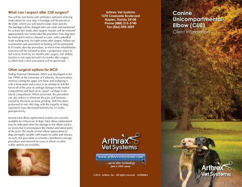
What can I expect after <strong>CUE</strong> surgery?<br />
You will be sent home with antibiotics and pain-relieving<br />
medications for your dog. A bandage will be placed on<br />
the limb, which you will need to keep clean and dry.<br />
The bandage will be changed after one week and maintained<br />
for at least two weeks after surgery. Sutures will be removed<br />
approximately two weeks after the procedure. Your dog must<br />
be restricted to rest in a kennel or crate, with controlled<br />
leash walking only, for eight weeks after surgery. Follow-up<br />
examination and assessment of healing will be performed<br />
8-10 weeks after the procedure, at which time rehabilitation<br />
exercises will be initiated to allow a progressive return to<br />
full activity levels by six months after surgery. Full athletic<br />
function is not expected until six months after surgery,<br />
at which time a final assessment will be performed.<br />
Other surgical options for MCD<br />
Sliding Humeral Osteotomy (SHO) was developed in the<br />
late 1990’s at the University of California. This procedure<br />
involves cutting the upper arm bone and realigning it<br />
with a bone plate and screws in an attempt to shift the<br />
forces off of the area of cartilage damage in the medial<br />
compartment and back on to ”good” cartilage in the<br />
lateral compartment. When successful, this procedure<br />
can also reduce or eliminate the pain and lameness<br />
caused by the bone-on-bone grinding. SHO has been<br />
performed in over 400 dogs with the majority of dogs<br />
reported to have decreased lameness by 12 weeks<br />
postoperatively.<br />
Several total elbow replacement systems are currently<br />
available for clinical use in dogs. Total elbow replacement<br />
may be indicated when the damage to the elbow joint is<br />
so severe that it encompasses the medial and lateral parts<br />
of the joint. The results of total elbow replacement in<br />
dogs are highly variable with respect to safety and efficacy.<br />
As such, this procedure is currently considered a salvage<br />
procedure and reserved for cases in which no other<br />
viable options are available.<br />
<strong>Arthrex</strong> <strong>Vet</strong> <strong>Systems</strong><br />
1370 Creekside Boulevard<br />
Naples, Florida 34108<br />
Phone (888) 215-3740<br />
Fax (866) 898-2059<br />
www.arthrexvetsystems.com<br />
...up-to-date technology<br />
just a click away<br />
©2012, <strong>Arthrex</strong>, Inc. All rights reserved. VLP0006A<br />
<strong>Canine</strong><br />
<strong>Unicompartmental</strong><br />
<strong>Elbow</strong> (<strong>CUE</strong>)<br />
Client Information
What is <strong>Canine</strong> <strong>Elbow</strong> Dysplasia?<br />
<strong>Elbow</strong> dysplasia is a general term used to identify the group<br />
of disorders that affect many dogs, especially large breeds<br />
and working/performance dogs. The three most common<br />
disorders included in the elbow dysplasia group are:<br />
• Fragmented Medial Coronoid Process (FMCP)<br />
• Osteochondrosis Dessicans (OCD)<br />
• Ununited Anconeal Process (UAP)<br />
While these disorders involve different areas of the elbow<br />
joint of affected dogs, they all result from abnormal<br />
development of the bone and cartilage of the elbow and<br />
lead to osteoarthritis.<br />
What are the symptoms of elbow dysplasia?<br />
<strong>Elbow</strong> dysplasia is the most common cause of forelimb<br />
lameness in dogs. The abnormalities start to occur when the<br />
puppy is going through a rapid growth phase (typically 4-10<br />
months of age), but often the symptoms are not noticed<br />
until the dog is much older. Changes in activity level, limping,<br />
swollen elbows, pain in the elbows, decreased performance,<br />
and behavioral changes are some of the visual signs you<br />
may observe if your dog has elbow dysplasia.<br />
Professional diagnosis of elbow dysplasia<br />
Your veterinarian will perform a careful orthopaedic<br />
examination of your dog to diagnose the problem. With<br />
elbow dysplasia, the doctor may note joint thickening and<br />
swelling, pain on joint manipulation, and loss of range<br />
of motion. X-rays should be done to confirm the findings<br />
and characterize the type and severity of elbow dysplasia.<br />
Advanced imaging, such as arthroscopy of the joint, CT scan,<br />
or MRI, may also be necessary to fully determine the extent<br />
of the problem and the most appropriate treatment options.<br />
Fragmented Medial Coronoid Process (FMCP) is by far the<br />
most common form of elbow dysplasia in dogs. In this<br />
disorder, the bone and cartilage on the medial part (inside<br />
portion) of the elbow joint develop cracks (microscopic or<br />
larger) which can cause the symptoms of elbow dysplasia<br />
and lead to osteoarthritis. The cracks may result in the<br />
formation of fragments of bone and cartilage that may remain<br />
in place or move in the joint like a pebble in your shoe.<br />
Arthroscopy performed by an experienced surgeon is an<br />
ideal way to deal with FMCP in dogs as it allows accurate<br />
and comprehensive diagnosis, as well as immediate treatment.<br />
Through two small incisions, the veterinary surgeon is able<br />
to carefully assess the joint, remove the fragments, and treat<br />
the surrounding cartilage as needed. This procedure typically<br />
takes between 20 and 45 minutes per elbow and many dogs<br />
can be treated on an outpatient basis. Arthroscopic treatment<br />
of FMCP has a high success rate of removing the abnormal<br />
cartilage and bone, slowing down the progression of<br />
osteoarthritis, and improving the symptoms of the disorder.<br />
However, it is not a cure and the osteoarthritis will still progress<br />
to some degree and needs to be monitored long term with a<br />
likely need for further treatments later in the dog’s life.<br />
As the dog ages, the osteoarthritis from FMCP and other forms<br />
of elbow dysplasia may result in complete loss of cartilage on<br />
the weight-bearing surfaces of the medial joint structures<br />
resulting in what veterinarians call Medial Compartment<br />
Disease (MCD). This is the “end stage” form of elbow dysplasia<br />
where the inside part of the joint collapses with eventual<br />
grinding of bone on bone. Interestingly and importantly,<br />
the larger lateral (outside) part of the elbow joint appears<br />
normal in the vast majority of patients.<br />
What are my treatment options for MCD?<br />
Options such as oral medications, joint injections, and<br />
physical therapy may be beneficial in many cases for at<br />
least a period of time and should be discussed with your<br />
veterinarian. When surgical treatment is deemed necessary,<br />
as is often the case, the <strong>Canine</strong> <strong>Unicompartmental</strong> <strong>Elbow</strong><br />
(<strong>CUE</strong>) is a safe and effective option to consider. The <strong>CUE</strong><br />
was developed as a treatment for MCD for dogs in which<br />
arthroscopic treatment and the nonsurgical options are<br />
no longer successful. By focusing on the specific<br />
area of disease (the medial compartment),<br />
the <strong>CUE</strong> implant provides a less invasive,<br />
bone-sparing option for resurfacing the<br />
bone-on-bone medial compartment<br />
while preserving the dog’s own<br />
“good” cartilage in the lateral<br />
compartment. This medial<br />
resurfacing procedure<br />
reduces or eliminates<br />
the pain and lameness<br />
that was caused by the<br />
bone-on-bone grinding.<br />
Arthroscopic image of<br />
severe MCD<br />
Arthroscopic image of <strong>CUE</strong><br />
seven months post-op<br />
<strong>CUE</strong> Humeral<br />
Implant<br />
Meshed titanium base<br />
promotes bone ingrowth<br />
<strong>CUE</strong> Ulnar<br />
Implant<br />
Post-op x-ray showing <strong>CUE</strong><br />
implants in place


