A founder mutation causing a severe ... - CENTOGENE
A founder mutation causing a severe ... - CENTOGENE
A founder mutation causing a severe ... - CENTOGENE
You also want an ePaper? Increase the reach of your titles
YUMPU automatically turns print PDFs into web optimized ePapers that Google loves.
Brief Communication<br />
A <strong>founder</strong> <strong>mutation</strong> <strong>causing</strong> a <strong>severe</strong> methylenetetrahydrofolate reductase (MTHFR)<br />
deficiency in Bukharian Jews<br />
Shay Ben-Shachar a , Tal Zvi a , Arndt Rolfs b,c , Andrea Breda Klobus c , Yuval Yaron a,d ,<br />
Anat Bar-Shira a , Avi Orr-Urtreger a,d, ⁎<br />
a The Genetic Institute & Prenatal Diagnosis Unit, Tel Aviv Sourasky Medical Center, Tel Aviv, Israel<br />
b Albrecht-Kossel-Institute for Neuroregeneration, Medical Faculty, University of Rostock, Germany<br />
c Centogene GmbH, The Rare Disease Company, Rostock, Germany<br />
d Sackler Faculty of Medicine, Tel Aviv University, Tel Aviv, Israel<br />
article info<br />
Article history:<br />
Received 30 June 2012<br />
Received in revised form 9 August 2012<br />
Accepted 9 August 2012<br />
Available online 18 August 2012<br />
Keywords:<br />
MTHFR<br />
Founder <strong>mutation</strong><br />
Bukhara<br />
1. Introduction<br />
abstract<br />
Homocystinuria due to methylenetetrahydrofolate reductase<br />
(MTHFR) deficiency (OMIM ID: 236250) is a rare inborn error of folate<br />
metabolism resulting in elevated homocysteine levels in plasma and<br />
urine [1]. Methylenetetrahydrofolate reductase catalyzes the conversion<br />
of 5,10-methylenetetrahydrofolate to 5-methyltetrahydrofolate, a cosubstrate<br />
for homocysteine remethylation to methionine. The <strong>severe</strong><br />
form of the disease is characterized by developmental delay, seizures and<br />
microcephaly [2], and may be associated with infantile epilepsy [3].The<br />
disease is autosomally recessively inherited, caused by <strong>mutation</strong>s in the<br />
MTHFR gene [4]. It has been suggested that early treatment with betaine<br />
may alleviate the effect of the disease to the brain in some cases [5].<br />
In this study we demonstrate the existence of a common splicing<br />
<strong>founder</strong> <strong>mutation</strong> in the MTHFR gene, <strong>causing</strong> a <strong>severe</strong> MTHFR deficiency<br />
among individuals of Jewish Bukharic ancestry.<br />
2. Material and methods<br />
2.1. Patient recruitment<br />
The study protocol and informed consent were approved by the<br />
local Institutional Ethical Committee. DNA and RNA samples were<br />
collected from affected individuals and their parents.<br />
⁎ Corresponding author at: The Genetic Institute, Tel Aviv Sourasky Medical Center,<br />
6 Weizmann St., Tel Aviv, 64239, Israel. Fax: +972 3 6974555.<br />
E-mail address: aviorr@tasmc.health.gov.il (A. Orr-Urtreger).<br />
1096-7192/$ – see front matter © 2012 Elsevier Inc. All rights reserved.<br />
http://dx.doi.org/10.1016/j.ymgme.2012.08.011<br />
Molecular Genetics and Metabolism 107 (2012) 608–610<br />
Contents lists available at SciVerse ScienceDirect<br />
Molecular Genetics and Metabolism<br />
journal homepage: www.elsevier.com/locate/ymgme<br />
Methylenetetrahydrofolate reductase (MTHFR) deficiency is a rare autosomal recessive disorder. A novel<br />
homozygous MTHFR c.474A>T (p.G158G) <strong>mutation</strong> was detected in two unrelated children of Jewish<br />
Bukharian origin. This <strong>mutation</strong> generates an abnormal splicing and early termination codon. A carrier<br />
frequency of 1:39 (5/196) was determined among unrelated healthy Bukharian Jews. Given the disease<br />
severity and allele frequency, a population screening for individuals of this ancestry is warranted in order to<br />
allow prenatal, or preimplantation diagnosis.<br />
© 2012 Elsevier Inc. All rights reserved.<br />
2.2. MTHFR gene sequencing<br />
MTHFR gene (NM_005957.4) was screened for <strong>mutation</strong>s using<br />
standard PCR and sequencing of both DNA strands of the coding<br />
region and the exon–intron boundaries followed the CMGS (Clinical<br />
Molecular Genetics Society) best practice guidelines (Centogene<br />
GmbH, Rostock, Germany). The <strong>mutation</strong> has been deposited in the<br />
NCBI dbSNP database (rs199476142).<br />
2.3. RNA analysis<br />
RT-PCR (reverse transcriptase PCR) was performed on cDNA<br />
generated from RNA extracted from peripheral blood leukocytes<br />
of the patients. Primers for the RT-PCR were located in exon 2<br />
and 4 of the reference sequence to verify whether exon 3 and/or<br />
intron 3 are included in the mutated transcript of the patient<br />
and the carrier parents. The primers' sequences are available upon<br />
request.<br />
2.4. Determination of the c.474A>T frequency among controls<br />
We examined 196 unaffected and unrelated Bukharian Jews<br />
and 176 Ashkenazi Jews, by an allelic discrimination test (TaqMan,<br />
custom-made test, Applied Biosystems, Carlsbad, CA, USA). The<br />
carrier status was further confirmed in all these individuals by Sanger<br />
sequencing.
3. Results<br />
A 13-month-old boy was referred for genetic work-up with the<br />
diagnosis of MTHFR deficiency. He had microcephaly, seizures and<br />
<strong>severe</strong> developmental delay. MTHFR deficiency was suspected due to<br />
elevated homocysteine in the blood and urine, and was confirmed by<br />
testing the specific activity of methylenetetrahydrofolate reductase<br />
in extract of the patient's fibroblasts (0.05 nmol/mg protein/h). The<br />
parents were non-consanguineous Jews of Bukharian ancestry. They<br />
previously had a daughter with similar manifestations, who died<br />
at 7 months of age. The analyses revealed a previously unreported<br />
homozygous variant c.474A>T (p.G158G) that caused a synonymous<br />
change. The parents were found to be heterozygous carriers of this<br />
change (Figs. 1A–C).<br />
The same homozygous <strong>mutation</strong> was detected in a 16-month-old<br />
girl, referred to our molecular genetics laboratory, with similar neurological<br />
and biochemical findings, suggestive of MTHFR deficiency.<br />
This girl was also an offspring of a non-consanguineous couple of<br />
Bukharian Jewish ancestry, unrelated to the family previously described.<br />
Her parents were found to be heterozygous carriers of the same<br />
<strong>mutation</strong>.<br />
As the transversion is located at the 3′ extremity of exon 3, and<br />
does not alter an amino acid, aberrant splicing was considered to be a<br />
consequence of this alteration.<br />
To test the hypothesis that the c.474A>T <strong>mutation</strong> causes an<br />
aberrant splicing, we performed an expression study by RT-PCR. From<br />
the cDNA of the affected individuals we found an amplicon of about<br />
1350 base pairs (bp), which was approximately 850 bp longer than<br />
the ~500 bp amplicon obtained in negative controls (Fig. 1F). The<br />
heterozygous parents had signs of both the wild-type allele and the<br />
mutant allele. Sanger sequencing of the large amplicon revealed<br />
the inclusion of the entire 838 bp sized intron 3 of the MTHFR gene<br />
A<br />
B<br />
C<br />
Genomic DNA<br />
Exon 3<br />
S. Ben-Shachar et al. / Molecular Genetics and Metabolism 107 (2012) 608–610<br />
Intron 3<br />
(Figs. 1D and E). An early stop codon was generated in the novel<br />
aberrant transcript 22 codons downstream to the original exon–<br />
intron boundary, confirming the truncation nature of the <strong>mutation</strong>.<br />
As these two affected individuals belong to the isolated ethnic group<br />
of the Jews coming from the province of Bukhara (Uzbekistan), we<br />
hypothesized that the c.474A>T <strong>mutation</strong> could be a <strong>founder</strong> allele. We<br />
therefore examined additional 196 unaffected and unrelated Bukharian<br />
Jews for the c.474A>T <strong>mutation</strong> and found it in five individuals,<br />
providing an estimated carrier frequency of 1:39. The <strong>mutation</strong> was<br />
not found among 176 Ashkenazi Jews. The prevalence of MTHFR<br />
deficiency due to this <strong>founder</strong> <strong>mutation</strong> in Bukharian Jews is expected<br />
to be ~1:6000.<br />
4. Discussion<br />
Our results show that a previously unreported <strong>mutation</strong> c.474A>T<br />
(p. G158G) is a common <strong>founder</strong> <strong>mutation</strong> among Jews of Bukharian<br />
ancestry, leading to aberrant splicing and resulting in generation of a<br />
premature termination codon. Currently, population-based screening<br />
programs are offered for a number of common genetic disorders<br />
world-wide, and are particularly prevalent in Israel [6]. Several<br />
criteria have been proposed to select genetic disorders for population<br />
screening. The agreed criteria include severity of disease, early onset,<br />
and high frequency of carriers, availability of genetic counseling and<br />
prenatal diagnosis, and an existence of a reliable test [7]. The MTHFR<br />
deficiency due to the c.474A>T <strong>mutation</strong> among couples of Bukharian<br />
Jewish origin clearly fulfills these criteria. Therefore, a populationbased<br />
screening for the disease among this population is warranted,<br />
and will be added to the panel of ethnicity-specific <strong>mutation</strong>s that<br />
are being offered in Israel. Furthermore, our results emphasize the<br />
importance of identifying rare <strong>founder</strong> <strong>mutation</strong>s that lead to <strong>severe</strong><br />
diseases among genetically-isolated populations. Such an approach<br />
Wild type<br />
Affected<br />
Carrier<br />
D<br />
E<br />
F<br />
1517bp<br />
1200bp<br />
1000bp<br />
500bp<br />
cDNA<br />
Exon 3 Exon 4<br />
Exon 3<br />
Intron 3<br />
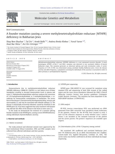
Fig. 1. Molecular analysis of the c.474A>T (G158G) <strong>mutation</strong>. On genomic DNA, compared with controls (A), affected individuals have a homozygous A>T transversion (B), while<br />
carrier individuals show the same change in the heterozygous state (C). The exon–intron junction is defined by the black vertical line. At the cDNA level, compared with unaffected<br />
controls (D), the original intron 3 is included in the cDNA of affected individuals immediately after exon 3 (E). RT-PCR of a cDNA fragment including exons 3 and 4 of the reference<br />
sequence revealed an expected ~500 bp band in controls and an enlarged ~1350 bp band in affected individuals. Both bands were detected in the heterozygous parents (F).<br />
609
610 S. Ben-Shachar et al. / Molecular Genetics and Metabolism 107 (2012) 608–610<br />
will assist in preventing and alleviating the burden of <strong>severe</strong> rare<br />
genetic disorders, in addition to the prevention of common <strong>severe</strong><br />
recessive diseases, such as spinal muscular atrophy (SMA) [8].<br />
References<br />
[1] S.H. Mudd, H.L. Levy, J.P. Kraus, Disorders of transsulfuration, in: C.R. Scriver, A.L.<br />
Beaudet, W.S. Sly, D. Valle (Eds.), The Metabolic and Molecular Bases of Inherited<br />
Disease, McGraw-Hill, Inc, New York, 2001, pp. 2007–2056.<br />
[2] F. Skovby, M. Gaustadnes, S.H. Mudd, A revisit to the natural history of homocystinuria<br />
due to cystathionine beta-synthase deficiency, Mol. Genet. Metab. 99 (2010) 1–3.<br />
[3] A.N. Prasad, C.A. Rupar, C. Prasad, Methylenetetrahydrofolate reductase (MTHFR)<br />
deficiency and infantile epilepsy, Brain Dev. 33 (2011) 758–769.<br />
[4] P. Goyette, J.S. Sumner, R. Milos, Human methylenetetrahydrofolate reductase:<br />
isolation of cDNA, mapping and <strong>mutation</strong> identification, Nature. Genet. (1994)<br />
195–200.<br />
[5] K.A. Strauss, D.H. Morton, E.G. Puffenberger, C. Hendrickson, D.L. Robinson, C.<br />
Wagner, S.P. Stabler, R.H. Allen, G. Chwatko, H. Jakubowski, M.D. Niculescu, S.H.<br />
Mudd, Prevention of brain disease from <strong>severe</strong> 5,10-methylenetetrahydrofolate<br />
reductase deficiency, Mol. Genet. Metab. (2007) 165–175.<br />
[6] G. Rosner, S. Rosner, A. Orr-Urtreger, Genetic testing in Israel: an overview, Annu.<br />
Rev. Genomics. Hum. Genet. 10 (2009) 175–192.<br />
[7] A.S.H.G., Ad Hoc Committee on Cystic Fibrosis Carrier Screening, Am. J. Hum. Genet.<br />
51 (1992) 1443–1444.<br />
[8] S. Ben-Shachar, A. Orr-Urtreger, E. Bardugo, R. Shomrat, Y. Yaron, Large-scale<br />
population screening for spinal muscular atrophy: clinical implications, Genet.<br />
Med. 13 (2011) 110–114.<br />
Web-based resources<br />
http://www.ncbi.nlm.nih.gov/omim<br />
http://www.ncbi.nlm.nih.gov/nuccore<br />
http://www.ncbi.nlm.nih.gov/projects/SNP/<br />
http://www.ensembl.org<br />
http://www.ncbi.nlm.nih.gov/projects/SNP/tranSNP/vsub.cgi?rm=view&set_id=23454


