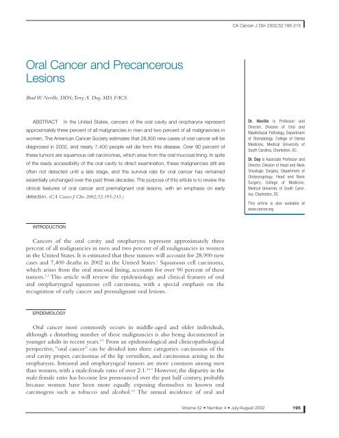
Oral Cancer and Precancerous Lesions - The Oral Cancer Foundation
Oral Cancer and Precancerous Lesions - The Oral Cancer Foundation
Oral Cancer and Precancerous Lesions - The Oral Cancer Foundation

You also want an ePaper? Increase the reach of your titles
YUMPU automatically turns print PDFs into web optimized ePapers that Google loves.
<strong>Oral</strong> <strong>Cancer</strong> <strong>and</strong> <strong>Precancerous</strong><br />
<strong>Lesions</strong><br />
Brad W. Neville, DDS;Terry A. Day, MD, FACS<br />
ABSTRACT In the United States, cancers of the oral cavity <strong>and</strong> oropharynx represent<br />
approximately three percent of all malignancies in men <strong>and</strong> two percent of all malignancies in<br />
women. <strong>The</strong> American <strong>Cancer</strong> Society estimates that 28,900 new cases of oral cancer will be<br />
diagnosed in 2002, <strong>and</strong> nearly 7,400 people will die from this disease. Over 90 percent of<br />
these tumors are squamous cell carcinomas, which arise from the oral mucosal lining. In spite<br />
of the ready accessibility of the oral cavity to direct examination, these malignancies still are<br />
often not detected until a late stage, <strong>and</strong> the survival rate for oral cancer has remained<br />
essentially unchanged over the past three decades. <strong>The</strong> purpose of this article is to review the<br />
clinical features of oral cancer <strong>and</strong> premalignant oral lesions, with an emphasis on early<br />
detection. (CA <strong>Cancer</strong> J Clin 2002;52:195-215.)<br />
INTRODUCTION<br />
<strong>Cancer</strong>s of the oral cavity <strong>and</strong> oropharynx represent approximately three<br />
percent of all malignancies in men <strong>and</strong> two percent of all malignancies in women<br />
in the United States. It is estimated that these tumors will account for 28,900 new<br />
cases <strong>and</strong> 7,400 deaths in 2002 in the United States. 1 Squamous cell carcinoma,<br />
which arises from the oral mucosal lining, accounts for over 90 percent of these<br />
tumors. 2-4 This article will review the epidemiology <strong>and</strong> clinical features of oral<br />
<strong>and</strong> oropharyngeal squamous cell carcinoma, with a special emphasis on the<br />
recognition of early cancer <strong>and</strong> premalignant oral lesions.<br />
EPIDEMIOLOGY<br />
<strong>Oral</strong> cancer most commonly occurs in middle-aged <strong>and</strong> older individuals,<br />
although a disturbing number of these malignancies is also being documented in<br />
younger adults in recent years. 5-7 From an epidemiological <strong>and</strong> clinicopathological<br />
perspective, “oral cancer” can be divided into three categories: carcinomas of the<br />
oral cavity proper, carcinomas of the lip vermilion, <strong>and</strong> carcinomas arising in the<br />
oropharynx. Intraoral <strong>and</strong> oropharyngeal tumors are more common among men<br />
than women, with a male:female ratio of over 2:1. 2,8-9 However, the disparity in the<br />
male:female ratio has become less pronounced over the past half century, probably<br />
because women have been more equally exposing themselves to known oral<br />
carcinogens such as tobacco <strong>and</strong> alcohol. 4,5 <strong>The</strong> annual incidence of oral <strong>and</strong><br />
CA <strong>Cancer</strong> J Clin 2002;52:195-215<br />
Dr. Neville is Professor <strong>and</strong><br />
Director, Division of <strong>Oral</strong> <strong>and</strong><br />
Maxillofacial Pathology, Department<br />
of Stomatology, College of Dental<br />
Medicine, Medical University of<br />
South Carolina, Charleston, SC.<br />
Dr. Day is Associate Professor <strong>and</strong><br />
Director, Division of Head <strong>and</strong> Neck<br />
Oncologic Surgery, Department of<br />
Otolaryngology, Head <strong>and</strong> Neck<br />
Surgery, College of Medicine,<br />
Medical University of South Carolina,<br />
Charleston, SC.<br />
This article is also available at<br />
www.cancer.org.<br />
Volume 52 • Number 4 • July/August 2002 195
<strong>Oral</strong> <strong>Cancer</strong> <strong>and</strong> <strong>Precancerous</strong> <strong>Lesions</strong><br />
FIGURE 1<br />
Age-adjusted Mortality Rates for <strong>Cancer</strong>s of the <strong>Oral</strong> Cavity <strong>and</strong> Pharynx<br />
Rate per 100,000<br />
12<br />
10<br />
8<br />
6<br />
4<br />
2<br />
0<br />
1950 1955 1960 1965 1970 1975 1980 1985 1990 1995<br />
Year of Death<br />
pharyngeal cancer in African Americans (12.4<br />
cases per 100,000 population) is higher than<br />
among whites (9.7 cases per 100,000);the highest<br />
incidence rate is among African-American<br />
males (20.5 cases per 100,000 population). 3,9<br />
In contrast to intraoral <strong>and</strong> oropharyngeal<br />
carcinomas, cancers of the lip vermilion are<br />
more akin epidemiologically to squamous cell<br />
carcinoma of the skin <strong>and</strong> occur primarily in<br />
white men. 2 <strong>The</strong>se lip tumors are most strongly<br />
associated with chronic sun exposure, although<br />
sometimes they have been related to the site<br />
where cigarettes or pipestems have habitually<br />
196 CA A <strong>Cancer</strong> Journal for Clinicians<br />
All Men<br />
African-American Women<br />
White Women<br />
White Men<br />
All Women<br />
African-American Men<br />
Over the past 50 years, the mortality rate for oral/pharyngeal cancer has slightly improved in white men, whereas it has significantly worsened<br />
for African-American men.<br />
been held. 10 <strong>The</strong>se malignancies are much<br />
more common in men, probably because men<br />
are more likely to have vocations <strong>and</strong>/or<br />
avocations that result in greater cumulative sun<br />
exposure. At one time, the lip was the most<br />
common site for oral cancer; however, the<br />
incidence of cancer in this location has<br />
decreased significantly over the past half<br />
century because fewer men hold outdoor<br />
occupations. 2,4<br />
Despite advances in surgery, radiation, <strong>and</strong><br />
chemotherapy, the five-year survival rate for<br />
oral cancer has not improved significantly over
the past several decades <strong>and</strong> it remains at about<br />
50 to 55 percent. 3,9 Unfortunately, African<br />
Americans have a significantly higher mortality<br />
rate when compared with whites (4.4 versus<br />
2.4 per 100,000 population), partly because<br />
among African Americans, tumors are more<br />
often discovered at an advanced stage (Figure<br />
1). 3,9,11,12 From 1985 to 1996, the five-year<br />
survival rate for carcinoma of the tongue in<br />
African-American men was 27 percent,<br />
compared with a 47 percent five-year survival<br />
rate among white men. 3 For floor of mouth<br />
cancers, the survival rate was 52 percent in<br />
whites, compared with only 33 percent among<br />
African Americans. When compared with<br />
intraoral carcinoma, the prognosis for lip<br />
cancer is quite good, with a five-year survival<br />
rate of 95 percent. 2,3<br />
RISK FACTORS<br />
<strong>The</strong> strong association between cancers of<br />
the oral cavity <strong>and</strong> pharynx with tobacco use is<br />
well established. Epidemiological studies show<br />
that the risk of developing oral cancer is five to<br />
nine times greater for smokers than for<br />
nonsmokers, <strong>and</strong> this risk may increase to as<br />
much as 17 times greater for extremely heavy<br />
smokers of 80 or more cigarettes per day. 2,13-17<br />
<strong>The</strong> percentage of oral cancer patients who<br />
smoke (approximately 80 percent) is two to<br />
three times greater than that of the general<br />
population. In addition, treated oral cancer<br />
patients who continue to smoke have a two to<br />
six times greater risk of developing a second<br />
malignancy of the upper aerodigestive tract<br />
than those who stop smoking. 10,18 Marijuana<br />
use is also considered to be a potential risk<br />
factor <strong>and</strong> may be partly responsible for the<br />
rise in oral cancers seen among young<br />
adults. 3,7,19 However, further epidemiological<br />
studies are necessary to confirm the purported<br />
association of marijuana <strong>and</strong> oral cancer in<br />
younger patients.<br />
Snuff <strong>and</strong> chewing tobacco have also been<br />
associated with an increased risk for oral<br />
cancer. 20 In one study of women in the<br />
southern United States, chronic users of snuff<br />
were estimated to have a four times greater risk<br />
of developing oral cancer. 21 In addition, a<br />
significant number of oral cancers in smokeless<br />
tobacco users develop at the site of tobacco<br />
placement. However, the use of smokeless<br />
tobacco appears to be associated with a much<br />
lower cancer risk than that associated with<br />
smoked tobacco. <strong>The</strong> incidence of oral cancer<br />
in West Virginia is below the national average,<br />
even though this state has the highest<br />
consumption of chewing tobacco in the<br />
United States. 22 Recent studies from<br />
Sc<strong>and</strong>inavia have suggested that the use of<br />
Swedish snuff (which is nonfermented <strong>and</strong> has<br />
lower nitrosamine levels) is not associated with<br />
an increased risk for oral cancer. 17,23<br />
Alcohol use has been identified as a major<br />
risk factor for cancers of the upper<br />
aerodigestive tract. In studies controlled for<br />
smoking, moderate-to-heavy drinkers have<br />
been shown to have a three to nine times<br />
greater risk of developing oral cancer. 13,14,16,17<br />
One study from France showed that extremely<br />
heavy drinkers (greater than 100 grams of<br />
alcohol per day) had a 30 times greater risk of<br />
developing oral <strong>and</strong> oropharyngeal cancer (a<br />
typical serving of beer, wine, or liquor contains<br />
ten to 15 grams of alcohol). 15 Of even greater<br />
significance is the synergistic effect of alcohol<br />
<strong>and</strong> smoking; some subsets of patients who are<br />
both heavy smokers <strong>and</strong> heavy drinkers can<br />
have over one hundred times greater risk for<br />
developing a malignancy. 15,16<br />
In India <strong>and</strong> Southeast Asia, the chronic use<br />
of betel quid (paan) in the mouth has been<br />
strongly associated with an increased risk for<br />
oral cancer. 24-26 <strong>The</strong> quid typically consists of<br />
a betel leaf that is wrapped around a mixture<br />
of areca nut <strong>and</strong> slaked lime, usually with<br />
tobacco <strong>and</strong> sometimes with sweeteners <strong>and</strong><br />
condiments. <strong>The</strong> slaked lime results in the<br />
CA <strong>Cancer</strong> J Clin 2002;52:195-215<br />
Volume 52 • Number 4 • July/August 2002 197
<strong>Oral</strong> <strong>Cancer</strong> <strong>and</strong> <strong>Precancerous</strong> <strong>Lesions</strong><br />
release of an alkaloid from the areca nut, which<br />
produces a feeling of euphoria <strong>and</strong> well-being<br />
in the user. Betel quid chewing often results in<br />
a progressive, scarring precancerous condition<br />
of the mouth known as oral submucous<br />
fibrosis. In India, one study showed a malignant<br />
transformation rate of 7.6 percent for oral<br />
submucous fibrosis. 25<br />
Recent evidence suggests that human<br />
papillomavirus (HPV) may be associated with<br />
some oral <strong>and</strong> oropharyngeal cancers. 27-31<br />
HPV-16 has been detected in up to 22 percent<br />
of oral cancers, <strong>and</strong> HPV-18 has been found in<br />
up to 14 percent of cases. 28 Dietary factors,<br />
such as a low intake of fruits <strong>and</strong> vegetables,<br />
may also be related to an increased cancer<br />
risk. 32,33 As previously indicated, chronic actinic<br />
exposure is associated with the development of<br />
carcinomas of the lip vermilion.<br />
A number of studies have suggested that oral<br />
lichen planus, especially the erosive form, may<br />
be associated with an increased cancer risk,<br />
although other investigators have questioned<br />
the strength of this association. 34-36 Iron<br />
deficiency anemia in combination with<br />
dysphagia <strong>and</strong> esophageal webs (known as<br />
Plummer-Vinson or Paterson-Kelly syndrome)<br />
is associated with an elevated risk for development<br />
of carcinoma of the oral cavity, oropharynx,<br />
<strong>and</strong> esophagus. 37,38 Immunosuppression<br />
appears to predispose some individuals to an<br />
increased risk for oral cancer. Carcinomas of the<br />
lip have been reported in a number of kidney<br />
transplant patients receiving immunosuppressive<br />
medications, <strong>and</strong> oral carcinomas have been<br />
documented in young AIDS patients. 39-42<br />
EARLY DIAGNOSIS<br />
Despite the great strides that have been<br />
198 CA A <strong>Cancer</strong> Journal for Clinicians<br />
made in recent decades to improve the<br />
prognosis for a number of cancers throughout<br />
the body, the prognosis for oral cancer has not<br />
experienced a similar improvement. 3,8,11<br />
Because five-year survival is directly related to<br />
stage at diagnosis, prevention <strong>and</strong> early<br />
detection efforts have the potential not only<br />
for decreasing the incidence, but also for<br />
improving the survival of those who develop<br />
this disease. Early diagnosis depends upon an<br />
astute clinician or patient who may identify a<br />
suspicious lesion or symptom while it is still at<br />
an early stage. However, it is apparent that<br />
many clinicians, including dentists <strong>and</strong><br />
physicians, may not be knowledgeable about<br />
the risk factors, diagnosis, <strong>and</strong> early detection<br />
of these cancers <strong>and</strong>/or are not performing<br />
routine oral cancer examinations. 43-49<br />
<strong>The</strong> Centers for Disease Control <strong>and</strong><br />
Prevention’s 1998 National Health Interview<br />
Survey (NHIS) Adult Prevention Supplement<br />
included questions regarding examinations for<br />
oral cancer. Participants were asked “Have you<br />
ever had a test for oral cancer in which the<br />
doctor or dentist pulls on your tongue,<br />
sometimes with gauze wrapped around it, <strong>and</strong><br />
feels under the tongue <strong>and</strong> inside the cheeks?”<br />
Only 16 percent of respondents reported that<br />
they ever had such an exam. This reported<br />
cumulative prevalence of oral cancer exams<br />
was higher in whites (18 percent) than in<br />
African Americans (10 percent), American<br />
Indians/Alaska Natives (8 percent), or<br />
Asian/Pacific Isl<strong>and</strong>ers (11 percent). Former<br />
smokers (21 percent) were more likely than<br />
current smokers (13 percent) or people who<br />
had never smoked (16 percent) to recall having<br />
ever had this examination. Among all<br />
individuals who reported having had an oral<br />
cancer exam, 70 percent reported that their last<br />
exam was within the past year. *<br />
*Vilma Cokkinides, PhD, (personal communication, May 2002), based on an analysis of the NHIS 1998 Adult Prevention<br />
Supplement Public Use Data Release accessed at www.ccdc.gov/nchs/nhis.htm.
Early oral cancers <strong>and</strong> precancerous lesions<br />
are often subtle <strong>and</strong> asymptomatic.<strong>The</strong>refore, it<br />
is important for the clinician to maintain a<br />
high index of suspicion, especially if risk<br />
factors such as tobacco use or alcohol abuse are<br />
present. Invasive oral squamous cell carcinoma<br />
is often preceded by the presence of clinically<br />
identifiable premalignant changes of the oral<br />
mucosa. <strong>The</strong>se lesions often present as either<br />
white or red patches, known as leukoplakia <strong>and</strong><br />
erythroplakia. As the cancer develops, the<br />
patient may notice the presence of a<br />
nonhealing ulcer. Later-stage symptoms<br />
include bleeding, loosening of teeth, difficulty<br />
wearing dentures, dysphagia, dysarthria,<br />
odynophagia, <strong>and</strong> development of a neck mass.<br />
<strong>The</strong> American <strong>Cancer</strong> Society recommends<br />
a cancer-related check-up annually for all<br />
individuals aged 40 <strong>and</strong> older, <strong>and</strong> every three<br />
years for those between the ages of 20 <strong>and</strong> 39,<br />
which “should include health counseling <strong>and</strong>,<br />
depending on a person’s age, might include<br />
examinations for cancers of the thyroid, oral<br />
cavity, skin, lymph nodes, testes, <strong>and</strong> ovaries.” 50<br />
According to the US Preventive Health<br />
Services Task Force (USPHSTF), “there is<br />
insufficient evidence to recommend for or<br />
against routine screening of asymptomatic<br />
persons for oral cancer by primary care<br />
clinicians … clinicians may wish to include an<br />
examination for cancerous <strong>and</strong> precancerous<br />
lesions of the oral cavity in the periodic health<br />
examination of persons who chew or smoke<br />
tobacco (or did so previously), older persons<br />
who drink regularly, <strong>and</strong> anyone with<br />
suspicious symptoms or lesions detected<br />
through self-examination. … Appropriate<br />
counseling should be offered to those persons<br />
who smoke cigarettes, pipes, or cigars, those<br />
who use chewing tobacco or snuff, <strong>and</strong> those<br />
who demonstrate evidence of alcohol abuse.” 51<br />
<strong>The</strong> USPHSTF document also notes that<br />
“…both the National <strong>Cancer</strong> Institute <strong>and</strong> the<br />
National Institute of Dental Research<br />
(subsequently renamed the National Institute<br />
of Dental <strong>and</strong> Craniofacial Research) support<br />
efforts to promote the early detection of oral<br />
cancers during routine dental examinations.”<br />
Clearly, the low prevalence of oral cancer<br />
screening reported in the NHIS indicates that<br />
most clinicians are not following ACS<br />
recommendations, <strong>and</strong> are not even following<br />
the USPHSTF suggestion for examinations in<br />
tobacco users <strong>and</strong> other high-risk individuals.<br />
Unfortunately, there has been little<br />
improvement in the early detection of oral<br />
cancer because many patients do not present<br />
for diagnosis <strong>and</strong> treatment until they have<br />
Stage III or Stage IV disease (Figure 2).<br />
<strong>The</strong>refore, in order to improve oral cancer<br />
survival, public education efforts are also<br />
necessary to encourage patients to avoid highrisk<br />
behaviors <strong>and</strong> to ask their health care<br />
providers about regular oral cancer screening<br />
examinations.<br />
LEUKOPLAKIA<br />
<strong>The</strong> term leukoplakia was first used by<br />
Schwimmer in 1877 to describe a white lesion<br />
of the tongue, which probably represented a<br />
syphilitic glossitis. 52 <strong>The</strong> definition of<br />
leukoplakia has often been confusing <strong>and</strong><br />
controversial—so much so, that some clinicians<br />
now avoid using this term in their lexicon. As<br />
defined by the World Health Organization,<br />
leukoplakia is “a white patch or plaque that<br />
cannot be characterized clinically or<br />
pathologically as any other disease.” 53 As such,<br />
leukoplakia should be used only as a clinical<br />
term; it has no specific histopathological<br />
connotation <strong>and</strong> should never be used as a<br />
microscopic diagnosis. 54 In the evaluation of<br />
the patient, leukoplakia is a clinical diagnosis of<br />
exclusion. If an oral white patch can be<br />
diagnosed as some other condition (e.g.,<br />
c<strong>and</strong>idiasis, lichen planus, leukoedema, etc.),<br />
then the lesion should not be considered to be<br />
an example of leukoplakia. Sometimes a white<br />
CA <strong>Cancer</strong> J Clin 2002;52:195-215<br />
Volume 52 • Number 4 • July/August 2002 199
<strong>Oral</strong> <strong>Cancer</strong> <strong>and</strong> <strong>Precancerous</strong> <strong>Lesions</strong><br />
FIGURE 2<br />
Trends in Diagnosis by Stage for <strong>Cancer</strong>s of the <strong>Oral</strong> Cavity <strong>and</strong> Pharynx for All Races<br />
Rate per 100,000<br />
6<br />
5<br />
4<br />
3<br />
2<br />
1<br />
0<br />
1973 1975 1977 1979 1981 1983 1985 1987 1989 1991 1993 1995 1997 1999<br />
Over the past 25 years, no significant improvement has been made in the early diagnosis of oral/pharyngeal cancer.<br />
patch is initially believed to represent<br />
leukoplakia, but the biopsy reveals another<br />
specific diagnosis. In such cases, the lesion<br />
should no longer be categorized as a<br />
leukoplakia.<br />
<strong>The</strong> usage of the term leukoplakia<br />
continues to undergo refinement. 55 Frequently,<br />
oral white patches are seen secondary to<br />
identifiable local irritation. For example,<br />
thickened hyperkeratotic changes are<br />
frequently found on the edentulous areas of<br />
the alveolar ridges, especially in patients who<br />
do not wear an overlying dental prosthesis<br />
200 CA A <strong>Cancer</strong> Journal for Clinicians<br />
Year<br />
Regional<br />
Localized<br />
Unstaged<br />
Distant<br />
(Figure 3). Because these exposed edentulous<br />
sites receive more irritation during<br />
mastication, there is a natural tendency for the<br />
epithelium to become more hyperkeratotic as<br />
a protective phenomenon, similar to a callus<br />
developing on one’s h<strong>and</strong>. Because such “ridge<br />
keratoses” rarely ever show any dysplastic<br />
changes or transform into carcinoma, most<br />
experts prefer placing them into a separate<br />
category (“frictional keratoses”), rather than<br />
considering them to be leukoplakias. 2,55<br />
Likewise, hyperkeratotic changes that develop<br />
secondary to chronic cheek chewing
(“morsicatio buccarum”) or tongue chewing<br />
(“morsicatio linguarum”) should not be<br />
classified as leukoplakia; such lesions are not<br />
premalignant <strong>and</strong> they are readily reversible if<br />
the irritation is avoided.<br />
Two specific tobacco-related lesions of the<br />
oral mucosa, nicotine stomatitis <strong>and</strong> tobacco<br />
pouch keratosis, have often been included<br />
under the broad umbrella of leukoplakia.<br />
However, because these lesions have a specific<br />
known cause <strong>and</strong> prognosis, we prefer to<br />
classify them separately from leukoplakia.<br />
Leukoplakia is seen most frequently in<br />
middle-aged <strong>and</strong> older men, with an increasing<br />
prevalence with age. 2,56 Fewer than one percent<br />
of men below the age of 30 have leukoplakia,<br />
but the prevalence increases to an alarming<br />
eight percent in men over the age of 70. 56 <strong>The</strong><br />
prevalence in women past the age of 70 is<br />
approximately two percent.<strong>The</strong> most common<br />
sites are the buccal mucosa, alveolar mucosa,<br />
<strong>and</strong> lower lip; however, lesions in the floor of<br />
mouth, lateral tongue, <strong>and</strong> lower lip are most<br />
likely to show dysplastic or malignant<br />
changes. 57<br />
Early or thin leukoplakia appears as a slightly<br />
elevated grayish-white plaque that may be<br />
either well defined or may gradually blend into<br />
the surrounding normal mucosa (Figure 4). 2,58<br />
As the lesion progresses, it becomes thicker <strong>and</strong><br />
whiter, sometimes developing a leathery<br />
appearance with surface fissures (homogeneous<br />
or thick leukoplakia) (Figure 5). Some<br />
leukoplakias develop surface irregularities <strong>and</strong><br />
are referred to as granular or nodular<br />
leukoplakias (Figure 6). Other lesions develop a<br />
papillary surface <strong>and</strong> are known as verrucous or<br />
verruciform leukoplakia (Figure 7).<br />
One uncommon variant, known as<br />
proliferative verrucous leukoplakia (PVL), is<br />
characterized by widespread, multifocal sites of<br />
involvement, often in patients without known<br />
risk factors. 59-63 <strong>The</strong> condition begins with<br />
conventional flat white patches that, over time,<br />
TABLE 1<br />
tend to become much thicker <strong>and</strong> papillary in<br />
nature (Figure 8). This papillary proliferation<br />
may progress to the point where the lesion can<br />
be categorized microscopically as a verrucous<br />
carcinoma. However, in spite of treatment, the<br />
lesions have a high recurrence rate <strong>and</strong> often<br />
eventually transform into more aggressive<br />
squamous cell carcinoma.<br />
In recent years, a number of oral white<br />
patches have been identified that appear to be<br />
related to the use of toothpastes or mouth<br />
rinses containing the herbal extract,<br />
sanguinaria. 64-66 Such lesions most frequently<br />
have been identified on the maxillary alveolar<br />
mucosa <strong>and</strong> buccal vestibule, although some<br />
patients have developed lesions on the<br />
m<strong>and</strong>ibular alveolar mucosa. Microscopically,<br />
these lesions usually show hyperkeratosis <strong>and</strong><br />
epithelial atrophy, sometimes in association<br />
with true dysplasia, although the potential for<br />
the development of cancer is uncertain.<br />
CA <strong>Cancer</strong> J Clin 2002;52:195-215<br />
Histopathological Nature of Leukoplakia by Site (3,360 Biopsy<br />
Specimens) 57<br />
% of<br />
Leukoplakias<br />
% of at this site that<br />
Leukoplakias showed dysplasia<br />
Site at this site or carcinoma<br />
Lips 10.3 24.0<br />
Maxillary mucosa <strong>and</strong> sulcus 10.7 14.8<br />
M<strong>and</strong>ibular mucosa <strong>and</strong> sulcus 25.2 14.6<br />
Palate 10.7 18.8<br />
Buccal mucosa 21.9 16.5<br />
Tongue 6.8 24.2<br />
Floor of mouth 8.6 42.9<br />
Retromolar 5.9 11.7<br />
Total 100.0 19.9<br />
(average for all sites)<br />
Source: Waldron CA, Shafer WG. Leukoplakia revisited: A clinicopathological<br />
study of 3,256 oral leukoplakias. <strong>Cancer</strong> 1975;36:1386-1392.<br />
Volume 52 • Number 4 • July/August 2002 201
<strong>Oral</strong> <strong>Cancer</strong> <strong>and</strong> <strong>Precancerous</strong> <strong>Lesions</strong><br />
3<br />
4<br />
5<br />
Figure 3 Frictional ridge keratosis. This rough, white change of<br />
the edentulous area of the alveolar ridge represents a frictional<br />
hyperkeratosis because this area now receives more irritation<br />
during mastication. This should not be mistaken for true<br />
leukoplakia, <strong>and</strong> biopsy is not indicated.<br />
Figure 4 Early or thin leukoplakia. This subtle white patch on<br />
the lateral soft palate showed severe epithelial dysplasia on biopsy.<br />
Figure 5 Thick leukoplakia. This thick white lesion on the<br />
lateral/ventral tongue showed moderate epithelial dysplasia.<br />
Thinner areas of leukoplakia are visible on the more posterior<br />
aspects of the lateral tongue <strong>and</strong> in the floor of mouth.<br />
202 CA A <strong>Cancer</strong> Journal for Clinicians<br />
6<br />
7<br />
8<br />
Figure 6 Granular leukoplakia. A small leukoplakic lesion with a<br />
rough, granular surface on the posterior lateral border of the<br />
tongue. <strong>The</strong> biopsy revealed early invasive squamous cell<br />
carcinoma. Such a lesion would be easily missed during an oral<br />
examination unless the tongue is pulled out <strong>and</strong> to the side to<br />
allow visualization of this high-risk site. (Courtesy of Neville BW,<br />
Damm DD, Allen CM, et al. <strong>Oral</strong> & Maxillofacial Pathology, ed 2,<br />
Philadelphia, WB Saunders, 2002.)<br />
Figure 7 Verruciform leukoplakia. <strong>The</strong> papillary component of<br />
this lesion on the left side of the picture (patient’s right) showed<br />
well-differentiated squamous cell carcinoma.<br />
Figure 8 Proliferative verrucous leukoplakia. This middle-aged<br />
gentleman has had a several year history of these recurring,<br />
spreading hyperkeratotic lesions that involve both the buccal <strong>and</strong><br />
lingual gingiva. Multiple biopsies have ranged from simple<br />
hyperkeratosis to moderate epithelial dysplasia.
9<br />
10<br />
11<br />
Figure 9 Speckled leukoplakia. This mixed white <strong>and</strong> red lesion<br />
of the buccal mucosa showed moderate epithelial dysplasia.<br />
Figure 10 Leukoplakia. A diffuse leukoplakia of the left lateral<br />
border of the tongue. A biopsy of the thick, rough zone at the<br />
anterior aspect of the lesion showed early invasive squamous cell<br />
carcinoma.<br />
Figure 11 Erythroplakia. This small, subtle red lesion on the right<br />
lateral border of the tongue showed carcinoma in situ on biopsy.<br />
Adjacent slight leukoplakic changes are also evident (erythroleukoplakia).<br />
(Courtesy of Neville BW, Damm DD, Allen CM, et al.<br />
<strong>Oral</strong> & Maxillofacial Pathology, ed 2, Philadelphia, WB Saunders,<br />
2002.)<br />
12<br />
13<br />
CA <strong>Cancer</strong> J Clin 2002;52:195-215<br />
Figure 12 Nicotine stomatitis. Rough, white, fissured<br />
appearance of the hard <strong>and</strong> soft palate in a heavy pipe smoker.<br />
<strong>The</strong> red, punctate areas represent the inflamed openings of the<br />
minor salivary gl<strong>and</strong> ducts.<br />
Figure 13 Tobacco pouch keratosis. A white, wrinkled change<br />
of the mucosa in the m<strong>and</strong>ibular buccal vestibule secondary to the<br />
use of chewing tobacco.<br />
Volume 52 • Number 4 • July/August 2002 203
<strong>Oral</strong> <strong>Cancer</strong> <strong>and</strong> <strong>Precancerous</strong> <strong>Lesions</strong><br />
TABLE 2<br />
Malignant Transformation of <strong>Oral</strong> Leukoplakia (Europe <strong>and</strong> United States)<br />
% of Patients<br />
with Malignant<br />
Source Country Year # of Patients Transformation<br />
Einhorn <strong>and</strong> Wersäll71 Sweden 1967 782 4.0<br />
Silverman70 United States 1968 117 6.0<br />
Pindborg et al. 67 Denmark 1968 248 4.4<br />
Kramer74 Engl<strong>and</strong> 1969 187 4.8<br />
Roed-Petersen75 Denmark 1971 331 3.6<br />
Bánóczy72 Hungary 1977 670 6.0<br />
Silverman et al. 76 United States 1984 247 17.5<br />
Lind77 Norway 1987 157 8.9<br />
Bouquot <strong>and</strong> Gorlin56 United States 1988 463 10.3<br />
Because sanguinaria-associated keratoses can<br />
be extensive or multifocal, sometimes they are<br />
misinterpreted as early proliferative verrucous<br />
leukoplakia.<br />
Some leukoplakias occur in combination<br />
with adjacent red patches or erythroplakia. If<br />
the red <strong>and</strong> white areas are intermixed, the<br />
lesion is called a speckled leukoplakia or<br />
speckled erythroplakia (Figure 9).<br />
<strong>The</strong> frequency of dysplastic or malignant<br />
alterations in oral leukoplakia has ranged from<br />
15.6 to 39.2 percent in several studies. 54,57,67-69 In<br />
one large, well known retrospective study that<br />
looked at approximately 3,300 biopsies of oral<br />
white lesions, Waldron <strong>and</strong> Shafer determined<br />
that 19.9 percent of leukoplakias showed some<br />
degree of epithelial dysplasia (Table 1). 57 In this<br />
group, 3.1 percent were unsuspected squamous<br />
cell carcinoma, 4.6 percent showed severe<br />
dysplasia or carcinoma in situ, <strong>and</strong> 12.2 percent<br />
showed mild-to-moderate epithelial dysplasia.<br />
Differences in the frequency of dysplastic<br />
changes in leukoplakia studies may reflect<br />
selection bias or differences in the clinical<br />
definition of oral leukoplakia. If white lesions<br />
such as frictional ridge keratoses <strong>and</strong> nicotine<br />
204 CA A <strong>Cancer</strong> Journal for Clinicians<br />
stomatitis are not included as examples of<br />
clinical leukoplakia, the percentage of cases<br />
showing dysplastic changes will be higher.<br />
<strong>The</strong> location of oral leukoplakia has a<br />
significant correlation with the frequency of<br />
finding dysplastic or malignant changes at<br />
biopsy. In the study by Waldron <strong>and</strong> Shafer, the<br />
floor of mouth was the highest-risk site, with<br />
42.9 percent of leukoplakias showing some<br />
degree of epithelial dysplasia, carcinoma in situ,<br />
or unsuspected invasive squamous cell<br />
carcinoma. 57 <strong>The</strong> tongue <strong>and</strong> lip were also<br />
identified as high-risk sites, with dysplasia or<br />
carcinoma present in 24.2 percent <strong>and</strong> 24.0<br />
percent of these cases, respectively.<br />
<strong>The</strong> clinical appearance of leukoplakia may<br />
also indicate some correlation with the<br />
likelihood that the lesion will show dysplastic<br />
or malignant features. In general, the thicker<br />
the leukoplakia, the greater the chance of<br />
finding dysplastic changes; therefore, a<br />
verrucous leukoplakia is more likely to show<br />
dysplasia than is a thick homogeneous<br />
leukoplakia, which, in turn, is more likely to<br />
show dysplasia than is a thin leukoplakia<br />
(Figure 10). 58 Leukoplakias with an intermixed
ed component (speckled leukoplakia or mixed<br />
leukoplakia/erythroplakia) are at greatest risk<br />
for showing dysplasia or carcinoma. Pindborg<br />
<strong>and</strong> associates found 14 percent of speckled<br />
leukoplakias to show carcinoma, whereas<br />
another 51 percent showed epithelial dysplasia.<br />
67 However, all leukoplakias should be<br />
viewed with suspicion because even small,<br />
subtle lesions can manifest significant dysplasia<br />
or unsuspected carcinoma. 57,70 <strong>The</strong>refore,<br />
directed conventional biopsy is recommended<br />
for any true oral leukoplakia.<br />
In addition to a small percentage of<br />
leukoplakias that will show invasive carcinoma<br />
when they are first sampled for biopsy, it is also<br />
recognized that currently non-carcinomatous<br />
leukoplakias are at risk for future malignant<br />
transformation. Several clinical studies have<br />
been conducted in Europe <strong>and</strong> the United<br />
States to assess the potential for malignant<br />
transformation of oral leukoplakia (Table<br />
2). 58,70-77 Most of the earlier studies showed a<br />
risk of malignant transformation in the range<br />
of 3.6 to 6.0 percent. However, several of the<br />
more recent studies have shown more alarming<br />
malignant transformation rates ranging from<br />
8.9 to 17.5 percent. 58,76,77 Although the reason<br />
for these results is unclear, it may be due to a<br />
more restrictive definition of what is<br />
considered clinical leukoplakia <strong>and</strong> further<br />
underscores the seriousness of “true<br />
leukoplakia.” <strong>The</strong> study by Silverman <strong>and</strong><br />
colleagues showed an overall malignant<br />
transformation of 17.5 percent. 76 In this study,<br />
only 6.5 percent of homogeneous leukoplakias<br />
underwent malignant change; however, 23.4<br />
percent of speckled leukoplakias <strong>and</strong> 36.4<br />
percent of leukoplakias with microscopic<br />
evidence of dysplastic changes transformed<br />
into cancer.<br />
When compared with “conventional<br />
leukoplakia,” proliferative verrucous leukoplakia<br />
is a particularly high-risk condition. In<br />
a follow-up study of 54 cases of proliferative<br />
verrucous leukoplakia, Silverman <strong>and</strong> Gorsky<br />
found that 70.3 percent of the patients<br />
subsequently developed squamous cell<br />
carcinoma. 62<br />
Although leukoplakia is more common in<br />
men than women, several studies have shown<br />
that women with leukoplakia have a higher risk<br />
of developing oral carcinoma. 70,72,75 Another<br />
disturbing finding is that leukoplakias in<br />
nonsmokers are more likely to undergo<br />
malignant transformation than leukoplakias in<br />
patients who do smoke. 71,72,75,76 This should<br />
not be interpreted to detract from the wellestablished<br />
role of tobacco in oral<br />
carcinogenesis, but may indicate that nonsmokers<br />
who develop leukoplakia do so as a<br />
result of other more potent carcinogenic factors.<br />
ERYTHROPLAKIA<br />
<strong>The</strong> term erythroplasia was originally used by<br />
Queyrat to describe a red, precancerous lesion<br />
of the penis. 78 <strong>The</strong> term erythroplakia is used for<br />
a clinically <strong>and</strong> histopathologically similar<br />
process that occurs on the oral mucosa. Similar<br />
to the definition for leukoplakia, erythroplakia<br />
is a clinical term that refers to a red patch that<br />
cannot be defined clinically or pathologically<br />
as any other condition. 53 This definition<br />
excludes inflammatory conditions that may<br />
result in a red clinical appearance.<br />
<strong>Oral</strong> erythroplakia occurs most frequently<br />
in older men <strong>and</strong> appears as a red macule or<br />
plaque with a soft, velvety texture (Figure 11). 2<br />
<strong>The</strong> floor of mouth, lateral tongue, retromolar<br />
pad, <strong>and</strong> soft palate are the most common sites<br />
of involvement. Often the lesion is well<br />
demarcated, but some examples may gradually<br />
blend into the surrounding mucosa. Some<br />
lesions may be intermixed with white areas<br />
(erythroleukoplakia). Erythroplakia is often<br />
asymptomatic, although some patients may<br />
complain of a sore, burning sensation.<br />
Although erythroplakia is not nearly as<br />
common as leukoplakia, it is much more likely<br />
CA <strong>Cancer</strong> J Clin 2002;52:195-215<br />
Volume 52 • Number 4 • July/August 2002 205
<strong>Oral</strong> <strong>Cancer</strong> <strong>and</strong> <strong>Precancerous</strong> <strong>Lesions</strong><br />
to show dysplasia or carcinoma. In a sister study<br />
to their large series of leukoplakia cases, Shafer<br />
<strong>and</strong> Waldron also analyzed their biopsy<br />
experience with 65 cases of erythroplakia. 79 All<br />
erythroplakia cases showed some degree of<br />
epithelial dysplasia; 51 percent showed invasive<br />
squamous cell carcinoma, 40 percent were<br />
carcinoma in situ or severe epithelial dysplasia,<br />
<strong>and</strong> the remaining 9 percent demonstrated<br />
mild-to-moderate dysplasia. <strong>The</strong>refore, true<br />
clinical erythroplakia is a much more<br />
worrisome lesion than leukoplakia. 80 Likewise,<br />
in a mixed erythroleukoplakia, the red<br />
component is more likely to demonstrate<br />
dysplastic changes than is the white component;<br />
when selecting an appropriate biopsy site in a<br />
mixed lesion, the clinician should make sure that<br />
the specimen includes the red component.<br />
NICOTINE STOMATITIS<br />
Nicotine stomatitis is a thickened,<br />
hyperkeratotic alteration of the palatal mucosa<br />
that is most frequently related to pipe smoking,<br />
but milder examples can also develop<br />
secondary to cigar smoking or, rarely, from<br />
cigarette smoking. 2,53 <strong>The</strong> palatal mucosa<br />
becomes thickened <strong>and</strong> hyperkeratotic,<br />
sometimes developing a fissured surface<br />
(Figure 12).<strong>The</strong> surface often develops papular<br />
elevations with red centers, which represent<br />
the inflamed openings of the minor salivary<br />
gl<strong>and</strong> ducts.<br />
<strong>The</strong> term nicotine stomatitis is actually a<br />
misnomer because it isn’t the nicotine that<br />
causes the changes; the changes are caused by<br />
the intense heat generated from the smoking.<br />
Nicotine stomatitis is seen more often in pipe<br />
smokers because of the great amount of heat<br />
that is generated from the pipestem. (Similar<br />
lesions have even been reported in patients<br />
who drink extremely hot beverages.) 81<br />
Although nicotine stomatitis is a tobaccorelated<br />
pathosis, it is not considered to be<br />
206 CA A <strong>Cancer</strong> Journal for Clinicians<br />
premalignant <strong>and</strong> it is readily reversible with<br />
discontinuation of the tobacco habit.<br />
However, in some Southeast Asian <strong>and</strong><br />
South American countries, individuals practice<br />
a habit known as reverse smoking in which the<br />
lit end of the cigarette or cigar is placed in the<br />
mouth. This habit creates a more severe heatrelated<br />
alteration of the palatal mucosa known<br />
as reverse smoker’s palate, which has been<br />
associated with a significant risk of malignant<br />
transformation. 10,82,83<br />
TOBACCO POUCH KERATOSIS<br />
Another specific tobacco-related oral<br />
mucosal alteration occurs in association with<br />
smokeless tobacco use, either from snuff or<br />
chewing tobacco. 2,84-87 Such lesions typically<br />
occur in the buccal or labial vestibule where<br />
the tobacco is held, but they can also extend<br />
onto the adjacent gingiva <strong>and</strong> buccal mucosa.<br />
Early lesions may show slight wrinkling that<br />
disappears when the tissues are stretched.<br />
Other lesions may appear as hyperkeratotic,<br />
granular patches. Advanced lesions exhibit<br />
greatly thickened zones of grayish white<br />
mucosa with well-developed folds <strong>and</strong> fissures<br />
(Figure 13). <strong>The</strong> degree of clinical alteration<br />
depends on the type <strong>and</strong> quantity of tobacco,<br />
the duration of tobacco usage, <strong>and</strong> host<br />
susceptibility.<br />
Tobacco pouch keratoses can occur at any<br />
age, even in children <strong>and</strong> adolescents. In<br />
Western cultures, these lesions currently are<br />
seen most frequently in young men <strong>and</strong> men<br />
older than 65 years of age; such lesions are less<br />
common among middle-aged men because the<br />
habit of using smokeless tobacco has not been<br />
as popular in this generation. 2 In some rural<br />
Southern populations, smokeless tobacco<br />
keratoses are seen with some degree of<br />
frequency in older women, who may have<br />
started their snuff-dipping habit in early<br />
childhood. 84 Overall, it is estimated that 15
percent of chewing tobacco users <strong>and</strong> 60<br />
percent of snuff users will develop clinical<br />
lesions, if mild examples are included. 2<br />
Microscopically, smokeless tobacco keratoses<br />
show hyperkeratosis <strong>and</strong> acanthosis of the<br />
mucosal epithelium.True epithelial dysplasia is<br />
uncommon; when dysplasia is found, it is<br />
usually mild in degree. 84 However, significant<br />
dysplasia or squamous cell carcinoma<br />
occasionally may be discovered.<br />
Most tobacco pouch keratoses are readily<br />
reversible within two to six weeks after<br />
cessation of the tobacco habit. 88 If the lesion<br />
does not resolve after the habit is stopped, then<br />
an incisional biopsy of the area should be<br />
performed <strong>and</strong> the patient managed<br />
accordingly. Some clinicians also recommend<br />
biopsy for lesions in patients who will not<br />
discontinue their tobacco habit.<br />
SQUAMOUS CELL CARCINOMA<br />
Early squamous cell carcinoma often<br />
presents as a white patch (leukoplakia), red<br />
patch (erythroplakia), or a mixed red <strong>and</strong> white<br />
lesion (erythroleukoplakia). With time,<br />
superficial ulceration of the mucosal surface<br />
may develop (Figure 14). As the lesion grows,<br />
it may become an exophytic mass with a<br />
fungating or papillary surface (Figure 15); other<br />
tumors have an endophytic growth pattern that<br />
is characterized by a depressed, ulcerated<br />
surface with a raised, rolled border (Figure<br />
16). 2,89 Pain is not a reliable indicator as to<br />
whether a particular lesion may be malignant;<br />
larger, advanced carcinomas will often be<br />
painful, but many early oral cancers will be<br />
totally asymptomatic or may be associated with<br />
only minor discomfort.<br />
<strong>The</strong> most common site for intraoral<br />
carcinoma is the tongue, which accounts for<br />
around 40 percent of all cases in the oral cavity<br />
proper.<strong>The</strong>se tumors most frequently occur on<br />
the posterior lateral border <strong>and</strong> ventral surfaces<br />
of the tongue. <strong>The</strong> floor of the mouth is the<br />
second most common intraoral location. Lesscommon<br />
sites include the gingiva, buccal<br />
mucosa, labial mucosa, <strong>and</strong> hard palate. 2,4<br />
<strong>The</strong> lateral tongue <strong>and</strong> floor of mouth (with<br />
extension back to the lateral soft palate <strong>and</strong><br />
tonsillar area) combine to form a horseshoeshaped<br />
region of the oral mucosa, which is at<br />
greatest risk for cancer development.<strong>The</strong>re are<br />
two major factors that may explain why this<br />
region is at high risk: first, any carcinogens will<br />
mix with saliva, pool in the bottom of the<br />
mouth, <strong>and</strong> constantly bathe these sites;<br />
secondly, these regions of the mouth are<br />
covered by a thinner, nonkeratinized mucosa,<br />
which provides less protection against<br />
carcinogens. 14<br />
It is important for the clinician to be aware of<br />
this high-risk region when examining the oral<br />
cavity. During an examination, if a tongue blade<br />
or other instrument is used simply to depress the<br />
tongue in order to see the rest of the mouth,<br />
then the two most common sites for intraoral<br />
cancer will be hidden. It is recommended that a<br />
cotton gauze be used to grasp the tip of the<br />
tongue, allowing it to be pulled upward <strong>and</strong> to<br />
each side so that the lateral tongue <strong>and</strong> oral floor<br />
can be adequately seen. 90<br />
In addition to the oral cavity proper,<br />
squamous cell carcinomas also often develop<br />
on the lip vermilion <strong>and</strong> the oropharynx.<br />
Vermilion carcinomas show a striking<br />
predilection for the lower lip, <strong>and</strong> usually occur<br />
in light-skinned individuals with a long history<br />
of actinic damage. <strong>The</strong> lesion usually arises in<br />
an actinic cheilosis, a premalignant condition<br />
that is akin to actinic keratosis of the skin.<br />
Actinic cheilosis is characterized by atrophy of<br />
the vermilion border, which may develop dry,<br />
scaly changes. As the condition progresses,<br />
ulcerated sites may appear which partially heal,<br />
only to recur at a later date (Figure 17). (<strong>The</strong><br />
patient often mistakes these recurring ulcerated<br />
lesions for “fever blisters.”) <strong>The</strong> evolving cancer<br />
slowly becomes a crusted, nontender, indurated<br />
CA <strong>Cancer</strong> J Clin 2002;52:195-215<br />
Volume 52 • Number 4 • July/August 2002 207
<strong>Oral</strong> <strong>Cancer</strong> <strong>and</strong> <strong>Precancerous</strong> <strong>Lesions</strong><br />
14<br />
15<br />
16<br />
Figure 14 Squamous cell carcinoma. Ulcerated lesion of the<br />
ventral tongue/floor of mouth.<br />
Figure 15 Squamous cell carcinoma. Exophytic, papillary mass<br />
of the buccal mucosa.<br />
Figure 16 Squamous cell carcinoma. Deeply invasive <strong>and</strong><br />
crater-like ulcer of the anterior floor of mouth <strong>and</strong> alveolar ridge.<br />
<strong>The</strong> lesion had eroded into the underlying m<strong>and</strong>ible.<br />
208 CA A <strong>Cancer</strong> Journal for Clinicians<br />
17<br />
18<br />
19<br />
Figure 17 Actinic cheilosis. Atrophic <strong>and</strong> ulcerated changes of<br />
the lower lip vermilion. Biopsy revealed early invasive squamous<br />
cell carcinoma.<br />
Figure 18 Squamous cell carcinoma. Crusted, ulcerated mass<br />
of the lower lip vermilion.<br />
Figure 19 Squamous cell carcinoma. Red, granular lesion of the<br />
left lateral soft palate <strong>and</strong> tonsillar region.
20<br />
21A<br />
21B<br />
ulcer or mass (Figure 18). 2,89<br />
Oropharyngeal carcinomas have a clinical<br />
appearance that is similar to cancers found in<br />
the oral cavity proper (Figure 19). Such tumors<br />
often arise on the lateral soft palate <strong>and</strong><br />
tonsillar region, but also may originate from<br />
the base of the tongue. Unfortunately, such<br />
tumors are typically larger <strong>and</strong> more advanced<br />
at the time of discovery than are more anterior<br />
cancers of the oral cavity. 2,3 Presenting<br />
symptoms often include difficulty in<br />
swallowing (dysphagia), pain during swallowing<br />
(odynophagia), <strong>and</strong> pain referred to the<br />
ear (otalgia).<br />
VERRUCOUS CARCINOMA<br />
Verrucous carcinoma is a low-grade variant<br />
of oral squamous cell carcinoma <strong>and</strong> comprises<br />
approximately three percent of all primary<br />
invasive carcinomas of the oral mucosa. 91 It is<br />
often associated with long-term use of<br />
smokeless tobacco, although examples also<br />
occur among nonusers. 92,93 This tumor occurs<br />
more often in older men, although many<br />
examples have also been documented in older<br />
women in areas of the country, such as the<br />
rural South, where the habit of snuff dipping<br />
has been popular among women. 20,93 Verrucous<br />
CA <strong>Cancer</strong> J Clin 2002;52:195-215<br />
Figure 20 Verrucous carcinoma. White, exophytic, warty mass<br />
of the maxillary alveolar ridge. (Courtesy of Neville BW, Damm DD,<br />
Allen CM, et al. <strong>Oral</strong> & Maxillofacial Pathology, ed 2, Philadelphia,<br />
WB Saunders, 2002.)<br />
Figure 21 Examination of the oral cavity. <strong>The</strong> tip of the tongue<br />
should be grasped with a piece of gauze (A) <strong>and</strong> pulled out to<br />
each side (B) to allow visualization of the posterior lateral borders<br />
<strong>and</strong> ventral surface of the tongue. This is the most common site<br />
for intraoral cancer.<br />
Volume 52 • Number 4 • July/August 2002 209
<strong>Oral</strong> <strong>Cancer</strong> <strong>and</strong> <strong>Precancerous</strong> <strong>Lesions</strong><br />
TABLE 3<br />
TNM Staging of <strong>Oral</strong> <strong>Cancer</strong><br />
Primary Tumor (T)<br />
TX Primary tumor cannot be assessed<br />
T0 No evidence of primary tumor<br />
Tis Carcinoma in situ<br />
T1 Tumor 2 cm or less in greatest dimension<br />
T2 Tumor more than 2 cm but not more than 4 cm in greatest<br />
dimension<br />
T3 Tumor more than 4 cm in greatest dimension<br />
T4 Tumor invades adjacent structures (e.g., through cortical bone, into<br />
maxillary sinus, skin, pterygoid muscle, deep muscle of tongue)<br />
Nodal Involvement (N)<br />
NX Regional lymph nodes cannot be assessed<br />
N0 No regional lymph node metastasis<br />
N1 Metastasis in a single ipsilateral lymph node, 3 cm or less in<br />
greatest dimension<br />
N2 Metastasis in a single ipsilateral lymph node, more than 3 cm but<br />
not more than 6 cm in greatest dimension; or in multiple ipsilateral<br />
lymph nodes, none more than 6 cm in greatest dimension; or in<br />
bilateral or contralateral lymph nodes, none more than 6 cm in<br />
greatest dimension<br />
N2a Metastasis in a single ipsilateral lymph node, more than 3 cm but<br />
not more than 6 cm in greatest dimension<br />
N2b Metastasis in multiple ipsilateral lymph nodes, none more than 6<br />
cm in greatest dimension<br />
N2c Metastasis in bilateral or contralateral lymph nodes, none more than<br />
6 cm in greatest dimension<br />
N3 Metastasis in a lymph node more than 6 cm in greatest dimension<br />
Distant Metastasis (M)<br />
MX Distant metastasis cannot be assessed<br />
M0 No distant metastasis<br />
M1 Distant metastasis<br />
Stage Grouping<br />
Stage 0 Tis N0 M0<br />
Stage I T1 N0 M0<br />
Stage II T2 N0 M0<br />
Stage III T3 N0 M0; T1 or T2 or T3 N1 M0<br />
Stage IV Any T4 lesion, or<br />
Any N2 or N3 lesions, or<br />
Any M1 lesion<br />
Modified from AJCC Manual for Staging of <strong>Cancer</strong>, 1997, Ed: Fleming ID, et al.<br />
Lippincott-Raven Publishers, Philadelphia, PA.<br />
210 CA A <strong>Cancer</strong> Journal for Clinicians<br />
carcinoma most commonly occurs on the<br />
buccal mucosa, the m<strong>and</strong>ibular or maxillary<br />
vestibule, <strong>and</strong> the m<strong>and</strong>ibular or maxillary<br />
alveolar ridge/gingiva—often corresponding<br />
to the site of tobacco placement within the<br />
mouth. <strong>The</strong> tumor presents as a diffuse,<br />
thickened plaque or mass with a warty or<br />
papillary surface (Figure 20). <strong>The</strong> lesion is<br />
usually white, although some examples with<br />
less keratinization may appear pink. In tobacco<br />
users, tobacco pouch keratosis may be seen on<br />
the adjacent mucosal surfaces; examples in<br />
nonusers of tobacco may arise from lesions of<br />
so-called proliferative verrucous leukoplakia. 59<br />
Because verrucous carcinoma is slow<br />
growing, exophytic, <strong>and</strong> well differentiated, it is<br />
associated with a much better prognosis than<br />
conventional squamous cell carcinoma of the<br />
mouth. 20,92 Treatment usually consists of<br />
surgical excision without the need for neck<br />
dissection because metastasis is rare. However,<br />
local recurrences may develop <strong>and</strong> require reexcision.<br />
Also, lesions that arise from<br />
proliferative verrucous leukoplakia may recur<br />
<strong>and</strong> undergo dedifferentiation into a more<br />
aggressive conventional squamous cell<br />
carcinoma. 59<br />
METASTASIS<br />
Metastases from oral squamous cell<br />
carcinomas most frequently develop in the<br />
ipsilateral cervical lymph nodes. Tumors from<br />
the lower lip <strong>and</strong> floor of mouth may initially<br />
involve the submental nodes. Contralateral or<br />
bilateral cervical metastases also can occur,<br />
especially in tumors of the base of tongue, in<br />
advanced tumors, <strong>and</strong> in tumors that occur<br />
near the midline. Involved nodes usually are<br />
enlarged, firm, <strong>and</strong> nontender to palpation. If<br />
the tumor has perforated the capsule of the<br />
involved node <strong>and</strong> invaded into the<br />
surrounding connective tissue (extracapsular
TABLE 4<br />
Five-year Relative Survival Rates for <strong>Oral</strong> <strong>and</strong> Oropharyngeal <strong>Cancer</strong> (SEER Data, 1992 to 1997) 9<br />
spread), the node will feel fixed <strong>and</strong><br />
immovable. As many as 30 percent of oral<br />
cancers have cervical metastases, either palpable<br />
or occult, at the time of initial evaluation. 94 In<br />
particular, the tongue has a rich blood supply<br />
<strong>and</strong> lymphatic drainage, which accounts for the<br />
fact that up to 66 percent of patients with<br />
primary tongue lesions have neck disease at the<br />
time of diagnosis. 95 Distant metastases are most<br />
common in the lungs, but any part of the body<br />
may be affected.<br />
Staging<br />
Staging of oral cancer is important for<br />
establishing proper treatment <strong>and</strong> determining<br />
prognosis. Tumors are staged using the TNM<br />
system, where T represents the size of the<br />
primary tumor, N indicates the status of the<br />
regional lymph nodes, <strong>and</strong> M indicates the<br />
presence or absence of distant metastases. This<br />
system is outlined in Table 3.<br />
Survival of patients with oral <strong>and</strong> oropharyngeal<br />
cancer is strongly related to the<br />
stage of disease at diagnosis. According to the<br />
1973-to-1988 SEER data from the National<br />
<strong>Cancer</strong> Institute, the five-year relative survival<br />
rate for patients with localized disease is 81.9<br />
percent. However, the survival rate drops to<br />
46.4 percent for patients with regional spread<br />
<strong>and</strong> to 21.1 percent for those with distant<br />
metastases (Table 4). 9<br />
CA <strong>Cancer</strong> J Clin 2002;52:195-215<br />
All Races Whites African Americans<br />
Stage Total Male Female Total Male Female Total Male Female<br />
Localized 81.9 81.3 82.9 82.3 82.0 82.8 71.5 65.2 80.6<br />
Regional Spread 46.4 46.0 47.3 48.4 48.5 48.1 28.8 26.0 37.7<br />
Distant Metastasis 21.1 19.8 24.3 21.4 21.8 20.6 17.6 9.9 38.7<br />
All Stages 55.8 53.9 59.8 58.4 57.3 60.6 34.3 28.3 50.5<br />
TABLE 5<br />
Components of an <strong>Oral</strong> <strong>Cancer</strong> Examination*<br />
1. Extraoral examination<br />
• Inspect head <strong>and</strong> neck.<br />
• Bimanually palpate lymph nodes <strong>and</strong> salivary gl<strong>and</strong>s.<br />
2. Lips<br />
• Inspect <strong>and</strong> palpate outer surfaces of lip <strong>and</strong> vermilion border.<br />
• Inspect <strong>and</strong> palpate inner labial mucosa.<br />
3. Buccal mucosa<br />
• Inspect <strong>and</strong> palpate inner cheek lining.<br />
4. Gingiva/alveolar ridge<br />
• Inspect maxillary/m<strong>and</strong>ibular gingiva <strong>and</strong> alveolar ridges on both the buccal<br />
<strong>and</strong> lingual aspects.<br />
5. Tongue<br />
• Have patient protrude tongue <strong>and</strong> inspect the dorsal surface.<br />
• Have patient lift tongue <strong>and</strong> inspect the ventral surface.<br />
• Grasping tongue with a piece of gauze <strong>and</strong> pulling it out to each side,<br />
inspect the lateral borders of the tongue from its tip back to the lingual<br />
tonsil region (Figure 21).<br />
• Palpate tongue.<br />
6. Floor of mouth<br />
• Inspect <strong>and</strong> palpate floor of mouth.<br />
7. Hard palate<br />
• Inspect hard palate.<br />
8. Soft palate <strong>and</strong> oropharynx<br />
• Gently depressing the patient’s tongue with a mouth mirror or tongue blade,<br />
inspect the soft palate <strong>and</strong> oropharynx.<br />
*A good oral examination requires an adequate light source, protective gloves,<br />
2x2 gauze squares, <strong>and</strong> a mouth mirror or tongue blade.<br />
Volume 52 • Number 4 • July/August 2002 211
<strong>Oral</strong> <strong>Cancer</strong> <strong>and</strong> <strong>Precancerous</strong> <strong>Lesions</strong><br />
DIAGNOSIS AND TREATMENT<br />
Because most individuals are seen more<br />
commonly by primary care physicians <strong>and</strong><br />
general dentists than by specialists, it is<br />
important for these clinicians to perform<br />
screening examinations to identify potential<br />
oral <strong>and</strong> pharyngeal cancers. Table 5<br />
summarizes the recommended components of<br />
an oral cancer examination (Figure 21). 90<br />
When a suspicious lesion is identified, a<br />
conventional biopsy using a scalpel or small<br />
biopsy forceps remains the best <strong>and</strong> most<br />
accurate means of assessing it. As stated by<br />
Alex<strong>and</strong>er et al., “Noninvasive screening<br />
techniques such as cytologic testing (including<br />
brush biopsy)… have many pitfalls <strong>and</strong> should<br />
not be considered as substitutes for biopsy<br />
when there is concern about malignancy.” 96<br />
<strong>The</strong> biopsy can be obtained by the primary<br />
caregiver or by referral to a head <strong>and</strong> neck<br />
specialist (e.g., otolaryngologist/head <strong>and</strong> neck<br />
surgeon, oral <strong>and</strong> maxillofacial surgeon, etc.).<br />
In addition to the need for improved early<br />
detection by clinicians, it is also important that<br />
the patient <strong>and</strong> general public are<br />
knowledgeable about the disease. 43,97 Delays in<br />
identification <strong>and</strong> recognition of suspicious<br />
lesions contribute to advanced stage at<br />
diagnosis <strong>and</strong> lower survival statistics. 98-105<br />
A complete, detailed discussion about the<br />
management of oral cancer <strong>and</strong> precancerous<br />
lesions is beyond the scope of this article.<br />
Generally speaking, it has been recommended<br />
that leukoplakias that show moderate epithelial<br />
dysplasia or worse be removed or destroyed if<br />
possible. 2 <strong>The</strong> management of lesions showing<br />
mild dysplasia depends on the size, location,<br />
<strong>and</strong> apparent cause of the lesion. Sometimes<br />
early dysplastic lesions may be reversible if the<br />
source of irritation (e.g., smoking) can be<br />
eliminated. Molecular markers, such as DNA<br />
content <strong>and</strong> loss of heterozygosity, hold the<br />
promise of becoming important tools for<br />
predicting the risk of malignant transformation<br />
for oral leukoplakias. 106-108<br />
212 CA A <strong>Cancer</strong> Journal for Clinicians<br />
<strong>The</strong> patient with invasive oral cancer is best<br />
managed by a coordinated, multidisciplinary<br />
team of health care professionals, which may<br />
include a head <strong>and</strong> neck surgeon, oral <strong>and</strong><br />
maxillofacial pathologist, general pathologist,<br />
radiation oncologist, neuroradiologist, reconstructive<br />
surgeon, medical oncologist, general<br />
dentist, oral <strong>and</strong> maxillofacial surgeon, maxillofacial<br />
prosthodontist, dental hygienist, nurse<br />
specialist, speech pathologist, nutritionist, <strong>and</strong><br />
tobacco cessation counselor. 109<br />
Up to 15 percent of individuals with oral<br />
cancer have been identified to harbor a second<br />
primary cancer; therefore, it is important that a<br />
complete head <strong>and</strong> neck examination,<br />
including the larynx, is performed. 110 Many<br />
clinicians perform an endoscopic examination<br />
to include the larynx, esophagus, trachea, <strong>and</strong><br />
lungs in order to identify other potential<br />
lesions in the high-risk patient. For patients<br />
who present with a neck mass but no obvious<br />
primary site (or if the neck mass is more<br />
amenable to biopsy than the primary tumor), a<br />
fine needle aspiration remains the diagnostic<br />
method of choice rather than an open biopsy,<br />
because open biopsy has been reported to be<br />
related to a lower survival rate when not<br />
accompanied by a simultaneous neck<br />
dissection. 111,112<br />
Imaging studies are now routinely<br />
performed to evaluate the primary tumor<br />
<strong>and</strong> neck disease. Both contrast-enhanced<br />
computed tomographic (CT) scans <strong>and</strong><br />
magnetic resonance imaging (MRI) may be<br />
utilized in determining the extent of the<br />
primary tumor, invasion, regional lymph<br />
node status, <strong>and</strong> distant metastatic disease,<br />
thereby providing important staging information.<br />
113,114 Positron emission tomography<br />
(PET) scans are also becoming an increasingly<br />
popular tool for the identification of primary,<br />
recurrent, <strong>and</strong> metastatic disease.<br />
Treatment options are variable <strong>and</strong> depend<br />
on the size <strong>and</strong> location of the primary tumor,<br />
lymph node status, presence or absence of distant<br />
metastases, the patient’s ability to tolerate
treatment, <strong>and</strong> the patient’s desires. Surgery<br />
<strong>and</strong>/or radiation therapy remain the gold<br />
st<strong>and</strong>ards for treatment of cancers of the lip <strong>and</strong><br />
oral cavity. Oropharyngeal cancer may be treated<br />
with surgery <strong>and</strong>/or radiation therapy for earlystage<br />
disease. For advanced-stage disease, surgery<br />
with adjuvant radiation therapy may be<br />
indicated, whereas recent evidence suggests that<br />
the addition of chemotherapy to radiation<br />
therapy may provide a survival advantage over<br />
radiation therapy alone in this population. 115,116 It<br />
is important to take into account disease status<br />
<strong>and</strong> prevalence of occult disease in the neck<br />
when evaluating primary cancers of the lip, oral<br />
cavity, <strong>and</strong> oropharynx. 117 Regardless of the<br />
treatment modality used, many patients will<br />
require consideration of problems related to<br />
airway protection, enteral feedings, xerostomia,<br />
mucositis, dysphagia, <strong>and</strong> voice change.<br />
REFERENCES<br />
1.American <strong>Cancer</strong> Society, <strong>Cancer</strong> facts <strong>and</strong> figures<br />
2002.Atlanta, GA:American <strong>Cancer</strong> Society;<br />
2002.<br />
2. Neville BW, Damm DD, Allen CM, et al. <strong>Oral</strong><br />
& maxillofacial pathology. 2nd ed. Phila., PA:<br />
Saunders; 2002;337-369.<br />
3. Silverman S Jr. Demographics <strong>and</strong> occurrence<br />
of oral <strong>and</strong> pharyngeal cancers. <strong>The</strong> outcomes,<br />
the trends, the challenge. J Am Dent Assoc<br />
2001;132:7S-11S.<br />
4. Silverman S Jr. Epidemiology. In: Silverman S<br />
Jr ed. <strong>Oral</strong> <strong>Cancer</strong>. 4th ed. Hamilton, Ontario,<br />
Canada: BC Decker Inc;1998;1-6.<br />
5. Chen JK, Katz RV, Krutchkoff DJ. Intraoral<br />
squamous cell carcinoma. Epidemiologic patterns<br />
in Connecticut from 1935 to 1985. <strong>Cancer</strong><br />
1990;66:1288-1296.<br />
6. Llewellyn CD, Johnson NW, Warnakulasuriya<br />
KA. Risk factors for squamous cell carcinoma of<br />
the oral cavity in young people—a comprehensive<br />
literature review. <strong>Oral</strong> Oncol 2001;37:401-<br />
418.<br />
7. Schantz SP,Yu GP. Head <strong>and</strong> neck cancer incidence<br />
trends in young Americans, 1973-1997,<br />
with a special analysis for tongue cancer. Arch<br />
Otolaryngol Head Neck Surg 2002;128:268-<br />
274.<br />
8. Swango PA. <strong>Cancer</strong>s of the oral cavity <strong>and</strong><br />
pharynx in the United States: An epidemiologic<br />
overview. J Public Health Dent 1996;56:309-318.<br />
9. Ries LAG, Hankey BF, Miller BA, et al. <strong>Cancer</strong><br />
CONCLUSIONS<br />
<strong>The</strong> ability to control oral <strong>and</strong> oropharyngeal<br />
cancer will depend on two<br />
cornerstones: prevention <strong>and</strong> early diagnosis.<br />
Continuing educational campaigns are needed<br />
on the local, state, <strong>and</strong> national level in order to<br />
educate the public about the risk factors <strong>and</strong><br />
early signs/symptoms associated with this<br />
disease. Individuals also need to be encouraged<br />
to seek regular professional oral examinations<br />
by a dentist <strong>and</strong>/or physician. Finally, health<br />
care workers must be encouraged to perform<br />
oral cancer examinations as part of their patient<br />
care regime, <strong>and</strong> to be knowledgeable about<br />
early signs of oral carcinoma. 118,119 CA<br />
Statistics Review 1973-1988. National <strong>Cancer</strong><br />
Institute, NIH Publication No. 91-2789, 1991.<br />
10. Silverman S Jr, Shillitoe EF. Etiology <strong>and</strong><br />
Predisposing Factors. In: Silverman S Jr ed. <strong>Oral</strong><br />
<strong>Cancer</strong>, 4th ed. Hamilton, Ontario, Canada: BC<br />
Decker Inc;1998, 7-24.<br />
11. Goldberg HI, Lockwood SA,Wyatt SW, et al.<br />
Trends <strong>and</strong> differentials in mortality from cancers<br />
of the oral cavity <strong>and</strong> pharynx in the United<br />
States, 1973-1987. <strong>Cancer</strong> 1994;74:565-572.<br />
12. Caplan DJ, Hertz-Picciotto I. Racial differences<br />
in survival of oral <strong>and</strong> pharyngeal cancer<br />
patients in North Carolina. J Public Health Dent<br />
1998;58:36-43.<br />
13. Mashberg A, Boffetta P, Winkelman R, et al.<br />
Tobacco smoking, alcohol drinking, <strong>and</strong> cancer<br />
of the oral cavity <strong>and</strong> oropharynx among U.S.<br />
veterans. <strong>Cancer</strong> 1993;72:1369-1375.<br />
14. Jovanovic A, Schulten EA, Kostense PJ, et al.<br />
Tobacco <strong>and</strong> alcohol related to the anatomical<br />
site of oral squamous cell carcinoma. J <strong>Oral</strong><br />
Pathol Med 1993;22:459-462.<br />
15.Andre K, Schraub S, Mercier M, et al. Role of<br />
alcohol <strong>and</strong> tobacco in the aetiology of head <strong>and</strong><br />
neck cancer: A case-control study in the Doubs<br />
region of France. <strong>Oral</strong> Oncol, Eur J <strong>Cancer</strong><br />
1995;31B:301-309.<br />
16. Blot WJ, McLaughlin JK, Winn DM, et al.<br />
Smoking <strong>and</strong> drinking in relation to oral <strong>and</strong><br />
pharyngeal cancer. <strong>Cancer</strong> Res 1988;48:3282-<br />
3287.<br />
17. Lewin F, Norell SE, Johansson H, et al.<br />
Smoking tobacco, oral snuff, <strong>and</strong> alcohol in the<br />
CA <strong>Cancer</strong> J Clin 2002;52:195-215<br />
etiology of squamous cell carcinoma of the head<br />
<strong>and</strong> neck.A population-based case-referent study<br />
in Sweden. <strong>Cancer</strong> 1998;82:1367-1375.<br />
18. Silverman S Jr, Griffith M. Smoking characteristics<br />
of patients with oral carcinoma <strong>and</strong> the<br />
risk for second oral primary carcinoma. J Am<br />
Dent Assoc 1972;85:637-640.<br />
19. Zhang ZF, Morgenstern H, Spitz MR, et al.<br />
Marijuana use <strong>and</strong> increased risk of squamous cell<br />
carcinoma of the head <strong>and</strong> neck. <strong>Cancer</strong><br />
Epidemiol Biomarkers Prev 1999;8:1071-1078.<br />
20. Brown RL, Suh JM, Scarborough JE, et al.<br />
Snuff dippers’ intraoral cancer: Clinical characteristics<br />
<strong>and</strong> response to therapy. <strong>Cancer</strong> 1965;18:<br />
2-13.<br />
21.Winn DM, Blot WJ, Shy CM, et al. Snuff dipping<br />
<strong>and</strong> oral cancer among women in the southern<br />
United States. N Engl J Med 1981;304:745-<br />
749.<br />
22. Bouquot JE, Meckstroth RL. <strong>Oral</strong> cancer in a<br />
tobacco-chewing U.S. population – no apparent<br />
increased incidence or mortality. <strong>Oral</strong> Surg <strong>Oral</strong><br />
Med <strong>Oral</strong> Pathol <strong>Oral</strong> Radiol Endod 1998;<br />
86:697-706.<br />
23. Johnson N. Tobacco use <strong>and</strong> oral cancer: A<br />
global perspective. J Dent Educ 2001;65:328-<br />
339.<br />
24. Pindborg JJ, Murti PR, Bhonsle RB, et al.<br />
<strong>Oral</strong> submucous fibrosis as a precancerous condition.<br />
Sc<strong>and</strong> J Dent Res 1984;92:224-229.<br />
25. Murti PR, Bhonsle RB, Pindborg JJ, et al.<br />
Malignant transformation rate in oral submucous<br />
fibrosis over a 17-year period. Community Dent<br />
Volume 52 • Number 4 • July/August 2002 213
<strong>Oral</strong> <strong>Cancer</strong> <strong>and</strong> <strong>Precancerous</strong> <strong>Lesions</strong><br />
<strong>Oral</strong> Epidemiol 1985;13:340-341.<br />
26. Murti PR, Bhonsle RB, Gupta PC, et al.<br />
Etiology of oral submucous fibrosis with special<br />
reference to the role of areca nut chewing. J <strong>Oral</strong><br />
Pathol Med 1995;24:145-152.<br />
27. Miller CS,White DK. Human papillomavirus<br />
expression in oral mucosa, premalignant conditions,<br />
<strong>and</strong> squamous cell carcinoma. A retrospective<br />
review of the literature. <strong>Oral</strong> Surg <strong>Oral</strong> Med<br />
<strong>Oral</strong> Pathol <strong>Oral</strong> Radiol Endod 1996;82:57-68.<br />
28. Sugerman PB, Shillitoe EJ. <strong>The</strong> high risk<br />
human papillomaviruses <strong>and</strong> oral cancer:<br />
Evidence for <strong>and</strong> against a causal relationship.<br />
<strong>Oral</strong> Dis 1997;3:130-147.<br />
29. Lindel K, Beer KT, Laissue J, et al. Human<br />
papillomavirus positive squamous cell carcinoma<br />
of the oropharynx: A radiosensitive subgroup of<br />
head <strong>and</strong> neck carcinoma. <strong>Cancer</strong> 2001;92:805-<br />
813.<br />
30. Gillison ML, Shah KV. Human papillomavirus-associated<br />
head <strong>and</strong> neck squamous cell<br />
carcinoma: Mounting evidence for an etiologic<br />
role for human papillomavirus in a subset of head<br />
<strong>and</strong> neck cancers. Curr Opin Oncol<br />
2001;13:183-188.<br />
31. Mork J, Lie AK, Glattre E, et al. Human papillomavirus<br />
infection as a risk factor for squamouscell<br />
carcinoma of the head <strong>and</strong> neck. N Engl J<br />
Med 2001;344:1125-1131.<br />
32.Winn DM, Ziegler RG, Pickle LW, et al. Diet<br />
in the etiology of oral <strong>and</strong> pharyngeal cancer<br />
among women from the southern United States.<br />
<strong>Cancer</strong> Res 1984;44:1216-1222.<br />
33.Winn DM. Diet <strong>and</strong> nutrition in the etiology<br />
of oral cancer. Am J Clin Nutr 1995;61:437S-<br />
445S.<br />
34. Silverman S Jr, Gorsky M, Lozada-Nur F, et al.<br />
A prospective study of findings <strong>and</strong> management<br />
in 214 patients with oral lichen planus. <strong>Oral</strong> Surg<br />
<strong>Oral</strong> Med <strong>Oral</strong> Pathol 1991;72:665-670.<br />
35. Barnard NA, Scully C, Eveson JW, et al. <strong>Oral</strong><br />
cancer development in patients with oral lichen<br />
planus. J <strong>Oral</strong> Pathol Med 1993;22:421-424.<br />
36. Eisenberg E. <strong>Oral</strong> lichen planus: A benign<br />
lesion. J <strong>Oral</strong> Maxillofac Surg 2000;58:1278-<br />
1285.<br />
37. Watts JM. <strong>The</strong> importance of the Plummer-<br />
Vinson syndrome in the etiology of carcinoma of<br />
the upper gastrointestinal tract. Postgrad Med J<br />
1961;37:523-533.<br />
38. Larsson LG, S<strong>and</strong>ström A, Westling P.<br />
Relationship of Plummer-Vinson disease to cancer<br />
of the upper alimentary tract in Sweden.<br />
<strong>Cancer</strong> Res 1975;35:3308-3316.<br />
39. de Visscher JG, Bouwes Bavinck JN, van der<br />
Waal I. Squamous cell carcinoma of the lower lip<br />
in renal-transplant recipients. Report of six cases.<br />
Int J <strong>Oral</strong> Maxillofac Surg 1997;26:120-123.<br />
40. van Zuuren EJ, de Visscher JGAM, Bouwes<br />
Bavinck JN. Carcinoma of the lip in kidney transplant<br />
recipients. J Amer Acad Dermatol<br />
1988;38:497-499.<br />
41. Flaitz CM, Nichols CM, Adler-Storthz K, et<br />
al. Intraoral squamous cell carcinoma in human<br />
immunodeficiency syndrome virus infection. A<br />
214 CA A <strong>Cancer</strong> Journal for Clinicians<br />
clinicopathologic study. <strong>Oral</strong> Surg <strong>Oral</strong> Med <strong>Oral</strong><br />
Pathol <strong>Oral</strong> Radiol Endod 1995;80:55-62.<br />
42. Flaitz CM, Silverman S Jr. Human<br />
Immunodeficiency Virus (HIV)-Associated<br />
Malignancies. In Silverman S Jr (ed). <strong>Oral</strong><br />
<strong>Cancer</strong>, 4th ed. Hamilton, Ontario, Canada: BC<br />
Decker, Inc; 1998:165-170.<br />
43.Yellowitz JA, Goodman HS. Assessing physicians’<br />
<strong>and</strong> dentists’ oral cancer knowledge, opinions<br />
<strong>and</strong> practices. J Am Dent Assoc 1995;126:53-<br />
59.<br />
44.Arvidson-Bufano UB, Blank LW,Yellowitz JA.<br />
Nurses’ oral health assessments of nursing home<br />
residents pre- <strong>and</strong> post-training: A pilot study.<br />
Spec Care Dentist 1996;16:58-64.<br />
45. Blank LW,Arvidson-Bufano UB,Yellowitz JA.<br />
<strong>The</strong> effect of nurses’ background on performance<br />
of nursing home resident oral health assessments<br />
pre- <strong>and</strong> post-training. Spec Care Dentist<br />
1996;16:65-70.<br />
46.Yellowitz J, Horowitz AM, Goodman HS, et al.<br />
Knowledge, opinions <strong>and</strong> practices of general<br />
dentists regarding oral cancer:A pilot survey. J Am<br />
Dent Assoc 1998;129:579-583.<br />
47.Yellowitz JA, Horowitz AM, Drury TF, et al.<br />
Survey of U.S. dentists’ knowledge <strong>and</strong> opinions<br />
about oral pharyngeal cancer. J Am Dent Assoc<br />
2000;131:653-661.<br />
48. Horowitz AM, Drury TF, Goodman HS, et al.<br />
<strong>Oral</strong> pharyngeal cancer prevention <strong>and</strong> early<br />
detection. Dentists’ opinions <strong>and</strong> practices. J Am<br />
Dent Assoc 2000;131:453-462.<br />
49. Horowitz AM, Siriphant P, Sheikh A, et al.<br />
Perspectives of Maryl<strong>and</strong> dentists on oral cancer.<br />
J Am Dent Assoc 2001;131:65-72.<br />
50. Smith RA, Cokkinides V, von Eschenbach<br />
AC, et al.American <strong>Cancer</strong> Society guidelines for<br />
the early detection of cancer. CA <strong>Cancer</strong> J Clin<br />
2002;52:8-22.<br />
51. United States Preventive Services Task Force<br />
(USPSTF). In: Guide to clinical preventive services:<br />
Report of the United States Preventive<br />
Services Task Force 2d ed. Baltimore, MD:<br />
Williams & Wilkins, 1996. Available at:<br />
http://www.ahcpr.gov/clinic/cpsix.htm.<br />
Accessed May 17, 2002.<br />
52. Schwimmer E. Die idiopathischen Schleimhautplaques<br />
der Mundhöhle (Leukoplakia buccalis).<br />
Arch Dermat Syph 1877;9:570-611.<br />
53. Kramer IR, Lucas RB, Pindborg, JJ, et al.<br />
WHO Collaborating Centre for <strong>Oral</strong><br />
<strong>Precancerous</strong> <strong>Lesions</strong>. Definition of leukoplakia<br />
<strong>and</strong> related lesions: An aid to studies on oral precancer.<br />
<strong>Oral</strong> Surg <strong>Oral</strong> Med <strong>Oral</strong> Pathol<br />
1978;46:518-539.<br />
54. Shafer WB, Waldron CA. A clinical <strong>and</strong><br />
histopathologic study of oral leukoplakia. Surg<br />
Gynecol Obstet 1961;112:411-420.<br />
55. Axéll T, Pindborg JJ, Smith CJ, et al. <strong>Oral</strong><br />
white lesions with special reference to precancerous<br />
<strong>and</strong> tobacco-related lesions: Conclusions of<br />
an international symposium held in Uppsala,<br />
Sweden, May 18-21 1994. International<br />
Collaborative Group on <strong>Oral</strong> White <strong>Lesions</strong> J<br />
<strong>Oral</strong> Pathol Med 1996;25:49-54.<br />
56. Bouquot JE, Gorlin RJ. Leukoplakia, lichen<br />
planus, <strong>and</strong> other oral keratoses in 23,616 white<br />
Americans over the age of 35 years. <strong>Oral</strong> Surg<br />
<strong>Oral</strong> Med <strong>Oral</strong> Pathol 1986;61:373-381.<br />
57.Waldron CA, Shafer WG. Leukoplakia revisited:A<br />
clinicopathologic study of 3256 oral leukoplakias.<br />
<strong>Cancer</strong> 1975;36:1386-1392.<br />
58. Bouquot JE,Whitaker SB. <strong>Oral</strong> leukoplakia—<br />
Rationale for diagnosis <strong>and</strong> prognosis of its clinical<br />
subtypes or “phases.” Quintessence Int<br />
1994:25:133-140.<br />
59. Hansen LS, Olson JA, Silverman S Jr.<br />
Proliferative verrucous leukoplakia. A long-term<br />
study of thirty patients. <strong>Oral</strong> Surg <strong>Oral</strong> Med <strong>Oral</strong><br />
Pathol 1985;60:285-298.<br />
60. Kahn MA, Dockter ME, Hermann-Petrin<br />
JM. Proliferative verrucous leukoplakia. Four<br />
cases with flow cytometric analysis. <strong>Oral</strong> Surg<br />
<strong>Oral</strong> Med <strong>Oral</strong> Pathol 1994;78:469-475.<br />
61. Zakrzewska JM, Lopes V, Speight P, et al.<br />
Proliferative verrucous leukoplakia: A report of<br />
ten cases. <strong>Oral</strong> Surg <strong>Oral</strong> Med <strong>Oral</strong> Pathol <strong>Oral</strong><br />
Radiol Endod 1996;82:396-401.<br />
62. Silverman S Jr, Gorsky M. Proliferative verrucous<br />
leukoplakia. A follow-up study of 54 cases.<br />
<strong>Oral</strong> Surg <strong>Oral</strong> Med <strong>Oral</strong> Pathol <strong>Oral</strong> Radiol<br />
Endod 1997;84:154-157.<br />
63. Fettig A, Pogrel MA, Silverman S Jr, et al.<br />
Proliferative verrucous leukoplakia of the gingiva.<br />
<strong>Oral</strong> Surg <strong>Oral</strong> Med <strong>Oral</strong> Pathol <strong>Oral</strong> Radiol<br />
Endod 2000;90:723-730.<br />
64. Damm DD, Curran A, White DK, et al.<br />
Leukoplakia of the maxillary vestibule—an association<br />
with Viadent? <strong>Oral</strong> Surg <strong>Oral</strong> Med <strong>Oral</strong><br />
Pathol <strong>Oral</strong> Radiol Endod 1999;87:61-66.<br />
65. Eversole LR, Eversole GM, Kopcik J.<br />
Sanguinaria-associated oral leukoplakia: Comparison<br />
with other benign <strong>and</strong> dysplastic leukoplakic<br />
lesions. <strong>Oral</strong> Surg <strong>Oral</strong> Med <strong>Oral</strong> Pathol<br />
<strong>Oral</strong> Radiol Endod 2000;89:455-464.<br />
66. Mascarenhas AK, Allen CM, Loudon J. <strong>The</strong><br />
association between Viadent use <strong>and</strong> oral leukoplakia.<br />
Epidemiology 2001;12:741-743.<br />
67. Pindborg JJ, Renstrup G, Poulsen HE, et al.<br />
Studies in oral leukoplakia.V. Clinical <strong>and</strong> histologic<br />
signs of malignancy. Acta Odont Sc<strong>and</strong><br />
1963;21:407-414.<br />
68. Bánóczy J, Csiba A. Occurrence of epithelial<br />
dysplasia in oral leukoplakia.Analysis <strong>and</strong> followup<br />
study of 12 cases. <strong>Oral</strong> Surg <strong>Oral</strong> Med <strong>Oral</strong><br />
Pathol 1976;42:766-774.<br />
69. Feller L, Altini M, Slabbert H. Pre-malignant<br />
lesions of the oral mucosa in a South African<br />
sample: A clinicopathologic study. J Dent Assoc<br />
South Africa 1991;46:261-265.<br />
70. Silverman S Jr. Observations on the clinical<br />
characteristics <strong>and</strong> natural history of oral leukoplakia.<br />
J Am Dent Assoc 1968;76:772-777.<br />
71. Einhorn J, Wersäll J. Incidence of oral carcinoma<br />
in patients with leukoplakia of the oral<br />
mucosa. <strong>Cancer</strong> 1967;20:2189-2193.<br />
72. Bánóczy J. Follow-up studies in oral leukoplakia.<br />
J Maxillofac Surg 1977;5:69-75.<br />
73. Pindborg JJ, Jφlst O, Renstrup G, et al. Studies<br />
on oral leukoplakia: A preliminary report on the
period prevalence of malignant transformation in<br />
leukoplakia based on a follow-up study of 248<br />
patients. J Am Dent Assoc 1968;76:767-771.<br />
74. Kramer IRH. <strong>Precancerous</strong> conditions of the<br />
oral mucosa: A computer-aided study. Ann R<br />
Coll Surg Eng 1969;45:340-356.<br />
75. Roed-Petersen B. <strong>Cancer</strong> development in<br />
oral leukoplakia: Follow-up of 331 patients. J<br />
Dent Res 1971;50:711.<br />
76. Silverman S Jr, Gorsky M, Lozada F. <strong>Oral</strong><br />
leukoplakia <strong>and</strong> malignant transformation: A follow-up<br />
study of 257 patients. <strong>Cancer</strong><br />
1984;53:563-568.<br />
77. Lind PO. Malignant transformation in oral<br />
leukoplakia. Sc<strong>and</strong> J Dent Res 1987;95:449-455.<br />
78. Queyrat L. Erythroplasie de gl<strong>and</strong>. Bull Soc<br />
Franc Derm Syph 1911;22:378-382.<br />
79. Shafer WG,Waldron CA. Erythroplakia of the<br />
oral cavity. <strong>Cancer</strong> 1975;36:1021-1028.<br />
80. Mashberg A, Samit A. Early diagnosis of<br />
asymptomatic oral <strong>and</strong> oropharyngeal squamous<br />
cancers. CA <strong>Cancer</strong> J Clin 1995;45:328-351.<br />
81. Rossie KM, Guggenheimer J. <strong>The</strong>rmally<br />
induced “nicotine” stomatitis. A case report. <strong>Oral</strong><br />
Surg <strong>Oral</strong> Med <strong>Oral</strong> Pathol 1990;70:597-599.<br />
82. Pindborg JJ, Mehta FS, Gupta PC, et al.<br />
Reverse smoking in Andhra Pradesh, India: A<br />
study of palatal lesions among 10,169 villagers. Br<br />
J <strong>Cancer</strong> 1971;25:10-20.<br />
83. Ortiz GM, Pierce AM, Wilson DF. Palatal<br />
changes associated with reverse smoking in<br />
Filipino women. <strong>Oral</strong> Dis 1996;2:232-237.<br />
84. Smith JF, Mincer HA, Hopkins KP, et al.<br />
Snuff-dipper’s lesion.A cytological <strong>and</strong> pathological<br />
study in a large population.Arch Otolaryngol<br />
1970;92:450-456.<br />
85. Greer RO Jr, Poulson TC. <strong>Oral</strong> tissue alterations<br />
associated with the use of smokeless tobacco<br />
by teen-agers. Part I. Clinical findings. <strong>Oral</strong><br />
Surg <strong>Oral</strong> Med <strong>Oral</strong> Pathol 1983;56:275-284.<br />
86. Grady D, Greene J, Daniels TE, et al. <strong>Oral</strong><br />
mucosal lesions found in smokeless tobacco users.<br />
J Am Dent Assoc 1990;121:117-123.<br />
87. Kaugars GE, Riley WT, Br<strong>and</strong>t RB, et al.<strong>The</strong><br />
prevalence of oral lesions in smokeless tobacco<br />
users <strong>and</strong> an evaluation of risk factors. <strong>Cancer</strong><br />
1992;70:2579-2585.<br />
88. Martin GC, Brown JP, Eifler CW, et al. <strong>Oral</strong><br />
leukoplakia status six weeks after cessation of<br />
smokeless tobacco use. J Am Dent Assoc<br />
1999;130:945-954.<br />
89. Silverman S Jr, Dillon WP, Fischbein NJ.<br />
Diagnosis In: Silverman S Jr ed. <strong>Oral</strong> <strong>Cancer</strong>. 4th<br />
ed. Hamilton, Ontario, Canada: BC Decker Inc;<br />
1998;41-66.<br />
90. National Institute of Dental <strong>and</strong> Craniofacial<br />
Research. Perform a death-defying act. <strong>The</strong> 90second<br />
oral cancer examination. J Am Dent Assoc<br />
2001;132:36S-40S.<br />
91. Bouquot JE. <strong>Oral</strong> verrucous carcinoma.<br />
Incidence in two US populations. <strong>Oral</strong> Surg <strong>Oral</strong><br />
Med <strong>Oral</strong> Pathol <strong>Oral</strong> Radiol Endod 1998;<br />
86:318-324.<br />
92.Ackerman LV.Verrucous carcinoma of the oral<br />
cavity. Surgery 1948;23:670-678.<br />
93. McCoy JM,Waldron CA.Verrucous carcinoma<br />
of the oral cavity.A review of forty-nine cases.<br />
<strong>Oral</strong> Surg <strong>Oral</strong> Med <strong>Oral</strong> Pathol 1981;52:623-<br />
629.<br />
94. Shah JP, C<strong>and</strong>ela FC, Poddar AK.<strong>The</strong> patterns<br />
of cervical lymph node metastasis from squamous<br />
carcinoma of the oral cavity. <strong>Cancer</strong><br />
1990;66:109-113.<br />
95. Ho CM, Lam KH, Wei WI, et al. Occult<br />
lymph node metastasis in small oral tongue cancers.<br />
Head Neck 1992;14:359-363.<br />
96. Alex<strong>and</strong>er RE, Wright JM, Thiebaud S.<br />
Evaluating, documenting <strong>and</strong> following up oral<br />
pathological conditions. A suggested protocol. J<br />
Am Dent Assoc 2001;132:329-335.<br />
97. Centers for Disease Control <strong>and</strong> Prevention.<br />
Preventing <strong>and</strong> controlling oral <strong>and</strong> pharyngeal<br />
cancer. Recommendations from a national strategic<br />
planning conference. MMWR 1998;47RR-<br />
14.<br />
98. Shafer WG. Initial mismanagement <strong>and</strong> delay<br />
in diagnosis of oral cancer. J Am Dent Assoc<br />
1975;90:1262-1264.<br />
99. Elwood JM, Gallagher RP. Factors influencing<br />
early diagnosis of cancers of the oral cavity. Can<br />
Med Assoc J 1985;133:651-656.<br />
100. Guggenheimer J,Verbin RS, Johnson JT, et<br />
al. Factors delaying the diagnosis of oral <strong>and</strong><br />
oropharyngeal carcinomas. <strong>Cancer</strong> 1989;64:932-<br />
935.<br />
101. Prout MN, Hereen TC, Barber CE, et al. Use<br />
of health services before diagnosis of head <strong>and</strong><br />
neck cancer among Boston residents. Am J Prev<br />
Med 1990;6:77-83.<br />
102. Schnetler JF. <strong>Oral</strong> cancer diagnosis <strong>and</strong> delays<br />
in referral. Br J <strong>Oral</strong> Maxillofac Surg<br />
1992;30:210-213.<br />
103. Smart CR. Screening for cancer of the<br />
aerodigestive tract. <strong>Cancer</strong> 1993;72:1061-1065.<br />
104. Carpenter RD,Yellowitz JA, Goodman HS.<br />
<strong>Oral</strong> cancer mortality in Maryl<strong>and</strong>. Maryl<strong>and</strong><br />
Med J 1993;42:1105-1109.<br />
105. Hollows P, McAndrew PG, Perini MG.<br />
Delays in the referral <strong>and</strong> treatment of oral squamous<br />
cell carcinoma. Br Dent J 2000;188:262-<br />
265.<br />
106. Rosin MP, Cheng X, Poh C, et al. Use of<br />
allelic loss to predict malignant risk for low-grade<br />
oral epithelial dysplasia. Clin <strong>Cancer</strong> Res<br />
2000;6:357-362.<br />
107. Sudbø J, Kildal W, Risberg B, et al. DNA<br />
content as a prognostic marker in patients with<br />
oral leukoplakia. N Engl J Med 2001;344:1270-<br />
1278.<br />
108. Lippman SM, Hong WK. Molecular markers<br />
of the risk of oral cancer. N Engl J Med<br />
2001;34:1323-1326.<br />
109. Ord RA, Blanchaert RH Jr. Current management<br />
of oral cancer. A multidisciplinary<br />
approach. J Am Dent Assoc 2001;132:19S-23S.<br />
110. Lippman SM, Hong WK. Second primary<br />
CA <strong>Cancer</strong> J Clin 2002;52:195-215<br />
tumors in head <strong>and</strong> neck squamous cell carcinoma:<br />
<strong>The</strong> overshadowing threat for patients with<br />
early-stage disease. Int J Radiat Oncol Biol Phys<br />
1989;17:691-694.<br />
111. Lefebvre JL, Coche-Dequeant B,Van JT, et<br />
al. Cervical lymph nodes from an unknown primary<br />
tumor in 190 patients. Am J Surg<br />
1990;160:443-446.<br />
112. Kleid S, Millar HS. <strong>The</strong> case against open<br />
neck biopsy. Aust N Z J Surg 1993;63:678-681.<br />
113. Som PM, Curtin HD, Mancuso AA. An<br />
imaging-based classification for the cervical<br />
nodes designed as an adjunct to recent clinically<br />
based nodal classifications. Arch Otolaryngol<br />
Head Neck Surg 1999;125:388-396.<br />
114. Robbins KT. Integrating radiological criteria<br />
into the classification of cervical lymph node disease.<br />
Arch Otolaryngol Head Neck Surg<br />
1999;125:385-387.<br />
115. Forastiere A, Goepfert H, Goffinet D, et al.<br />
NCCN practice guidelines for head <strong>and</strong> neck<br />
cancer. National Comprehensive <strong>Cancer</strong><br />
Network Proceedings. Oncology 1998;12:39-<br />
247.<br />
116. Calais G, Alfonsi M, Bardet E, et al.<br />
R<strong>and</strong>omized trial of radiation therapy versus<br />
concomitant chemotherapy <strong>and</strong> radiation therapy<br />
for advanced-stage oropharynx carcinoma. J Natl<br />
<strong>Cancer</strong> Inst 1999;91:2081-2086.<br />
117. Robbins KT, Atkinson JL, Byers RM, et al.<br />
<strong>The</strong> use <strong>and</strong> misuse of neck dissection for head<br />
<strong>and</strong> neck cancer. J Am Coll Surg 2001;193:91-<br />
102.<br />
118. Horowitz AM, Goodman HS,Yellowitz JA,<br />
et al.<strong>The</strong> need for health promotion in oral cancer<br />
prevention <strong>and</strong> early detection. J Public<br />
Health Dent 1996;56:319-330.<br />
119. Goodman HS,Yellowitz JA, Horowitz AM.<br />
<strong>Oral</strong> cancer prevention.<strong>The</strong> role of family practitioners.<br />
Arch Fam Med 1995;4:628-636.<br />
Volume 52 • Number 4 • July/August 2002 215