Mini TightRope® For Hallux Valgus Correction and Lisfranc ...
Mini TightRope® For Hallux Valgus Correction and Lisfranc ...
Mini TightRope® For Hallux Valgus Correction and Lisfranc ...

You also want an ePaper? Increase the reach of your titles
YUMPU automatically turns print PDFs into web optimized ePapers that Google loves.
c<br />
d<br />
5 6<br />
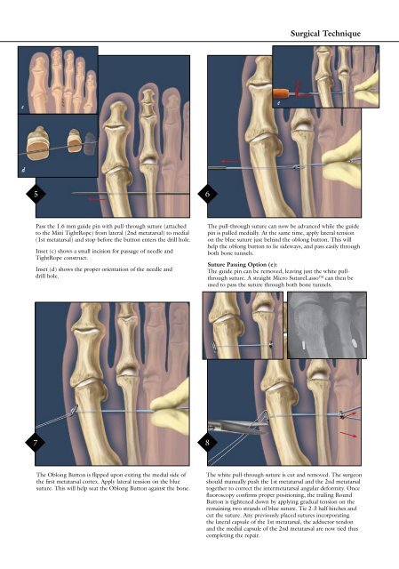
Pass the 1.6 mm guide pin with pull-through suture (attached<br />
to the <strong>Mini</strong> TightRope) from lateral (2nd metatarsal) to medial<br />
(1st metatarsal) <strong>and</strong> stop before the button enters the drill hole.<br />
Inset (c) shows a small incision for passage of needle <strong>and</strong><br />
TightRope construct.<br />
Inset (d) shows the proper orientation of the needle <strong>and</strong><br />
drill hole.<br />
7 8<br />
The Oblong Button is flipped upon exiting the medial side of<br />
the first metatarsal cortex. Apply lateral tension on the blue<br />
suture. This will help seat the Oblong Button against the bone.<br />
e<br />
Surgical Technique<br />
The pull-through suture can now be advanced while the guide<br />
pin is pulled medially. At the same time, apply lateral tension<br />
on the blue suture just behind the oblong button. This will<br />
help the oblong button to lie sideways, <strong>and</strong> pass easily through<br />
both bone tunnels.<br />
Suture Passing Option (e):<br />
The guide pin can be removed, leaving just the white pull-<br />
through suture. A straight Micro SutureLasso can then be<br />
used to pass the suture through both bone tunnels.<br />
The white pull-through suture is cut <strong>and</strong> removed. The surgeon<br />
should manually push the 1st metatarsal <strong>and</strong> the 2nd metatarsal<br />
together to correct the intermetatarsal angular deformity. Once<br />
fluoroscopy confirms proper positioning, the trailing Round<br />
Button is tightened down by applying gradual tension on the<br />
remaining two str<strong>and</strong>s of blue suture. Tie 2-3 half hitches <strong>and</strong><br />
cut the suture. Any previously placed sutures incorporating<br />
the lateral capsule of the 1st metatarsal, the adductor tendon<br />
<strong>and</strong> the medial capsule of the 2nd metatarsal are now tied thus<br />
completing the repair.