The surgical anatomy of the aortic root - Multimedia Manual Cardio ...
The surgical anatomy of the aortic root - Multimedia Manual Cardio ...
The surgical anatomy of the aortic root - Multimedia Manual Cardio ...

You also want an ePaper? Increase the reach of your titles
YUMPU automatically turns print PDFs into web optimized ePapers that Google loves.
2<br />
R.H. Anderson / <strong>Multimedia</strong> <strong>Manual</strong> <strong>of</strong> <strong>Cardio</strong>thoracic Surgery / doi:10.1510/mmcts.2006.002527<br />
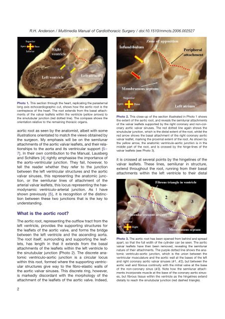
Photo 1. This section through <strong>the</strong> heart, replicating <strong>the</strong> parasternal<br />
long axis echocardiographic cut, shows how <strong>the</strong> <strong>aortic</strong> <strong>root</strong> is <strong>the</strong><br />
centrepiece <strong>of</strong> <strong>the</strong> heart. <strong>The</strong> <strong>root</strong> extends from <strong>the</strong> basal attachments<br />
<strong>of</strong> <strong>the</strong> valvar leaflets within <strong>the</strong> ventricle (yellow arrows) to<br />
<strong>the</strong> sinutubular junction (red dotted line). <strong>The</strong> compass shows <strong>the</strong><br />
orientation relative to <strong>the</strong> remaining thoracic organs.<br />
<strong>aortic</strong> <strong>root</strong> as seen by <strong>the</strong> anatomist, albeit with some<br />
illustrations orientated to match <strong>the</strong> views obtained by<br />
<strong>the</strong> surgeon. My emphasis will be on <strong>the</strong> semilunar<br />
attachments <strong>of</strong> <strong>the</strong> <strong>aortic</strong> valvar leaflets, and <strong>the</strong>ir relationships<br />
to <strong>the</strong> aorta and its ventricular support w5–<br />
7x. In <strong>the</strong>ir own contribution to <strong>the</strong> <strong>Manual</strong>, Lausberg<br />
and Schäfers w4x rightly emphasise <strong>the</strong> importance <strong>of</strong><br />
<strong>the</strong> aorto-ventricular junction. <strong>The</strong>y fail, however, to<br />
tell <strong>the</strong> reader whe<strong>the</strong>r <strong>the</strong>y refer to <strong>the</strong> junction<br />
between <strong>the</strong> left ventricular structures and <strong>the</strong> <strong>aortic</strong><br />
valvar sinuses, this representing <strong>the</strong> anatomic junction,<br />
or <strong>the</strong> semilunar lines <strong>of</strong> attachment <strong>of</strong> <strong>the</strong><br />
arterial valvar leaflets, this locus representing <strong>the</strong> haemodynamic<br />
ventriculo-arterial junction. As I have<br />
shown previously w5x, it is recognition <strong>of</strong> <strong>the</strong> distinction<br />
between <strong>the</strong>se two junctions that is <strong>the</strong> key to<br />
understanding.<br />
What is <strong>the</strong> <strong>aortic</strong> <strong>root</strong>?<br />
<strong>The</strong> <strong>aortic</strong> <strong>root</strong>, representing <strong>the</strong> outflow tract from <strong>the</strong><br />
left ventricle, provides <strong>the</strong> supporting structures for<br />
<strong>the</strong> leaflets <strong>of</strong> <strong>the</strong> <strong>aortic</strong> valve, and forms <strong>the</strong> bridge<br />
between <strong>the</strong> left ventricle and <strong>the</strong> ascending aorta.<br />
<strong>The</strong> <strong>root</strong> itself, surrounding and supporting <strong>the</strong> leaflets,<br />
has length in that it extends from <strong>the</strong> basal<br />
attachments <strong>of</strong> <strong>the</strong> leaflets within <strong>the</strong> left ventricle to<br />
<strong>the</strong> sinutubular junction (Photo 2). <strong>The</strong> discrete anatomic<br />
ventriculo-<strong>aortic</strong> junction is a circular locus<br />
within this <strong>root</strong>, formed where <strong>the</strong> supporting ventricular<br />
structures give way to <strong>the</strong> fibro-elastic walls <strong>of</strong><br />
<strong>the</strong> <strong>aortic</strong> valvar sinuses. This discrete ring, however,<br />
is markedly discordant with <strong>the</strong> morphology <strong>of</strong> <strong>the</strong><br />
attachment <strong>of</strong> <strong>the</strong> leaflets <strong>of</strong> <strong>the</strong> <strong>aortic</strong> valve. Indeed,<br />
Photo 2. This close-up <strong>of</strong> <strong>the</strong> section illustrated in Photo 1 shows<br />
<strong>the</strong> extent <strong>of</strong> <strong>the</strong> <strong>aortic</strong> <strong>root</strong>, and reveals <strong>the</strong> semilunar attachments<br />
<strong>of</strong> <strong>the</strong> valvar leaflets supported by <strong>the</strong> right coronary and non-coronary<br />
<strong>aortic</strong> valvar sinuses. <strong>The</strong> red dotted line again shows <strong>the</strong><br />
sinutubular junction, which is <strong>the</strong> distal extent <strong>of</strong> <strong>the</strong> <strong>root</strong>, whilst <strong>the</strong><br />
red arrow shows <strong>the</strong> basal attachment <strong>of</strong> <strong>the</strong> right coronary <strong>aortic</strong><br />
valvar leaflet, marking <strong>the</strong> proximal extent <strong>of</strong> <strong>the</strong> <strong>root</strong>. As shown by<br />
<strong>the</strong> yellow arrow, <strong>the</strong> anatomic ventriculo-<strong>aortic</strong> junction is in <strong>the</strong><br />
middle part <strong>of</strong> <strong>the</strong> <strong>root</strong>, and is crossed by <strong>the</strong> hinge-lines <strong>of</strong> <strong>the</strong><br />
valvar leaflets (see Photo 3).<br />
it is crossed at several points by <strong>the</strong> hingelines <strong>of</strong> <strong>the</strong><br />
valvar leaflets. <strong>The</strong>se lines, semilunar in structure,<br />
extend throughout <strong>the</strong> <strong>root</strong>, running from <strong>the</strong>ir basal<br />
attachments within <strong>the</strong> left ventricle to <strong>the</strong>ir distal<br />
Photo 3. <strong>The</strong> <strong>aortic</strong> <strong>root</strong> has been opened from behind and spread<br />
apart, so that <strong>the</strong> full width <strong>of</strong> <strong>the</strong> cylinder can be seen. <strong>The</strong> <strong>aortic</strong><br />
valvar leaflets have <strong>the</strong>n been removed, revealing <strong>the</strong> semilunar<br />
nature <strong>of</strong> <strong>the</strong>ir attachments. <strong>The</strong> purple dotted line shows <strong>the</strong> anatomic<br />
ventriculo-<strong>aortic</strong> junction, which is <strong>the</strong> union between <strong>the</strong><br />
ventricular musculature and <strong>the</strong> <strong>aortic</strong> wall at <strong>the</strong> bases <strong>of</strong> <strong>the</strong> left<br />
and right coronary <strong>aortic</strong> valvar sinuses (1, 2), but between <strong>the</strong><br />
<strong>aortic</strong> wall and fibrous continuity with <strong>the</strong> mitral valve at <strong>the</strong> base<br />
<strong>of</strong> <strong>the</strong> non-coronary sinus (3). Note how <strong>the</strong> semilunar attachments<br />
incorporate muscle at <strong>the</strong> base <strong>of</strong> <strong>the</strong> coronary <strong>aortic</strong> sinuses,<br />
but fibrous tissue within <strong>the</strong> ventricle as <strong>the</strong> hingelines extend<br />
distally to reach <strong>the</strong> sinutubular junction (red dashed triangle).