The surgical anatomy of the aortic root - Multimedia Manual Cardio ...
The surgical anatomy of the aortic root - Multimedia Manual Cardio ...
The surgical anatomy of the aortic root - Multimedia Manual Cardio ...
You also want an ePaper? Increase the reach of your titles
YUMPU automatically turns print PDFs into web optimized ePapers that Google loves.
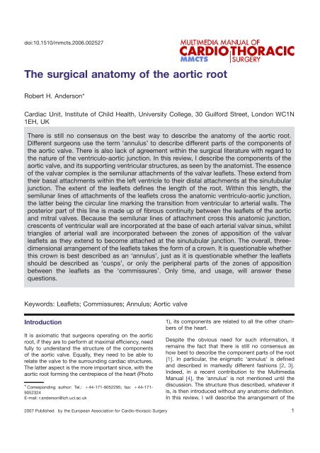
doi:10.1510/mmcts.2006.002527<br />
<strong>The</strong> <strong>surgical</strong> <strong>anatomy</strong> <strong>of</strong> <strong>the</strong> <strong>aortic</strong> <strong>root</strong><br />
Robert H. Anderson*<br />
Cardiac Unit, Institute <strong>of</strong> Child Health, University College, 30 Guilford Street, London WC1N<br />
1EH, UK<br />
<strong>The</strong>re is still no consensus on <strong>the</strong> best way to describe <strong>the</strong> <strong>anatomy</strong> <strong>of</strong> <strong>the</strong> <strong>aortic</strong> <strong>root</strong>.<br />
Different surgeons use <strong>the</strong> term ‘annulus’ to describe different parts <strong>of</strong> <strong>the</strong> components <strong>of</strong><br />
<strong>the</strong> <strong>aortic</strong> valve. <strong>The</strong>re is also lack <strong>of</strong> agreement within <strong>the</strong> <strong>surgical</strong> literature with regard to<br />
<strong>the</strong> nature <strong>of</strong> <strong>the</strong> ventriculo-<strong>aortic</strong> junction. In this review, I describe <strong>the</strong> components <strong>of</strong> <strong>the</strong><br />
<strong>aortic</strong> valve, and its supporting ventricular structures, as seen by <strong>the</strong> anatomist. <strong>The</strong> essence<br />
<strong>of</strong> <strong>the</strong> valvar complex is <strong>the</strong> semilunar attachments <strong>of</strong> <strong>the</strong> valvar leaflets. <strong>The</strong>se extend from<br />
<strong>the</strong>ir basal attachments within <strong>the</strong> left ventricle to <strong>the</strong>ir distal attachments at <strong>the</strong> sinutubular<br />
junction. <strong>The</strong> extent <strong>of</strong> <strong>the</strong> leaflets defines <strong>the</strong> length <strong>of</strong> <strong>the</strong> <strong>root</strong>. Within this length, <strong>the</strong><br />
semilunar lines <strong>of</strong> attachments <strong>of</strong> <strong>the</strong> leaflets cross <strong>the</strong> anatomic ventriculo-<strong>aortic</strong> junction,<br />
<strong>the</strong> latter being <strong>the</strong> circular line marking <strong>the</strong> transition from ventricular to arterial walls. <strong>The</strong><br />
posterior part <strong>of</strong> this line is made up <strong>of</strong> fibrous continuity between <strong>the</strong> leaflets <strong>of</strong> <strong>the</strong> <strong>aortic</strong><br />
and mitral valves. Because <strong>the</strong> semilunar lines <strong>of</strong> attachment cross this anatomic junction,<br />
crescents <strong>of</strong> ventricular wall are incorporated at <strong>the</strong> base <strong>of</strong> each arterial valvar sinus, whilst<br />
triangles <strong>of</strong> arterial wall are incorporated between <strong>the</strong> zones <strong>of</strong> apposition <strong>of</strong> <strong>the</strong> valvar<br />
leaflets as <strong>the</strong>y extend to become attached at <strong>the</strong> sinutubular junction. <strong>The</strong> overall, threedimensional<br />
arrangement <strong>of</strong> <strong>the</strong> leaflets takes <strong>the</strong> form <strong>of</strong> a crown. It is questionable whe<strong>the</strong>r<br />
this crown is best described as an ‘annulus’, just as it is questionable whe<strong>the</strong>r <strong>the</strong> leaflets<br />
should be described as ‘cusps’, or only <strong>the</strong> peripheral parts <strong>of</strong> <strong>the</strong> zones <strong>of</strong> apposition<br />
between <strong>the</strong> leaflets as <strong>the</strong> ‘commissures’. Only time, and usage, will answer <strong>the</strong>se<br />
questions.<br />
Keywords: Leaflets; Commissures; Annulus; Aortic valve<br />
Introduction<br />
It is axiomatic that surgeons operating on <strong>the</strong> <strong>aortic</strong><br />
<strong>root</strong>, if <strong>the</strong>y are to perform at maximal efficiency, need<br />
fully to understand <strong>the</strong> structure <strong>of</strong> <strong>the</strong> components<br />
<strong>of</strong> <strong>the</strong> <strong>aortic</strong> valve. Equally, <strong>the</strong>y need to be able to<br />
relate <strong>the</strong> valve to <strong>the</strong> surrounding cardiac structures.<br />
<strong>The</strong> latter aspect is <strong>the</strong> more important since, with <strong>the</strong><br />
<strong>aortic</strong> <strong>root</strong> forming <strong>the</strong> centrepiece <strong>of</strong> <strong>the</strong> heart (Photo<br />
* Corresponding author: Tel.: q44-171-9052295; fax: q44-171-<br />
9052324<br />
E-mail: r.anderson@ich.ucl.ac.uk<br />
2007 Published by <strong>the</strong> European Association for <strong>Cardio</strong>-thoracic Surgery<br />
1), its components are related to all <strong>the</strong> o<strong>the</strong>r chambers<br />
<strong>of</strong> <strong>the</strong> heart.<br />
Despite <strong>the</strong> obvious need for such information, it<br />
remains <strong>the</strong> fact that <strong>the</strong>re is still no consensus as<br />
how best to describe <strong>the</strong> component parts <strong>of</strong> <strong>the</strong> <strong>root</strong><br />
w1x. In particular, <strong>the</strong> enigmatic ‘annulus’ is defined<br />
and described in markedly different fashions w2, 3x.<br />
Indeed, in a recent contribution to <strong>the</strong> <strong>Multimedia</strong><br />
<strong>Manual</strong> w4x, <strong>the</strong> ‘annulus’ is not mentioned until <strong>the</strong><br />
discussion. <strong>The</strong> structure thus described, whatever it<br />
is, is <strong>the</strong>n introduced without any anatomic definition.<br />
In this review, I will describe <strong>the</strong> arrangement <strong>of</strong> <strong>the</strong><br />
1
2<br />
R.H. Anderson / <strong>Multimedia</strong> <strong>Manual</strong> <strong>of</strong> <strong>Cardio</strong>thoracic Surgery / doi:10.1510/mmcts.2006.002527<br />
Photo 1. This section through <strong>the</strong> heart, replicating <strong>the</strong> parasternal<br />
long axis echocardiographic cut, shows how <strong>the</strong> <strong>aortic</strong> <strong>root</strong> is <strong>the</strong><br />
centrepiece <strong>of</strong> <strong>the</strong> heart. <strong>The</strong> <strong>root</strong> extends from <strong>the</strong> basal attachments<br />
<strong>of</strong> <strong>the</strong> valvar leaflets within <strong>the</strong> ventricle (yellow arrows) to<br />
<strong>the</strong> sinutubular junction (red dotted line). <strong>The</strong> compass shows <strong>the</strong><br />
orientation relative to <strong>the</strong> remaining thoracic organs.<br />
<strong>aortic</strong> <strong>root</strong> as seen by <strong>the</strong> anatomist, albeit with some<br />
illustrations orientated to match <strong>the</strong> views obtained by<br />
<strong>the</strong> surgeon. My emphasis will be on <strong>the</strong> semilunar<br />
attachments <strong>of</strong> <strong>the</strong> <strong>aortic</strong> valvar leaflets, and <strong>the</strong>ir relationships<br />
to <strong>the</strong> aorta and its ventricular support w5–<br />
7x. In <strong>the</strong>ir own contribution to <strong>the</strong> <strong>Manual</strong>, Lausberg<br />
and Schäfers w4x rightly emphasise <strong>the</strong> importance <strong>of</strong><br />
<strong>the</strong> aorto-ventricular junction. <strong>The</strong>y fail, however, to<br />
tell <strong>the</strong> reader whe<strong>the</strong>r <strong>the</strong>y refer to <strong>the</strong> junction<br />
between <strong>the</strong> left ventricular structures and <strong>the</strong> <strong>aortic</strong><br />
valvar sinuses, this representing <strong>the</strong> anatomic junction,<br />
or <strong>the</strong> semilunar lines <strong>of</strong> attachment <strong>of</strong> <strong>the</strong><br />
arterial valvar leaflets, this locus representing <strong>the</strong> haemodynamic<br />
ventriculo-arterial junction. As I have<br />
shown previously w5x, it is recognition <strong>of</strong> <strong>the</strong> distinction<br />
between <strong>the</strong>se two junctions that is <strong>the</strong> key to<br />
understanding.<br />
What is <strong>the</strong> <strong>aortic</strong> <strong>root</strong>?<br />
<strong>The</strong> <strong>aortic</strong> <strong>root</strong>, representing <strong>the</strong> outflow tract from <strong>the</strong><br />
left ventricle, provides <strong>the</strong> supporting structures for<br />
<strong>the</strong> leaflets <strong>of</strong> <strong>the</strong> <strong>aortic</strong> valve, and forms <strong>the</strong> bridge<br />
between <strong>the</strong> left ventricle and <strong>the</strong> ascending aorta.<br />
<strong>The</strong> <strong>root</strong> itself, surrounding and supporting <strong>the</strong> leaflets,<br />
has length in that it extends from <strong>the</strong> basal<br />
attachments <strong>of</strong> <strong>the</strong> leaflets within <strong>the</strong> left ventricle to<br />
<strong>the</strong> sinutubular junction (Photo 2). <strong>The</strong> discrete anatomic<br />
ventriculo-<strong>aortic</strong> junction is a circular locus<br />
within this <strong>root</strong>, formed where <strong>the</strong> supporting ventricular<br />
structures give way to <strong>the</strong> fibro-elastic walls <strong>of</strong><br />
<strong>the</strong> <strong>aortic</strong> valvar sinuses. This discrete ring, however,<br />
is markedly discordant with <strong>the</strong> morphology <strong>of</strong> <strong>the</strong><br />
attachment <strong>of</strong> <strong>the</strong> leaflets <strong>of</strong> <strong>the</strong> <strong>aortic</strong> valve. Indeed,<br />
Photo 2. This close-up <strong>of</strong> <strong>the</strong> section illustrated in Photo 1 shows<br />
<strong>the</strong> extent <strong>of</strong> <strong>the</strong> <strong>aortic</strong> <strong>root</strong>, and reveals <strong>the</strong> semilunar attachments<br />
<strong>of</strong> <strong>the</strong> valvar leaflets supported by <strong>the</strong> right coronary and non-coronary<br />
<strong>aortic</strong> valvar sinuses. <strong>The</strong> red dotted line again shows <strong>the</strong><br />
sinutubular junction, which is <strong>the</strong> distal extent <strong>of</strong> <strong>the</strong> <strong>root</strong>, whilst <strong>the</strong><br />
red arrow shows <strong>the</strong> basal attachment <strong>of</strong> <strong>the</strong> right coronary <strong>aortic</strong><br />
valvar leaflet, marking <strong>the</strong> proximal extent <strong>of</strong> <strong>the</strong> <strong>root</strong>. As shown by<br />
<strong>the</strong> yellow arrow, <strong>the</strong> anatomic ventriculo-<strong>aortic</strong> junction is in <strong>the</strong><br />
middle part <strong>of</strong> <strong>the</strong> <strong>root</strong>, and is crossed by <strong>the</strong> hinge-lines <strong>of</strong> <strong>the</strong><br />
valvar leaflets (see Photo 3).<br />
it is crossed at several points by <strong>the</strong> hingelines <strong>of</strong> <strong>the</strong><br />
valvar leaflets. <strong>The</strong>se lines, semilunar in structure,<br />
extend throughout <strong>the</strong> <strong>root</strong>, running from <strong>the</strong>ir basal<br />
attachments within <strong>the</strong> left ventricle to <strong>the</strong>ir distal<br />
Photo 3. <strong>The</strong> <strong>aortic</strong> <strong>root</strong> has been opened from behind and spread<br />
apart, so that <strong>the</strong> full width <strong>of</strong> <strong>the</strong> cylinder can be seen. <strong>The</strong> <strong>aortic</strong><br />
valvar leaflets have <strong>the</strong>n been removed, revealing <strong>the</strong> semilunar<br />
nature <strong>of</strong> <strong>the</strong>ir attachments. <strong>The</strong> purple dotted line shows <strong>the</strong> anatomic<br />
ventriculo-<strong>aortic</strong> junction, which is <strong>the</strong> union between <strong>the</strong><br />
ventricular musculature and <strong>the</strong> <strong>aortic</strong> wall at <strong>the</strong> bases <strong>of</strong> <strong>the</strong> left<br />
and right coronary <strong>aortic</strong> valvar sinuses (1, 2), but between <strong>the</strong><br />
<strong>aortic</strong> wall and fibrous continuity with <strong>the</strong> mitral valve at <strong>the</strong> base<br />
<strong>of</strong> <strong>the</strong> non-coronary sinus (3). Note how <strong>the</strong> semilunar attachments<br />
incorporate muscle at <strong>the</strong> base <strong>of</strong> <strong>the</strong> coronary <strong>aortic</strong> sinuses,<br />
but fibrous tissue within <strong>the</strong> ventricle as <strong>the</strong> hingelines extend<br />
distally to reach <strong>the</strong> sinutubular junction (red dashed triangle).
R.H. Anderson / <strong>Multimedia</strong> <strong>Manual</strong> <strong>of</strong> <strong>Cardio</strong>thoracic Surgery / doi:10.1510/mmcts.2006.002527<br />
Schematic 1. <strong>The</strong> cartoon shows a bisected <strong>aortic</strong> <strong>root</strong>, and illustrates<br />
how <strong>the</strong> semilunar attachment <strong>of</strong> <strong>the</strong> valvar leaflets incorporates<br />
<strong>aortic</strong> wall in <strong>the</strong> intersinusal triangles, and ventricular tissues<br />
at <strong>the</strong> base <strong>of</strong> each <strong>of</strong> <strong>the</strong> coronary <strong>aortic</strong> sinuses.<br />
attachments at <strong>the</strong> sinutubular junction (Photo 3). <strong>The</strong><br />
<strong>root</strong> as thus defined, <strong>the</strong>refore, is a cylinder, its walls<br />
being made up <strong>of</strong> <strong>the</strong> <strong>aortic</strong> valvar sinuses along with<br />
<strong>the</strong> interdigitating intersinusal fibrous triangles, and<br />
with two small crescents <strong>of</strong> ventricular muscle incorporated<br />
at its proximal end (Schematic 1).<br />
It is <strong>the</strong> semilunar attachments <strong>of</strong> <strong>the</strong> leaflets within<br />
<strong>the</strong> valvar sinuses that form <strong>the</strong> haemodynamic junction<br />
between <strong>the</strong> left ventricle and <strong>the</strong> aorta. All structures<br />
on <strong>the</strong> distal side <strong>of</strong> <strong>the</strong>se attachments are<br />
subject to arterial pressures, whereas all parts proximal<br />
to <strong>the</strong> attachments are subjected to ventricular<br />
pressures. In functional terms, all three sinuses <strong>of</strong> <strong>the</strong><br />
<strong>root</strong>, and <strong>the</strong>ir contained leaflets, are identical. Anatomically,<br />
however, it is necessary to distinguish<br />
between <strong>the</strong> three components. This is best achieved<br />
by noting that two <strong>of</strong> <strong>the</strong> valvar sinuses give rise to<br />
<strong>the</strong> coronary arteries (Photo 4). <strong>The</strong>se can be nominated<br />
as <strong>the</strong> right and left coronary <strong>aortic</strong> sinuses.<br />
<strong>The</strong>se two sinuses, for <strong>the</strong>ir greater part, are made up<br />
<strong>of</strong> <strong>the</strong> <strong>aortic</strong> wall. But, because <strong>the</strong> semilunar attachments<br />
<strong>of</strong> <strong>the</strong> leaflets cross <strong>the</strong> anatomic ventriculo<strong>aortic</strong><br />
junction, a crescent <strong>of</strong> ventricular musculature<br />
is incorporated at <strong>the</strong> base <strong>of</strong> each <strong>of</strong> <strong>the</strong>se two sinuses<br />
(Photo 3, Schematic 1). <strong>The</strong> third sinus does not<br />
give rise to a coronary artery, and hence, can be designated<br />
as <strong>the</strong> non-coronary <strong>aortic</strong> sinus. <strong>The</strong>re is no<br />
muscular crescent at <strong>the</strong> base <strong>of</strong> this sinus, since this<br />
sinus has exclusively fibrous walls, <strong>the</strong> basal part<br />
beneath <strong>the</strong> anatomic ventriculo-<strong>aortic</strong> junction being<br />
part <strong>of</strong> <strong>the</strong> important continuity between <strong>the</strong> leaflets<br />
<strong>of</strong> <strong>the</strong> <strong>aortic</strong> and mitral valves that is a feature <strong>of</strong> <strong>the</strong><br />
outflow tract <strong>of</strong> <strong>the</strong> left ventricle (Photo 5).<br />
<strong>The</strong> areas between <strong>the</strong> basal attachments <strong>of</strong> <strong>the</strong> <strong>aortic</strong><br />
sinuses within <strong>the</strong> ventricle itself, which extend distally<br />
to <strong>the</strong> level <strong>of</strong> <strong>the</strong> sinutubular junction, are<br />
triangular extensions <strong>of</strong> <strong>the</strong> left ventricular outflow<br />
tract. <strong>The</strong>y are thinned fibrous areas <strong>of</strong> <strong>the</strong> <strong>aortic</strong> wall.<br />
Photo 4. <strong>The</strong> heart has been dissected by removing <strong>the</strong> atrial chambers<br />
and <strong>the</strong> arterial trunks, and is photographed from above, looking<br />
down on <strong>the</strong> atrioventricular and ventriculo-arterial junctions. It<br />
is orientated as it may be seen by <strong>the</strong> surgeon. <strong>The</strong> dissection<br />
shows how two <strong>of</strong> <strong>the</strong> <strong>aortic</strong> valvar sinuses (1, 2) give rise to<br />
coronary arteries, and can be nominated as <strong>the</strong> left and right coronary<br />
<strong>aortic</strong> sinuses, respectively. <strong>The</strong> third sinus (3) does not give<br />
rise to a coronary artery, and hence, is <strong>the</strong> non-coronary <strong>aortic</strong><br />
sinus. Note again that <strong>the</strong> <strong>aortic</strong> valve forms <strong>the</strong> cardiac<br />
centrepiece.<br />
Photo 5. <strong>The</strong> <strong>aortic</strong> <strong>root</strong> has been opened from <strong>the</strong> front, and <strong>the</strong><br />
leaflets <strong>of</strong> <strong>the</strong> <strong>aortic</strong> valve removed. <strong>The</strong> dissection shows how <strong>the</strong><br />
interleaflet triangle between <strong>the</strong> non-coronary and left coronary <strong>aortic</strong><br />
sinuses (purple dashed line) is part <strong>of</strong> <strong>the</strong> area <strong>of</strong> fibrous continuity<br />
with <strong>the</strong> <strong>aortic</strong> leaflet <strong>of</strong> <strong>the</strong> mitral valve. <strong>The</strong> red dotted line<br />
marks <strong>the</strong> anatomic ventriculo-<strong>aortic</strong> junction.<br />
3
4<br />
R.H. Anderson / <strong>Multimedia</strong> <strong>Manual</strong> <strong>of</strong> <strong>Cardio</strong>thoracic Surgery / doi:10.1510/mmcts.2006.002527<br />
Photo 6. <strong>The</strong> heart is viewed from <strong>the</strong> right and behind, having<br />
pulled apart <strong>the</strong> aorta and <strong>the</strong> anterior atrial walls to show <strong>the</strong> transverse<br />
pericardial sinus. <strong>The</strong> interleaflet triangle between <strong>the</strong> nonand<br />
right coronary <strong>aortic</strong> sinuses has also been removed, showing<br />
how it communicates with <strong>the</strong> transverse sinus.<br />
Removing <strong>the</strong>se triangular extensions puts <strong>the</strong> most<br />
distal parts <strong>of</strong> <strong>the</strong> left ventricle in direct communication<br />
ei<strong>the</strong>r with <strong>the</strong> pericardial space or, in <strong>the</strong> case <strong>of</strong><br />
<strong>the</strong> triangle between <strong>the</strong> two coronary <strong>aortic</strong> valvar<br />
sinuses, with <strong>the</strong> fibroadipose plane <strong>of</strong> tissue between<br />
<strong>the</strong> back <strong>of</strong> <strong>the</strong> subpulmonary infundibulum and <strong>the</strong><br />
front <strong>of</strong> <strong>the</strong> aorta (Photos 6–8). As shown in Photo 5,<br />
<strong>the</strong> triangle between <strong>the</strong> left coronary and <strong>the</strong> noncoronary<br />
<strong>aortic</strong> valvar sinuses is part <strong>of</strong> <strong>the</strong> extensive<br />
curtain <strong>of</strong> <strong>aortic</strong>-to-mitral valvar fibrous continuity.<br />
This triangle, when removed, creates a window to <strong>the</strong><br />
transverse pericardial sinus, <strong>the</strong> latter being <strong>the</strong> space<br />
between <strong>the</strong> back <strong>of</strong> <strong>the</strong> <strong>aortic</strong> <strong>root</strong> and <strong>the</strong> anterior<br />
atrial walls (Photo 6).<br />
<strong>The</strong> triangle between <strong>the</strong> non-coronary and <strong>the</strong> right<br />
coronary <strong>aortic</strong> valvar sinuses is directly continuous<br />
with <strong>the</strong> membranous part <strong>of</strong> <strong>the</strong> ventricular septum.<br />
<strong>The</strong> basal part <strong>of</strong> this fibrous wall is crossed on its<br />
right side by <strong>the</strong> hinge <strong>of</strong> <strong>the</strong> tricuspid valve, dividing<br />
<strong>the</strong> membranous septum itself into atrioventricular<br />
and interventricular components. <strong>The</strong> apical part <strong>of</strong><br />
Photo 7. <strong>The</strong> heart has been opened through <strong>the</strong> right atrium and<br />
ventricle, and is viewed from <strong>the</strong> right side. <strong>The</strong> fibrous triangle<br />
between <strong>the</strong> non-coronary and right coronary <strong>aortic</strong> sinuses has<br />
been removed, showing how it abuts on <strong>the</strong> area <strong>of</strong> <strong>the</strong> membranous<br />
septum, but extends distally so as to be in potential communication<br />
with <strong>the</strong> pericardial cavity above <strong>the</strong> supraventricular<br />
crest <strong>of</strong> <strong>the</strong> right ventricle (yellow asterisks).<br />
<strong>the</strong> triangle extends to <strong>the</strong> sinutubular junction.<br />
Removal <strong>of</strong> this part creates a window between <strong>the</strong><br />
left ventricular outflow tract and <strong>the</strong> right side <strong>of</strong> <strong>the</strong><br />
transverse pericardial sinus, opening externally above<br />
<strong>the</strong> attachment <strong>of</strong> <strong>the</strong> supraventricular crest <strong>of</strong> <strong>the</strong><br />
right ventricle (Photo 7).<br />
<strong>The</strong> third triangle, which separates <strong>the</strong> two coronary<br />
<strong>aortic</strong> valvar sinuses, is <strong>the</strong> least extensive <strong>of</strong> <strong>the</strong><br />
three. To show <strong>the</strong> location <strong>of</strong> this triangle, it is first<br />
necessary to remove <strong>the</strong> free-standing muscular subpulmonary<br />
infundibulum. Once this has been done,<br />
<strong>the</strong>n it can be seen that removal <strong>of</strong> <strong>the</strong> triangle itself<br />
creates a window between <strong>the</strong> sub<strong>aortic</strong> outflow tract<br />
and <strong>the</strong> plane <strong>of</strong> tissue which separates <strong>the</strong> <strong>aortic</strong> <strong>root</strong><br />
from <strong>the</strong> infundibulum (Photo 8).<br />
<strong>The</strong> semilunar attachment <strong>of</strong> <strong>the</strong> valvar leaflets, <strong>the</strong>refore,<br />
divides <strong>the</strong> <strong>aortic</strong> <strong>root</strong> into supravalvar and sub-
R.H. Anderson / <strong>Multimedia</strong> <strong>Manual</strong> <strong>of</strong> <strong>Cardio</strong>thoracic Surgery / doi:10.1510/mmcts.2006.002527<br />
Photo 8. This dissection has been made first by removing <strong>the</strong> freestanding<br />
subpulmonary infundibulum, and <strong>the</strong>n by removing <strong>the</strong> triangle<br />
between <strong>the</strong> two coronary <strong>aortic</strong> sinuses. As can be seen,<br />
<strong>the</strong> tip <strong>of</strong> <strong>the</strong> triangle ‘points’ to <strong>the</strong> tissue plane between <strong>the</strong> back<br />
<strong>of</strong> <strong>the</strong> infundibulum and <strong>the</strong> <strong>aortic</strong> <strong>root</strong>.<br />
valvar components. <strong>The</strong> supravalvar components, <strong>the</strong><br />
<strong>aortic</strong> sinuses, are primarily <strong>aortic</strong> in structure, but<br />
contain structures <strong>of</strong> ventricular origin at <strong>the</strong>ir base.<br />
<strong>The</strong> supporting subvalvar parts are primarily ventricular,<br />
but extend as thin-walled fibrous triangles to <strong>the</strong><br />
level <strong>of</strong> <strong>the</strong> sinutubular junction. <strong>The</strong> sinutubular junction<br />
itself forms <strong>the</strong> discrete distal boundary <strong>of</strong> <strong>the</strong><br />
<strong>root</strong>. <strong>The</strong> valvar leaflets are attached peripherally at<br />
this level, and hence, <strong>the</strong> junction is an integral part<br />
<strong>of</strong> <strong>the</strong> valvar mechanism. Any significant dilation at <strong>the</strong><br />
level <strong>of</strong> <strong>the</strong> sinutubular junction will produce valvar<br />
incompetence. It is moot, <strong>the</strong>refore, whe<strong>the</strong>r stenosis<br />
at this level should be labelled as ‘supra<strong>aortic</strong>’, since<br />
<strong>the</strong> sinutubular junction is just as crucial a component<br />
<strong>of</strong> <strong>the</strong> overall valvar mechanism as are <strong>the</strong> leaflets and<br />
<strong>the</strong>ir supporting sinuses w8x. Anatomically, <strong>the</strong> sinutubular<br />
junction is no more than <strong>the</strong> distal extent <strong>of</strong><br />
<strong>the</strong> overall valvar complex.<br />
Is <strong>the</strong>re a valvar annulus?<br />
<strong>The</strong> answer to this question, <strong>the</strong> major ongoing<br />
conundrum for <strong>the</strong> cardiac surgeon, depends very<br />
much on <strong>the</strong> structure nominated to represent <strong>the</strong><br />
‘annulus’. If we take refuge in <strong>the</strong> dictionary, and seek<br />
etymological origins, <strong>the</strong>n we find that an annulus is<br />
no more than a little ring. In this regard, it is certainly<br />
<strong>the</strong> case that <strong>the</strong> entirety <strong>of</strong> <strong>the</strong> <strong>aortic</strong> <strong>root</strong> can be<br />
removed from <strong>the</strong> heart, and can be slipped on <strong>the</strong><br />
finger in <strong>the</strong> form <strong>of</strong> a ring. As far as I am aware,<br />
however, no surgeon defines <strong>the</strong> entirety <strong>of</strong> <strong>the</strong> <strong>root</strong><br />
as <strong>the</strong> <strong>aortic</strong> valvar annulus. Most surgeons seem to<br />
nominate <strong>the</strong> remnants <strong>of</strong> <strong>the</strong> removed valvar leaflets<br />
as <strong>the</strong>ir annulus w3, 9x. As I have described, however,<br />
by virtue <strong>of</strong> <strong>the</strong>ir semilunar position, <strong>the</strong>se structures<br />
are supported in crown-like fashion when viewed in<br />
<strong>the</strong> three-dimensional context <strong>of</strong> <strong>the</strong> overall <strong>aortic</strong> <strong>root</strong><br />
(Schematic 2). O<strong>the</strong>r surgeons, in contrast, define <strong>the</strong><br />
virtual basal ring constructed by joining toge<strong>the</strong>r <strong>the</strong><br />
most proximal parts <strong>of</strong> each leaflet as <strong>the</strong> ‘annulus’<br />
w2x. It is certainly this diameter that is typically analysed<br />
by <strong>the</strong> echocardiographer when providing<br />
measurements <strong>of</strong> <strong>the</strong> diameter <strong>of</strong> <strong>the</strong> purported structure<br />
(Schematic 3).<br />
In view <strong>of</strong> <strong>the</strong>se ongoing discrepancies, it is my own<br />
belief that <strong>the</strong> <strong>aortic</strong> <strong>root</strong> would be best understood if<br />
divorced from <strong>the</strong> concept <strong>of</strong> <strong>the</strong> ‘annulus’. This is<br />
unlikely to happen. We need to understand, <strong>the</strong>refore,<br />
that <strong>the</strong> <strong>aortic</strong> <strong>root</strong> itself is cylindrical, with <strong>the</strong> valvar<br />
leaflets supported within <strong>the</strong> <strong>root</strong> in crown-like, ra<strong>the</strong>r<br />
than circular, fashion (Schematic 2). We should also<br />
take note that <strong>the</strong>re can be marked differences in<br />
diameter <strong>of</strong> its component parts, not only in <strong>the</strong> nor-<br />
Schematic 2. <strong>The</strong> cartoon shows an idealised <strong>aortic</strong> <strong>root</strong>. <strong>The</strong><br />
attachments <strong>of</strong> <strong>the</strong> valvar leaflets, shown in red, extend through <strong>the</strong><br />
entire length <strong>of</strong> <strong>the</strong> <strong>root</strong>, from <strong>the</strong> sinutubular junction, in blue, to<br />
<strong>the</strong> virtual basal ring, shown in green, and produced by joining<br />
toge<strong>the</strong>r <strong>the</strong> basal attachments <strong>of</strong> <strong>the</strong> leaflets. <strong>The</strong> crown-like<br />
attachments <strong>of</strong> <strong>the</strong> leaflets cross <strong>the</strong> anatomic ventriculo-<strong>aortic</strong><br />
junction, shown in yellow.<br />
5
6<br />
R.H. Anderson / <strong>Multimedia</strong> <strong>Manual</strong> <strong>of</strong> <strong>Cardio</strong>thoracic Surgery / doi:10.1510/mmcts.2006.002527<br />
Schematic 3. <strong>The</strong> cartoon shows how measurement <strong>of</strong> <strong>the</strong> basal<br />
ring provides information relating only to <strong>the</strong> entrance <strong>of</strong> <strong>the</strong> <strong>aortic</strong><br />
<strong>root</strong>. To provide full details, measurements should be taken also <strong>of</strong><br />
<strong>the</strong> diameter <strong>of</strong> <strong>the</strong> sinutubular junction, and at mid-sinusal level.<br />
None <strong>of</strong> <strong>the</strong>se measurements take account <strong>of</strong> <strong>the</strong> diameter at <strong>the</strong><br />
anatomic ventriculo-<strong>aortic</strong> junction.<br />
mal patient, but particularly in <strong>the</strong> setting <strong>of</strong> disease<br />
w10, 11x.<br />
<strong>The</strong> relationships <strong>of</strong> <strong>the</strong> <strong>aortic</strong> <strong>root</strong><br />
When viewed in attitudinally correct orientation w12,<br />
13x, <strong>the</strong> <strong>aortic</strong> <strong>root</strong> is positioned to <strong>the</strong> right and posterior<br />
relative to <strong>the</strong> subpulmonary infundibulum (Photo<br />
9).<br />
<strong>The</strong> subpulmonary infundibulum itself is a complete<br />
muscular funnel, supporting in uniform fashion <strong>the</strong><br />
leaflets <strong>of</strong> <strong>the</strong> pulmonary valve. <strong>The</strong> leaflets <strong>of</strong> <strong>the</strong> aor-<br />
Photo 9. <strong>The</strong> cavities <strong>of</strong> <strong>the</strong> heart have been cast in blue for <strong>the</strong><br />
right side, and red for <strong>the</strong> left side. As can be seen, when positioned<br />
in attitudinally appropriate fashion, <strong>the</strong> <strong>aortic</strong> <strong>root</strong> is posterior and<br />
to <strong>the</strong> right <strong>of</strong> <strong>the</strong> pulmonary valve.<br />
tic valve, in contrast, are attached only in part to <strong>the</strong><br />
muscular walls <strong>of</strong> <strong>the</strong> left ventricle, since so as to fit<br />
<strong>the</strong> orifices <strong>of</strong> both <strong>aortic</strong> and mitral valves within <strong>the</strong><br />
circular pr<strong>of</strong>ile <strong>of</strong> <strong>the</strong> left ventricle, <strong>the</strong>re is no muscle<br />
between <strong>the</strong>m in <strong>the</strong> ventricular ro<strong>of</strong>. <strong>The</strong> <strong>aortic</strong> <strong>root</strong>,<br />
fur<strong>the</strong>rmore, is wedged between <strong>the</strong> orifices <strong>of</strong> <strong>the</strong><br />
two atrioventricular valves (Photo 4). As already discussed,<br />
<strong>the</strong> <strong>root</strong> is related to all four cardiac chambers.<br />
<strong>The</strong>se relationships can be well recognised in<br />
<strong>the</strong> clinical situation. <strong>The</strong> proximity <strong>of</strong> <strong>the</strong> <strong>root</strong> to <strong>the</strong><br />
anterior interatrial groove is now appreciated by those<br />
who have inserted devices via ca<strong>the</strong>ters to close<br />
defects <strong>of</strong> <strong>the</strong> oval fossa, only to find <strong>the</strong> arms <strong>of</strong> <strong>the</strong><br />
devices eroding into <strong>the</strong> aorta. <strong>The</strong> relationship to <strong>the</strong><br />
subpulmonary infundibulum is well demonstrated by<br />
<strong>the</strong> spread <strong>of</strong> bacterial infection from <strong>the</strong> valve, or by<br />
aneurysmal dilation <strong>of</strong> <strong>the</strong> right coronary <strong>aortic</strong> sinus<br />
<strong>of</strong> Valsalva. <strong>The</strong> most important <strong>surgical</strong> relationship,<br />
none<strong>the</strong>less, is probably to <strong>the</strong> atrioventricular node<br />
and <strong>the</strong> penetrating atrioventricular bundle. <strong>The</strong> node,<br />
located in <strong>the</strong> wall <strong>of</strong> <strong>the</strong> right atrium at <strong>the</strong> apex <strong>of</strong><br />
<strong>the</strong> triangle <strong>of</strong> Koch, is relatively distant from <strong>the</strong> <strong>root</strong>.<br />
As <strong>the</strong> conduction axis penetrates through <strong>the</strong> central<br />
fibrous body, however, it is positioned at <strong>the</strong> base <strong>of</strong><br />
<strong>the</strong> interleaflet triangle between <strong>the</strong> non- and right<br />
coronary <strong>aortic</strong> sinuses (Photo 9). Having penetrated<br />
through <strong>the</strong> fibrous plane providing atrioventricular<br />
insulation, <strong>the</strong> bundle <strong>the</strong>n branches on <strong>the</strong> crest <strong>of</strong><br />
<strong>the</strong> muscular ventricular septum, <strong>the</strong> left bundle<br />
branch fanning out on <strong>the</strong> smooth left ventricular side,<br />
whilst <strong>the</strong> cord-like right bundle branch penetrates<br />
back through <strong>the</strong> muscular septum, emerging on <strong>the</strong><br />
septal surface in <strong>the</strong> environs <strong>of</strong> <strong>the</strong> medial papillary<br />
muscle. In this position, <strong>the</strong>refore, <strong>the</strong> muscular axis<br />
responsible for atrioventricular conduction should be<br />
relatively distant from most <strong>surgical</strong> manoeuvres carried<br />
out to replace or repair <strong>the</strong> <strong>aortic</strong> valve and its<br />
supporting structures (Schematic 4).<br />
Clinical implications<br />
<strong>The</strong>re are several inferences from <strong>the</strong> complex interplay<br />
<strong>of</strong> ventricular and arterial structures which make<br />
up <strong>the</strong> <strong>aortic</strong> <strong>root</strong> that are important in <strong>the</strong> clinical context.<br />
I have already emphasised that, when seen in<br />
long axis section, <strong>the</strong> diameter <strong>of</strong> <strong>the</strong> <strong>root</strong> varies<br />
markedly through its short length. <strong>The</strong> <strong>root</strong> is much<br />
wider at <strong>the</strong> midpoint <strong>of</strong> <strong>the</strong> sinuses than at ei<strong>the</strong>r <strong>the</strong><br />
sinutubular junction or at <strong>the</strong> basal attachment <strong>of</strong> <strong>the</strong><br />
leaflets, whilst <strong>the</strong> basal diameter can be up to onefifth<br />
wider than <strong>the</strong> outlet at <strong>the</strong> sinutubular junction<br />
w10, 11x. This becomes <strong>of</strong> significance when considering<br />
measurements <strong>of</strong> <strong>the</strong> ‘annulus’. As already discussed,<br />
as I understand <strong>the</strong> situation, most surgeons
R.H. Anderson / <strong>Multimedia</strong> <strong>Manual</strong> <strong>of</strong> <strong>Cardio</strong>thoracic Surgery / doi:10.1510/mmcts.2006.002527<br />
Schematic 4. <strong>The</strong> cartoon shows <strong>the</strong> location <strong>of</strong> <strong>the</strong> atrioventricular<br />
conduction axis as it would be seen by <strong>the</strong> surgeon looking down<br />
through <strong>the</strong> <strong>aortic</strong> <strong>root</strong>.<br />
consider <strong>the</strong> crown-like hinges <strong>of</strong> <strong>the</strong> leaflets to represent<br />
this structure, and <strong>the</strong>se extend through all<br />
three levels <strong>of</strong> <strong>the</strong> <strong>root</strong>. Proper values can only be<br />
provided when measurements are made at <strong>the</strong> bottom<br />
<strong>of</strong> <strong>the</strong> valvar attachments, at <strong>the</strong> widest point <strong>of</strong> <strong>the</strong><br />
sinuses, and also at <strong>the</strong> sinutubular junction (Schematic<br />
3). When considering this feature in <strong>the</strong> context<br />
<strong>of</strong> cardiac surgery, it is obviously necessary to remove<br />
<strong>the</strong> native valvar leaflets along <strong>the</strong>ir semilunar attachments<br />
during <strong>the</strong> process <strong>of</strong> valvar replacement.<br />
Some pros<strong>the</strong>ses <strong>the</strong>n used for <strong>the</strong> purposes <strong>of</strong><br />
replacement have a truly circular sewing ring. Should<br />
<strong>the</strong> stitches used for securing this ring be placed within<br />
<strong>the</strong> semilunar remnants <strong>of</strong> <strong>the</strong> removed valvar leaflets,<br />
<strong>the</strong>n <strong>the</strong>re will be some distortion when <strong>the</strong> valve<br />
is ‘seated’, albeit that this does not usually compromise<br />
its subsequent function. When autopsied hearts<br />
are examined subsequent to valvar replacement, <strong>the</strong><br />
circular sewing ring is usually found to be located at<br />
<strong>the</strong> anatomic ventriculo-arterial junction w6x. Itisan<br />
appreciation <strong>of</strong> <strong>the</strong> normal discrepancy between this<br />
junction and <strong>the</strong> haemodynamic junction which is <strong>the</strong><br />
key to understanding <strong>the</strong> clinical <strong>anatomy</strong> <strong>of</strong> <strong>the</strong> <strong>aortic</strong><br />
<strong>root</strong>. Whe<strong>the</strong>r it is appropriate to describe <strong>the</strong> semilunar<br />
attachments as <strong>the</strong> valvar ‘annulus’ <strong>the</strong>n<br />
depends very much on philosophies concerning communication<br />
and <strong>the</strong> usage <strong>of</strong> words.<br />
Words and how we use <strong>the</strong>m<br />
For better or worse, it is now an inescapable fact that<br />
American English has become <strong>the</strong> ‘lingua franca’ <strong>of</strong><br />
<strong>the</strong> scientific world. It is surprising, <strong>the</strong>refore, that in<br />
<strong>the</strong> field <strong>of</strong> cardiac surgery we employ so many words<br />
in a fashion that is foreign to <strong>the</strong>ir vernacular use.<br />
Consider <strong>the</strong> word ‘cusp’. If we consult any dictionary,<br />
we find that this means a point, or an elevation. This<br />
is how <strong>the</strong> word is used appropriately in <strong>anatomy</strong> to<br />
describe <strong>the</strong> surfaces <strong>of</strong> <strong>the</strong> molar teeth. Is it appropriate,<br />
however, to use this word to describe <strong>the</strong> components<br />
<strong>of</strong> <strong>the</strong> skirts <strong>of</strong> tissue that guard <strong>the</strong><br />
atrioventricular and ventriculo-arterial junctions? In<br />
my opinion, <strong>the</strong>se structures, serving <strong>the</strong> same function<br />
at both junctions, are best described as leaflets.<br />
<strong>The</strong>n consider <strong>the</strong> current use <strong>of</strong> ‘commissure’. When<br />
defined literally, this is <strong>the</strong> zone <strong>of</strong> apposition between<br />
adjacent anatomic structures, and is used in this fashion<br />
to describe <strong>the</strong> junctions <strong>of</strong> bones in <strong>the</strong> skull, or<br />
<strong>the</strong> lines <strong>of</strong> opening and closing <strong>of</strong> <strong>the</strong> eyes and<br />
mouth. When used in <strong>the</strong> setting <strong>of</strong> <strong>the</strong> cardiac valves,<br />
<strong>the</strong>refore, <strong>the</strong> ‘commissure’ should account for <strong>the</strong><br />
entirety <strong>of</strong> <strong>the</strong> zones <strong>of</strong> apposition between <strong>the</strong> valvar<br />
leaflets. Currently, we use <strong>the</strong> word to describe only<br />
<strong>the</strong> peripheral ends <strong>of</strong> <strong>the</strong>se zones <strong>of</strong> apposition.<br />
What, <strong>the</strong>n, <strong>of</strong> <strong>the</strong> ‘annulus’? As already discussed,<br />
this is no more than a little ring. Is <strong>the</strong> crown-like configuration<br />
<strong>of</strong> <strong>the</strong> semilunar valves and <strong>the</strong>ir supporting<br />
sinuses best described as an annulus? Only time will<br />
tell.<br />
Acknowledgements<br />
Many <strong>of</strong> <strong>the</strong> illustrations used for this review are modified<br />
from those appearing in ‘Surgical Anatomy <strong>of</strong> <strong>the</strong><br />
Heart’, published by Cambridge University Press. We<br />
thank <strong>the</strong> co-authors <strong>of</strong> this book, Drs Benson Wilcox<br />
and Andrew Cook, for permission to modify <strong>the</strong> images<br />
for <strong>the</strong> purposes <strong>of</strong> our current work.<br />
References<br />
w1x Antunes MJ. <strong>The</strong> <strong>aortic</strong> valve: an everlasting<br />
mystery to surgeons. Eur J <strong>Cardio</strong>thorac Surg<br />
2005;28:855–856.<br />
w2x Thubrikar MJ, Labrosse MR, Zehr KJ, Robicsek F,<br />
Gong GG, Fowler BL. Aortic <strong>root</strong> dilatation may<br />
alter <strong>the</strong> dimensions <strong>of</strong> <strong>the</strong> valve leaflets. Eur J<br />
<strong>Cardio</strong>thorac Surg 2005;28:850–855.<br />
w3x Pretre R, Kadner A, Dave H, Bettex D, Genoni M.<br />
Tricuspidisation <strong>of</strong> <strong>the</strong> <strong>aortic</strong> valve with creation <strong>of</strong><br />
a crown-like annulus is able to restore a normal<br />
valve function in bicuspid <strong>aortic</strong> valves. Eur J<br />
<strong>Cardio</strong>thorac Surg 2006;29:1001–1006.<br />
w4x Lausberg HF, Schafers H-J. Valve sparing <strong>aortic</strong><br />
replacement – <strong>root</strong> remodeling. Multimed Man<br />
<strong>Cardio</strong>thorac Surg doi:10.1510/mmcts.2006.<br />
001982.<br />
7
8<br />
R.H. Anderson / <strong>Multimedia</strong> <strong>Manual</strong> <strong>of</strong> <strong>Cardio</strong>thoracic Surgery / doi:10.1510/mmcts.2006.002527<br />
w5x Anderson RH. Editorial note: <strong>The</strong> <strong>anatomy</strong> <strong>of</strong><br />
arterial valvar stenosis. Int J <strong>Cardio</strong>l 1990;26:355–<br />
360.<br />
w6x Sutton JP III, Ho SY, Anderson RH. <strong>The</strong> forgotten<br />
interleaflet triangles: a review <strong>of</strong> <strong>the</strong> <strong>surgical</strong><br />
<strong>anatomy</strong> <strong>of</strong> <strong>the</strong> <strong>aortic</strong> valve. Ann Thorac Surg<br />
1995;59:419–427.<br />
w7x Anderson RH. Clinical <strong>anatomy</strong> <strong>of</strong> <strong>the</strong> <strong>aortic</strong> <strong>root</strong>.<br />
Heart 2000;84:670–673.<br />
w8x Stamm C, Li J, Ho SY, Redington AN, Anderson<br />
RH. <strong>The</strong> <strong>aortic</strong> <strong>root</strong> in supravalvular <strong>aortic</strong><br />
stenosis: <strong>the</strong> potential <strong>surgical</strong> relevance <strong>of</strong><br />
morphologic findings. J Thorac <strong>Cardio</strong>vasc Surg<br />
1997;114:16–24.<br />
w9x Yacoub MH, Kilner PJ, Birks EJ, Misfeld M. <strong>The</strong><br />
<strong>aortic</strong> outflow and <strong>root</strong>: a tale <strong>of</strong> dynamism and<br />
crosstalk. Ann Thorac Surg 1999;68:(3 Suppl)<br />
S37–43.<br />
w10x Reid K. <strong>The</strong> <strong>anatomy</strong> <strong>of</strong> <strong>the</strong> sinus <strong>of</strong> Valsalva.<br />
Thorax 1970;25:79–85.<br />
w11x Kunzelman KS, Grande J, David TE, Cochran RP,<br />
Verrier ED. Aortic <strong>root</strong> and valve relationships:<br />
impact on <strong>surgical</strong> repair. J Thorac <strong>Cardio</strong>vasc<br />
Surg 1994;107:162–170.<br />
w12x McAlpine WA. Heart and coronary arteries. An<br />
anatomical atlas for clinical diagnosis, radiological<br />
investigation, and <strong>surgical</strong> treatment. Berlin:<br />
Springer-Verlag 1995.<br />
w13x Cook AC, Anderson RH. Editorial. Attitudinally<br />
correct nomenclature. Heart 2002;87:503–506.


