Preliminary experience with the tangoRS: polyaxial, percutaneous ...
Preliminary experience with the tangoRS: polyaxial, percutaneous ...
Preliminary experience with the tangoRS: polyaxial, percutaneous ...

You also want an ePaper? Increase the reach of your titles
YUMPU automatically turns print PDFs into web optimized ePapers that Google loves.
<strong>Preliminary</strong> <strong>experience</strong> <strong>with</strong> <strong>the</strong> <strong>tangoRS</strong>: <strong>polyaxial</strong>, <strong>percutaneous</strong>, cement<br />
augmenting pedicle screw system.<br />
John C Sutcliffe, FRCS *, Farzam Vahzifehdan, MD **, Marcus Richter, MD**<br />
* The London Spine Clinic, 119 Harley Street London, England<br />
** Spine Center, St. Josefs-Hospital, Solmsstrasse 15, 65189 Wiesbaden, Germany<br />
Abstract<br />
Multiple pedicle screw systems have evolved over <strong>the</strong> last three decades. All have<br />
certain attractions and all have drawbacks. The tendency to move more to<br />
<strong>percutaneous</strong> placement and minimal access surgery has led to a new generation of<br />
systems. A series of cases, compromising <strong>the</strong> initial <strong>experience</strong> <strong>with</strong> <strong>the</strong> <strong>tangoRS</strong><br />
system [ulrich, Ulm, Germany] is presented and <strong>the</strong> advantages over <strong>the</strong> existing<br />
systems are discussed.<br />
Introduction<br />
There are innumerable pedicle screw systems available to <strong>the</strong> spine surgeon, choices<br />
usually being made on historical and loyalty grounds. The mere fact that <strong>the</strong>re are so<br />
many demonstrates that no one system contains all <strong>the</strong> benefits and none of <strong>the</strong><br />
drawbacks. Every spinal surgeon will have a view on <strong>the</strong> “best” system, but may not<br />
have had <strong>experience</strong> of all <strong>the</strong> available options. The <strong>tangoRS</strong> system has been<br />
designed by surgeons and engineers toge<strong>the</strong>r to combine all <strong>the</strong> necessary features of<br />
a pedicle screw system, <strong>with</strong> ergonomic instrumentation and a user-friendly system<br />
for <strong>the</strong> <strong>the</strong>atre nursing staff.<br />
Materials and methods<br />
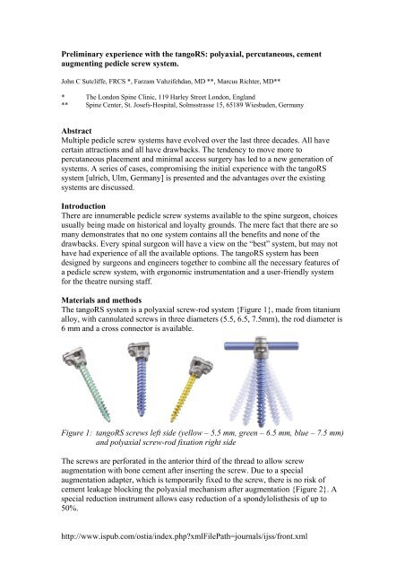
The <strong>tangoRS</strong> system is a <strong>polyaxial</strong> screw-rod system {Figure 1}, made from titanium<br />
alloy, <strong>with</strong> cannulated screws in three diameters (5.5, 6.5, 7.5mm), <strong>the</strong> rod diameter is<br />
6 mm and a cross connector is available.<br />
Figure 1: <strong>tangoRS</strong> screws left side (yellow – 5.5 mm, green – 6.5 mm, blue – 7.5 mm)<br />
and <strong>polyaxial</strong> screw-rod fixation right side<br />
The screws are perforated in <strong>the</strong> anterior third of <strong>the</strong> thread to allow screw<br />
augmentation <strong>with</strong> bone cement after inserting <strong>the</strong> screw. Due to a special<br />
augmentation adapter, which is temporarily fixed to <strong>the</strong> screw, <strong>the</strong>re is no risk of<br />
cement leakage blocking <strong>the</strong> <strong>polyaxial</strong> mechanism after augmentation {Figure 2}. A<br />
special reduction instrument allows easy reduction of a spondylolis<strong>the</strong>sis of up to<br />
50%.<br />
http://www.ispub.com/ostia/index.php?xmlFilePath=journals/ijss/front.xml
Figure 2. Cement augmentation through <strong>the</strong> implanted pedicle screws <strong>with</strong><br />
fenestrations in <strong>the</strong> anterior third of <strong>the</strong> screws, left side sawbone model in<br />
<strong>the</strong> overview and detail, right side intraoperative<br />
The three spine surgeons operated upon a consecutive series of 34 patients requiring<br />
pedicle screw fixation, over a three-month period. Patients ranged in age from 23 to<br />
83 years, 17 were male. The indications for fusion were:<br />
Diagnosis Number of patients<br />
degenerative instability and stenosis 15<br />
degenerative scoliosis 3<br />
degenerative disc disease 3<br />
spondylolis<strong>the</strong>sis 5<br />
failed back surgery syndrome 4<br />
pathological # (metastasis) 2<br />
spondylodiscitis 2<br />
http://www.ispub.com/ostia/index.php?xmlFilePath=journals/ijss/front.xml
Figure 3. X-rays of a<br />
four level procedure<br />
augmented <strong>with</strong> TLIF<br />
PEEK-cages<br />
The <strong>tangoRS</strong> instrumentation was utilised, including guide-wire <strong>percutaneous</strong><br />
placement, when indicated. Cement augmentation, utilising <strong>the</strong> multiple fenestrations<br />
near <strong>the</strong> tip of <strong>the</strong> screws, was utilised in 8 patients, to reinforce osteoporotic vertebral<br />
bodies. 24 patients underwent single level fusion, 5 two level, 3 three level and in two<br />
a 4 level procedure {Figure 3} was performed. The postero-lateral fusion was<br />
augmented by a TLIF procedure in 23, PLIF in 2, ALIF in 2 and vertebral body<br />
replacement in 2.<br />
Results<br />
At follow up, <strong>the</strong>e were no signs of screw loosening. Fusion was augmented <strong>with</strong> rh-<br />
BMP-2 [Medtronic, Minneapolis, USA] in 4 cases. There were no neurological<br />
complications, no CSF leaks and no instrument failures. To date <strong>the</strong>re have been no<br />
cases of infection or wound breakdown. Mean operating time was 150 minutes (40 –<br />
240); mean blood loss was 220 ml (50 – 400).<br />
Discussion<br />
As <strong>with</strong> most developments in spinal surgery, <strong>the</strong>re has been a huge proliferation of<br />
designs of instrumentation, for pedicle screw fixation of <strong>the</strong> lumbar spine. Initial<br />
designs from <strong>the</strong> 1970’s {5} were simple systems allowing solid fixation between<br />
adjacent vertebrae. To facilitate reduction of spondylolis<strong>the</strong>sis and fracture,<br />
modifications were made {6}, leading to a widening of <strong>the</strong> indications, but also an<br />
increase in <strong>the</strong> complexity of <strong>the</strong> systems and trays. To facilitate fixation <strong>with</strong> less<br />
need for rod bending, <strong>polyaxial</strong> screw systems were developed {3} in <strong>the</strong> early<br />
1990’s, allowing easier rod placement, but bringing <strong>with</strong> <strong>the</strong>m a new tier of concerns<br />
http://www.ispub.com/ostia/index.php?xmlFilePath=journals/ijss/front.xml
for <strong>the</strong> operating surgeon: would <strong>the</strong> fixation take <strong>the</strong> forces involved and would <strong>the</strong><br />
stability be maintained over time? {1} In <strong>the</strong> 2000’s, minimally invasive spinal<br />
surgery began to receive mainstream attention and <strong>the</strong> ability to place pedicle screws<br />
safely, through smaller and smaller incisions became <strong>the</strong> goal {2}. A wide variety of<br />
systems and applications followed. During this time, <strong>the</strong> design of <strong>the</strong> screw itself<br />
went through a renaissance. Screws which once resembles woodscrews now had selftapping<br />
threads, self-drilling threads, cylindrical threads on conical shafts and tapered<br />
threads, widening at <strong>the</strong> base of <strong>the</strong> screw to conform better to <strong>the</strong> anatomy of <strong>the</strong><br />
pedicle and to afford a more secure fit.<br />
Cement vertebral augmentation has been used for many years, in cases of osteoporotic<br />
bone, by inserting cement into <strong>the</strong> drilled or tapped pedicle and <strong>the</strong>n quickly inserting<br />
<strong>the</strong> screw to allow <strong>the</strong> cement to harden around it, ra<strong>the</strong>r like a rawlplug. This is<br />
known to have <strong>the</strong> effect of increasing <strong>the</strong> pullout strength of <strong>the</strong> screw {4}.<br />
The <strong>tangoRS</strong> system has been designed jointly by engineers and surgeons, to allow<br />
this single system all <strong>the</strong> advantages a spinal surgeon would expect from a pedicle<br />
screw system and yet to keep it simple. The system is ergonomic, <strong>with</strong> padded<br />
contoured handles and has an appropriate range of <strong>the</strong> necessary screws, rods and<br />
connectors. The screws have a cylindrical thread on a conical shaft to give highest<br />
stability in <strong>with</strong>in <strong>the</strong> pedicle, have thickened threads in <strong>the</strong> intra-pedicular region to<br />
increase <strong>the</strong> surface area in contact <strong>with</strong> <strong>the</strong> bone. The screws are cannulated to allow<br />
passage over a guide wire, a vital teaching aid and a useful adjunct for <strong>percutaneous</strong><br />
placement, as well as allowing injection of cement into <strong>the</strong> vertebral body after screw<br />
placement. This allows maximal screw-to-bone contact and yet gives <strong>the</strong> surgeon <strong>the</strong><br />
confidence that a potentially weak osteoporotic bone has been adequately reinforced,<br />
reducing <strong>the</strong> risk of collapse or screw loosening.<br />
References<br />
1 Shepard MF, Davies MR, Abayan A, Kabo JM, Wang JC. Effects of <strong>polyaxial</strong><br />
pedicle screws on lumbar construct rigidity. J Spinal Disord Tech. 2002<br />
Jun;15(3):233-6.<br />
2 Foley KT, Gupta SK. Percutaneous pedicle screw fixation of <strong>the</strong> lumbar spine:<br />
preliminary clinical results. J Neurosurg Spine. 2002 Jul;97(1):7-12.<br />
3 Fogel GR, Reitman CA, Liu W, Esses SI. Physical characteristics of<br />
<strong>polyaxial</strong>-headed pedicle screws and biomechanical comparison of load <strong>with</strong><br />
<strong>the</strong>ir failure. Spine. 2003 Mar 1;28(5):470-3.<br />
4 Zindrick MR, Wiltse LL, Widell EH, Thomas JC, Holland WR, Field BT,<br />
Spencer CW. A biomechanical study of intrapeduncular screw fixation in <strong>the</strong><br />
lumbosacral spine. Clin Orthop. 1986 Feb;(203):99-112.<br />
5 Steffee AD, Biscup RS, Sitkowski DJ. Segmental spine plates <strong>with</strong> pedicle<br />
screw fixation. A new internal fixation device for disorders of <strong>the</strong> lumbar and<br />
thoracolumbar spine. Clin Orthop. 1986 Feb;(203):45-53.<br />
6 Guyer DW, Wiltse LL, Peek RD. The Wiltse pedicle screw fixation system.<br />
Orthopedics. 1988 Oct;11(10):1455-60.<br />
http://www.ispub.com/ostia/index.php?xmlFilePath=journals/ijss/front.xml
Corresponding Author:<br />
Marcus Richter, MD, PhD<br />
Head of <strong>the</strong> Spine Center<br />
St. Josefs-Hospital<br />
Solmsstrasse 15<br />
D 65189 Wiesbaden<br />
Germany<br />
E-mail: mrichter@joho.de<br />
Internet: www.joho.de<br />
Phone: +49-611-177-3701<br />
Fax: +49-611-177-3702<br />
http://www.ispub.com/ostia/index.php?xmlFilePath=journals/ijss/front.xml