Marrow stimulation techniques
Marrow stimulation techniques
Marrow stimulation techniques

Create successful ePaper yourself
Turn your PDF publications into a flip-book with our unique Google optimized e-Paper software.
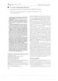
Injury, Int. J. Care Injured (2008) 39S1, S26–S31<br />
www.elsevier.com/locate/injury<br />
<strong>Marrow</strong> <strong>stimulation</strong> <strong>techniques</strong><br />
MR Steinwachs 1 , Th Guggi 1 , PC Kreuz 2<br />
1 Schulthess Clinic, Dept. of Orthobiologics & Cartilage Repair, Zürich, Switzerland<br />
2 University Hospital Freiburg, Dept. of Orthopaedic and Trauma Surgery, Freiburg, Germany<br />
KEYWORDS:<br />
Cartilage, cartilage<br />
repair, marrow <strong>stimulation</strong>,<br />
microfracture,<br />
autologous chondrocyte<br />
transplantation,<br />
autologous matrix<br />
associated chondroneogenesis,<br />
AMIC,<br />
stem cells, Chondro-<br />
Gide ® , collagen<br />
membrane<br />
Summary 1 Due to the very low intrinsic activity of human adult cartilage, healing<br />
of chondral and osteochondral defects in patients cannot be expected. In<br />
treating symptomatic cartilage damage, marrow <strong>stimulation</strong> methods belong to<br />
the most frequently used methods, along with autologous chondrocyte transplantation<br />
(ACT) and mosaicplasty. These arthroscopic procedures are generally easy<br />
and the marrow <strong>stimulation</strong> treatment costs relatively little. In recent years,<br />
Pridie drilling has been increasingly replaced by the microfracture technique.<br />
This modification relies on the same biological principles of promoting resurfacing<br />
with the formation of fibro-cartilaginous repair tissue. For the treatment<br />
of smaller cartilage defects (< 2.5 cm²), microfracture still remains the first<br />
choice for treatment. The clinical results after microfracture in the knee are age<br />
dependent. Younger and active patients (< 40 years) with smaller isolated traumatic<br />
lesions on the femoral condyles have the best long-term results. The deterioration<br />
of the clinical results begins after 18 months and is significantly more<br />
pronounced in older patients with defects on the patella-femoral joint and tibia.<br />
The inferior quality of the repair tissue, partially incomplete defect filling and<br />
new bone formation in the defect area seem to be limitations of these methods.<br />
The AMIC ® (autologous matrix induced chondrogenesis) technique was developed<br />
to enable treatment of larger defects by the application of a collagen Type III/I<br />
membrane (Geistlich Pharma, Wolhusen, Switzerland), in particular when cellengaged<br />
procedures such as ACT cannot be used for financial reasons or because<br />
it is not indicated. AMIC ® seems to be particularly suitable for treating damaged<br />
retropatellar cartilage, which is an advantage because these defects can be hard<br />
to treat with standard microfracturing alone. The results of the ongoing studies<br />
are awaited to establish whether better results with this technology are achievable<br />
in the long term.<br />
Introduction<br />
The use of magnetic resonance tomography has<br />
clearly simplified the diagnosis of joint cartilage<br />
damage. With the help of cartilage-specific sequences,<br />
eg, flash 3-D, a good representation of<br />
1 Abstracts in German, French, Italian, Spanish, Japanese,<br />
and Russian are printed at the end of this supplement.<br />
the joint surfaces and the subchondral bone plate<br />
is possible [2, 5, 27]. Treatment of symptomatic<br />
cartilage lesions must be discussed for each patient’s<br />
individual situation. The response of the<br />
organism to damage of the joint cartilage depends<br />
on the patient’s age as well as on the type and size<br />
of the defect. Adults show very low potential for<br />
regeneration because the resident differentiated<br />
chondrocytes have no mitotic activity. The matrix<br />
encapsulated chondrocytes are not able to initiate<br />
0020–1383/$—see front matter © 2008 Published by Elsevier Ltd.<br />
doi:10.1016/j.injury.2008.01.042
<strong>Marrow</strong> <strong>stimulation</strong> <strong>techniques</strong><br />
S27<br />
an effective repair process. Additionally, the cartilage<br />
tissue is apparently unable to recruit local<br />
sources of progenitor cells at the articular surface<br />
and the synovial lining of the joint cavity [10].<br />
The relationship between decreasing numbers<br />
of mesenchymal stem cells (MSC) and aging is still<br />
unclear [7, 8]. The progression of these defects<br />
to osteoarthritis is proven and well documented<br />
[26, 35, 40]. Due to the very low intrinsic activity,<br />
healing of symptomatic full thickness chondral and<br />
osteochondral defects in adult patients cannot be<br />
expected [26]. The treatment of such defects is only<br />
successful if the concomitant injuries are treated as<br />
well [6, 12, 39].<br />
In treating symptomatic cartilage damage, marrow<br />
<strong>stimulation</strong> methods are among the most<br />
frequently used methods, along with ACT and<br />
mosaicplasty. These arthroscopic procedures are<br />
generally easy and the treatment costs relatively<br />
little. In recent years, older <strong>techniques</strong> such as<br />
Pridie drilling [31] or abrasion [18, 19, 20] have<br />
been increasingly replaced by the microfracture<br />
technique [32, 37, 38]. All marrow <strong>stimulation</strong><br />
methods base on the penetration of the subchondral<br />
bone plate at the bottom of the cartilage<br />
defect. Different instruments such as the bent awls<br />
used in microfracturing create persisting holes in<br />
the bone plate. The outflowing bone marrow blood<br />
contains the pluripotent stem cells (hMSC) which<br />
are stabilised by the clot formation in the defect.<br />
The number of these highly proliferative stem cells<br />
during this procedure is very low and the concentration<br />
in the bone marrow is age dependent [7,<br />
8]. The hMSC which are able to differentiate into<br />
fibrochondrocytes, result in fibrocartilage repair<br />
with varying amounts of type I, II and III collagen<br />
[32, 34, 35, 37, 40].<br />
All the different bone marrow <strong>stimulation</strong> <strong>techniques</strong><br />
rely on the same biological principles of<br />
promoting resurfacing with the formation of fibrocartilaginous<br />
repair tissue [Fig. 3] with inferior biomechanical<br />
qualities [1, 18, 19, 20, 30]. The method<br />
can be applied in smaller isolated cartilage defects<br />
(1 3 cm²) in young, active patients. Meanwhile, use<br />
of this technique in joints other than the knee has<br />
been published, ie, in the shoulder, hip and ankle<br />
[3, 9, 36].<br />
The mechanism of the repair tissue formation<br />
using the microfracture technique is based on the<br />
activation of an endogenous stem cell tool [16,<br />
3, 38]. The human mesenchymal stem/progenitor<br />
cells (MSPC) are located in the bone marrow in low<br />
concentration. They are pluripotent and the precursors<br />
for marrow stroma, bone, cartilage, muscle<br />
and connective tissues. The potential of human<br />
mesenchymal stem cells (hMSC) to differentiate<br />
into various types of mesenchymal tissue, such as<br />
chondrocytes, makes them a potential cell source<br />
in cartilage repair. Adult human mesenchymal stem<br />
cells have been derived from a variety of tissues and<br />
have shown the potential to participate in repair<br />
processes in vivo and in vitro. This tissue formation<br />
has been the subject of numerous animal studies<br />
that observed the formation of a fibrous cartilage<br />
with diminished biomechanical and limited longterm<br />
qualities [15, 16, 28, 32, 33, 35]. Clinical studies<br />
in humans showed viable results for the Pridie<br />
drillings and the abrasion arthroplasty [13, 25, 31].<br />
Clinically, clearly superior results were published by<br />
Steadman for microfracturing [37, 38]. The potential<br />
to repair damaged or diseased tissues with an<br />
autologous cell source has resulted in a great deal<br />
of interest in these cells to provide the basis for<br />
strategies in regenerative medicine.<br />
The AMIC ® (autologous matrix induced chondrogenesis)<br />
technique was developed to enable treatment<br />
of larger defects by application of a collagen<br />
Type III/I membrane (Geistlich Pharma, Wolhusen,<br />
Switzerland), particularly when cell-engaged procedures<br />
such as ACT cannot be used for financial<br />
reasons or because they are not indicated [1, 4].<br />
AMIC ® is particularly suitable for treating damaged<br />
retropatellar cartilage and has become a real alternative,<br />
since these defects can be hard to treat with<br />
standard microfracturing alone. Our own first pilot<br />
cases were done in Freiburg in 2000 and showed good<br />
clinical results. A complementary animal study using<br />
a sheep model originated from the Nehrer study<br />
group in Vienna. In this direct comparison of ACT<br />
technology with AMIC ® , the histological superiority<br />
of ACT was demonstrated [11].<br />
Microfracturing technique<br />
The destroyed and unstable cartilage is removed<br />
arthroscopically in a first step, carefully using the<br />
shaver, curette and spoon. In particular, the tidemark<br />
zone should not be disturbed and the cartilage<br />
should be prepared resulting in a well-contained<br />
defect. Microfractures are generated with specially<br />
bent awls (Karl Storz ® , Zimmer ® ) by creating V-<br />
shaped perforation holes with a diameter of 1.5 2<br />
mm at a distance of 3 mm (3 4 holes/cm²) [Fig. 1].<br />
After shutting off the water influx, bone marrow<br />
bleeding from the perforation holes can be checked.<br />
In isolated cases, re-reaming of the perforations may<br />
be necessary [Fig. 2]. Protruding osseous particles<br />
must be removed carefully with the shaver. Insertion<br />
of a drainage tube without suction completes<br />
the procedure.
S28<br />
M Steinwachs et al<br />
AMIC ® -Technique<br />
Using minimal open knee surgery with a standard<br />
small anterior approach, the destroyed and unstable<br />
cartilage is removed using a scalpel, curette and<br />
spoon, until a well-contained defect results. Utilising<br />
microfracture instruments, V-shaped perforation<br />
holes with a diameter of 1.5 2 mm at a distance of 3<br />
mm (3 4 holes/cm²) are created. An imprint of the<br />
defect is taken using an aluminium template. The<br />
Chondro-Gide ® collagen membrane is cut slightly<br />
smaller than this template. Application of ringer salt<br />
solution to the membrane will later increase the size<br />
by approximately 10%. The membrane can be placed<br />
precisely with the rough side to the preserved bone<br />
plate using fibrin glue (Tissucoll; Baxter, Vienna). To<br />
prevent delamination, laying the membrane edges<br />
over the rim of the cartilage should be avoided. A<br />
mixture of commercial fibrin and autologous serum<br />
can also be used according to [4]. If the defect is<br />
too large for gluing or the location of the defect is<br />
critical from a biomechanical point of view, sutures<br />
(6/0 PDS II Ethicon) can be easily used [Fig. 5]. The<br />
stable position of the membrane can be established<br />
by bending and extending the knee five times. The<br />
tourniquet may then be opened if the membrane is<br />
still in place. Insertion of a drainage tube, careful<br />
haemostasis and suturing of the wound complete<br />
the surgery.<br />
Rehabilitation following microfracturing/<br />
AMIC ®<br />
Rehabilitation begins with 24 hours of bed rest and<br />
fixed full-leg extension. Starting on the first postoperative<br />
day, mobilisation of the patient includes<br />
walking with light foot contact for approximately<br />
6 weeks. The range of motion during this time period<br />
Fig 1: Perforation of the subchondral bone plate during a<br />
microfracture procedure.<br />
Fig. 3: Repair tissue one year after microfracture.<br />
Fig 2: Outflow of bone marrow blood after microfracture.<br />
Fig.4: New bone formation in the completely filled defect<br />
on the medial condyle one year after microfracture.
<strong>Marrow</strong> <strong>stimulation</strong> <strong>techniques</strong><br />
S29<br />
is limited as a function of the defect localisation (typically<br />
0/0/90° for the femur condyle/tibia; 0/0/30°,<br />
0/0/60° and 0/0/90° for patella/trochlea, increasing<br />
in 2-week steps, respectively). Physiotherapy three<br />
times a week with isometric muscle activation and<br />
exercises in a closed chain are our standard. Low<br />
molecular weight heparin and lymphatic drainage<br />
are important in the patient’s postoperative management.<br />
6 hours of CPM daily are necessary. This plays<br />
an important role in the resulting quality of the repair<br />
tissue [32, 37]. After the initial 6-week period with<br />
partial weight bearing, the patients increase loading<br />
up to full body weight over a further 2 weeks. As<br />
would be expected, intensive muscle and coordinative<br />
training are required.<br />
Results and discussion<br />
Steadman [38] produced good and very good clinical<br />
results in his microfracture study using the Lysholm,<br />
Tegner, WOMAC and SF-36 scores with a follow up of<br />
7 17 years (mean 11 years). The average defect size<br />
was 2.7 cm² in 72 patients. In this study, only young<br />
patients with smaller (< 4 cm²) traumatic cartilage<br />
damage were treated. Unfortunately, follow up MRIs<br />
and second-look histology were absent in this study<br />
of selected patients and no cartilage sensitive score<br />
such as proposed by the ICRS (Internationally Cartilage<br />
Repair Society) was used.<br />
In the cohort study of Miethöfer [29], clinical and<br />
MRI results were analysed in 52 patients over a period<br />
of up to 48 months. The average defect size was<br />
Fig.5: AMIC ® procedure on the trochlea.<br />
4.8 cm². In contrast to the Steadman study, Miethöfer<br />
used the ICRS Score for the clinical evaluation. The<br />
results showed, that after an initial improvement<br />
up to 18 months, the clinical outcome decreased<br />
between 18 36 months. In addition to the deterioration<br />
of the ICRS score after 18 months, almost half<br />
the patients had incomplete defect filling in the MRI<br />
and 25% showed new bone formation in the defect<br />
[Fig. 4].<br />
In our prospective cohort study [23, 24], with the<br />
currently highest number of patients, we also used<br />
the sensitive ICRS score and the modified Cincinnati<br />
score for patient evaluation. We showed results<br />
similar to Miethöfer [29] in our own study of 85 patients<br />
with a follow up of 36 months. [23, 24]. Both<br />
scores revealed significant improvement 18 months<br />
after microfracturing (P < .0001). During the second<br />
18 months following surgery, there was a significant<br />
deterioration in the ICRS score (P < .0001). In addition,<br />
patients over 40-years-of-age presented with<br />
significantly poorer clinical results after 36 months<br />
than younger patients.<br />
In the evidence level I study by Knutsen [21], there<br />
was no significant deterioration of the clinical scores<br />
seen after 2 years in the direct comparison between<br />
ACT-P and microfracture in treating isolated defects<br />
on the condyle. Without using the ICRS score, clinical<br />
deterioration was not detectable, just as was seen<br />
in the Steadman study. In the ACT group a tendency<br />
for better histological tissue quality was seen in the<br />
histological scoring system compared to the microfracture<br />
group. But the detected differences did not<br />
reach a statistically significant level because of a<br />
too low number of samples with a drop out rate of<br />
20% of the biopsies in the ACT group (32/40). In the<br />
Knutsen study [21] as in the Steadman study [38],<br />
a MRI follow up was not done. Incomplete defect<br />
filling and new bone formation as it was seen in the<br />
MRI Study of Miethöfer [29] and Kreuz [23], was not<br />
detected,<br />
A new evidence level I Study by Saris [34] with<br />
118 patients presented completely different results<br />
from Knutsen. In this study, the histological tissue<br />
quality in the ICRS II Histo Score was shown to be<br />
significantly superior in the ACT group after one year<br />
compared to the microfracture group [34]. Similar to<br />
the Knutsen study, no significant differences in the<br />
clinical outcome were seen during the short follow<br />
up time.<br />
AMIC ® combines microfracturing with the application<br />
of a porcine collagen type-III/I, bi-layer matrix to<br />
host the MSC and to stabilise the blood clot. AMIC ®<br />
as a 1-step procedure enables the reasonable treatment<br />
of larger (> 2 cm²) cartilage defects. Kramer<br />
and co-workers [22] showed that from the unique<br />
attachment of a Chondro-Gide ® collagen membrane
S30<br />
M Steinwachs et al<br />
(Geistlich Pharma, Wolhusen, Switzerland) to the microfractured<br />
bone plate, suitable stem cells (hMSC)<br />
can be cultivated. With this autologous regenerative<br />
approach [22,38], the stem cells (hMSC) available in<br />
the bone marrow are brought to the surface by microfracturing<br />
and so become available for cartilage repair.<br />
The collagen matrix serves as a natural scaffold<br />
for cell binding and should stimulate differentiation<br />
processes [13]. The first clinical results of 32 patients<br />
rating clinical functional improvement, pain reduction<br />
and patient satisfaction (ICRS functional status,<br />
Cincinnati score, Lysholm score, VES) as well as the<br />
demonstrated good defect filling in MRI are promising<br />
[1] [Fig. 6]. The outcome was evaluated with a<br />
follow-up of 6 24 months. The mean defect size was<br />
3.9 cm² (1.0 6.8 cm²). Microfracturing in combination<br />
with a collagen matrix (AMIC ® ) is a minimally<br />
invasive, effective technique for the repair of focal<br />
cartilage defects of the knee joint.<br />
Conclusion<br />
The clinical results after microfracturing in the knee<br />
are age dependent. Younger, active patients (< 40<br />
years) with smaller isolated traumatic lesions on the<br />
femoral condyles have the best long-term results.<br />
The deterioration of the clinical results begins after<br />
18 months and is significantly more pronounced in<br />
older patients with defects on the patella-femoral<br />
joint and tibia. For the treatment of smaller cartilage<br />
defects (< 2.5 cm²), microfracturing is a good<br />
Fig. 6: Complete defect filling one year after AMIC ® procedure<br />
on the patella.<br />
first line procedure because it is a minimal invasive<br />
method which does not interfere with other cartilage<br />
repair <strong>techniques</strong>. The AMIC ® procedure seems to be<br />
a promising, cost effective method with good clinical<br />
results in the short term follow up. This procedure<br />
possibly enables better clinical long-term results<br />
in the treatment of larger cartilage defects of the<br />
patello-femoral joint.<br />
References<br />
1. Anders S, Wiech O, Schaumburger J, et al. Autologous matrix<br />
induced chondrogenesis (AMIC®) for focal chondral defects<br />
of the knee First Result. Abstract EFFORT. 2007 Florence,<br />
Italy, Abstract CD<br />
2. Bachmann G, Heinrichs C, Jürgensen I, et al. Comparison of<br />
different MRT <strong>techniques</strong> in the diagnosis of degenerative<br />
cartilage diseases. In vitro study of 50 joint specimens of the<br />
knee at T1.5.Fortschr Rontgenstr. 1997 166: 429−436<br />
3. Becher C, Thermann H. Results of microfracture in the treatment<br />
of articular cartilage defects of the talus. Foot Ankle<br />
Int. 2005 Aug;26 (8):583−9<br />
4. Behrens P. Matrixgekoppelte Mikrofrakturierung. Arthroskopie.<br />
2005 18:193−197<br />
5. Bohndorf K. Injuries at the articulating surfaces of bone<br />
(chondral, osteochondral, subchondral fractures and osteochondrosis<br />
dissecans ). Eur J Radiol. 1996 22: 22−29<br />
6. Brittberg M. Lindahl A, Nilsson A, et al. Treatment of deep<br />
cartilage defects in the knee with autologous chondrocyte<br />
transplantation. N Engl J Med. 1994 331: 889−895<br />
7. Brittberg M. A critical analysis of cartilage repair. Acta Orthop<br />
Scand. 1997 68: 186−191<br />
8. Caplan AI, Fink DJ, Goto T, et al. Mesenchymal stem cells<br />
and tissue repair. In: Jackson DW, Arnoczky SP, Frank CB,<br />
Woo SL-YY, Simon TM, The anterior cruciate ligament: current<br />
and future concepts. Raven Press, New York. 1993<br />
405−417<br />
9. Crawford K, Philippon MJ, Sekiya JK, et al. Microfracture of<br />
the hip in athletes. Clin Sports Med. 2006 Apr;25(2):327−35<br />
10. Dowthwaite GP, Bishop JC, Redman SN, et al. The surface<br />
of articular cartilage contains a progenitor population cell.<br />
J Cell Sci. 2004 29;117(Pt 6):889−97. Epub 2004 Feb 3<br />
11. Dorotka R, Bindreiter U, Macfelda K, et al. <strong>Marrow</strong> <strong>stimulation</strong><br />
and chondrocyte transplantation using a collagen matrix for<br />
cartilage repair Osteoarthritis Cartilage. 2005 13(8):655−64<br />
12. Erggelet C, Steinwachs M, Reichelt A. Die Behandlung von<br />
Gelenkknorpeldefekten. Dtsch Arztebl. 1998 95: 1397−1382<br />
13. Friedman MJ, Berasi CC, Fox JM, et al. Preliminary results<br />
with abrasion arthroplasty in the osteoarthritis knee. Clin<br />
Orthop. 1984 182: 200−205<br />
14. Fuss M, Ehlers EM. Characteristics of human chondrocytes,<br />
osteoblasts and fibroblasts seeded onto a type I / III collagen<br />
sponge under different culture conditions. A light scanning<br />
and transmission electron microscopy study, Anat Ann. 2000<br />
182(4): 303−310
<strong>Marrow</strong> <strong>stimulation</strong> <strong>techniques</strong><br />
S31<br />
15. Goymann V. Abrasion arthroplasty. Orthopäde. 1999 28(1):<br />
11−18<br />
16. Grande DA, Pitman MI, Peterson L, et al. The repair of experimentally<br />
produced defects in rabbit articular cartilage<br />
by autologous chondrocyte transplantation. J Orthop Res.<br />
1989 7(2): 208−218<br />
17. Johnson LL. Diagnostic and surgical arthroscopy. The knee<br />
and other joints. 3rd ed. C.V.Mosby Co: St.Louis 1986<br />
18. Johnson LL. Characteristics of the immediate postarthroscopic<br />
blood dot formation in the knee joint. Arthroscopy.<br />
1991 7:14−23<br />
19. Johnson LL. Arthroscopic abrasions arthroplasty. In: McGinty<br />
JB(ED) Operative Arthroscopy. Raven Press: New York. 1991<br />
341−360<br />
20. Kim HK, Moran ME, Salter RB. The potential for regeneration<br />
of articular cartilage in defects created by chondral shaving<br />
and subchondral abrasion. An experimental investigation in<br />
rabbits. J Bone Joint Surg Am. 1991 73(9): 1301−15<br />
21. Knutsen G, Engebretsen L, Ludvigsen TC, et al. Autologous<br />
chondrocyte implantation compared with microfracture in<br />
the knee. A randomized trial. J Bone Joint Surg. 2004 86-<br />
A(3):455−464<br />
22. Kramer J, Böhrnsen F, Lindner U, et al. In vivo matrix-guided<br />
human mesenchymal stem cells. Cell Mol Life Sci. 2006 Mar;<br />
63 (5):616−26<br />
23. Kreuz PC, Steinwachs MR, Erggelet C, et al. Results after<br />
microfracture of full-thickness chondral defects in different<br />
compartments in the knee. Osteoarthritis Cartilage. 2006<br />
Nov;14(11):1119−25. Epub 2006 Jul 11<br />
24. Kreuz PC, Erggelet C, Steinwachs MR, et al. Is microfracture<br />
of chondral defects in the knee associated with different<br />
results in patients aged 40 years or younger? Arthroscopy.<br />
2006 22(11):1180−6<br />
25. Magnuson PB. Joint debridement surgical treatment of degenerative<br />
arthritis Surg Gynecol Obstet. 1941 73: 1−9<br />
26. Mankin HJ. The response of articular cartilage to mechanical<br />
injury. J Bone Joint Surg Am 1982 64(3): 460−466<br />
27. Mc Cauley T, Disler. DMRI of articular cartilage. Radiology.<br />
1998 209: 629−640<br />
28. Messner K, Maletius W. The long-term prognosis for severe<br />
damage of to weight-bearing cartilage in the knee. Acta<br />
Orthop Scand. 1996 67: 165−168<br />
29. Mithoefer K, Williams RJ, Warren RF, et al. The microfracture<br />
technique for the treatment of articular cartilage lesions in<br />
the knee. A prospective cohort study. J Bone Joint Surg Am.<br />
2005 87(9):1911−20<br />
30. Peterson L, Brittberg M, Kiviranta I, et al. Autologous<br />
Chondrocyte Transplantation, Biomechanics and Long-Term<br />
Durability. Am J Sports Med. 200230/1: 2−12<br />
31. Pridie KH. A method of resurfacing osteoarthritic knee joints.<br />
J Bone Joint Surg [Br]. 1959 41:618−619<br />
32. Rodrigo JJ, Steadman JR, Silliman JF, et al. Improvement<br />
in full thickness chondral defect healing in the human knee<br />
after debridement and microfracture using continuous passive<br />
motion. Am J Knee Surg. 1994 7:109−116<br />
33. Russlies M, Rüther P, Kurz B, et al. Histological and biomechanical<br />
results of 3 different cartilage repair technique in a<br />
sheep model. ICRS 2002, Toronto, Canada<br />
34. Saris T. Characterized chondrocyte implantation results in<br />
better structural repair when treating symptomatic cartilage<br />
defects of the knee in a randomized controlled trial versus<br />
microfracture. AJSM 2007 in press<br />
35. Shapiro F, Koide S, Glimcher MJ. Cell origin and differentiation<br />
in the repair of full-thickness defects of articular cartilage.<br />
J Bone Joint Surg Am. 1993 75(4): 532−53<br />
36. Siebold R, Lichtenberg S, Habermeyer P. Combination of<br />
microfracture and periostal-flap for the treatment of focal<br />
full thickness articular cartilage lesions of the shoulder: a<br />
prospective study Knee Surg Sports Traumatol Arthrosc. 2003<br />
11(3):183−9. Epub 2003 Apr 29<br />
37. Steadman JR, Rodkey WG, Briggs KK. Microfracture to treat<br />
full-thickness chondral defects: surgical technique, rehabilitation<br />
and outcomes. J Knee Surg. 2002 15(3):170−176<br />
38. Steadman JR, Briggs KK, Rodrigo JJ, et al. Outcomes of<br />
microfracture for traumatic chondral defects of the knee:<br />
average 11-year follow-up. Arthroscopy. 2003 19(5):477−484<br />
39. Steinwachs MR, Erggelet C, Lahm A, et al. Clinical and cell<br />
biology aspects of autologous chondrocytes transplantation.<br />
Unfallchirurg. 1999 102 (11): 855−60<br />
40. Wakitani S, Goto T, Pineda SJ, et al. Mesenchymal cell-based<br />
repair of large, full-thickness defects of articular cartilage.<br />
J Bone Joint Surg Am. 1994 76(4): 579−92<br />
Corresponding author:<br />
Prof. h.c. PD Dr. med. Matthias Reinhard<br />
Steinwachs<br />
Schulthess Clinic<br />
Dept. of Orthobiologics & Cartilage Repair<br />
Lengghalde 2, CH-8008 Zürich, Switzerland<br />
Phone: 0041 44 385 7464<br />
Fax: 0041 44 385 7594<br />
e-mail: matthias.steinwachs@kws.ch<br />
This paper has been written entirely by the authors, and<br />
has received no external funding. The authors have no<br />
significant financial interest or other relationship.