Marrow stimulation techniques
Marrow stimulation techniques
Marrow stimulation techniques

You also want an ePaper? Increase the reach of your titles
YUMPU automatically turns print PDFs into web optimized ePapers that Google loves.
<strong>Marrow</strong> <strong>stimulation</strong> <strong>techniques</strong><br />
S29<br />
is limited as a function of the defect localisation (typically<br />
0/0/90° for the femur condyle/tibia; 0/0/30°,<br />
0/0/60° and 0/0/90° for patella/trochlea, increasing<br />
in 2-week steps, respectively). Physiotherapy three<br />
times a week with isometric muscle activation and<br />
exercises in a closed chain are our standard. Low<br />
molecular weight heparin and lymphatic drainage<br />
are important in the patient’s postoperative management.<br />
6 hours of CPM daily are necessary. This plays<br />
an important role in the resulting quality of the repair<br />
tissue [32, 37]. After the initial 6-week period with<br />
partial weight bearing, the patients increase loading<br />
up to full body weight over a further 2 weeks. As<br />
would be expected, intensive muscle and coordinative<br />
training are required.<br />
Results and discussion<br />
Steadman [38] produced good and very good clinical<br />
results in his microfracture study using the Lysholm,<br />
Tegner, WOMAC and SF-36 scores with a follow up of<br />
7 17 years (mean 11 years). The average defect size<br />
was 2.7 cm² in 72 patients. In this study, only young<br />
patients with smaller (< 4 cm²) traumatic cartilage<br />
damage were treated. Unfortunately, follow up MRIs<br />
and second-look histology were absent in this study<br />
of selected patients and no cartilage sensitive score<br />
such as proposed by the ICRS (Internationally Cartilage<br />
Repair Society) was used.<br />
In the cohort study of Miethöfer [29], clinical and<br />
MRI results were analysed in 52 patients over a period<br />
of up to 48 months. The average defect size was<br />
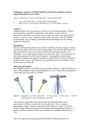
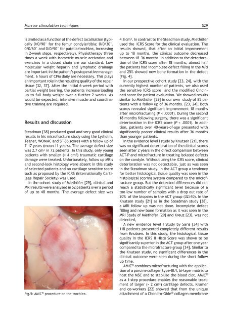
Fig.5: AMIC ® procedure on the trochlea.<br />
4.8 cm². In contrast to the Steadman study, Miethöfer<br />
used the ICRS Score for the clinical evaluation. The<br />
results showed, that after an initial improvement<br />
up to 18 months, the clinical outcome decreased<br />
between 18 36 months. In addition to the deterioration<br />
of the ICRS score after 18 months, almost half<br />
the patients had incomplete defect filling in the MRI<br />
and 25% showed new bone formation in the defect<br />
[Fig. 4].<br />
In our prospective cohort study [23, 24], with the<br />
currently highest number of patients, we also used<br />
the sensitive ICRS score and the modified Cincinnati<br />
score for patient evaluation. We showed results<br />
similar to Miethöfer [29] in our own study of 85 patients<br />
with a follow up of 36 months. [23, 24]. Both<br />
scores revealed significant improvement 18 months<br />
after microfracturing (P < .0001). During the second<br />
18 months following surgery, there was a significant<br />
deterioration in the ICRS score (P < .0001). In addition,<br />
patients over 40-years-of-age presented with<br />
significantly poorer clinical results after 36 months<br />
than younger patients.<br />
In the evidence level I study by Knutsen [21], there<br />
was no significant deterioration of the clinical scores<br />
seen after 2 years in the direct comparison between<br />
ACT-P and microfracture in treating isolated defects<br />
on the condyle. Without using the ICRS score, clinical<br />
deterioration was not detectable, just as was seen<br />
in the Steadman study. In the ACT group a tendency<br />
for better histological tissue quality was seen in the<br />
histological scoring system compared to the microfracture<br />
group. But the detected differences did not<br />
reach a statistically significant level because of a<br />
too low number of samples with a drop out rate of<br />
20% of the biopsies in the ACT group (32/40). In the<br />
Knutsen study [21] as in the Steadman study [38],<br />
a MRI follow up was not done. Incomplete defect<br />
filling and new bone formation as it was seen in the<br />
MRI Study of Miethöfer [29] and Kreuz [23], was not<br />
detected,<br />
A new evidence level I Study by Saris [34] with<br />
118 patients presented completely different results<br />
from Knutsen. In this study, the histological tissue<br />
quality in the ICRS II Histo Score was shown to be<br />
significantly superior in the ACT group after one year<br />
compared to the microfracture group [34]. Similar to<br />
the Knutsen study, no significant differences in the<br />
clinical outcome were seen during the short follow<br />
up time.<br />
AMIC ® combines microfracturing with the application<br />
of a porcine collagen type-III/I, bi-layer matrix to<br />
host the MSC and to stabilise the blood clot. AMIC ®<br />
as a 1-step procedure enables the reasonable treatment<br />
of larger (> 2 cm²) cartilage defects. Kramer<br />
and co-workers [22] showed that from the unique<br />
attachment of a Chondro-Gide ® collagen membrane