A COMPARATIVE PRIMATE ANATOMY - University of Cape Town
A COMPARATIVE PRIMATE ANATOMY - University of Cape Town
A COMPARATIVE PRIMATE ANATOMY - University of Cape Town
You also want an ePaper? Increase the reach of your titles
YUMPU automatically turns print PDFs into web optimized ePapers that Google loves.
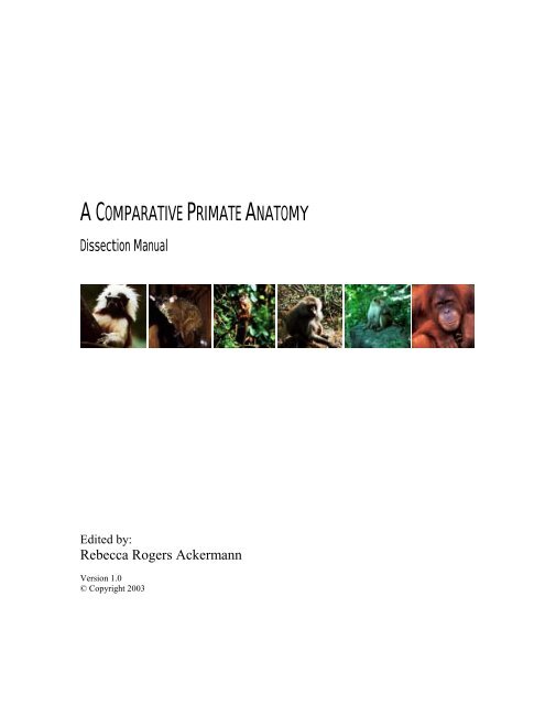
A <strong>COMPARATIVE</strong> <strong>PRIMATE</strong> <strong>ANATOMY</strong><br />
Dissection Manual<br />
Edited by:<br />
Rebecca Rogers Ackermann<br />
Version 1.0<br />
© Copyright 2003
FOREWARD<br />
This dissection manual was prepared in conjunction with a course in Comparative Primate Anatomy taught<br />
jointly by Pr<strong>of</strong>essors J Cheverud, G Conroy, and J Phillips-Conroy, at Washington <strong>University</strong> in St. Louis in the<br />
mid 1990’s. The dissections in this course focused on the skeleton and musculature <strong>of</strong> the limbs, specifically in<br />
a comparative context. As most dissection guides are Homo-centric, Homo sapiens is used as a reference<br />
species against which we compared the anatomy <strong>of</strong> the non-human primates. It also includes brief sections on<br />
behavior and ecology, as well as summary comparative anatomy sections. The choice <strong>of</strong> species was solely a<br />
function <strong>of</strong> availability, and included three New World monkeys (Saguinus oedipus, Saimiri sciureus, Ateles<br />
sp.), one African prosimian (Galago crassicaudatus), two southeast Asian macaques (Macaca fascicularis, M.<br />
mulatta), one African baboon (Papio hamadryas), and one great ape (Pongo pygmaeus). This guide is by no<br />
means a comprehensive guide, and is instead intended as a working document. As such, any comments or<br />
additional contributions to this guide are welcome, and will be included in updated versions.<br />
Importantly, this dissection guide was a collective effort, and a number <strong>of</strong> graduate and undergraduate students<br />
at Washington <strong>University</strong> contributed text and graphics to this work. These contributors include: Rebecca<br />
Rogers Ackermann, Jeff Baliff, Susan Foxman, Jennifer Helbig, Jonathan Lesser, Mark Mosbacher, Sam<br />
Senturia, Debbie Sklar, Patricia Sothman. Without this group cooperation (a good primate trait!) and the<br />
support <strong>of</strong> the faculty at Washington <strong>University</strong>, this manual would not have happened.<br />
REBECCA ROGERS ACKERMANN<br />
MARCH 2003
Comparative Primate Anatomy Behavior & Ecology<br />
______________________________<br />
COTTON-TOP TAMARIN<br />
Saguinus oedipus<br />
______________________________<br />
© Copyright 2003<br />
SECTION 1:<br />
BEHAVIOR AND ECOLOGY<br />
The cotton-top tamarin inhabits the forested areas <strong>of</strong> northern Columbia. These diurnal primates feed largely on<br />
fruits, insects, and spiders. Occasionally the tamarin will eat buds, s<strong>of</strong>t young leaves, and vertebrates. While<br />
knowledge <strong>of</strong> their social structure is sketchy, they seem to be basically monogamous, although recent research<br />
suggests that there may be a “divorce” element to this monogamy, or that perhaps the structure is actually<br />
polyandrous. They reside in concentrations <strong>of</strong> 30-180 per square kilometer, with a home range <strong>of</strong> about 10 ha.<br />
These home ranges overlap, but contact is agnostic. Additionally, they tend to remain in extended family<br />
groups (1-19 individuals), and <strong>of</strong>ten form larger groups (20-40 individuals). These groups contain a dominant<br />
mated pair, their new young, and a group <strong>of</strong> subordinate young. Each troop has a defended territory.<br />
Tamarins are approximately 9 inches long and weigh about 450g (1 lb). They are easily identifiable by their flat<br />
faces and distinctive markings; the cotton-top tamarin has brown, black, and white body hair, with a plume <strong>of</strong><br />
white hair on the top <strong>of</strong> its head. There is no sexual dimorphism. They reproduce seasonally (Jan-June).<br />
Typical gestation length is 140-145 days, with twinning as the norm. The father assists at birth and both parents<br />
participate in the care <strong>of</strong> the infants, which ride on their backs for the first 6-7 weeks <strong>of</strong> life. Tamarins spend<br />
much <strong>of</strong> their time grooming.<br />
Tamarins are arboreal quadrupeds who spend a great deal <strong>of</strong> time walking on branches. However, they also<br />
spend much <strong>of</strong> their time climbing trunks, and even occasionally leaping/hopping. Adaptations for this<br />
locomotor behavior are apparent in their anatomy.<br />
Kavanagh, M. (1983) A Complete Guide to Monkeys, Apes, and Other Primates. New York: The Viking Press.<br />
Napier, J.R. and P.H. Napier (1985) The Natural History <strong>of</strong> the Primates. Cambridge, Mass.: The MIT Press.<br />
Nowak, R.M. (1991) Walker’s Mammals <strong>of</strong> the World. Baltimore and London: The John Hopkins <strong>University</strong><br />
Press, 5th ed, Vol. 1.<br />
1
Comparative Primate Anatomy Behavior & Ecology<br />
______________________________<br />
THICK-TAILED BUSHBABY<br />
Galago crassicaudatus<br />
______________________________<br />
The thick-tailed bushbaby is the largest <strong>of</strong> all the galagos. The average weight <strong>of</strong> males (1,510g n=8) is slightly<br />
higher than females (1,258g n=9) (Petter and Petter-Rousseaux, 1979). This species is nocturnal.<br />
This species has been observed throughout eastern and southern Africa. Highest population densities are found<br />
in humid subtropical evergreen forests in which fruiting trees are plentiful. Populations are also found in dense<br />
riparian vegetation, subtropical orchards, and open woodland (Charles-Dominique and Bearder, 1979).<br />
The thick-tailed bushbaby, while anatomically suited to leaping (Fleagle, 1988), actually exhibits a tendency<br />
towards quadrupedalism. In fact, the musculature and skeleton <strong>of</strong> this species gives no clues to indicate that<br />
this animal locomotes via arboreal running and walking. Yet locomotor studies reveal that quadrupeal running<br />
and walking occurs arboreally, along the top <strong>of</strong> horizontal branches, as well as terrestrially. Saltation is rare.<br />
Jumping between trees is minimized by either climbing between connecting terminal branches or descending to<br />
the ground. No patterns have been observed in preference <strong>of</strong> using supports at a particular height. This species<br />
is flexible to individual forest structure despite its preference for horizontal supports (Charles-Dominique and<br />
Bearder, 1979).<br />
Gum, fruit, nectar, seeds, and insects comprise the annual diet <strong>of</strong> the thick-tailed bushbaby, yet this diet varies<br />
seasonally. Gums are eaten throughout the year, but become important during the dry season when fruit and<br />
nectar are scarce. At one study area Charles-Dominique and Bearder (1979) reported the following annual<br />
percentages for food intake: gum 62%; fruit 21%; flower secrections 8%; seeds 4%; insects and other items 5%.<br />
Lorisiformes do not live in social groups typical <strong>of</strong> diurnal primates. Instead, contact is limited to shared<br />
sleeping sites, brief encounters while foraging, and mother-infant relationships. At sleeping sites, animals have<br />
been observed huddling, allogrooming, and playing. Communication away from sleeping sites is maintained<br />
through a variety <strong>of</strong> mechanisms. Olfactory communication (e.g., urine washing) is used by both sexes to<br />
convey information without direct contact. Vocal communication is also frequent, and the loud cry <strong>of</strong> this<br />
species is the inspiration for the common name <strong>of</strong> “bushbaby.”<br />
Charles-Dominique, P. and S. K. Bearder (1979) Field studies <strong>of</strong> lorisid behavior: Methodological aspects. In<br />
The Study <strong>of</strong> Prosimian Behavior, ed. G.A. Doyle and R.D. Martin, pp. 567-630. New York: Academic Press.<br />
Fleagle, J.G. (1988) Primate Adaptation and Evolution. New York: Academic Press.<br />
Petter, J.-J., and A.Petter-Rousseaux (1979) Classification <strong>of</strong> the prosimians. In The Study <strong>of</strong> Prosimian<br />
Behavior, ed. G.A. Doyle and R.D. Martin, pp. 1-44. New York: Academic Press.<br />
______________________________<br />
SQUIRREL MONKEY<br />
Saimiri sciureus<br />
______________________________<br />
The genus Saimiri is attributed to the subfamily Cebinae within the family Cebidae and the infraorder<br />
Platyrrhini. There is some debate as to the number <strong>of</strong> species, but most researchers recognize two Saimiri<br />
© Copyright 2003<br />
2
Comparative Primate Anatomy Behavior & Ecology<br />
species: Saimiri sciureus and Saimiri oerstedi. Saimiri is the smallest <strong>of</strong> the cebines with a body mass that<br />
ranges from 0.6 to 1.1 kg. They are sexually dimorphic with the males usually weighing more than the females.<br />
The head and body length is 260-360 mm. The tail <strong>of</strong> the squirrel monkey is very long (350-425 mm in adults)<br />
and prehensile only in infants.<br />
Geographically, Saimiri ranges from Costa Rica and Panama in the north, to central Brazil and Bolivia in the<br />
south, and from central and northeastern Brazil in the east, to just east <strong>of</strong> the Andes in Peru in the west. While<br />
these platyrrhines are found throughout the rain forests <strong>of</strong> Central and South America, they prefer riverine and<br />
secondary forests to primary forests. The squirrel monkey can be found primarily in the lower levels <strong>of</strong> the<br />
forest. Terborgh (1983) reported a home range <strong>of</strong> more than 250 hectares for Saimiri sciureus. Saimiri has been<br />
reported to have a day range as large as 4 km.<br />
Squirrel monkeys are <strong>of</strong>ten seen in polyspecific groups with Cebus monkeys. Terborgh (1983) reported that the<br />
mean troop size <strong>of</strong> Saimiri studied at Coch Cashu in Peru was 35 individuals ranging from 30-40. Thorington<br />
(1968) studied a troop <strong>of</strong> 18 individuals <strong>of</strong> Saimiri in Colombia which increased to 22 during a 10-week period<br />
due to births. During the day the group divided into smaller groups <strong>of</strong> pregnant females, females with young,<br />
and adult males, but during the night they stayed together in the large group. The population densities <strong>of</strong> this<br />
genus ranges from 20-30 individuals per km2 in Colombia to 151-528 individuals per km2 in Peru.<br />
Fleagle and Mittermeier (1980) define Saimiri as a frugivore that spends much time foraging for insects while<br />
travelling between fruit trees. Janson and Boinski (1992) noted that squirrel monkeys eat vertebrate prey and<br />
but seldom feed on animals that are greater than 50g. Janson and Boinski also noted that caterpillars make up<br />
about half <strong>of</strong> the faunal component <strong>of</strong> Saimiri’s diet. Terborgh (1983) reported that Saimiri sciureus’s diet<br />
consisted <strong>of</strong> 18% fruit and 82% prey.<br />
Overall Saimiri is classified as an arboreal quadruped that <strong>of</strong>ten leaps. Fleagle and Mittermieier (1980) reported<br />
that when Saimiri sciureus travelled between feeding locations it engaged in slightly more bouts <strong>of</strong> arboreal<br />
quadrupedalism (55%) than bouts <strong>of</strong> leaping (42%). However, when feeding, the difference between these two<br />
modes <strong>of</strong> locomotion was much greater (quadrupedalism - 87% and leaping - 11%).<br />
Eisenberg, JF (1989) Mammals <strong>of</strong> the Neotropics: The Northern Neotropics, Volume 1. <strong>University</strong> <strong>of</strong> Chicago<br />
Press: Chicago.<br />
Fleagle, JG (1988) Primate Adaptation and Evolution. Academic Press: San Diego.<br />
Fleagle, JG and RA Mittermeier (1980) Locomotor behavior, body size, and comparative ecology <strong>of</strong> seven<br />
Surinam monkeys. American Journal <strong>of</strong> Physical Anthropology 52:301-314.<br />
Janson, CH and S Boinski (1992) Morphological and behavioral adaptations for foraging in generalist<br />
primates: The case <strong>of</strong> the cebines. American Journal <strong>of</strong> Physical Anthropology 88:483-498.<br />
Nowak, RM (1991) Walker’s Mammals <strong>of</strong> the World: Fifth Edition. Johns Hopkins <strong>University</strong> Press:<br />
Baltimore.<br />
Terborgh, J (1983) Five New World Primates: A Study in Comparative Ecology. Princeton <strong>University</strong> Press:<br />
Princeton.<br />
Thorington, RW (1968) Observations <strong>of</strong> squirrel monkeys in a Colombian forest. In RA Rosenblum and RW<br />
Cooper (Eds.): The Squirrel Monkey. Academic Press: New York, pp. 69-85.<br />
© Copyright 2003<br />
3
Comparative Primate Anatomy Behavior & Ecology<br />
______________________________<br />
LONG-TAILED, OR CRAB-EATING MACAQUE<br />
Macaca fascicularis<br />
______________________________<br />
The long-tailed, or crab-eating macaque is found throughout southeast Asia from Burma to the Philippines and<br />
further southward through Indochina, Malaysia, and on all the islands <strong>of</strong> Indonesia except for Celebes<br />
(Sulawesi). M. fascicularis is considered to be an ecologically opportunistic species which is able to thrive in<br />
habitats ranging from primary to secondary forests, and from riverine to coastal forests. The long-tailed<br />
macaque is also a very successful edge species which has learned to inhabit, and exploit plantations, parks, and<br />
gardens. In fact, it has been noted that population densities <strong>of</strong> the long-tailed macaque are sometimes higher in<br />
disturbed habitats than in undisturbed habitats.<br />
Group size in M. fascicularis ranges from 10 to 50 individuals, although groups containing nearly 100<br />
individuals have been reported. Home range size varies between 40 and 100 ha, and day ranges are reportedly<br />
less than one km. Groups are matrilineal, and males transfer between groups frequently.<br />
M. fascicularis is a sexually dimorphic species. Males weigh 5 kg, and females weigh 3 kg on average. Social<br />
grooming accounts for the majority <strong>of</strong> social interactions, and grooming is primarily solicited and provided by<br />
the females. Grooming is directed toward close relatives, and close relatives provide support during agonistic<br />
encounters. Playful behavior occurs between infants, and between juveniles. Adult males are the least socially<br />
active group members.<br />
Like other macaques, M. fascicularis possesses a generalized anatomy adapted for quadrupedal locomotion.<br />
However, the long-tailed macaque is a highly arboreal species which spends the bulk <strong>of</strong> its feeding and<br />
traveling time in the mid-canopy <strong>of</strong> the forests it inhabits. Though the majority <strong>of</strong> the long-tailed macaques’<br />
locomotion is in the form <strong>of</strong> quadrupedal walking and running, some leaping has been observed, and hind-limb<br />
hanging is used during feeding. The long-tailed, or crab-eating macaque is a frugivore. It supplements its diet<br />
with leaves, with a considerable proportion <strong>of</strong> invertebrates (crabs and termites), and with small vertebrates.<br />
Chivers, D.J. (1973) An introduction to the socio-ecology <strong>of</strong> Malayan forest primates. In Comparative Ecology<br />
and Behavior <strong>of</strong> Primates. Michael, P.P. and Crook, J.H. (eds.)<br />
Crockett, C.M. and Wilson, W.L. (1980) The ecological seperation <strong>of</strong> M. nemenstrina and M. fascicularis in<br />
Sumatra. In: Lindberg, D.G. (ed.)The Macaques. Van Nostrand Reinhold, New York.<br />
Fleagle, J.G. (1988) Primate Adaptation and Evolution. Academic Press. New York.<br />
Goosen, C. (1991) Social Grooming in Primates. In: The Order Primates. Stephens, M.E. (ed.) Kendall/Hunt.<br />
Iowa.<br />
______________________________<br />
RHESUS MACAQUE<br />
Macaca mulatta<br />
______________________________<br />
The rhesus macaque, Macaca mulatta (subfamily Cercopithecinae), ranges from Afghanistan eastward through<br />
South and Southeast Asia. Data obtained from a study on rhesus monkeys in Nepal noted the high adaptive<br />
© Copyright 2003<br />
4
Comparative Primate Anatomy Behavior & Ecology<br />
flexibility <strong>of</strong> the rhesus, considered to be the greatest <strong>of</strong> any nonhuman primate species. The macaques occupy<br />
habitats including temple grounds and parkland, heavily populated towns, lowland monsoon forest, and upper<br />
montane forest. Various studies have found that rhesus can feed on natural forest vegetation as well as take<br />
advantage <strong>of</strong> human food sources provided by agriculture, enabling them to coexist with man as well as on their<br />
own. Moreover, behavioral data obtained from temple ground-living populations found 20% <strong>of</strong> rhesus<br />
aggression involved other species (i.e. dogs, humans); <strong>of</strong> this total, 80% was directed from other species toward<br />
the monkeys, 20% from the monkeys toward other species, with population density remaining stable during the<br />
study period. Teas et. al (1980) conclude that this indicates the notable ability <strong>of</strong> rhesus monkeys to deal with<br />
adverse conditions <strong>of</strong> crowding and competition, particularly in areas <strong>of</strong> human occupation. Troop size ranges<br />
from 20-51 individuals, averaging 32 individuals. Troop composition is generally 8% male, 35% female, 30%<br />
juveniles, 15% yearlings, and 13% infants. These figures vary with season, year, and particular ecological<br />
conditions, however. The population density <strong>of</strong> a lowland monsoon region in Nepal was 0.29 rhesus groups per<br />
square kilometer. Home range varies from 2.5 to 24 hectares, depending on habitat, with overlap <strong>of</strong> 10-20% in<br />
some areas, up to 80% in more crowded areas. Despite such extensive home range overlap, each group<br />
maintains a core area <strong>of</strong> exclusive use.<br />
Teas, J., Richie, T., Taylor, H., and Southwick, C. (1980) Population patterns and behavioral ecology <strong>of</strong> rhesus<br />
monkeys (Macaca mulatta) in Nepal. In Lindburg, D. (ed.), The Macaques: Studies <strong>of</strong> Ecology, Behavior, and<br />
Evolution, Van Nostrand Reinhold, pp. 247-262.<br />
______________________________<br />
HAMADRYAS BABOON<br />
Papio hamadryas<br />
______________________________<br />
Hamadryas baboons (Papio hamadryas) are Old World monkeys belonging to the subfamily Cercopithecinae.<br />
P. hamadryas is one <strong>of</strong> the 5 species within Papio (in addition to P. anubis, P. papio, P. cynocephalus, P.<br />
ursinus). However, the specific status <strong>of</strong> the other 4 species is debated. P. hamadryas and P. anubis have<br />
formed an apparently stable hybrid zone where their ranges meet in Ethiopia, but the two are still considered<br />
distinct species.<br />
In P. hamadryas, head and body length is 610-762 mm and tail length is 362-610 mm. Adult body weights<br />
range between 12,000 grams and 21,000 grams, with considerable size sexual dimorphism; females are almost<br />
half the size <strong>of</strong> males. Pelage in the young animals is brown, and this becomes ash gray with age. Older males<br />
possess a heavy mane around the neck and shoulders. Hymadryas baboons, who are terrestrial quadrupeds,<br />
have similar upper and lower limb lengths; intermembral indices center around 95.<br />
P. hamadryas is geographically distributed throughout upper Egypt, northeastern Sudan, eastern Ethiopia,<br />
northern Somalia, and the southwestern Arabian Peninsula. They inhabit open woodland, savannahs, grassland,<br />
and rocky hill country. The diurnal hymadryas baboons forage and travel primarily by terrestrial, quadrupedial<br />
walking and running. However, the animals <strong>of</strong>ten climb trees or rocky cliffs for sleeping and resting. They<br />
generally occur at low densities, with recorded densities at 1.8-3.4 animals per square kilometer. Day ranges<br />
are considerable (6-19 km), and home ranges may range up to 28 square kilometers. Home ranges <strong>of</strong><br />
neighboring bands may overlap by 50%.<br />
Hamadryas baboons eat a variety <strong>of</strong> vegetable and fruit matter, but seem to concentrate on whatever is easily<br />
available at a given time. They consume ripe fruits, roots, tubers, grass seeds, and leaves. In addition, they are<br />
opportunistic faunivores, taking small mammals, invertebrate and insects.<br />
The basic social unit is the one-male group (OMG), which consists <strong>of</strong> one or two males, several females, and<br />
their <strong>of</strong>fspring. Usually, two or three OMG are associated in autonomous foraging groups called bands (30-85<br />
© Copyright 2003<br />
5
Comparative Primate Anatomy Behavior & Ecology<br />
animals). Up to three or four bands may congregate at one rock or sleeping cliff to form a troop. The males <strong>of</strong><br />
the OMG defend the same set <strong>of</strong> females from the advances <strong>of</strong> other males, and actually herd the females by<br />
tracking any stray females and biting them in the neck and back to keep the group together. Both males and<br />
females may depart from their natal unit, at about 2 years in males and 3.5 years in females. Breeding occurs<br />
throughout the year, although there are birth peaks in Ethiopia in May-June and November-December. The<br />
average estrous cycle is 30 days, gestation is 170-173 days, and single births are the norm. The age <strong>of</strong> sexual<br />
maturity is 7 years in males and 5 years in females.<br />
Fleagle, JG. (1988) Primate adaptation and evolution. San Diego: Academic Press Inc.<br />
Nowak, RM. (1991) Walker’s Mammals <strong>of</strong> the World. Volume 1. Baltimore: Johns Hopkins <strong>University</strong> Press.<br />
Stammbach, E. (1987) Desert, forest, and montane baboons: multilevel societies. In BB Smuts, et. al., Primate<br />
Societies. Chicago: The <strong>University</strong> <strong>of</strong> Chicago Press.<br />
______________________________<br />
SPIDER MONKEY<br />
Ateles<br />
______________________________<br />
Spider monkeys (Ateles) are diurnal Cebids from the subfamily Atelinae whose distribution ranges from the<br />
southern Amazon in Brazil to the Neotropical regions <strong>of</strong> Mexico. This is a vast area consisting <strong>of</strong> rainforests <strong>of</strong><br />
various types in which many other primate species thrive, including Howler monkeys (Alloutta), Squirrel<br />
monkeys (Saimiri), and Owl monkeys (Aotus).<br />
The genus Ateles consists <strong>of</strong> four species, A. ge<strong>of</strong>froyi, A. fusciceps, A. belzebuth, and A. paniscus, all <strong>of</strong> which<br />
are allopatric. All species <strong>of</strong> spider monkeys are frugivorous, complementing their diet with insects, leaves, and<br />
other items.<br />
Spider monkeys are aboreal and have an extremely dexterous prehensile tail which allows them to hang from<br />
trees and use their hands for the collection <strong>of</strong> fruit. Their range is primarily in the upper to mid canopy in<br />
mature forests, yet they can locomote on the ground if necessary. Locomotion in Ateles is extremely variable,<br />
ranging from semi-brachiation to brief moments <strong>of</strong> bipedal walking. However, the majority <strong>of</strong> locomotion is a<br />
composite <strong>of</strong> arboreal quadrupedalism and semi-brachiation (suspensory locomotion). The semi-brachiation<br />
used by Ateles differs from that <strong>of</strong> the true brachiators, genus Hylobates primarily in the use <strong>of</strong> the tail. The tail<br />
is used virtually all <strong>of</strong> time during the various modes <strong>of</strong> locomotion and is employed during postural behavior as<br />
well. Spider monkeys <strong>of</strong>ten feed while suspended using various combinations <strong>of</strong> the upper and hind limbs and<br />
the tail, an important feature that affects the musculoskeletal system greatly.<br />
Spider monkeys can be differentiated from other Platyrrhines on the basis <strong>of</strong> their social organization.<br />
Members <strong>of</strong> the genus Ateles form large polygamous groups characterized by a fission-fusion social<br />
organization lacking strict dominance heirarchies. This social organization is very similar to that <strong>of</strong><br />
Chimpanzees (genus Pan). In Ateles, there are two major levels <strong>of</strong> organization: the group and the subgroup.<br />
Groups consist <strong>of</strong> up to 35 individuals who scatter throughout the day yet occasionally regroup at night.<br />
Subgroups are fluctuating units in which various individuals forage together. Membership in the subgroup is<br />
not constant and changes on a daily basis or even more frequently. However, despite the apparent randomnesss,<br />
male spider monkeys <strong>of</strong>ten form stable subgroups which are cohesive, reflecting a tendency towards male<br />
affiliation. Females migrate in and out <strong>of</strong> subgroups individually or with their young.<br />
© Copyright 2003<br />
6
Comparative Primate Anatomy Behavior & Ecology<br />
Cant, J.G.H. Ecology, Locomotion, and Social Organization <strong>of</strong> Spider Monkeys (Ateles ge<strong>of</strong>froyi). Ph.D.<br />
Thesis, <strong>University</strong> <strong>of</strong> California, Davis, 1977.<br />
Eisenberg, J.F. Communication Mechanisms and Social Integration in the Black Spider Monkey, Ateles<br />
fusciceps and Related Species. Smithsonian Contr. Zoo, 113 (1989).<br />
Kellogg, R. and E.A.Goldman. Review <strong>of</strong> Spider Monkeys. Proceedings U.S. National Museum, 96: 1-45, 1944.<br />
Symington, M.M. Food Competition and Foraging Party Size in the Black Spider Monkey. Behavior, 105<br />
(1988), 117-34.<br />
______________________________<br />
ORANGUTAN<br />
Pongo pygmaeus<br />
______________________________<br />
The orangutan’s main habitat are the rain forests <strong>of</strong> insular South East Asia, specifically the islands <strong>of</strong> Borneo<br />
and Sumatra. There are two recognized subspecies <strong>of</strong> the orangutan which correspond to the particular islands;<br />
Pongo pygmaeus pygmaeus from Borneo and Pongo pygmaeus abelii found on Sumatra. The orangutan is the<br />
largest primate which is almost completely arboreal (males weigh about 70 kg and females approximately 37<br />
kg). Because <strong>of</strong> its large size, the orangutan does not brachiate while moving in their arboreal habitat. The<br />
main form <strong>of</strong> arboreal locomotion for the orangutan is quadrumanus climbing, used to achieve maximal weight<br />
distribution across as many supports as possible. While terrestrial, the orangutan utilizes a particular form <strong>of</strong><br />
quadrupedism known as “fist-walking” where they use the sides <strong>of</strong> their hands and feet as the main bodily<br />
support. Orangutans exhibit that modification <strong>of</strong> quadrupedism because <strong>of</strong> their elongated phalanges.<br />
The diet <strong>of</strong> the orangutan consists mainly <strong>of</strong> fruit: mangoes, figs and durian being the most common elements;<br />
however, they do supplement their diet with insects such as ants, termites and honeybees. Also some predation<br />
on young gibbons has been observed. During the rainy season, many <strong>of</strong> the fruits are not as abundant and the<br />
orangutans eat more bark, leaves and pith to make up for the lack <strong>of</strong> fruit.<br />
Unlike most primates, the orangutans are relatively unsocial animals. The most common group observed are<br />
females with their <strong>of</strong>fspring. Solitary adult males have home ranges which overlap the home ranges <strong>of</strong> several<br />
adult females. The adult males are highly territorial and actively maintain their domains by displays and loud<br />
calls. The females do not display swellings during estrus and are receptive throughout their cycles; however,<br />
some swellings are seen during pregnancy. The gestation period is approximately 264 days, weaning occurs<br />
around 3 years and sexual maturity around 6-7 years. Orangutans have life spans <strong>of</strong> approximately 50 years.<br />
Galdikas BM (1988) Orangutan diet, range and activity at Tanjung Puting, Central Borneo. Int. J. Primat. 9:<br />
1-35.<br />
Hamburg DA and ER McCown (eds.) (1979) The Great Apes. Reading, MA: The Benjamin/Cummings<br />
Publishing Company.<br />
Napier JR and PH Napier (1994) The Natural History <strong>of</strong> the Primate Cambridge, MA: MIT Press.<br />
Richard AF (1985) Primates in Nature. New York: W.H. Freeman and Company.<br />
Tuttle RH (1986) Apes <strong>of</strong> the World: Their Social Behavior, Communication, Mentality, and Ecology. Park<br />
Ridge, NJ: Noyes Publications.<br />
© Copyright 2003<br />
7
Comparative Primate Anatomy Back, Shoulder & Forelimb<br />
______________________________<br />
INTRODUCTION<br />
Homo sapiens<br />
______________________________<br />
© Copyright 2003<br />
SECTION 2:<br />
BACK, SHOULDER AND FORELIMB<br />
The major muscles responsible for the movement <strong>of</strong> the arm and pectoral girdle in humans can be divided into<br />
five groups based on the movements they produce; muscles that elevate the arm, lower the arm, abduct and flex<br />
the humerus, adduct and extend the humerus, and rotatie and stabilize the shoulder. Some muscles are<br />
responsible for movements in more than one <strong>of</strong> these categories.<br />
The muscles that elevate the arm are the trapezius, serratus anterior, supraspinatus, deltoid and pectoralis major.<br />
When the arm is lowered, the levator scapulae, rhomboids, latissimus dorsi, and pectoralis minor muscles are<br />
engaged. Abduction and flexion <strong>of</strong> the humerus are accomplished by the deltoid, supraspinatus, and pectoralis<br />
major. Adduction and extension <strong>of</strong> the humerus is carried out by latissimus dorsi, pectoralis major, and the<br />
deltoid muscles. The shoulder is rotated and stabilized by the infraspinatus, teres minor, subscapularis, teres<br />
major, and coracobrachialis. (Each muscle description includes medial/proximal attachments, lateral/distal<br />
attachments, and the main actions when engaged.)<br />
Trapezius<br />
--medial attachment - medial third <strong>of</strong> superior nuchal line, external occipital protuberance, ligamentum nuchae<br />
spinous processes <strong>of</strong> C7 to T12 vertebrae<br />
--lateral attachment - lateral third <strong>of</strong> clavicle, acromion, spine <strong>of</strong> scapula<br />
--main actions - superior fibers elevate the arm, middle fibers retract, inferior fibers depress the scapula; the<br />
superior and inferior fibers act together in superior rotation <strong>of</strong> the scapula.<br />
Serratus Anterior<br />
--proximal attachment - external surfaces <strong>of</strong> the lateral parts <strong>of</strong> the first through eighth ribs<br />
--distal attachment - anterior surface <strong>of</strong> the medial border <strong>of</strong> the scapula<br />
--main actions - protracts scapula and hold it against thoracic wall, rotates scapula<br />
Supraspinatus (rotator cuff muscle)<br />
--medial attachment - supraspinous fossa <strong>of</strong> scapula<br />
--lateral attachment - superior facet on greater tubercle <strong>of</strong> humerus<br />
--main actions - helps deltoid to abduct arm and acts with rotator cuff muscles<br />
Deltoid<br />
--proximal attachment - lateral third <strong>of</strong> clavicle, acromion, and spine <strong>of</strong> scapula<br />
--distal attachment - deltoid tuberosity <strong>of</strong> humerus<br />
--main actions - anterior part flexes and medially rotates arm, middle part abducts arm, posterior part extends<br />
and laterally rotates arm<br />
8
Comparative Primate Anatomy Back, Shoulder & Forelimb<br />
Pectoralis Major<br />
--proximal attachment - clavicular head <strong>of</strong> the muscle attaches on the anterior surface <strong>of</strong> the medial half <strong>of</strong> the<br />
clavicle; sternocostal head attaches on the anterior surface <strong>of</strong> the sternum, superior six costal cartilages and the<br />
aponeurosis <strong>of</strong> the external oblique muscle<br />
--distal attachment - lateral lip <strong>of</strong> the intertubercular groove <strong>of</strong> the humerus<br />
--main actions - adducts and medially rotates humerus, draws scapula anteriorly and inferiorly, the clavicular<br />
head acting alone flexes humerus, and sternoclaviuclar head extends humerus<br />
Levator Scapulae<br />
--medial attachment - posterior tubercles <strong>of</strong> the transverse processes <strong>of</strong> C1 to C4 vertebrae<br />
--lateral attachment - superior part <strong>of</strong> the medial border <strong>of</strong> the scapula<br />
--main actions - elevates scapula and tilts its glenoid cavity inferiorly by rotating scapula<br />
Rhomboid Major and Minor<br />
--medial attachment - minor attaches at the ligamentum nuchae and spinous processes <strong>of</strong> C7 and T1 vertebrae<br />
--lateral attachment - medial border <strong>of</strong> scapula from level <strong>of</strong> spine to inferior angle<br />
--main actions - retracts scapula and rotates it to depress the glenoid cavity, and fixes scapula to thoracic wall.<br />
Latissimus dorsi<br />
--medial attachment - spinous process <strong>of</strong> the inferior six thoracic vertebrae, thoracolumbar fascia, iliac crest,<br />
and inferior 3 or 4 ribs.<br />
--lateral attachment - floor <strong>of</strong> intertubercular groove <strong>of</strong> humerus<br />
--main actions - extends, adducts, and medially rotates humerus (raises body toward arms during climbing)<br />
Pectoralis Minor<br />
--proximal attachment - third through fifth ribs near costal cartilages<br />
--distal attachment - medial border and superior surface <strong>of</strong> coracoid process <strong>of</strong> scapula<br />
--main actions - stabilizes scapula by drawing it inferiorly and anteriorly against thoracic wall<br />
Infraspinatus (rotator cuff muscle)<br />
--medial attachment - infraspinous fossa <strong>of</strong> scapula<br />
--lateral attachment - middle facet on greater tubercle <strong>of</strong> humerus<br />
--main actions - laterally rotates arm, helps to hold humeral head in glenoid cavity <strong>of</strong> scapula<br />
Teres Minor (rotator cuff muscle)<br />
--medial attachment - superior part <strong>of</strong> lateral border <strong>of</strong> scapula<br />
--lateral attachment - inferior facet on greater tubercle <strong>of</strong> humerus<br />
--main actions - laterally rotates arm, helps to hold humeral head in glenoid cavity <strong>of</strong> scapula<br />
Subscapularis (rotator cuff muscle)<br />
--medial attachment - subscapular fossa<br />
--lateral attachment - lesser tubercle <strong>of</strong> humerus<br />
--main actions - medially rotates arm and adducts it , helps to hold humeral head in glenoid cavity<br />
Teres Major<br />
--medial attachment - dorsal surface <strong>of</strong> inferior angle <strong>of</strong> scapula<br />
--lateral attachment - medial lip <strong>of</strong> intertubercular groove <strong>of</strong> humerus<br />
--main actions - adducts and medially rotates arm<br />
Coracobrachialis<br />
--proximal attachment - tip <strong>of</strong> coracoid process <strong>of</strong> scapula<br />
--distal attachment - middle third <strong>of</strong> medial surface <strong>of</strong> humerus<br />
--main actions - helps to flex and adduct arm<br />
The muscles that move the forearm in humans can be divided into those groups that flex the elbow, extend the<br />
elbow, pronate the forearm, and supinate the forearm. The biceps brachii, brachialis, and brachioradialis<br />
© Copyright 2003<br />
9
Comparative Primate Anatomy Back, Shoulder & Forelimb<br />
muscles produce flexion at the elbow, while the triceps brachii and anconeus muscles extend the elbow.<br />
Pronation <strong>of</strong> the forearm is carried out by the pronator teres and pronator quadratus. Supination <strong>of</strong> the forearm<br />
is produced by the biceps brachii and supinator muscles.<br />
Biceps Brachii<br />
--proximal attachment - short head attaches on the tip <strong>of</strong> the coracoid process <strong>of</strong> the scapula, long head<br />
originates at the supraglenoid tubercle <strong>of</strong> the scapula<br />
--distal attachment - radial tuberosity and fascia <strong>of</strong> arm via the bicipital aponeurosis<br />
--main actions - supinates forearm and when supine flexes forearm<br />
Brachialis<br />
--proximal attachment - distal half <strong>of</strong> anterior surface <strong>of</strong> humerus<br />
--distal attachment - coronoid process and tuberosity <strong>of</strong> ulna<br />
--main actions - flexes forearm in all positions<br />
Brachioradialis<br />
--proximal attachment - proximal two-thirds <strong>of</strong> the supracondylar ridge <strong>of</strong> the humerus<br />
--distal attachment - lateral surface <strong>of</strong> distal end <strong>of</strong> radius<br />
--main actions - flexes forearm<br />
Triceps Brachii<br />
--proximal attachment - long head begins at infraglenoid tubercle <strong>of</strong> glenoid, lateral head is at the posterior<br />
surface <strong>of</strong> humerus superior to radial groove, medial head originates on the posterior surface <strong>of</strong> the humerus<br />
inferior to the radial groove<br />
--distal attachment - proximal end <strong>of</strong> olecranon <strong>of</strong> ulna, fascia <strong>of</strong> forearm<br />
--main actions - extends forearm (chief extensor <strong>of</strong> forearm), long head steadies head <strong>of</strong> abducted humerus<br />
Anconeus<br />
--proximal attachment - lateral epicondyle <strong>of</strong> humerus<br />
--distal attachment - lateral surface <strong>of</strong> olecranon and superior part <strong>of</strong> posterior surface <strong>of</strong> ulna<br />
--main actions - assists triceps in extending forearm, stabilizes elbow joint, abducts ulna during pronation<br />
Pronator Teres<br />
--proximal attachment - medial epicondyle <strong>of</strong> humerus, coronoid process <strong>of</strong> ulna<br />
--distal attachment - middle <strong>of</strong> lateral surface <strong>of</strong> radius<br />
--main actions - pronates forearm and flexes it<br />
Pronator Quadratus<br />
--proximal attachment - distal fourth anterior surface <strong>of</strong> ulna<br />
--distal attachment - distal fourth anterior surface <strong>of</strong> radius<br />
--main actions - pronates forearm, deep fibers bind ulna and radius together<br />
Supinator<br />
--proximal attachment - lateral epicondyle <strong>of</strong> humerus, radial collateral and anular ligaments, supinator fossa,<br />
crest <strong>of</strong> ulna<br />
--distal attachment - lateral, posterior, and anterior surfaces <strong>of</strong> proximal third <strong>of</strong> radius<br />
--main actions - supinates forearm<br />
The muscles that move the hand in humans are divided into the long flexors and the long extensors <strong>of</strong> the hand<br />
and digits. The flexor group includes flexor carpi radialis, flexor carpi ulnaris, palmaris longus, flexor<br />
digitorum pr<strong>of</strong>undus, and flexor pollicus longus. The extensors are extensor carpi radialis longus, extensor<br />
carpi radialis brevis, extensor carpi ulnaris, extensor digitorum, extensor digiti minimi, extensor indicus,<br />
extensor pollicus longus, extensor pollicus brevis, and abductor pollicus longus.<br />
Flexor Carpi Radialis<br />
--proximal attachment - medial epicondyle <strong>of</strong> humerus<br />
© Copyright 2003<br />
10
Comparative Primate Anatomy Back, Shoulder & Forelimb<br />
--distal attachment - base <strong>of</strong> second metacarpal bone<br />
--main actions - flexes hand and abducts it<br />
Flexor Carpi Ulnaris<br />
--proximal attachment - humeral head originates at medial epicondyle <strong>of</strong> humerus, ulnar head originates at<br />
olecranon and posterior border <strong>of</strong> ulna<br />
--distal attachment - pisiform bone, hook <strong>of</strong> hamate bone, fifth metacarpal bone<br />
--main actions - flexes hand and adducts it<br />
Palmaris Longus<br />
--proximal attachment - medial epicondyle <strong>of</strong> humerus<br />
--distal attachment - distal half <strong>of</strong> flexor retinaculum, palmar aponeurosis<br />
--main actions - flexes hand and tightens palmar aponeurosis<br />
Flexor Digitorum Superficialis<br />
--proximal attachment - humeroulnar head originates at medial epicondyle <strong>of</strong> humerus, ulnar collateral<br />
ligament, and coronoid process <strong>of</strong> the ulna, radial head originates on superior half <strong>of</strong> anterior border <strong>of</strong> radius<br />
--distal attachment - bodies <strong>of</strong> the middle phalanges <strong>of</strong> medial four digits<br />
--main actions - flexes middle phalanges <strong>of</strong> medial four digits (stronger action - flexes proximal phalanges and<br />
hand)<br />
Flexor Digitorum Pr<strong>of</strong>undus<br />
--proximal attachment - proximal three-fourths <strong>of</strong> the medial and anterior surfaces <strong>of</strong> the ulna, interosseous<br />
membrane<br />
--distal attachment - bases <strong>of</strong> distal phalanges <strong>of</strong> medial four digits<br />
--main actions - flexes distal phalanges <strong>of</strong> medial four digits<br />
Flexor Pollicus Longus<br />
--proximal attachment - anterior surface <strong>of</strong> radius, interosseous membrane<br />
--distal attachment - base <strong>of</strong> distal phalanx <strong>of</strong> thumb<br />
--main actions - flexes phalanges <strong>of</strong> thumb<br />
Extensor Carpi Radialis Longus<br />
--proximal attachment - lateral supracondylar ridge <strong>of</strong> humerus<br />
--distal attachment - base <strong>of</strong> second metacarpal bone<br />
--main actions - extend and abduct hand at wrist joint<br />
Extensor Carpi Radialis Brevis<br />
--proximal attachment - lateral epicondyle <strong>of</strong> humerus<br />
--distal attachment - base <strong>of</strong> third metacarpal<br />
--main actions - extend and abduct hand at wrist joint<br />
Extensor Carpi Ulnaris<br />
--proximal attachment - lateral epicondyle <strong>of</strong> humerus and posterior border <strong>of</strong> ulna<br />
--distal attachment - base <strong>of</strong> fifth metacarpal bone<br />
--main actions - extends and adducts hand at wrist<br />
Extensor Digitorum<br />
--proximal attachment - lateral epicondyle <strong>of</strong> humerus<br />
--distal attachment - extensor expansions <strong>of</strong> medial four digits<br />
--main actions - extends medial four digits at metacarpophalangeal joints, extends at wrist joints<br />
Extensor Digiti Minimi<br />
--proximal attachment - lateral epicondyle <strong>of</strong> humerus<br />
--distal attachment - extensor expansion <strong>of</strong> fifth digit<br />
--main actions - extends fifth digit at metacarpophalangeal and interphalangeal joints<br />
© Copyright 2003<br />
11
Comparative Primate Anatomy Back, Shoulder & Forelimb<br />
Extensor Indicus<br />
--proximal attachment - posterior surface <strong>of</strong> ulna, interosseous membrane<br />
--distal attachment - extensor expansion <strong>of</strong> second digit (index finger)<br />
--main actions - extends second digit and helps to extend hand<br />
Extensor Pollicus Longus<br />
--proximal attachment - posterior surface <strong>of</strong> middle third <strong>of</strong> ulna, interosseous membrane<br />
--distal attachment - base <strong>of</strong> distal phalanx <strong>of</strong> thumb<br />
--main actions - extends distal phalanx <strong>of</strong> thumb at metacarpophlangeal and interphalangeal joints<br />
Extensor Pollicus Brevis<br />
--proximal attachment - posterior surface <strong>of</strong> radius, interosseous membrane<br />
--distal attachment - base <strong>of</strong> proximal phalanx <strong>of</strong> thumb<br />
--main actions - extends proximal phalanx <strong>of</strong> thumb at carpometacarpal joint<br />
Abductor Pollicus Longus<br />
-- proximal attachment - posterior surfaces <strong>of</strong> ulna and radius, interosseous membrane<br />
--distal attachment - base <strong>of</strong> first metacarpal bone<br />
--main actions - abducts thumb and extends it at carpometacarpal joint<br />
The deep muscles <strong>of</strong> the back can be grouped one category as erectors. This region <strong>of</strong> the body is where one<br />
would expect to find strong contrasts between the human and other primates (especially those with tails). The<br />
muscle included here are the iliocostalis, longissimus, and spinalis. Together, these make up the erector spinae<br />
muscle group. The main actions <strong>of</strong> this muscle group are to act as the chief extensor <strong>of</strong> the vertebral column,<br />
bend vertebral column posteriorly, and to control movement during flexion <strong>of</strong> the vertebral column.<br />
Iliocostalis<br />
--origin - broad tendon attached inferiorly to the posterior part <strong>of</strong> the iliac crest, the posterior aspect <strong>of</strong> the<br />
sacrum, the sacroiliac ligaments, and the sacral and inferior lumbar spinous process<br />
--insertion - angles <strong>of</strong> the ribs<br />
-- main actions - acting unilaterally it laterally flexes the head or the vertebral column, bilaterally it extends the<br />
head and part or all <strong>of</strong> the vertebral column<br />
Longissimus<br />
--origin - same as iliocostalis<br />
--insertion - transverse processes <strong>of</strong> thoracic and cervical vertebrae, mastoid process<br />
--main actions - same as iliocostalis<br />
Spinalis<br />
--origin - same as iliocostalis and longissimus<br />
--insertion - extends from spinous processes in the superior lumbar and inferior thoracic regions to the spinous<br />
processes in the superior thoracic region<br />
--main actions - same as iliocostalis and longissimus<br />
Aiello, Leslie and Dean, Christopher (1990). An Introduction to Human Evolutionary Anatomy. Academic<br />
Press: London.<br />
Moore, Keith L. (1992). Clincally Oriented Anatomy. Williams and Wilkins: Baltimore.<br />
© Copyright 2003<br />
12
Comparative Primate Anatomy Back, Shoulder & Forelimb<br />
______________________________<br />
COTTON-TOP TAMARIN<br />
Saguinus oedipus<br />
______________________________<br />
One <strong>of</strong> the main differences between the forelimb <strong>of</strong> a tamarin and that <strong>of</strong> a human involves the medial-lateral<br />
lengthening <strong>of</strong> the scapula which helps to properly position the humerus for quadrupedal locomotion. For the<br />
most part, the muscles and their insertions are the same in the two animals, however, the osteological changes<br />
necessary for the different locomotor forms may reposition the muscles relative to the joints, thereby altering<br />
their functions. These differences, in turn, cause relative changes in the proportions <strong>of</strong> the muscles. I will<br />
discuss both the absolute changes in muscles and the relative changes in their form due to function in a specific<br />
order: those muscles that move the arm and pectoral girdle, those muscles that move the forearm, and those<br />
muscles that move the wrist and hand.<br />
The tamarin has only three muscles that are not found in the arm and pectoral girdle <strong>of</strong> the human-atlantoscapularis<br />
anterior, atlantoscapularis posterior, and pectoralis abdominus. Atlantoscapularis<br />
anterior originates from the spinous process <strong>of</strong> the atlas vertebra and inserts on the acromion <strong>of</strong> the scapula. It<br />
functions as a upward rotator <strong>of</strong> the scapula. Atlantoscapularis posterior originates on the transverse process<br />
<strong>of</strong> the atlas and inserts on the anterior medial border <strong>of</strong> the scapula, and functions as a replacement for levator<br />
scapulae which raises the scapula. The pectoralis abdominus was not observable, as it was removed when the<br />
specimen was eviscerated. However, it should arise from the sheath <strong>of</strong> rectus abdominus, insert into the<br />
humerus, and flex the arm.<br />
Some <strong>of</strong> these muscles that act on the arm and shoulder in the tamarin have different origin and insertion sites<br />
and areas compared with humans, largely due to relative differences in the sizes <strong>of</strong> the muscles. The origin <strong>of</strong><br />
trapezius does not extend as far up the spine in tamarins as in humans, with only a few fibers reaching the<br />
nuchal crest, and the muscle in general is less substantial. Latissimus dorsi is also less extensive, as there is<br />
no portion <strong>of</strong> latissimus dorsi which originates from the iliac crest. Teres major is robust compared with<br />
humans, as is serratus anterior, while the origin <strong>of</strong> the rhomboids is more extensive, traveling up into the<br />
cervical region. Also, the deltoid is not very large, but is distinctly divided into its three functional regions-anterior,<br />
middle, and posterior.<br />
© Copyright 2003<br />
Figure 1: Cotton-top tamarin back. (A)<br />
deltoid muscle, (B) atlantoscapularis anterior,<br />
(C) trapezius.<br />
13
Comparative Primate Anatomy Back, Shoulder & Forelimb<br />
As a whole, these differences in the muscle sizes and attachment areas result from functional differences.<br />
Tamarins, as arboreal quadrupeds, don't use their arms above their heads. Therefore, their arm-raising muscles-<br />
-deltoid, trapezius--are relatively underdeveloped. (While serratus anterior is used in arm-raising, its good<br />
development in the tamarin is probably due to the fact that it is also used to support the rib cage in quadrupeds<br />
and functions as a sling.) In contrast, the muscles which are used for quadrupedal propulsion--latissimus dorsi,<br />
teres major, and presumably pectoralis major (if my animal had one)--are relatively overdeveloped.<br />
The muscles that move the forearm are the same in tamarins and humans with one exception. The<br />
dorsoepitrochlearis originates from the tendon <strong>of</strong> latissimus dorsi and inserts into the olecranon process <strong>of</strong> the<br />
ulna. It functions as an extensor <strong>of</strong> the elbow.<br />
The arm flexors <strong>of</strong> the tamarin are highly developed (biceps brachii and brachioradialis), relative to humans.<br />
The extensors <strong>of</strong> the arm, in particular triceps brachii, are also highly developed, as well as being more<br />
extensive. As an arboreal quadruped, the elbow <strong>of</strong> the tamarin remains in a constant state <strong>of</strong> flexion, with both<br />
the flexors and extensors <strong>of</strong> the arm almost continuously active during motion. Therefore, the muscular<br />
morphology <strong>of</strong> the tamarin arm reflects its mode <strong>of</strong> locomotion.<br />
The muscles that move the hand and wrist in the tamarin are the same as in humans. There are, however, some<br />
relative differences in the size <strong>of</strong> these muscles. As a whole, the flexors <strong>of</strong> the hand are extremely well<br />
developed. This is probably a reflection <strong>of</strong> the fact that, as arboreal quadrupeds, they spend much <strong>of</strong> their time<br />
not only balancing on branches, but actively grasping these branches both when walking <strong>of</strong> them and when<br />
climbing.<br />
______________________________<br />
THICK-TAILED BUSHBABY<br />
Galago crassicaudatus<br />
______________________________<br />
© Copyright 2003<br />
Figure 2: The tamarin arm. (A) triceps<br />
brachii, (B) extensor group.<br />
The well developed musculature <strong>of</strong> the forelimbs <strong>of</strong> G. crassicaudatus indicates that the forelimbs are used in<br />
locomotion. This could either indicate that this species moves quadrupedally or by leaping. (Crompton (1984)<br />
distinguishes between leaping, in which the animal includes use <strong>of</strong> its forelimbs, and hopping, in which the<br />
animal locomotes entirely with its hindlimbs. It is important to note that not all saltation requires use <strong>of</strong> the<br />
forelimbs).<br />
14
Comparative Primate Anatomy Back, Shoulder & Forelimb<br />
Superficial back and shoulder:<br />
The muscles <strong>of</strong> Galago crassicaudatus in the superficial back and shoulder are primarily similar to Homo.<br />
The omohyoid is not considered a back or shoulder muscle in Homo , yet in G. crassicaudatus it originates at<br />
the vertebral border <strong>of</strong> the scapula, near the scapular spine. The levator scapula muscle in this species can be<br />
demarcated into more distinct muscles than the single levator scapula in Homo. The levator scapula ventralis<br />
originates on the spine <strong>of</strong> the scapula near the acromion and inserts onto the body <strong>of</strong> C1. The levator scapula<br />
dorsalis originates from the vertebral border <strong>of</strong> the scapula, and inserts onto the transverse processes <strong>of</strong> C1 to<br />
C5 or C6. The deltoid is clearly divided into three functional groups (posterior, middle, and anterior) and is<br />
relatively small.<br />
Differences between G. crassicaudatus and Homo in muscle size relative to body size can be accounted for by<br />
functional differences in postural behavior: the thick-tailed bushbaby is an arboreal quadruped, and the human<br />
is a biped. Unlike Homo,, G. crassicaudatus utilizes its superficial back and shoulder muscles to maintain<br />
balance, locomote, and support its body weight. The serratus anterior is relatively robust, forming a weight<br />
bearing sling around the thorax. This muscle, in conjunction with the equally robust rhomboids (major, minor,<br />
and capitus) and the trapezius, stabilizes the scapula when the forelimbs are bearing weight. The trapezius is<br />
not as relatively well developed as other superficial back muscles because galagos do not use them to raise their<br />
arms above their heads -- one <strong>of</strong> this muscle’s functions in humans. The latissimus dorsi, teres major, and<br />
pectoralis major are used to transfer the weight <strong>of</strong> the animal on to the arm when standing still, to adduct the<br />
arm in order to balance on arboreal supports, and to propel the animal when locomoting. They are,<br />
understandable, relatively well developed. In addition to these muscles <strong>of</strong> the "sling," G. crassicaudatus has an<br />
expansive pectoralis abdominus muscles. This muscle originates along the aponeurosis <strong>of</strong> the rectus<br />
abdominus at the mid-line, and inserts onto the medial aspect <strong>of</strong> the deltoid tuberosity.<br />
© Copyright 2003<br />
Figure 3: Galago chest: (latissimus dorsi, (B) serratus anterior, (C) pectoralis major, (E) deltoid.<br />
15
Comparative Primate Anatomy Back, Shoulder & Forelimb<br />
Upper arm and shoulder:<br />
G. crassicaudatus has all <strong>of</strong> the upper arm and shoulder muscles that Homo has, and has one additional<br />
muscle: epitrochlearis. Epitrochlearis takes origin from the olecranon process <strong>of</strong> the ulna and distal tendon <strong>of</strong><br />
the triceps and inserts onto the latissimus dorsi, near its insertion on the humerus. This muscle is an additional<br />
flexor at the elbow. All <strong>of</strong> the upper arm muscles are well developed as a result <strong>of</strong> the functional demands <strong>of</strong><br />
arboreal quadrupedalism. Those muscles responsible for flexion and extension <strong>of</strong> the humerus at the shoulder<br />
joint propel the body during locomotion. The flexors and extensors <strong>of</strong> the ulna at the elbow are always being<br />
used to stabilize the continuously flexed elbow, as well as propel the body. The biceps are also used in<br />
supination. This motion is important in an arboreal setting where the supports may be skewed in relation to the<br />
long axis <strong>of</strong> the body.<br />
The forearm, wrist, and hand:<br />
The muscles <strong>of</strong> the forearm, wrist, and hand are also relatively well developed as a result <strong>of</strong> the functional<br />
demands <strong>of</strong> arboreal quadrupedalism. All <strong>of</strong> the muscles found in Homo are also present in G. crassicaudatus<br />
with the exception <strong>of</strong> extensor digiti minimi. In the galago, this muscle is absent. Its functional equivalent is<br />
found in the extensor digitorum communis. This muscle is like the extensor digitorum <strong>of</strong> Homo --inserting<br />
onto the first phalange <strong>of</strong> digits 2-4, yet extensor digitorum communis also attaches to digit 5. The exacting<br />
manual dexterity exhibited by Homo is not necessary to G. crassicaudatus This species can afford to move<br />
digits 2-5 more as a functional unit.<br />
Crompton, R.H. (1984). Foraging, habitat structure and locomotion in two species <strong>of</strong> Galago. In Adaptations<br />
for Foraging in Non-human Primates, ed. P.S. Rodman and J.G.H. Cant, pp. 73-111. New York: Columbia<br />
<strong>University</strong> Press.<br />
Stevens, J.L., Edgerton, V.R., Haines, D.E., and D. M. Meyer (1981). An Atlas and Source Book <strong>of</strong> the Lesser<br />
Bushbaby, Galago senegalensis. Boca Raton, Florida: CRC Press, Inc.<br />
______________________________<br />
SQUIRREL MONKEY<br />
Saimiri<br />
______________________________<br />
Back and shoulder:<br />
With few exceptions, the muscles <strong>of</strong> Saimiri originate and insert in the same fashion as in Homo. Saimiri has<br />
two muscles not possessed by Homo: the dorsoepitrochlearis and the atlantoscapularis. The<br />
dorsoepitrochlearis is actually part <strong>of</strong> the triceps group that runs from the olecranon fascia and the medial<br />
epicondyle <strong>of</strong> the humerus to the latissimus dorsi where it attaches. This muscle is important for the arboreally<br />
adapted quadrupedal squirrel monkey because it aids in extension <strong>of</strong> the arm at the shoulder and extension <strong>of</strong><br />
the forearm at the elbow. The altlantoscapularis originates on the transverse process <strong>of</strong> the atlas and inserts on<br />
the medial part <strong>of</strong> the cranial border <strong>of</strong> the scapula. The main function <strong>of</strong> this muscle is to move the shoulder<br />
cranially.<br />
There are a few more differences between Saimiri and Homo in the back and shoulder. Saimiri is characterized<br />
by undifferentiated rhomboids (major and minor), absence <strong>of</strong> the serratus posterior inferior, and a more<br />
robust serratus anterior. The rhomboides major and minor are either undifferentiated or the rhomboides<br />
minor may be absent. The serratus posterior inferior is occasionally absent in humans. The relatively thick<br />
serratus anterior is easily understood when one considers the morphological adaptation <strong>of</strong> the squirrel<br />
© Copyright 2003<br />
16
Comparative Primate Anatomy Back, Shoulder & Forelimb<br />
monkey. Because this taxon is an arboreal quadruped, this muscle acts as a sling holding up the torso when the<br />
monkey is in a quadrupedal stance. In addition, it works with the trapezius and the rhomboids to fix the the<br />
vertebral border <strong>of</strong> the scapula. This muscle can also aid in respiration.<br />
The deltoid and the pectoralis group have important roles for Saimiri. In the squirrel monkey, the deltoid<br />
serves a major role in protraction, retraction and abduction <strong>of</strong> the arm. All <strong>of</strong> these actions are very important<br />
for the quadruped. The pectoralis group is relatively robust, serves as an adductor and medial rotator <strong>of</strong> the<br />
humerus, and helps to support the torso on the arm. The pectoralis minor acts as a depressor and inward<br />
rotator <strong>of</strong> the scapula. One major difference in the pectoral group is that Saimiri possesses a pectoralis<br />
abdominalis. This muscle is not present in Homo. The muscle originates on both the costal region and the<br />
xiphoid process and inserts on the upper humerus. The main function <strong>of</strong> the pectoralis abdominalis, which is<br />
very well developed in Saimiri, is to adduct the arm at the shoulder joint.<br />
Upper arm and shoulder:<br />
© Copyright 2003<br />
Figure 4: Saimiri arm. (A)<br />
brachioradialis, (B), deltoid, (C)<br />
unknown muscle.<br />
Saimiri differs from Homo in upper arm muscular morphology. Saimiri is characterized by a relatively more<br />
robust teres major and a well developed and large triceps group (particularly the long head <strong>of</strong> the triceps).<br />
The teres major and triceps morphology can best be explained by their function; they are both extensors <strong>of</strong> the<br />
arm at the shoulder joint. Arm extension plays an important role in the locomotion <strong>of</strong> the quadruped.<br />
The flexor group in the upper arm <strong>of</strong> Saimiri is essentially the same as in Homo. The main differences lie in the<br />
relative sizes <strong>of</strong> the muscles. The coracobrachialis is relatively smaller in Saimiri and functions as an adductor<br />
and flexor <strong>of</strong> the humerus at the shoulder joint. The biceps brachii is well developed in the squirrel monkey<br />
and has two important functions for this arboreal quadruped: it acts as a flexor <strong>of</strong> the humerus at the shoulder<br />
joint, and also as a powerful supinator <strong>of</strong> the forearm. This is extremely important in an arboreal environment.<br />
Another difference in the upper arm musculature is the well developed and robust brachialis. This muscle acts<br />
as a powerful forearm flexor in Saimiri and is important for arboreal quadrupedalism. The brachioradialis is<br />
well developed in the squirrel monkey. Functionally the brachioradialis serves to flex the forearm, an<br />
important function for the arboreal quaduped. There is one additional muscle that this specimen <strong>of</strong> Saimiri<br />
possesses. This muscle appears to originate from the deltoid muscle near its attachment on the humerus and<br />
runs distally down the arm. It has a slight connection at the anterior bend <strong>of</strong> the elbow and then continues<br />
distally down the arm riding just above the brachioradialis and has a common attachment with this muscle.<br />
17
Comparative Primate Anatomy Back, Shoulder & Forelimb<br />
The forearm, wrist and hand:<br />
Both the flexor and extensor groups <strong>of</strong> the forearm and hand are essentially the same in Saimiri and Homo. The<br />
only major difference is that both <strong>of</strong> these muscle groups are a little more developed and relatively more robust<br />
in Saimiri in comparison with Homo.<br />
Hartman, CG & WL Strauss (1961). The Anatomy <strong>of</strong> the Rhesus Monkey (Macaca mulatta). New York: Hafner<br />
Publishing.<br />
Larson, SG (1994). Functional morphology <strong>of</strong> the shoulder in primates. In DL Gebo (ed.): Postcranial<br />
Adaptations in Nonhuman Primates. DeKalb: NIU Press. pp. 45-69.<br />
______________________________<br />
LONG-TAILED OR CRAB-EATING MACAQUE<br />
Macaca fascicularis<br />
______________________________<br />
Although the long-tailed macaque relies on quadrupedalism in both arboreal and terrestrial settings, the over all<br />
patterns <strong>of</strong> muscle origin and insertion resembles the musculature in humans. Resemble, however, does not<br />
mean identical, and as we shall see the differences that do exist in Macaca fascicularis help optimize the<br />
locomotor pattern <strong>of</strong> the species. Other muscles do exist i.e., panniculus carnosus (see below), that do not<br />
appear in humans and have little, if any, influence on the movement <strong>of</strong> the limbs.<br />
Panniculus: The panniculus muscles are subcutaneous muscles which move the skin. These muscles originate<br />
from the superficial fascia and insert onto the internal surface <strong>of</strong> the skin. Most <strong>of</strong> the panniculus muscles do<br />
not exert any force, or influence on the limbs <strong>of</strong> the primate body. The panniculus carnosus does assist in the<br />
movement <strong>of</strong> the humerus in the long-tailed macaque. The panniculus carnosus, characterized by a thin, fan<br />
shape exists in two portions. The caudal part originates from the superficial fascia overlying the anterior<br />
surface <strong>of</strong> the thigh and gluteal region, and from the lumbar fascia. The thoracic part originates from the dorsal<br />
and caudal portions <strong>of</strong> the skin, and ultimately interdigates with the fibers from the caudal part. While the<br />
caudal part inserts onto the skin covering the thorax, the thoracic part inserts by a tendon on to the humerus.<br />
The thoracic portion assists the pectoralis minor in lowering the arm.<br />
Superficial back and shoulder:<br />
Trapezius (cervical and thoracic portions): The trapezius orginates and inserts at the same locations as in<br />
humans. However, where as the clavicular insertion is usually missing in humans, it is always present in<br />
macaques. The cervical portion <strong>of</strong> the trapezius aids in elevation <strong>of</strong> the scapula while the thoracic portion<br />
adducts, depresses, and stabalizes the scapula. Functionally, the presence <strong>of</strong> the clavicular insertion may assist<br />
in, or increase the level <strong>of</strong> elevation, or caudal to cranial movement, <strong>of</strong> the scapula. In quadrupedal locomotion<br />
the scapula moves in a caudal to cranial direction with the spine in a, more or less, horizontal orientation to the<br />
ground. This differs from bipedal locomotion where the scapula is oriented in the sagittal plane. Elevation <strong>of</strong><br />
the scapula for a bipedal animal occurs when the arm is raised, and not during locomotion. Elevation <strong>of</strong> the<br />
scapula does not occur during quadrupedal locomotion, rather the scapula moves in a caudal to cranial direction,<br />
and perhaps the clavicular insertion <strong>of</strong> the trapezius aids in this particular movement.<br />
Atlantoscapularis anterior/posterior: The levator scapulae is the functionally equivalent muscle in humans.<br />
In the long-tailed macaque the two portions <strong>of</strong> the atlantoscapularis lie directly underneath the cervical portion<br />
<strong>of</strong> the trapezius. Both the anterior and posterior portions <strong>of</strong> the atlantoscapularis orginate from the transverse<br />
© Copyright 2003<br />
18
Comparative Primate Anatomy Back, Shoulder & Forelimb<br />
process <strong>of</strong> the atlas. The anterior portion, inserting on the superior and lateral part <strong>of</strong> the scapular spine as well<br />
as on the acromion, assists in the elevation <strong>of</strong> the scapula. The posterior part, inserting on the vertebral border<br />
<strong>of</strong> the scapula superior to the scapular spine, assists in the medial rotation and elevation <strong>of</strong> the scapula.<br />
Rhomboid capitis/cervicis/dorsi: Humans posses rhomboid major and minor which are equivalent to<br />
rhomboid dorsi and rhomboid cervicis in macaques, respectively. The equivalent to rhomboid capitis<br />
occasionally occurs in humans, and is called rhomboid occipitalis when it does. The capitis part originates<br />
from the medial secton <strong>of</strong> the superior nuchal line. The cervicis originates from the ligamentum nuchae. The<br />
dorsi part originates from the spinous processes <strong>of</strong> the cervical and the first six or seven thoracic vertebrae. The<br />
three parts <strong>of</strong> the rhomboid insert along the vertebral border <strong>of</strong> the scapula with the capitis portion most<br />
cranial, and the dorsi portion most caudal. The rhomboids adduct and rotate the scapula.<br />
Latissimus dorsi: The latissimus dorsi does not extend to the iliac spine in Macaca fascicularis as it does in<br />
humans. The origin, insertion, and action <strong>of</strong> the latissimus dorsi are the same in humans and in Macaca<br />
fascicularis. Unlike humans, where the more medial and caudal portion <strong>of</strong> the trapezius overlays the latissimus<br />
doris, in the long-tailed macaque the more medial and cranial portion <strong>of</strong> the latissimus dorsi overlays the<br />
trapezius. It appears as if the latissimus dorsi forms a pocket around a portion <strong>of</strong> the trapezius, and the<br />
covered portion <strong>of</strong> the trapezius does not attach to the spinous processes <strong>of</strong> the vertebral column.<br />
Serratus anterior: The serratus anterior in the long-tailed macaque derives its origin from the transverse<br />
processes <strong>of</strong> the cervical vertebrae, and the inferior border <strong>of</strong> the first nine to ten ribs. In humans, the serratus<br />
anterior does not originate from the cervical vertebrae, and it attaches to the superior portion <strong>of</strong> the ribs. The<br />
insertion on the vertebral border <strong>of</strong> the scapula is identical in humans and the long-tail. As in humans, the<br />
serratus anterior aids in respiration, and abducts the scapula. Unlike humans, the serratus anterior in the<br />
long-tailed macaque forms a sling around the the ribs and helps in supporting the chest cavity.<br />
The remaining muscles <strong>of</strong> the back and shoulder (teres major, teres minor, supra and infraspinatus, and the<br />
subscapularis) in Macaca fascicularis do not differ from their orientation in humans.<br />
Secondary muscles <strong>of</strong> respiration:<br />
Serratus posterior superior originates from the spinous processes <strong>of</strong> the thoracic vertebra, and inserts on the<br />
first through fifth ribs. The muscle consists <strong>of</strong> three thin, ovoid, shaped bellies and aids in respiration. The<br />
serratus posterior inferior is approximately five times as large as the serratus posterior superior. The muscle<br />
consists <strong>of</strong> six ovoid bellies, each attached to the ribs by a long, thin tendon; each belly is connected to the next<br />
by a layer <strong>of</strong> fascia. The muscle fibers run superior laterally, and it also aids in respiration.<br />
© Copyright 2003<br />
Figure 5: M.<br />
fascicularis<br />
medial arm. (A)<br />
flexor carpi<br />
radialis, (B)<br />
palmaris longus,<br />
(C) biceps brachii<br />
19
Comparative Primate Anatomy Back, Shoulder & Forelimb<br />
Shoulder:<br />
The muscles responsible for the motions <strong>of</strong> the shoulder (the deltoid, pectoralis major, pectoralis minor, and<br />
pectoralis abdominalis) do not signifcantly differ from their orientation in humans. Most notably, the<br />
pectoralis major in Macaca fascicularis and Macaca mulatta lacks the clavicular origin which occurs in<br />
humans, as well as in other primates. The lack <strong>of</strong> the origin on the clavicle, and the stronger origin from the<br />
entire sternum is related to the quadrupedal locomotor pattern <strong>of</strong> the macaque. In quadrupedalism the limbs<br />
need to move in, and remain in a para-sagital orientation; thus the pectoralis major in macaques aids in holding<br />
the upper limbs in position in the para-sagital plane. A second, minor difference is noted in the deltoid. Unilke<br />
humans, and unlike some other primates i.e., Saimiri, the separation <strong>of</strong> the deltoid into three distinct parts in<br />
relation to its spinous, acromial, and clavicular insertions is very distinct in the long-tailed macaque.<br />
Upper arm :<br />
The upper arm in Macaca fascicularis greatly resembles the upper arm in humans. The brachialis and the<br />
coracobrachialis are reduced in comparison to humans. The pronators, pronator teres and pronator<br />
quadratus, and the supinator do not differ from their orientation in humans. The greatest differences lie in the<br />
triceps complex. The long and lateral heads <strong>of</strong> the triceps are proportionally much larger than in humans. The<br />
extent to which the triceps are enlarged may be directly related to the extension <strong>of</strong> the forearm, and the the<br />
propulsion <strong>of</strong> the animals during quadrupedal locomotion. The dorso-epitrochlearis, a part <strong>of</strong> the triceps<br />
complex, occurs in Macaca fascicularis and not in humans. This thin, sheet-like muscle which orginates from<br />
the tendonous insertion <strong>of</strong> the latissimus dorsi, wraps around the posterior and medial portion <strong>of</strong> the triceps<br />
group to insert, by an a tendonous sheet, on to the olecranon fascia. The dorsi-epitrochlearis assists in the<br />
extension <strong>of</strong> the forearm. The brachioradalis in the long-tailed macaque has a higher point <strong>of</strong> origin on the<br />
deltoid ridge <strong>of</strong> the humerus than in humans. The insertion and action are the same in the long-tail as they are<br />
in humans. However, the extent <strong>of</strong> flexion in long-tailed macaques is limited by the shape and orientation <strong>of</strong><br />
the brachioradalis. When trying to extend the forearm, the brachioradalis becomes taut and for full extension<br />
would need to be cut. Thus, the forearm remains in partial flexion.<br />
Flexors <strong>of</strong> the hand:<br />
There are no great differences between the flexors <strong>of</strong> the hand in Macaca fascicularis and in humans. Both the<br />
flexor carpi radialis, and the palmaris longus show no differences from humans. The flexor carpi ulnaris in<br />
Macaca fascicularis is proportionally much larger than in humans. The flexor digitorum superficialis lacks<br />
the radial origin which humans possess. The muscles listed above are responsible for the flexion <strong>of</strong> the wrist,<br />
and the flexion <strong>of</strong> the proximal phalanges, not including the thumb. The flexor digitorum pr<strong>of</strong>undus<br />
eliminates the inidividual muscle <strong>of</strong> the flexor policis longus by incorporating it into the body <strong>of</strong> the pr<strong>of</strong>undus<br />
group. As the tendon <strong>of</strong> the flexor digitorum pr<strong>of</strong>undus separates, the tendons insert into the distal phalanges<br />
<strong>of</strong> all five <strong>of</strong> the digits, and when stimulated causes a closed fist to form.<br />
Extensors <strong>of</strong> the hand:<br />
The extensors in Macaca fuscicularis are very similar to those in humans. Extensor carpi radialis longus,<br />
extensor carpi radialis brevis, and extensor carpi ulnaris show no discernible differences from their<br />
orientation in humans. The communis group <strong>of</strong> extensors separates into two groups. The extensor digitorum<br />
communis originates, as do the other extensors, from the lateral epicondyle <strong>of</strong> the humerus, and inserts on the<br />
dorsal side <strong>of</strong> the first through fourth digits. The tendonous insertions differ from humans in the communis<br />
group. At first, they appear as four distinct tendons deriving from the communis muscle. The four tendons join<br />
together as they pass over the metacarpals. Then, as the tendonous sheet approaches the digits, it separates into<br />
four tendons and they insert on to the digits. A second belly <strong>of</strong> the communis group, the extensor digitorum<br />
quatri and quinti proprius, extends <strong>of</strong>f <strong>of</strong> the larger group and leads to two separate tendons which insert on to<br />
the fourth and fifth digits. The other extensors; extensor digiti minimi, extensor indicis, extensor pollicus<br />
longus, extensor pollicus brevis are all present and do not exhibit any great differences from humans.<br />
© Copyright 2003<br />
20
Comparative Primate Anatomy Back, Shoulder & Forelimb<br />
Agur, M.R. (1991) Grant’s atlas <strong>of</strong> anatomy. Gardner, J.N. (ed.) Williams and Wilkins. Baltimore, Md.<br />
Aiello, L., and Dean, C. (1990) An introduction to human evolutionary anatomy. Acedemic Press. San Diego,<br />
Ca.<br />
Berringer, O.M., Browing, F.M., and Schroeder, C.R. (1968) An atlas and dissection manual <strong>of</strong> rhesus monkey<br />
anatomy. Animal Laboratory Aids. Tallahassee, Fl.<br />
Netter, F.H. (1989) Atlas <strong>of</strong> human anatomy. Colacino, S. (ed.) Ciba-Geiba Corporation. Summit, N.J.<br />
______________________________<br />
RHESUS MACAQUE<br />
Macaca mulatta<br />
______________________________<br />
Muscles <strong>of</strong> the superficial back and shoulder:<br />
When considering the muscles <strong>of</strong> the superficial back and shoulder in the rhesus macaque, one must keep in<br />
mind the macaque’s quadrupedal locomotion. Most <strong>of</strong> the muscles, decribed below in detail, act upon the<br />
proximal humerus differently than in humans. Most importantly, they are more directly involved in retration<br />
and adduction <strong>of</strong> the arm, as a result <strong>of</strong> their altered insertion points when compared to Homo (see below). This<br />
is to be expected in quadrupedalism, as the arm must necessarily be kept close to the body and must be able to<br />
retract in the parasagittal plain with propulsion coming from the legs muscles.<br />
Unlike Homo, the rhesus macaque pectoralis major has no origin from the medial clavicle. Rather, the muscle<br />
is composed <strong>of</strong> the pars capsularis and pars sternalis. The pars capsularis arises from the sternoclavicular joint<br />
and manubrium, while the pars sternalis originates along the length <strong>of</strong> the sternum running deep to the pars<br />
capsularis. As in Homo, both insert upon the anterior border <strong>of</strong> the intertubercular sulcus <strong>of</strong> the humerus below<br />
the synovial joint capsule <strong>of</strong> the shoulder.<br />
Action: contrary to Homo, the pectoralis major has less <strong>of</strong> a role in flexion <strong>of</strong> the upper limb since it has no<br />
clavicular origin. Rather, it acts in adduction f the limb, especially from an elevated position.<br />
Whereas the pectoralis minor inserts upon the coracoid process <strong>of</strong> the scapula in humans, in rhesus macaques<br />
it inserts into an aponeurotic sheet that extends long the intertubercular sulcus <strong>of</strong> the humerus upward over the<br />
lesser tuberosity to the shoulder joint capsule.<br />
Action: although the insertion is slightly different from Homo, the muscle produces the same action <strong>of</strong> rotating<br />
the scapula and shoulder joint inward and downward (caudalward), working in conjunction with the rhomboids<br />
and trapezius.<br />
The pectoralis abdominalis, absent in Homo, originates from the sheath <strong>of</strong> the rectus abdominis caudalward<br />
to the xiphoid process and lateral to the midline and inserts upon the humerus by an aponeurosis just distal to<br />
the insertion <strong>of</strong> the pectoralis minor, proximal to the panniculus carnosus insertion.<br />
Action: contraction produces the adduction <strong>of</strong> the upper limb, aiding the pectoralis major and minor. Also<br />
functions as an arm flexor.<br />
© Copyright 2003<br />
21
Comparative Primate Anatomy Back, Shoulder & Forelimb<br />
© Copyright 2003<br />
Figure 6: Rhesus macaque<br />
superficial arm. Reproduced from<br />
Hartman & Straus (1933).<br />
The panniculus carnosus, absent in Homo runs superficial to the other trunk muscles, originating from the<br />
anterolateral surface <strong>of</strong> the thigh and gluteal region and from the abdominal wall. The fibers continue cranially<br />
along the side <strong>of</strong> the trunk to insert into the deep pectoral aponeurosis, with the pectoral abdominalis insertion<br />
just proximal.<br />
Action: mainly causes skin movement. Whereas Hill (1974) does not consider it effective in upper limb<br />
movement, Hartman and Strauss (1933) note that it aids the pectoralis major in depressing the arm from an<br />
elevated position.<br />
Of the superficial back muscles, the trapezius does not significantly differ from that <strong>of</strong> Homo. The latissimus<br />
dorsi differs from Homo in that there are no fibers which originate from the crest <strong>of</strong> the ilium, either fleshily or<br />
through an aponeurotic attachment.<br />
Whereas Homo has the rhomboid major and minor, the rhesus macaque has three components composing the<br />
rhomboideus:<br />
1) pars capitis, arising from the occipital bone and inserting upon the vertebral border <strong>of</strong> the scapula<br />
just above the scapular spine.<br />
2) pars cervicis, arising from the ligamentum nuchae and inserting caudal to the pars capitis.<br />
Equivalent to the rhomboid minor in Homo.<br />
3) pars dorsi, arising caudal to the pars cervicis from the upper thoracic spinous processes and<br />
inserting caudal to the same muscle on the vertebral border <strong>of</strong> the scapula, extending to the glenovertebral<br />
angle. Equivalent to the rhomboid major in Homo.<br />
Action: despite the slightly different musculature, the rhesus rhomboideus functions to adduct the scapula and<br />
play a role in scapular rotation, as is the case in Homo.<br />
The atlantoscapularis posterior resembles the levator scapulae <strong>of</strong> Homo in that it originates from the dorsal<br />
aspect <strong>of</strong> the transverse process <strong>of</strong> the atlas, inserting upon the vertebral border <strong>of</strong> the scapula cranial to the<br />
scapular spine. The rhesus differs markedly from Homo in the atlantoscapularis anterior, arising from the<br />
22
Comparative Primate Anatomy Back, Shoulder & Forelimb<br />
ventral border <strong>of</strong> the transverse process <strong>of</strong> the atlas and inserting upon the lateral half <strong>of</strong> the scapular spine and<br />
acromion. Homo lacks anything resembling the atlantoscapularis anterior.<br />
Action: the atlantoscapularis anterior and posterior both function to rotate the scapula cranially, the former<br />
muscle being more effective.<br />
Whereas both the deltoid and pectoralis major have a clavicular origin in Homo, the deltoid dominates the<br />
clavicle in Macaca mulatta, with the pectoralis major arising from the manubrium and sternum. Unlike Homo,<br />
the most dorsal aspect <strong>of</strong> the deltoid arises from an aponeurosis that extends from the scapular spine to the<br />
glenovertebral angle <strong>of</strong> the scapula; in Homo, this portion <strong>of</strong> the muscle arises from an aponeurotic attachment<br />
both to the spine and superior border <strong>of</strong> the scapula. Thus, the dorsal portion <strong>of</strong> the rhesus deltoid is positioned<br />
more laterocaudally than in Homo.<br />
Action: the deltoid function in the protraction, retraction, and abduction <strong>of</strong> the humerus.<br />
The serratus anterior, supraspinatus, infraspinatus, subscapularis, teres minor, teres major muscles are<br />
all similar to those <strong>of</strong> Homo.<br />
The coracobrachialis differs from Homo in the presence <strong>of</strong> two component muscles, the coracobrachialis<br />
pr<strong>of</strong>undus and coracobrachialis medius. Homo lacks the former. The coracobrachialis pr<strong>of</strong>undus arises partly<br />
from the coracoid process and from the deep surface <strong>of</strong> the common coracoid tendon to insert upon the surgical<br />
neck <strong>of</strong> the humerus.<br />
Action: the coracobrachialis pr<strong>of</strong>undus rotates the upper limb medially.<br />
Muscles <strong>of</strong> the upper arm:<br />
Overall, the musculature <strong>of</strong> the upper arm is similar in the macaque and Homo. The main differences arise due<br />
to the macaque’s quadrupedal locomotion; muscles aiding in active and/or maintained extension <strong>of</strong> the forearm<br />
are emphasized both in size and specific insertion sites.<br />
The brachioradialis originates closer to the insertion <strong>of</strong> the deltoid on the humerus than in Homo. As a<br />
powerful forearm flexor, the muscle probably aids the macaque in manipulation <strong>of</strong> objects with the forelimbs.<br />
Perhaps the greatest difference in arm musculature from Homo is in the macaque triceps brachii; this<br />
difference is manifest not in origin/insertion sites, but in sheer size. The macaque has extremely robust triceps<br />
due to the necessity <strong>of</strong> supporting its upper body solely on its arms when in the quadrupedal posture. As part <strong>of</strong><br />
this group, and absent in humans, the dorso-epitrochlearis aids in this function. It originates from the tendon<br />
<strong>of</strong> the latissimus dorsi and runs along the medial aspect <strong>of</strong> the humerus to attach by an aponeurosis to the<br />
superficial olecranon fascia and the medial epicondyle <strong>of</strong> the humerus. The remainder <strong>of</strong> the upper arm<br />
muscles--biceps brachii, brachialis, anconeus lateralis, pronator teres, pronator quadratus, and supinator-<br />
-are all similar to that <strong>of</strong> Homo.<br />
Long flexors <strong>of</strong> the hand and digits:<br />
The superficial flexor muscles <strong>of</strong> the forearm are similar to those in Homo in function, origins, and insertions.<br />
They differ as far as size is concerned--the flexor carpi ulnaris and palmaris longus are proportionately larger<br />
and more developed in the rhesus macaque. The flexor carpi radialis is similar to that in Homo. One level<br />
deeper, the flexor digitorum sublimis differs from the human flexor digitorum superficialis in the absense <strong>of</strong> a<br />
radial origin. Moreover, there is a deep insertion <strong>of</strong> some fibers into the medial margin <strong>of</strong> the radial portion <strong>of</strong><br />
the flexor digitorum pr<strong>of</strong>undus. This latter muscle differs from Homo in that it incorporates the flexor pollicis<br />
longus in one <strong>of</strong> its two bellies: 1) the radial portion arises from the medial apect <strong>of</strong> the radius, and from the<br />
interosseous membrane; it provides the tendons for digits I-III; 2) the ulnar portion originates from the lateral<br />
and volar spects <strong>of</strong> the ulna and also from the interosseous membrane; it provides the tendons for digits III-V.<br />
Long extensors <strong>of</strong> the hand and digits:<br />
© Copyright 2003<br />
23
Comparative Primate Anatomy Back, Shoulder & Forelimb<br />
The superficial extensors <strong>of</strong> the wrist--extensor carpi radialis longus, extensor carpi radialis brevis, and<br />
extensor carpi ulnaris--are similar to Homo. The extensor digitorum communis, equivalent to the human<br />
extensor digitorum, differs from Homo in that the four separate tendons are united as far distally as the<br />
metacarpal heads, after which they diverge and progress toward digits II-V. Moreover, there is not marked<br />
continuation <strong>of</strong> the tendons to the terminal phalanx <strong>of</strong> each digit, as the case is in Homo. The macaque<br />
extensor digiti quarti proprius and extensor digiti quinti proprius originate from the lateral epicondyle <strong>of</strong><br />
the humerus and have a common tendon which divides in two parts at variable positions in the forearm. The<br />
tendon <strong>of</strong> the e.d. quarti proprius inserts upon the dorsum <strong>of</strong> the fourth finger; this muscle is absent in Homo.<br />
The tendon <strong>of</strong> the e.d. quinti proprius inserts upon the dorsum <strong>of</strong> the fifith finger; this muscle is equivalent to<br />
the human extensor digiti minimi.<br />
Deep to the above, the abductor pollic longus, and extensor pollicis longus are similar to that in Homo; the<br />
extensor pollicis brevis is absent in the macaque. The extensor digiti secundi proprius is an equivalent<br />
muscle to the human extensor indicus, inserting upon the dorsum <strong>of</strong> digit II; however, the macaque also has an<br />
extensor digiti tertii proprius which arises with the previous muscle from the lateral border <strong>of</strong> the distal ulna,<br />
and inserts via a tendon upon the dorsum <strong>of</strong> the third finger on the basal phalanx. Overall, then, it seems that<br />
the macaque has tendons inserting upon digits II-V from the communis group and from the individual proprius<br />
extensors mentioned above. A suggested hypothesis for this difference may be the habitual quadrupedalism <strong>of</strong><br />
the macaque, with the constant extension <strong>of</strong> the wrist and fingers.<br />
Hartman, C.G. & Straus, W.L. Jr. (1933). The Anatomy <strong>of</strong> the Rhesus Monkey. Baltimore: Williams & Wilkins<br />
Co.; pp. 101-105, 116-120, 128-131.<br />
______________________________<br />
HAMADRYAS BABOON<br />
Papio hamadryas:<br />
______________________________<br />
In P. hamadryas the shoulder and forelimb is subject mostly to compressive forces associated with terrestrial<br />
weight bearing and locomotion. Locomotor patterns involve the movement <strong>of</strong> the upper limb in the sagittal<br />
plane, with no need for extensive rotation and abduction <strong>of</strong> the arm. P. hamadryas differs from Homo mainly in<br />
the musculature related to shoulder/arm mobility and scapular rotation. For terrestrial quadrupedal primates<br />
like P. hamadryas, the musculature shows efficient positioning for producing a greater degree <strong>of</strong> protraction (vs.<br />
rotation) <strong>of</strong> the scapula and retraction and protraction (vs. abduction) <strong>of</strong> the humerus. In addition, there is less<br />
demand on the pronators, supinators, and wrist deviators in P. hamadryas, who do not extensively use these<br />
muscles for climbing and limb grasping.<br />
Muscles that move the arm and pectoral girdle:<br />
P. hamadryas possess four muscles in this region that are not found in Homo. 1) Panniculus carnosus: This<br />
muscles overlies the latissimus dorsi. Muscle fibers arise along the gluteal and lateral thoracic region, are<br />
thickest ventrally, and form a tendon that attaches to the proximal part <strong>of</strong> the humerus, deep to the pectoralis<br />
musculature. The panniculus carnosus, relatively large in P. hamadryas, allows the hamadryas baboon to move<br />
its skin and aids the pectoralis major in depressing the arm when it is in an elevated position. 2) Pectoralis<br />
abdominus: This muscles originates from the sheath <strong>of</strong> the rectus abdominus and inserts into the proximal<br />
humerus. This relatively large muscle in P. hamadryas adducts and medially rotates the humerus. 3)<br />
Atlantoscapularis anterior: This muscle originates on the ventral part <strong>of</strong> the atlas and inserts into the cranial<br />
margin <strong>of</strong> the acromion and lateral scapular spine. 4) Atlantoscapularis posterior: This muscle originates at<br />
the dorsal part <strong>of</strong> the transverse process <strong>of</strong> the atlas and inserts at the vertebral border <strong>of</strong> the scapula between the<br />
© Copyright 2003<br />
24
Comparative Primate Anatomy Back, Shoulder & Forelimb<br />
spine and superior angle. The levator scapulae muscle <strong>of</strong> Homo is represented by these last two muscles. These<br />
muscles move the scapula cranially (anterior) and move the superior angle <strong>of</strong> the scapula laterally (posterior).<br />
In addition to the four muscles described above, there are a number <strong>of</strong> differences between P. hamadryas and<br />
Homo musculature in the superior back and shoulder region. 1) Latissimus dorsi: The origin is the same as<br />
Homo, but in addition to its insertion on the humerus, P. hamadryas displays a cranial portion <strong>of</strong> the muscle that<br />
attaches to the teres major, which helps produce more efficient retraction <strong>of</strong> the humerus in the sagittal plane.<br />
In addition, Homo also has more extensive posterior attachment at the iliac crest, lumbodorsal fascia, and on the<br />
inferior angle <strong>of</strong> the scapula.<br />
2) Triceps brachii: The triceps is divided into three heads: long, lateral and medial. The attachment <strong>of</strong> the<br />
long head differs from Homo. In P. hamadryas, the long head arises from an extensive area along the axillary<br />
border <strong>of</strong> the scapula (it is limited to the infraglenoid tubercle <strong>of</strong> the scapula in Homo). The three parts<br />
converge about mid-shaft to form a broad tendon and continues to receive fibers from the humerus nearly as far<br />
as the olecranon fossa in P. hamadryas. Again, the extensive attachment <strong>of</strong> the long head affords more<br />
retraction <strong>of</strong> the humerus and extension <strong>of</strong> the forearm for quadrupedal locomotion.<br />
3) Deltoid: The deltoid is composed <strong>of</strong> three parts: clavicular, acromial, and spinal, and is easily divisible into<br />
its three parts in P. hamadryas. The clavicular portion has an extensive attachment to the clavicle and limits the<br />
pectoralis major to the region <strong>of</strong> the sternoclavicular joint. The cephalic vein intervenes between the caudal<br />
border <strong>of</strong> the deltoid and the cranial border <strong>of</strong> the pectoralis major, but the division is not a precise as in Homo.<br />
The attachment <strong>of</strong> the deltoid on the humerus (on the deltoid turberosity) is more proximal in P. hamadryas than<br />
in Homo and brachiating primates. In the terrestrial quadrupedal P. hamadryas, it is a powerful protractor and<br />
retractor <strong>of</strong> the arm. The more distal placement in brachiators is interpreted as <strong>of</strong>fering a greater mechanical<br />
advantage in lifting the humerus as they move through the trees. Homo also displays a more brachiator like<br />
pattern in the ability to lift the humerus.<br />
4) Rhomboids: The rhomboideus major and minor are not as easily divisible in P. hamadryas as in humans.<br />
In Homo, these muscles adducts and rotates the scapula, and scapular rotation is not as important for P.<br />
hamadryas' terrestrial lifestyle.<br />
© Copyright 2003<br />
Figure 7: Baboon right shoulder: (A) atlantoscapularis anterios, (B) infraspinatus, (C) teres minor,<br />
(D) triceps brachii, (E) teres minor, (F) latissimus dorsi, (G) dorsoepitrochlearis.<br />
25
Comparative Primate Anatomy Back, Shoulder & Forelimb<br />
Muscles that move the forearm:<br />
P. hamadryas possess one muscle in this region that is not found in Homo. 1) Dorsoepitrochlearis: This<br />
muscle is a part <strong>of</strong> the triceps that has become secondarily attached to the latissimus dorsi. The muscle arises<br />
near beginning <strong>of</strong> the tendon <strong>of</strong> the latissimus dorsi, accompanies the long head <strong>of</strong> the triceps to the elbow and<br />
passes to the medial epicondyle <strong>of</strong> the humerus. The action <strong>of</strong> this muscle is to extend the forearm.<br />
In addition to dorsoepitrochlearis, there are a number <strong>of</strong> differences between P. hamadryas and Homo arm<br />
musculature. 1) Biceps brachii: This muscle, a flexor and supinator <strong>of</strong> the forearm, has similar attachments in<br />
both Homo and P. hamadryas. However, the relative proportions <strong>of</strong> the biceps and triceps differ between the<br />
two - P. hamadryas has relatively larger triceps and Homo has relatively larger biceps. This reflects the need in<br />
terrestrial quadrupedal locomotion for powerful extension (retraction) <strong>of</strong> the arm at the shoulder.<br />
2) Anconeus lateralis: In humans, this muscle is simply called anconeus. This muscle, which originates on<br />
the posterolateral side <strong>of</strong> the elbow and attaches just below the elbow on the lateral ulna, is relatively larger in<br />
P. hamadryas. Anconeus lateralis aids in extending the elbow.<br />
3) Pronator teres: P. hamadryas has only one head <strong>of</strong> origin (medial epicondyle <strong>of</strong> the humerus), while<br />
humans have an additional origin on the medial border <strong>of</strong> the ulna.<br />
Mucles that move the wrist and hand:<br />
P. hamadryas possess four muscles in this region that are rare or absent in Homo. 1) Extensor digiti quatri<br />
proprius: This muscle is rarely found in humans, and when it occurs, it is normally referred to as extensor<br />
digiti angularis. This muscle originates on the lateral epicondyle <strong>of</strong> the humerus and inserts into the dorsal<br />
surface <strong>of</strong> the proximal phalanx <strong>of</strong> digit IV. Its action is to extend the fourth metacarpophalangeal joint. 2)<br />
Extensor digiti tertii proprius: This muscle is usually absent in humans, but when it does occur, it is referred<br />
to as extensor digiti medius. It originates in common with the extensor digit secundi proprius on the dorsal<br />
surface <strong>of</strong> the ulna and attaches to the dorsum <strong>of</strong> the proximal phalanx <strong>of</strong> digit III, which it extends.<br />
In addition to the two muscles described above, there are a number <strong>of</strong> differences between P. hamadryas and<br />
Homo musculature <strong>of</strong> the forearm. 1) Brachioradialis: The proximal attachment <strong>of</strong> this muscle is higher<br />
(more proximal) in P. hamadryas than Homo; it extend up to the insertion <strong>of</strong> the deltoid.). Brachioradialis is a<br />
forearm flexor.<br />
2) Palmaris longus: This muscle, frequently absent in Homo, originates from the medial epicondyle <strong>of</strong> the<br />
humerus and attaches into the palmar aponerosis.<br />
3) Flexor digitorum superficialis: Both P. hamadryas and Homo have origins <strong>of</strong> flexor digitorum<br />
superficialis on the medial epicondyle <strong>of</strong> the humerus, but Homo also has an ulnar and radial head. Both<br />
species show attachments to the middle phalanges <strong>of</strong> digits II-V.<br />
4) Extensor digitorum: The origin is similar to Homo, but in P. hamadryas, the insertions are in the sides <strong>of</strong><br />
the proximal phalanges and bases <strong>of</strong> middle phalanges <strong>of</strong> digits II-V. This contrasts humans which have<br />
insertions into the dorsal surface <strong>of</strong> all three phalanges <strong>of</strong> digits II-V.<br />
5) Extensor digiti quinti proprius: This muscle, which extends the fifth metacarpophalangeal joint is<br />
commonly called extensor digiti minimus in humans.<br />
6) Extensor digiti secundi proprius: In humans, this muscle is called extensor digiti indicus. It originates<br />
on the distal aspect <strong>of</strong> the dorsal surface <strong>of</strong> the ulna in common with the extensor digiti tertii proprius and<br />
attaches onto the proximal phalanx (dorsal surface) <strong>of</strong> digit II, which it extends.<br />
Extensor pollicus brevis and flexor pollicus longus, each normally a differentiated muscle in Homo are not<br />
found in P. hamadryas.<br />
© Copyright 2003<br />
26
Comparative Primate Anatomy Back, Shoulder & Forelimb<br />
______________________________<br />
SPIDER MONKEY<br />
Ateles<br />
______________________________<br />
As with most other primates, the musculoskeletal anatomy <strong>of</strong> the genus Ateles greatly resembles that <strong>of</strong> the<br />
modern humans, genus Homo. The primary differences lie in the relative sizes <strong>of</strong> specific muscle groups and in<br />
the origins and insertions <strong>of</strong> the various muscles. Also, certain muscles found in Ateles (and other nonhuman<br />
primates) are lacking in Homo while other muscles distinct in humans are either fused or undifferentiated in the<br />
spider monkeys.<br />
As a whole, the upper limb, back, and shoulder regions in Ateles are very similar to the corresponding regions<br />
in Homo. The muscles are well differentiated and well developed for suspension. The shoulder, elbow, and<br />
wrist joints are very flexible and have the pronounced ability to support the mass <strong>of</strong> the animal in a hanging<br />
position below limbs as well as above the limb. The muscles and joints are well adapted to provide for minor<br />
modifications in the suspensory position <strong>of</strong> the animal. This is particularly important in a genus whose<br />
members use their tails and both the upper and lower limbs in varying combinations while both in locomotion<br />
and nonlocomotory behavior. The muscles and joints <strong>of</strong> the lower limb have similar abilities and functions with<br />
regard to suspensory behavior. However, the lower limb is used much less for locomotion and therefore the<br />
relative sizes <strong>of</strong> the muscles are smaller than in most primates, including humans. The muscles are also fused in<br />
many instances and have origins and insertions on the tendons <strong>of</strong> other muscles.<br />
Overall, the musculature <strong>of</strong> the upper limb and the back and shoulder regions is much more developed and<br />
differentiated than in the lower limb, hip, and thigh regions due to the emphasis placed upon locomotion using<br />
the upper body. The propulsion normally provided by the hindlimbs is instead accomplished using the tail.<br />
This includes the "leaping" used to cross gaps in the trees, where the power arises from the tail. Thus, the<br />
hindlimb musculature is less developed. Correspondingly, the intermembral index for Ateles is 105, reflecting<br />
the practice <strong>of</strong> semi-brachiation (arboreal quadrupeds normally have shorter upper limbs in order to lower the<br />
center <strong>of</strong> gravity close to the support). The brachial index is high and thus evident <strong>of</strong> the increased use <strong>of</strong> the<br />
forearms for suspension.<br />
Back and Shoulder:<br />
With the exception <strong>of</strong> the pectorals, the muscles <strong>of</strong> these regions are well developed and closely correspond to<br />
those in humans. However, those muscles which elevate the arm are relatively larger as are those which<br />
function to retract the arm. Those muscles which are relatively larger or have more extensive origins and<br />
insertions include Latissimus dorsi, Trapezius, Serratus anterior, Teres major, Atlantoscapularis, and the<br />
rhomboids. Latissimus dorsi differs considerably in its increased thickness over that in humans and its<br />
increased iliac insertion. Interestingly, spider monkeys have an iliac origin more extensive than that <strong>of</strong> humans<br />
and less than that <strong>of</strong> the great apes while the true brachiators, genus Hylobates, have no insertion onto the ilium.<br />
Another differenence in this muscle is the lack <strong>of</strong> a small slip <strong>of</strong> origin on the point <strong>of</strong> the scapula. Trapezius<br />
differs from its human morphology in the increased size but decreased vertebral attachment. The muscle<br />
originates most inferiorly on the vertebrae attached to the true ribs rather than the floating ribs. Serratus<br />
anterior, Teres major, Atlantoscapularis, and the rhomboids have similar origins and insertions to humans<br />
but are relatively thicker and larger.<br />
Other differences in the back and shoulder region between Ateles and Homo are the decreased size <strong>of</strong> the<br />
pectoral muscles, the absence <strong>of</strong> Serratus posterior inferior, and the additional origin <strong>of</strong> Pectoralis minor on<br />
the humeral head in addition to the coracoid process <strong>of</strong> the scapula.<br />
© Copyright 2003<br />
27
Comparative Primate Anatomy Back, Shoulder & Forelimb<br />
Upper Arm and Shoulder:<br />
The morphology <strong>of</strong> the upper arm musculature is largely similar to that <strong>of</strong> Homo. The major difference lies in<br />
the more prominent muscles which extend the forearm. Triceps brachii differs in that the medial head is<br />
attached more proximally to the humerus. Additionally, the long head <strong>of</strong> Triceps is attached to Teres major by<br />
connective tissue and extends more anteriorly to the medial side <strong>of</strong> the proximal humerus. Triceps brachii is<br />
complemented by the presence <strong>of</strong> an additional muscle not present in humans, Dorsoepitrochlearis. This<br />
muscle originates from the tendon <strong>of</strong> Latissimus dorsi and inserts into the humerus on the medial epicondyle.<br />
Among the flexors <strong>of</strong> the forearm, few differences arise between the morphology <strong>of</strong> Ateles and that <strong>of</strong> Homo.<br />
Biceps brachii differs from its human morphology only in the attachment to the Pronator teres by thick<br />
connective tissue. Brachialis morphology differs from that in humans only in the more proximal humeral<br />
origin. Brachioradialis inserts more distally onto the styloid process <strong>of</strong> the radius. Additionally, this muscle is<br />
much larger in Ateles.<br />
Forearm:<br />
The forearm region <strong>of</strong> the spider monkey is largely the same as that <strong>of</strong> humans with the important exception<br />
that no pollex is present in Ateles. This lack <strong>of</strong> an opposable thumb is thought to have arisen to reduce<br />
interference during brachiation. Interestingly, the absence <strong>of</strong> a pollex also occurs in the genus Colobus while<br />
gibbons and siamangs do retain the thumb. This would seem to refute the theory that a reduced or absent pollex<br />
is an adaptation to brachiation. The other major difference in the forearm region is the large size <strong>of</strong> many <strong>of</strong> the<br />
flexors, extensors, and rotators in the spider monkey due to the increased use <strong>of</strong> the forearm during locomotion.<br />
Within the hand region, the four remaining digits form functional hooks for suspending below branches.<br />
The flexors <strong>of</strong> the hand in Ateles differ in few ways from those in Homo. Flexor carpi ulnaris originates<br />
normally but remains attached to the ulna along its entire length. This tendonus attachment is fused with that <strong>of</strong><br />
Flexor digitorum superficialis from the common flexor origin to the distal end <strong>of</strong> the ulna. Flexor digitorum<br />
pr<strong>of</strong>undus has a morphology which differs from the human form due to the division <strong>of</strong> the muscle into two<br />
major parts which are connected at their tendons. The portion originating from the radius inserts into the<br />
second digit. The portion originating from the olecranon process <strong>of</strong> the ulna inserts into the phalanges <strong>of</strong> the<br />
second through fifth digits. Finally, the absence <strong>of</strong> a pollex has resulted in the absence <strong>of</strong> Flexor pollicus<br />
longus.<br />
The extensors <strong>of</strong> the hand are largely the same with one major exception regarding supination <strong>of</strong> the hand.<br />
Extensor carpi ulnaris has an additional origin from the olecranon process <strong>of</strong> the ulna. Extensor digiti<br />
minimi has an additional insertion into the ulna near the wrist. Extensor indicus originates along virtually all<br />
<strong>of</strong> the ulnar border and inserts into the second through fifth digits. Finally, the major distinction regarding the<br />
extensors deals with the lack <strong>of</strong> the pollex. Extensor pollicus longus and Extensor Pollicus Brevis are both<br />
absent while an extensive abductor has become the predominant muscle in the region. Abductor pollicus<br />
originates from the radius and ulna very proximally near the elbow and inserts onto the metacarpal <strong>of</strong> the first<br />
digit (the only remnant <strong>of</strong> the pollex). The attachments to the radius and ulna extend along the entire lengths <strong>of</strong><br />
the two bones, thus providing an extremely large and mechanically efficient supinator <strong>of</strong> the hand. This<br />
increased ability to laterally rotate the hand assists in suspensory type locomotion and posturing.<br />
______________________________<br />
ORANGUTAN<br />
Pongo pygmaeus<br />
______________________________<br />
Back and shoulder:<br />
Basically, most <strong>of</strong> the back and shoulder musculature in the orangutan are identical to those found in humans.<br />
The differences arise in the extent <strong>of</strong> origins and insertions <strong>of</strong> muscles and the resulting activity differences.<br />
© Copyright 2003<br />
28
Comparative Primate Anatomy Back, Shoulder & Forelimb<br />
Non-human hominoids have two muscles that are not found in humans: the atlantoclavicularis and pectoralis<br />
abdominis (the latter <strong>of</strong> these is also found in other primates). The atlantoclavicularis originates on the<br />
spinous process <strong>of</strong> C2 and inserts on the lateral margin <strong>of</strong> the clavicle. It functions as a clavicular elevator. (On<br />
this particular specimen, due to an autopsy, the clavicles were broken and the muscles around them damaged, so<br />
this particular muscle is not clearly defined.) The pectoralis abdominis originates on the caudal portion <strong>of</strong> the<br />
rib cage and inserts onto the humerus near the insertion point <strong>of</strong> the other pectoralis muscles, and it functions as<br />
an arm flexor. (Again, due to the autopsy, most <strong>of</strong> the pectoralis muscles have been damaged and the rib cage<br />
cut so this particular muscle could not be found in my specimen.)<br />
The muscles discussed in this paragraph have different functions or attachments than their counterparts in<br />
humans. The trapezius has a more extensive (thicker) cranial portion than humans, as well as originating from<br />
more thoracic vertebrae (on this particular specimen, the trapezius originated from ~T8/9) than in humans. The<br />
serratus anterior has a large caudal portion with muscle fibers arising from a greater number <strong>of</strong> ribs with the<br />
most inferior fibers oriented almost directly craniocaudally. The size and orientation <strong>of</strong> the fibers indicate that<br />
the serratus anterior functions as a powerful scapular elevator/rotator in the orangutan. The latissimus dorsi<br />
is a laterally expanded muscle in the orangutan with the lateral fibers originating directly on the iliac crest. This<br />
muscle functions as an arm retractor and helps in forward movement/propulsion <strong>of</strong> the lower limb when<br />
climbing (or in suspensory postures) due to the direct attachment to the iliac crest. Both the supraspinatus and<br />
infraspinatus are relatively larger muscles in the orangutan, contributing to the power/shoulder mobility seen<br />
in this species. The large teres minor seems to have a slightly different function in orangutans, based upon<br />
EMG studies; it does not function in arm elevation; rather, it is an arm retractor. The deltoid is a large muscle<br />
which helps in maintenance <strong>of</strong> arm elevation. The subscapularis in orangutans functions as a strong medial<br />
arm rotator. The rhomboids have an extended origination in the orangutan when compared to humans. They<br />
originate from more thoracic vertebrae (past T1) than in humans. The atlantoscapularis anterior and posterior<br />
(found in non-hominoids) are represented in the orangutan as levator scapulae (the form seen in humans). For<br />
the most part, the muscles that are larger in the orangutan can be associated with demands related to bodily<br />
support during suspensory and/or climbing activities.<br />
Upper arm:<br />
© Copyright 2003<br />
Figure 8: The<br />
orangutan arm.<br />
(A) biceps<br />
brachii, (B)<br />
brachialis, (C)<br />
brachioradialis.<br />
Similar to the back and shoulder musculature, the muscles found in the upper arm in the orangutan are mostly<br />
identical to humans, with main distinctions lying in the attachments, size and sometimes the functions <strong>of</strong><br />
muscles. The orangutan, unlike humans, has a dorsiepitrochlearis. The dorsiepitrochlearis originates from<br />
29
Comparative Primate Anatomy Back, Shoulder & Forelimb<br />
the tendon <strong>of</strong> the latissimus dorsi and inserts onto the medial epicondyle <strong>of</strong> the humerus and functions as an<br />
arm fascia tensor.<br />
The orangutan (like most apes) demonstrates larger and more powerful arm flexors and supinators. The<br />
robusticity in these muscles (when compared to humans) relates to the inclusion <strong>of</strong> the forearms in positional<br />
and locomotor behavior. The radioulnar joint allows for a great range <strong>of</strong> supination and pronation <strong>of</strong> the<br />
forearm in orangutans (almost as much as what is seen in humans). The arm flexors (brachialis,<br />
brachioradialis and biceps brachii) as a group are more powerful than the extensors (triceps brachii and<br />
anconeus). The arm pronators (pronator teres and pronator quadratus) are also rather large muscles in the<br />
orangutan. These differences can be noticed through the relative sizes <strong>of</strong> those muscles in the orangutan. The<br />
brachioradialis has a higher insertion in the orangutan than non-hominoids.<br />
Elbow and forearm:<br />
Much like the previous two muscular groups within this section, the orangutan shares much <strong>of</strong> its musculature<br />
with humans. Again, there are some differences which are noted below. The orangutan (like all non-human<br />
primates) lacks a distinct flexor pollicis longus tendenous insertion onto the distal thumb phalanx which is seen<br />
in humans. The flexor digitorum superficialis <strong>of</strong> the orangutan has three heads (radial, ulnar and humeral)<br />
like the form in humans, unlike the form in other primates. The orangutan also lacks an extensor digiti quarti<br />
proprius, which is found in other primates.<br />
The flexor digitorum pr<strong>of</strong>undus has two bellies. The radial belly (flexor digitorum radialis) originates from<br />
the ventral portion <strong>of</strong> the proximal radius and usually has tendenous attachments onto digits I - III (thumb and<br />
index). The ulnar belly (flexor digitorum ulnaris) originates from the medial and ventral portions <strong>of</strong> the<br />
proximal ulna and interosseus and inserts onto digits IV-V. The radial belly is known as the flexor pollicis<br />
longus in humans (and can be called that in apes, but they have different tendenous attachments). Similar to<br />
most <strong>of</strong> the arm musculature, the long flexors <strong>of</strong> the orangutans’ hands are better developed and more powerful<br />
than in humans, due to the orangutans postural and locomotor behaviors (suspension and climbing).<br />
Aiello L and C Dean (1990) An Introduction to Human Evolutionary Anatomy. New York: Academic.<br />
Larson SG (1994) Functional morphology <strong>of</strong> the shoulder in primates. In DL Gebo (ed.): Postcranial<br />
Adaptations in Nonhuman Primates. DeKalb: NIU Press. pp.45-69.<br />
Tuttle RH and GW Cortright (1988) Positional behavior, adaptive complexes, and evolution. In JH Schwartz<br />
(ed.): Orangu-utan Biology. New York: Oxford. pp.311-330.<br />
______________________________<br />
<strong>COMPARATIVE</strong> MUSCULATURE<br />
The Forelimb<br />
______________________________<br />
The locomotor adaptations <strong>of</strong> primates are reflected in the orientations, proportions, and functions <strong>of</strong> the<br />
musculature in the shoulder and forelimb. We dissected nine primate species: Saquinis oedipus, Saimiri<br />
scuireus, Ateles ge<strong>of</strong>froyi, Galago crassicaudatus, Macaca fascicularis, M. mulatta, Papio hamadryas, Pongo<br />
pygmaeus, and Homo sapiens. These primates represent the range <strong>of</strong> locomotor patterns from arboreal<br />
quadrupeds (S. oedipus, S. scuireus, G. crassicaudatus, M. fascicularis), to terrestrial quadrupeds (M. mulatta,<br />
P. hamadryas), to semi-brachiators and quadrumanus, suspensory climbers (A. ge<strong>of</strong>froyi, and P. pygmaeus,<br />
respectively), to bipeds (H. sapiens). This section will discuss the differences in musculature <strong>of</strong> the primate<br />
shoulder and forelimb, and will relate the differences to each primate’s respective locomotor category.<br />
© Copyright 2003<br />
30
Comparative Primate Anatomy Back, Shoulder & Forelimb<br />
Muscles which move the shoulder: trapezius, latissimus dorsi, levator scapulae (hominoids),<br />
atlantoscapularis anterior and posterior (non-hominoids), rhomboids, supraspinatus, infraspinatus, teres<br />
major, and minor, subscapularis, serratus anterior, pectoralis major, minor, and abdominalis, and the<br />
deltoid.<br />
A major distinction between hominoids and non-hominoids in the shoulder is the latter’s lack <strong>of</strong> a levator<br />
scapulae. Non-hominoids, all primates that are not apes or humans, posses the atlantoscapularis anterior and<br />
posterior. The muscles seem to be functionally equivalent (both levator scapulae, and the atlantoscapularis<br />
muscles assist in the elevation <strong>of</strong> the scapula); thus, perhaps the atlantoscapularis muscles are<br />
symplesiomorphies for all lower primates. A second distintion between hominoids and non-hominoids is the<br />
latter’s possession <strong>of</strong> a pectoralis abdominalis. This muscle aids in the adduction <strong>of</strong> the upperlimb, and is not<br />
present in humans or the orangutan.<br />
The latissimus dorsi, trapezius, and rhomboids in arboreal and terrestrial quadrupeds tend to be reduced in<br />
size in comparison to humans and the orangutan. However, the latissimus dorsi in G. crassicaudatus was<br />
noted to be more developed than in humans. The reduction in the size <strong>of</strong> these muscles appear to be related to<br />
quadrupedal adaptations. A quadruped lacks the need to habitually raise its arms over its head, and the muscles<br />
(latissimus dorsi, trapezius, rhomboids) which assist in the rotation and elevation <strong>of</strong> the scapula and arm are<br />
correspondingly reduced. The relative robustness <strong>of</strong> the latissimus dorsi in G. crassicaudatus is most likely<br />
related to the galago’s ability to engage in vertical clinging and leaping as well as in quadrupedalism. Arboreal<br />
and terrestrial quadrupeds also appear to have more robust development in the teres major, pectoralis major,<br />
and serratus anterior. The teres major and the pectoralis major adducts and medially rotates the humerus in<br />
the shoulder joint; these motions keep the forelimb close to the body in the para-sagittal plane. A quadruped’s<br />
limbs are always oriented in the para-sagittal plane. The enlargement <strong>of</strong> the serratus anterior is related to its<br />
function as a sling to support the ribcage and thorax in quadrupeds.<br />
The shoulder musculature in A. ge<strong>of</strong>froyi and P. pygmaeus is proportionally larger than in humans and the<br />
quadrupedal primates. It was noted in the Ateles and Pongo sections that the shoulder muscles were extremely<br />
robust, and that their size was related to the expanded range <strong>of</strong> motion, and strength required by semibrachiating<br />
and quadrumanus, suspensory climbing. The orangutan, and the spider-monkey are similar to<br />
humans in that the deltoid in all three taxa cannot be separated into three (spinous, acromial, clavicular) distinct<br />
sections. The deltoid in the quadrupedal primates could be divided into three sections.<br />
Muscles which move the arm: the tricep and bicep groups.<br />
The most distinctive difference between the primates studied was the possession <strong>of</strong> a dorso-epitrochlearis in<br />
all the primates except the human. The dorso-epitrochlearis assists the triceps group in extending the forearm.<br />
In the smaller arboreal quadrupeds (S. oedipus, S. scuireus, and G. crassicaudatus) the triceps and biceps<br />
groups were proportionally larger than in humans. The larger arboreal quadruped (M. fascicularis), and the<br />
larger terrestrial quadrupeds (M. mulatta, and P. hamadryas) were disinguished by having very robust triceps in<br />
comparison to their moderately reduced biceps groups. In all <strong>of</strong> these quadrupedal primates, the triceps is<br />
enlarged for the rapid and powerful extension <strong>of</strong> the forearm during locomotion. The larger biceps group in the<br />
smaller arboreal quadrupeds may be indicative <strong>of</strong> their occasional leaping behavior.<br />
Ateles also has well developed triceps and biceps muscles. Remember that Ateles is known for its eclectic<br />
locomotor behavior, and perhaps the well developed triceps and biceps in the species is related to the use <strong>of</strong><br />
many locomotor behaviors. P. pygmaeus has proportionally huge biceps muscles. The hypertrophy <strong>of</strong> the<br />
biceps group in the orangutan is expected because the orangutan constantly hangs, and lifts its own body weight<br />
in suspensory behavior.<br />
The flexors in P. pygmaeus are proportionally much larger than any primate discussed here. Again, the<br />
development <strong>of</strong> the flexors in the orangutan is due to the orangutans suspensory climbing behavior. Ateles also<br />
displays some hypertrophy <strong>of</strong> its flexors. The arboreal and terrestrial quadrupeds also have more developed<br />
flexors in comparison to their extensors.<br />
© Copyright 2003<br />
31
Comparative Primate Anatomy Back, Shoulder & Forelimb<br />
The shoulder and upper limb in primates differs in proportion from humans. The muscular proportions relate to<br />
the locomotor adaptations <strong>of</strong> primates. Quadrupeds have reduced shoulder musculature, enlarged triceps, and<br />
more developed flexors in comparison to humans. Ateles has more developed shoulder and arm musculature,<br />
and the orangutan exhibits muscular hypertrophy in all areas <strong>of</strong> the shoulder, and upper limb except for the<br />
extensors <strong>of</strong> the hand. When examining muscles <strong>of</strong> any animal, always relate what you see in the animal to the<br />
locomotor behavior <strong>of</strong> the animal.<br />
______________________________<br />
<strong>COMPARATIVE</strong> OSTEOLOGY<br />
The Shoulder<br />
______________________________<br />
The shoulder joint is comprised <strong>of</strong> three bones, the clavicle, scapula and humerus. Between these bones there<br />
are four articulations: (1) glenohumeral (shoulder), (2) sternoclavicular, (3) acromioclavicular (scapula and<br />
clavicle) and (4) scapulothoracic. The last one is not a joint proper since the two bony regions never articulate<br />
directly; however, the scapula does move in relation to the thoracic vertebrae. Each <strong>of</strong> the four articulations can<br />
move independently, but act together to produce overall shoulder movements.<br />
The clavicle is an S-shaped bone which provides the only bony articulation between the upper limb and the<br />
body. The superior surface <strong>of</strong> the clavicle is relatively smooth, with the only morphology being the deltoid<br />
tuberosity. This tuberosity is a roughened, raised area on the anterior surface <strong>of</strong> the clavicle which depicts the<br />
most medial attachment <strong>of</strong> the deltoid muscle. The inferior surface <strong>of</strong> the clavicle displays more morphological<br />
landmarks. This area provides attachment points for shoulder stabilizing ligaments and the subclavius muscle<br />
(which also functions in joint stability).<br />
© Copyright 2003<br />
Figure 9: Primate scapulae. Clockwise from upper left: Saguinus, Galago, Saimiri, Macaca<br />
fascicularis, M. Mulatta, Papio, Ateles, Homo.<br />
32
Comparative Primate Anatomy Back, Shoulder & Forelimb<br />
The scapula is an irregularly shaped bone which can be roughly divided into two areas: (1) scapular blade and<br />
(2) glenoid. Viewed from the dorsal surface, the most notable feature on the scapula is the spine. It runs<br />
horizontally across the bone from the medial (vertebral) border and ends in the acromion process. The region<br />
above the spine is called the supraspinatus fossa and provides the origin for the supraspinatus muscle. The<br />
region below the spine is called the infraspinatus and provides the origin for the infraspinatus muscle. The<br />
ventral portion <strong>of</strong> the scapula is concave and is called the subscapular fossa. The muscles which are responsible<br />
for upward and downward scapular rotation insert along the blade edges. The glenoid region contains two main<br />
features: (1) coracoid process and (2) the glenoid fossa. The coracoid process projects laterally in front <strong>of</strong> the<br />
glenoid fossa. The coracoid provides area for ligament and muscular attachment. The ligaments aid in joint<br />
stability and the muscles in arm movement. The glenoid fossa is a shallow concave region which articulates<br />
with the humeral head.<br />
The proximal humeral anatomy includes the head, greater and lesser tubercles (tuberosities) and the bicipital<br />
groove. The head is rounded and articulates with the glenoid fossa to form the shoulder joint proper. The<br />
greater and lesser tubercles provide attachment area for the insertion <strong>of</strong> the rotator cuff muscles. The bicipital<br />
groove separates the greater and lesser tubercles.<br />
Locomotor Behavior:<br />
The shoulder joint in most primates provides an integral aspect <strong>of</strong> locomotion. Many <strong>of</strong> the contrasts in the<br />
anatomy are made using the dichotomy <strong>of</strong> suspensory verses quadruped. For this section, the only primate<br />
which displays any suspensory morphology is Ateles. The most contrast within the shoulder morphology<br />
should be between both Macaca species and Papio verses Ateles. Saguinus, Galago and Saimiri should look<br />
more like the quadrupeds.<br />
General scapular shape is different when comparing Ateles to more traditional quadrupeds. These differences<br />
are related to scapular rotation. More suspensory primates have wider ranges <strong>of</strong> motion at the shoulder. In<br />
order to facilitate more power within shoulder rotation the insertions for the trapezius and serratus anterior are<br />
further apart. Osteologically, this muscular difference is reflected in elongation <strong>of</strong> the vertebral border. The<br />
relative sizes <strong>of</strong> the supra- and infraspinatus fossae differ between quadrupeds and more suspensory primates.<br />
In suspensory primates, these fossae are larger, due to craniocaudal widening.<br />
Another feature <strong>of</strong> the scapula which displays different morphology between suspensory and quadrupedal<br />
primates is the degree <strong>of</strong> lateral projection <strong>of</strong> the acromion. In more suspensory primates, the acromion projects<br />
further laterally than in quadrupeds. The degree <strong>of</strong> acromion projection has been linked with deltoid muscle<br />
function in glenohumeral elevation. Other features <strong>of</strong> the glenoid region which displays different morphology<br />
between suspensory and quadrupeds are the shape and orientation <strong>of</strong> the glenoid fossa. Quadruped primates<br />
have an elongated glenoid fossa in the cranio-caudal dimension. This cranio-caudal elongation allows a better<br />
range <strong>of</strong> motion within the parasagittal plane. The cranial margin <strong>of</strong> the glenoid fossa in quadrupeds also<br />
displays lipping, which prevents glenohumeral dislocation during extreme humeral retraction. The glenoid<br />
fossa in suspensory primates is more ovate, which allows for a greater range <strong>of</strong> motion. The glenoid fossa in<br />
suspensory primates also is more cranially oriented to facilitate a larger range <strong>of</strong> motion.<br />
In order for the arm to function normally, the humerus needs to face forward. In suspensory primates, the<br />
thoracic cavity is medio-laterally broadened and the scapulae rest on the back. In order for the humeral head to<br />
properly articulate with the scapula, either the arms face laterally or the head faces medially. Suspensory<br />
primates display medial torsion <strong>of</strong> the humeral head. Quadrupeds’ humeral heads face directly posteriorly. In<br />
suspensory primates, the humeral head is also more spherical, allowing for circumduction. In quadrupeds, the<br />
humeral head is flatter and narrow.<br />
Another region <strong>of</strong> contrast between suspensory and quadrupedal primates is the relative heights <strong>of</strong> the humeral<br />
head and greater tubercle. In suspensory primates, the greater tubercle is small in comparison with the humeral<br />
head. In quadrupeds, the greater tubercle projects above the humeral head. The differences in the relative<br />
heights <strong>of</strong> these two structures relates to the mechanical advantage <strong>of</strong> the supraspinatus. Increasing the size <strong>of</strong><br />
the greater tubercle increases the lever arm, thus reducing the amount <strong>of</strong> force needed from the supraspinatus<br />
© Copyright 2003<br />
33
Comparative Primate Anatomy Back, Shoulder & Forelimb<br />
during arm elevation. In suspensory primates with lower greater tubercle height, larger supraspinatus muscles<br />
compensate for the lack <strong>of</strong> mechanical advantage.<br />
______________________________<br />
<strong>COMPARATIVE</strong> OSTEOLOGY<br />
The Elbow Joint<br />
______________________________<br />
The elbow joint is defined as the articulation among the distal humerus, the proximal radius and the proximal<br />
ulna. This joint allows for flexion and extension as well as pronation and supination. Aiello and Dean (1990)<br />
describe the elbow as a third class lever, indicating that it is most efficient at moving <strong>of</strong>ten and fast but is only<br />
able to displace a small amount <strong>of</strong> weight.<br />
The morphology <strong>of</strong> the three bones that comprise the elbow joint will be briefly reviewed here. The distal<br />
humerus has both a medial epicondyle and a lateral epicondyle. The medial epicondyle is where almost all <strong>of</strong><br />
the wrist flexors originate as well as some finger flexors. The lateral epicondyle is the source for the wrist<br />
extensors and some finger extensors originate here also. The trochlea is located on the anteriomedial aspect <strong>of</strong><br />
the distal humerus and is the location where the ulna articulates with the distal humerus. The capitulum can be<br />
found on the anteriolateral aspect <strong>of</strong> the distal humerus. The capitulum is usually rounded and is the point <strong>of</strong><br />
articulation between the humerus and the radial head <strong>of</strong> the radius. The olecranon fossa is located on the<br />
posterior aspect <strong>of</strong> the distal humerus. This is the location where the olecranon process <strong>of</strong> the ulna contacts that<br />
distal humerus.<br />
The two major features <strong>of</strong> the proximal radius are 1) the head <strong>of</strong> the radius and 2) the tuberosity <strong>of</strong> the radius.<br />
The radial head is the part <strong>of</strong> the radius that articulates with the capitulum <strong>of</strong> the humerus. The radial tuberosity<br />
is the point <strong>of</strong> insertion <strong>of</strong> the biceps brachii muscle.<br />
The proximal ulna has the following features: 1) olecranon process, 2) trochlear notch, 3) coronoid process, 4)<br />
radial notch. The olecranon process is located on the most proximal portion <strong>of</strong> the ulna. The olecranon process<br />
in the point <strong>of</strong> insertion for the triceps brachii muscle, and in some primates it is also the location where the<br />
dorsoepitrochlearis muscle inserts. The trochlear notch <strong>of</strong> the proximal ulna articulates directly with the<br />
trochlea <strong>of</strong> the distal humerus. The coronoid process <strong>of</strong> the ulna marks the most distal portion <strong>of</strong> the trochlear<br />
notch. The radial notch <strong>of</strong> the proximal ulna is the point <strong>of</strong> articulation between the ulna and the radial head.<br />
Locomotor Behavior:<br />
As is evident from the anatomical description above, locomotor adaptations are reflected in the osteology <strong>of</strong> the<br />
elbow region. Generally speaking primates that engage in suspensory locomotion have relatively large and<br />
medially extensive medial epicondyles. This is the result <strong>of</strong> having large and powerful wrist flexors. Note the<br />
medial epicondyle morphology <strong>of</strong> Ateles. This taxon primarily moves about by suspensory locomotion.<br />
Arboreal quadrupeds also have a large and medially projecting medial epicondyle, but it is not as robust the<br />
medial epicondyle possessed by suspensory primates. The hands and wrists <strong>of</strong> arboreal quadrupeds are <strong>of</strong>ten in<br />
different degrees <strong>of</strong> pronation and supination. Their large medial epicondyle provides leverage for the flexors<br />
<strong>of</strong> the wrists and hands in all degrees <strong>of</strong> pronation and supination. Observe the medial epicondyle in the<br />
following arboreal quadrupeds: Galago crassicaudatus, Saguinus oedipus, Saimiri and Macaca fascicularis.<br />
Contrast these species with the suspesory primates. Because the hand <strong>of</strong> a terrestrial quadruped is pronated<br />
while in locomotion, their medial epicondyle is pointed dorsally and does not have an extensive medial<br />
projection. This orientation and morphology <strong>of</strong> the medial epicondyle aids the wrist and hand flexors while the<br />
hand is in pronation. Note the medial epicondyle <strong>of</strong> Macaca mulatta and Papio hamadryas and compare these<br />
species to the primates that engage in arboreal quadrupedalism and suspension.<br />
© Copyright 2003<br />
34
Comparative Primate Anatomy Back, Shoulder & Forelimb<br />
© Copyright 2003<br />
Figure 10: Primate humerii. From left: Saguinus, Galago, Saimiri, Macaca fascicularis, M.<br />
Mulatta, Papio, Ateles, Homo.<br />
The relative sizes <strong>of</strong> the capitulum and trochlea <strong>of</strong> the distal humerus can also be informative about the<br />
locomotor adaptations <strong>of</strong> terrestrial quadrupeds. Terrestrial quadrupeds have a relatively large capitulum and a<br />
narrow trochlea. The capitulum, oriented distally, transmits force to the radius, a major weight bearing bone.<br />
Note the capitulum size and orientation in both Macaca mulatta and Papio hamadryas. Contrast the articular<br />
region <strong>of</strong> the distal humerus <strong>of</strong> these terrestrial quadrupeds with this same region <strong>of</strong> the arboreal quadrupeds.<br />
The morphology <strong>of</strong> the olecranon process <strong>of</strong> the ulna and the olecranon fossa <strong>of</strong> the distal humerus reflects<br />
locomotor adaptations <strong>of</strong> different groups <strong>of</strong> primates. Suspensory primates have short olecranon processes.<br />
Note the condition <strong>of</strong> this feature in Ateles. Arboreal quadrupeds have a long olecranon process and a shallow<br />
olecranon fossa. The long olecranon process allows for more powerful forces to be exerted by the triceps<br />
brachii muscle and the dorsoepitrochlearis muscle. The shallow olecranon fossa <strong>of</strong> arboreal quadrupeds reflects<br />
the fact that the forearm <strong>of</strong> these primates is seldom fully extended. For the most part, the arboreal quadrupeds'<br />
forearm remains slightly flexed during locomotion. Note the olecranon fossa and olecranon process on the<br />
following primates: Galago crassicaudatus, Saguinus oedipus, Saimiri and Macaca fascicularis. The<br />
morphology <strong>of</strong> the olecranon process <strong>of</strong> terrestrial quadrupeds is indicative <strong>of</strong> their mode <strong>of</strong> locomotion. The<br />
process extends posteriorly to allow for maximum leverage <strong>of</strong> the triceps while the forearm is straight as<br />
opposed to flexed (as it is commonly in arboreal quadrupeds). The olecranon fossa <strong>of</strong> the distal humerus is<br />
relatively deep in terrestrial quadrupeds. Again, this is the result <strong>of</strong> a forearm that is commonly fully extended.<br />
Note these two features on the following two terrestrial quadrupeds: Macaca mulatta and Papio hamadryas.<br />
Contrast this region among these three locomotor categories.<br />
Fleagle, JG (1988). Primate Adaptation and Evolution. San Diego: Academic Press.<br />
Rose, MD (1988). Another look at the anthropoid elbow. Journal <strong>of</strong> Human Evolution 17:193-224.<br />
Senturia, SJ (in press). The morphometry and allometry <strong>of</strong> the primate humerus. Primates.<br />
Szalay, FS and Dagosto, M (1980). Locomotor adaptations as reflected on the humerus <strong>of</strong> Paleogene primates.<br />
Folia Primatologica 34:1-45.<br />
35
Comparative Primate Anatomy Tail, Hip & Hindlimb<br />
______________________________<br />
INTRODUCTION<br />
Homo sapiens<br />
______________________________<br />
© Copyright 2003<br />
SECTION 3:<br />
TAIL, HIP AND HINDLIMB<br />
The muscles <strong>of</strong> the hip and thigh in humans can be divided in a fashion similar to those in the shoulder and<br />
pectoral girdle. The five different muscle actions include flexion, extension, abduction, adduction, and lateral<br />
rotation. The flexors are the iliopsoas, rectus femoris, tensor fascia latae, gluteus maximus, and the sartorius.<br />
Extension is controlled by the biceps femoris, semimembranosus, semitendinosus, and the gluteus maximus.<br />
The abductors include the gluteus medius and gluteus minimus, while the adductors are made up by the<br />
adductor magnus, adductor longus, adductor brevis, pectineus, and the gracilis. Lateral rotation is achieved<br />
through the actions <strong>of</strong> the piriformis, obturator externus, obturator internus, superior gemellus, inferior<br />
gemellus, and quadratus femoris.<br />
Iliopsoas<br />
--proximal attachment - psoas major attaches at sides <strong>of</strong> T12 to L5 vertebrae and intervertebral discs between<br />
them, iliacus attaches at the iliac crest, the iliac fossa, the ala <strong>of</strong> the sacrum and the anterior sacroiliac ligaments<br />
--distal attachment - psoas major inserts in the lesser trochanter <strong>of</strong> the femur, iliacus inserts at the tendon <strong>of</strong> the<br />
psoas major and body <strong>of</strong> the femur inferior to the lesser trochanter<br />
--main actions - flexes thigh at hip joint and acts as a stabilizer <strong>of</strong> the joint<br />
Rectus Femoris<br />
--proximal attachment - anterior inferior iliac spine, groove superior to acetabulum<br />
--distal attachment - base <strong>of</strong> patella, via patellar ligament to tibial tuberosity<br />
--main actions - extends leg at knee joint, steadies hip joint and helps iliopsoas to flex thigh<br />
Tensor Fascia Latae<br />
--proximal attachment - anterior superior iliac spine, anterior part <strong>of</strong> external lip <strong>of</strong> iliac crest<br />
--distal attachment - iliotibial tract that attaches to lateral condyle <strong>of</strong> tibia<br />
--main actions - abducts, medially rotates, flexes thigh, helps to keep knee extended, and steadies trunk on thigh<br />
Gluteus Maximus<br />
--proximal attachment - external surface <strong>of</strong> ala <strong>of</strong> ilium, including iliac crest, dorsal surface <strong>of</strong> sacrum and<br />
coccyx, and sacrotuberous ligament<br />
--distal attachment - iliotibial tract (inserts into lateral condyle <strong>of</strong> tibia), gluteal tuberosity <strong>of</strong> femur<br />
--main actions - extends thigh and assists in its lateral rotation, also steadies thigh and assists in raising trunk<br />
from a flexed position and stabilizes it<br />
Sartorius<br />
--proximal attachment - anterior superior iliac spine and superior part <strong>of</strong> notch inferior to it<br />
36
Comparative Primate Anatomy Tail, Hip & Hindlimb<br />
--distal attachment - superior part <strong>of</strong> medial surface <strong>of</strong> tibia<br />
--main actions - flexes, abducts, and laterally rotates thigh at hip joint<br />
Biceps Femoris<br />
--proximal attachment - long head is at ischial tuberosity, short head begins at lateral lip <strong>of</strong> linea aspera and<br />
lateral supracondylar line<br />
--distal attachment - lateral side <strong>of</strong> head <strong>of</strong> fibula, tendon is split at this site by fibular collateral ligament <strong>of</strong><br />
knee joint<br />
--main actions - flexes leg and rotates it laterally, extends thigh (when starting to walk)<br />
Semimembranosus<br />
--proximal attachment - ischial tuberosity<br />
--distal attachment - posterior part <strong>of</strong> medial condyle <strong>of</strong> tibia<br />
--main actions - extend thigh, flex leg and rotate it medially, when thigh and leg are flexed, extends trunk<br />
Semitendinosus<br />
--proximal attachment - ischial tuberosity<br />
--distal attachment - medial surface <strong>of</strong> superior part <strong>of</strong> tibia<br />
--main actions - same as semimembranosus<br />
Gluteus Medius<br />
--proximal attachment - external surface <strong>of</strong> ilium between anterior and posterior gluteal lines<br />
--distal attachment - lateral surface <strong>of</strong> greater trochanter <strong>of</strong> femur<br />
--main actions - abducts and medially rotates thigh, steadies pelvis<br />
Gluteus Minimus<br />
--proximal attachment - external surface <strong>of</strong> ilium between anterior and inferior gluteal lines<br />
--distal attachment - anterior surface <strong>of</strong> greater trochanter <strong>of</strong> femur<br />
--main actions - same as gluteus medius<br />
Adductor Magnus<br />
--proximal attachment - inferior ramus <strong>of</strong> pubis, ramus <strong>of</strong> ischium (adductor part), and ischial tuberosity<br />
--distal attachment - gluteal tuberosity, linea aspera medial, supracondylar line (adductor part), and adductor<br />
tubercle <strong>of</strong> femur (hamstring part)<br />
--main actions - adducts thigh, its adductor part also flexes thigh, and its hamstring part extends thigh<br />
Adductor Longus<br />
--proximal attachment - body <strong>of</strong> pubis inferior to pubic crest<br />
--distal attachment - middle third <strong>of</strong> linea aspera <strong>of</strong> femur<br />
--main actions - adducts thigh<br />
Adductor Brevis<br />
--proximal attachment - body and inferior ramus <strong>of</strong> pubis<br />
--distal attachment - pectineal line, proximal part <strong>of</strong> linea aspera <strong>of</strong> femur<br />
--main actions - adducts thigh and somewhat flexes it<br />
Pectineus<br />
--proximal attachment - pectineal line <strong>of</strong> pubis<br />
--distal attachment - pectineal line <strong>of</strong> femur<br />
--main actions - adducts and flexes thigh<br />
Gracilis<br />
--proximal attachment - body and inferior ramus pubis<br />
--distal attachment - superior part <strong>of</strong> medial surface <strong>of</strong> tibia<br />
--main actions - adducts thigh, flexes leg, and helps to rotate it medially<br />
© Copyright 2003<br />
37
Comparative Primate Anatomy Tail, Hip & Hindlimb<br />
Piriformis<br />
--proximal attachment - anterior surface <strong>of</strong> sacrum, sacrotuberous ligament<br />
--distal attachment - superior border <strong>of</strong> greater trochanter <strong>of</strong> femur<br />
--main actions - laterally rotates extended thigh and abducts flexed thigh, steadies femoral head in acetabulum<br />
Obturator Externus<br />
--proximal attachment - margins <strong>of</strong> obturator foramen, obturator membrane<br />
--distal attachment - trochanteric fossa <strong>of</strong> femur<br />
-- main actions - laterally rotates thigh, steadies head <strong>of</strong> femur in acetabulum<br />
Obturator Internus<br />
--proximal attachment - pelvic surface <strong>of</strong> obturator membrane and surrounding bones<br />
--distal attachment - medial surface <strong>of</strong> greater trochanter <strong>of</strong> femur<br />
--main actions - laterally rotates extended thigh and abducts flexed thigh, steadies femoral head in acetabulum<br />
Superior Gemellus<br />
--proximal attachment - ischial spine<br />
--distal attachment - medial surface <strong>of</strong> greater trochanter <strong>of</strong> femur<br />
--main actions - same as obturator internus<br />
Inferior Gemellus<br />
--proximal attachment - ischial tuberosity<br />
--distal attachment - medial surface <strong>of</strong> greater trochanter <strong>of</strong> femur<br />
--main actions - same as obturator internus and superior gemellus<br />
Quadratus Femoris<br />
--proximal attachment - lateral border <strong>of</strong> ischial tuberosity<br />
--distal attachment - quadrate tubercle on intertrochanteric crest <strong>of</strong> femur, inferior to it<br />
--main actions - laterally rotates thigh and steadies femoral head in acetabulum<br />
The muscles that move only the lower leg are divided into flexors and extensors. The muscle responsible for<br />
flexion is the popliteus. The extensor group is made up <strong>of</strong> the vastus lateralis, vastus medialis, and vastus<br />
intermedius.<br />
Popliteus<br />
--proximal attachment - lateral surface <strong>of</strong> lateral condyle <strong>of</strong> femur, lateral meniscus<br />
--distal attachment - posterior surface <strong>of</strong> tibia, superior to soleal line<br />
--main actions - weakly flexes knee and unlocks it<br />
Vastus Lateralis<br />
--proximal attachment - greater trochanter and lateral lip <strong>of</strong> linea aspera <strong>of</strong> femur<br />
--distal attachment - base <strong>of</strong> patella and via patellar ligament to tibial tuberosity<br />
--main actions - extends leg at knee joint<br />
Vastus Medialis<br />
--proximal attachment - intertrochanteric line and medial lip <strong>of</strong> linea aspera <strong>of</strong> femur<br />
--distal attachment same as vastus lateralis<br />
--main actions - same as vastus lateralis<br />
Vastus Intermedius<br />
--proximal attachment - anterior and lateral surfaces <strong>of</strong> body <strong>of</strong> femur<br />
--distal attachment - same as vastus lateralis and medius<br />
--main actions - same as vastus lateralis and medius<br />
Muscles responsible for movement <strong>of</strong> the foot are divided into five categories bases on the movements they<br />
control. Dorsiflexion (extension <strong>of</strong> the foot) is controlled by the tibialis anterior, extensor hallicus longus,<br />
© Copyright 2003<br />
38
Comparative Primate Anatomy Tail, Hip & Hindlimb<br />
extensor digitorum longus, and peroneus tertius. Plantar flexion (flexion <strong>of</strong> the foot ) is achieved by the<br />
gastrocnemius, soleus, and plantaris. The muscle responsible for inversion is the tibialis posterior. The muscles<br />
involved in the action <strong>of</strong> eversion are the peroneus longus, peroneus brevis, and sometimes the peroneus tertius.<br />
Flexion <strong>of</strong> toes is controlled by the flexor hallicus longus and the flexor digitorum longus. Extension <strong>of</strong> the toes<br />
is the responsibility <strong>of</strong> the extensor hallicus longus and extensor digitorum longus.<br />
Tibialis Anterior<br />
--proximal attachment - lateral condyle, superior half <strong>of</strong> lateral surface <strong>of</strong> tibia<br />
--distal attachment - medial and inferior surfaces <strong>of</strong> medial cuneiform bone, base <strong>of</strong> first metatarsal bone<br />
--main actions - dorsiflexes and inverts foot<br />
Extensor Hallicus Longus<br />
--proximal attachment - middle part <strong>of</strong> anterior surface <strong>of</strong> fibula, interosseous membrane<br />
--distal attachment - dorsal aspect <strong>of</strong> base <strong>of</strong> distal phalanx <strong>of</strong> great toe<br />
--main actions - extends great toe and dorsiflexes foot<br />
Extensor Digitorum Longus<br />
--proximal attachment - lateral condyle <strong>of</strong> tibia, superior three-fourths <strong>of</strong> anterior surface <strong>of</strong> fibula, and<br />
interosseous membrane<br />
--distal attachment - middle and distal phalanges <strong>of</strong> lateral four digits<br />
Peroneus Tertius<br />
--proximal attachment - inferior third <strong>of</strong> anterior surface <strong>of</strong> fibula, interosseous membrane<br />
--distal attachment - dorsum <strong>of</strong> base <strong>of</strong> fifth metatarsal bone<br />
--main actions - dorsiflexes foot and aids in eversion <strong>of</strong> it<br />
Gastrocnemius<br />
--proximal attachment - lateral head attaches at lateral aspect <strong>of</strong> lateral condyle <strong>of</strong> femur, medial head originates<br />
at popliteal surface <strong>of</strong> femur superior to medial condyle<br />
--distal attachment - posterior surface <strong>of</strong> calcaneus via tendo calcaneus<br />
--main actions - plantarflexes foot, raises heel during walking, flexes knee joint<br />
Soleus<br />
--proximal attachment - posterior aspect <strong>of</strong> head <strong>of</strong> fibula, superior fourth <strong>of</strong> posterior surface <strong>of</strong> fibula, soleal<br />
line, medial border <strong>of</strong> tibia<br />
--distal attachment - same as gastrocnemius<br />
--main actions - plantarflexes foot, steadies leg on foot<br />
Plantaris<br />
--proximal attachment - Inferior end <strong>of</strong> lateral supracondylar line <strong>of</strong> femur, oblique popliteal ligament<br />
--distal attachment - same as gastrocnemius and soleus<br />
--main actions - weakly assists gastrocnemius in plantarflexing foot and flexing knee joint<br />
Tibialis Posterior<br />
--proximal attachment - interosseous membrane, posterior surface <strong>of</strong> tibia inferior to soleal line, posterior<br />
surface <strong>of</strong> fibula<br />
--distal attachment - tuberosity <strong>of</strong> navicular, cuneiform, and cuboid bones, bases <strong>of</strong> second, third, and fourth<br />
metatarsal bones<br />
--main actions - plantarflexes and inverts foot<br />
Peroneus Longus<br />
--proximal attachment - head and superior two-thirds <strong>of</strong> lateral surface <strong>of</strong> fibula<br />
--distal attachment - base <strong>of</strong> first metatarsal bone, medial cuneiform bone<br />
--main actions - eversion, steadies leg on foot<br />
Peroneus Brevis<br />
© Copyright 2003<br />
39
Comparative Primate Anatomy Tail, Hip & Hindlimb<br />
--proximal attachment - inferior two-thirds <strong>of</strong> lateral surface <strong>of</strong> fibula<br />
--distal attachment - dorsal surface <strong>of</strong> tuberosity on the lateral side <strong>of</strong> base <strong>of</strong> fifth metatarsal bone<br />
--main actions - everts foot and weakly plantarflexes it<br />
Flexor Hallicus Longus<br />
--proximal attachment - inferior two-thirds <strong>of</strong> posterior surface <strong>of</strong> fibula, inferior part <strong>of</strong> interosseous membrane<br />
--distal attachment - base <strong>of</strong> distal phalanx <strong>of</strong> great toe<br />
--main actions - flexes great toe at all joints, plantarflexes foot, supports longitudinal arch <strong>of</strong> foot<br />
Flexor Digitorum Longus<br />
--proximal attachment - medial part <strong>of</strong> posterior surface <strong>of</strong> tibia, inferior to soleal line, and by a broad<br />
aponeurosis to fibula<br />
--distal attachment - bases <strong>of</strong> distal phalanges <strong>of</strong> lateral four digits<br />
--main actions - flexes lateral four digits and plantarflexes foot, supports longitudinal arch <strong>of</strong> foot.<br />
Aiello, Leslie and Dean, Christopher (1990). An Introduction to Human Evolutionary Anatomy. Academic<br />
Press: London.<br />
Moore, Keith L. (1992). Clincally Oriented Anatomy. Williams and Wilkins: Baltimore.<br />
© Copyright 2003<br />
40
Comparative Primate Anatomy Tail, Hip & Hindlimb<br />
______________________________<br />
COTTON-TOP TAMARIN<br />
Saguinus oedipus<br />
______________________________<br />
The muscles <strong>of</strong> the deep back and tail, thigh, and leg vary between tamarins and humans both in the types <strong>of</strong><br />
muscles themselves and in the relative proportions <strong>of</strong> the shared muscles. Both unique muscles and shared, but<br />
different, muscles can be explained in terms <strong>of</strong> the functional anatomy <strong>of</strong> the tamarin.<br />
The tamarin tail has three unique muscles not found in humans. The sacrococcygeus dorsalis lateralis,<br />
sacrococcygeus dorsalis medialis , and intertransversarius dorsalis medialis. While the tamarin tail is not<br />
prehensile, it is still quite muscular, no doubt because <strong>of</strong> its use in balance. All <strong>of</strong> the erector spinae muscles are<br />
well-developed because they not only extend the spinal column but also aid in extending the tail. The tail<br />
muscles are also quite well-developed, although, for obvious reasons, they are not comparable tothe human tail!<br />
The muscles that move the hip and thigh in the tamarin were hard to view in this specimen. Both <strong>of</strong> the femora<br />
were removed post-mortem--a procedure which destroyed most <strong>of</strong> the muscles <strong>of</strong> the medial thigh and pelvis, as<br />
well as damaging any muscles which attach to the femur. I will do my best to describe fully all <strong>of</strong> the<br />
interesting features in the tamarin thigh, and will note when they are presumed but unobservable.<br />
The major differences in the observable muscles <strong>of</strong> the tamarin thigh involve relative proportions <strong>of</strong> these<br />
muscles. Gluteus maximus takes a very different form in the tamarin relative to the human condition. It is<br />
divided into two distinct parts--gluteus maximus proprious and ischi<strong>of</strong>emoralis. Both arise via aponeurosis<br />
from the distal sacrum and upper caudal vertebrae. Gluteus maximus proprious inserts into the iliotibial tract.<br />
It is a thin, small muscle compared with its counterpart in humans, and functions as an abductor and lateral<br />
rotator. Ischi<strong>of</strong>emoralis inserts along the lateral length <strong>of</strong> the femur. It is quite robust in the tamarin and<br />
functions as an extensor <strong>of</strong> the hip joint. Gluteus medius is quite robust in tamarins, as are both the flexors and<br />
extensors <strong>of</strong> the hip and thigh.<br />
These differences in the tamarin musculature are not surprising when you take into account the differences<br />
between bipedal and quadrupedal locomotion. Humans have extremely large gluteus maximus muscles which<br />
are used in bipedal locomotion such as climbing and running, as well as in balancing the trunk. The tamarin<br />
gluteus maximus, because <strong>of</strong> the 'relatively flexed' angle <strong>of</strong> the hip joint in quadrupeds, functions somewhat<br />
differently. Although gluteus maximus proprious is homologous woth the human gluteus maximus, it is<br />
relatively underdeveloped, as its function <strong>of</strong> abduction and lateral rotation are not very important in tamarin<br />
locomotion. The robustness <strong>of</strong> the ischi<strong>of</strong>emoralis, however, is a function <strong>of</strong> the constant tension <strong>of</strong> the hip<br />
flexors <strong>of</strong> the quadrupedal tamarin. Additionally, in humans gluteus maximus is much larger than gluteus<br />
medius. In tamarins, gluteus medius is quite robust compared with gluteus maximus because they are also<br />
primary extensors <strong>of</strong> the hip. In fact, the robustness <strong>of</strong> all <strong>of</strong> the extensors and flexors results from the fact that<br />
these muscles are continuously active in a standing quadruped.<br />
© Copyright 2003<br />
Figure 11: The tamarin lower leg.<br />
Note that the leg (including the<br />
primary calf muscles, in tweezers) is<br />
less robust relative to the forelimb.<br />
41
Comparative Primate Anatomy Tail, Hip & Hindlimb<br />
The long muscles that move the lower leg and foot are the same in tamarins and humans with one exception.<br />
Abductor hallicus longus originates as part <strong>of</strong> the tibialis anterior and inserts on the base <strong>of</strong> the first metatarsal.<br />
It functions to abduct the big toe in the tamarin.<br />
The other muscles in the leg <strong>of</strong> the tamarin, while remarkably well defined, are similar to the muscles in the leg<br />
<strong>of</strong> humans. It is interesting to note that overall the muscles <strong>of</strong> the leg in tamarins are relatively less developed<br />
than those <strong>of</strong> the arm. This is no doubt a reflection <strong>of</strong> the fact that tamarins spend much <strong>of</strong> their time climbing<br />
upwards--an activity which requires very strong arms.<br />
______________________________<br />
THICK-TAILED BUSHBABY<br />
Galago crassicaudatus<br />
______________________________<br />
The musculature <strong>of</strong> the lower limb <strong>of</strong> G. crassicaudatus is typical <strong>of</strong> a saltatory primate.<br />
Deep back and tail:<br />
The deep back muscles found in G. crassicaudatus are virtually identical to Homo. They are well developed.<br />
Leaping primates have relatively long lumbar regions that allow for flexion and extension <strong>of</strong> the back during<br />
take-<strong>of</strong>f and landing, and these muscles aid in that process.<br />
Unlike Homo, the galago has a tail. This important appendage is used for balance, and is especially important<br />
for maintaining control in an arboreal environment. Sacrococcygeus dorsalis lateralis, sacrococcygeus<br />
dorsalis medialis, and intertransversarius dorsalis coccygeus encircle the length <strong>of</strong> the caudal vertebrae in<br />
the tail. These muscles are uniformly well developed, indicating that the tail is used frequently and for<br />
balancing when the animal is in any position. These muscles do not provide any clues as to the specific mode<br />
<strong>of</strong> arboreal locomotion.<br />
Hip and thigh:<br />
All muscles found in the hip and thigh <strong>of</strong> Homo are also found in G. crassicaudatus with one exception:<br />
tensor fascia latae. No trace <strong>of</strong> this muscle could be found in the galago. In addition, the galago has a clear<br />
division <strong>of</strong> the psoas muscle into a major and minor which is not found in humans. No functional differences<br />
are created by this division.<br />
Arboreal quadrupeds <strong>of</strong>ten locomote with their hindlimbs abducted in order to bring their centers <strong>of</strong> gravity<br />
closer to the support. Leapers prefer to keep their hindlimbs in a parasagital plane. By limiting motion to<br />
flexion and extension, leaping primates are able to move with more control during the take-<strong>of</strong>f and landing <strong>of</strong><br />
powerful jumps. With this in mind, the musculature <strong>of</strong> the hindlimb <strong>of</strong> G. crassicaudatus resembles that<br />
expected in a leaping primate.<br />
All muscles responsible for extension <strong>of</strong> the hip and knee are enormous. Most notably, the vastus lateralis and<br />
the sartorius are incredibly robust. All <strong>of</strong> these muscles are used in the powerful take<strong>of</strong>fs, as well as in<br />
support upon landing. Muscles used for abduction and adduction are uniformly robust. This indicates that G.<br />
crassicaudatus does not seem to carry its hindlimbs in the abducted posture <strong>of</strong> an arboreal quadruped. Rather,<br />
these muscles are used to stabilize the leg in a parasagital plane during take<strong>of</strong>f and landing. Robust flexors are<br />
also present and are necessary to counteract the tremendous force <strong>of</strong> take-<strong>of</strong>fs and landings.<br />
© Copyright 2003<br />
42
Comparative Primate Anatomy Tail, Hip & Hindlimb<br />
Lower leg and foot:<br />
© Copyright 2003<br />
Figure 12: Galago superficial lateral thigh. (A) vastus lateralis, (B)<br />
sartorius, (C) gluteus maximus, (D) gastrocnemius, (E) peroneus<br />
longus, (F) semimembranosus.<br />
All <strong>of</strong> the muscles <strong>of</strong> the lower leg and foot <strong>of</strong> G. crassicaudatus are well developed. They, too, are<br />
responsible for providing power and control during leaps. Of note, plantaris is a well developed muscle, with a<br />
fleshy muscular belly from origin to insertion. This is drastically different from its form in Homo where there<br />
is very little muscle, and a very long tendon. The tibialis posterior, responsible for inverting the foot, is equal<br />
in mass to the peroneus longus and the peroneus brevis, which are responsible for everting the foot. This<br />
symmetry is necessary for stabilizing the foot while leaping. These muscles are also important in that they<br />
allow the foot to be placed on unstable, angled supports found in an arboreal environment.<br />
All those muscles found in the lower leg and foot <strong>of</strong> Homo are present in G. crassicaudatus. The galago, like<br />
most non-human primates, also has an abductor hallucis longus muscle. This muscle originates from the<br />
lateral tibial condyle to the tibial tuberosity, and inserts at the base <strong>of</strong> the phylanx <strong>of</strong> the first digit.<br />
Stevens, J.L., Edgerton, V.R., Haines, D.E., and D. M. Meyer (1981). An Atlas and Source Book <strong>of</strong> the Lesser<br />
Bushbaby, Galago senegalensis. Boca Raton, Florida: CRC Press, Inc.<br />
43
Comparative Primate Anatomy Tail, Hip & Hindlimb<br />
______________________________<br />
SQUIRREL MONKEY<br />
Saimiri<br />
______________________________<br />
In many respects, the muscular anatomy <strong>of</strong> the hind limb <strong>of</strong> Saimiri is similar to Homo. The differences<br />
between these two genera can best be understood through their locomotor behavior. Saimiri is an arboreal<br />
quadruped that commonly leaps while Homo is an obligate biped.<br />
The hip and thigh:<br />
Generally, the muscles <strong>of</strong> the hip and thigh <strong>of</strong> Saimiri originate and insert in the same manner as in Homo. The<br />
main difference between these two genera is the remodeling <strong>of</strong> the pelvis in Homo for bipedality. The gluteal<br />
muscles in Saimiri reflect this taxon's adaptation to quadrupedal locomotion. The gluteus superficialis muscle<br />
has a rather long insertion on the posterior lateral aspect <strong>of</strong> the proximal half <strong>of</strong> the femur. This muscle<br />
functions both as a hip extensor and as an abductor <strong>of</strong> the thigh. The gluteus superficialis muscle plays an<br />
important role in quadrupedal running and may aid in leaping. The gluteus medius muscle originates at the<br />
gluteal plane <strong>of</strong> the ilium and inserts on the caudal edge <strong>of</strong> the trochanter <strong>of</strong> the femur. It functions as a medial<br />
rotator <strong>of</strong> the thigh at the hip joint. The origin and insertion <strong>of</strong> the tensor fascia latae muscle is essentially the<br />
same in Saimiri as it is in Homo except that in Saimiri it is fused to the gluteus superficialis muscle. In<br />
addition, the tensor fascia latae muscle is relatively more robust in Saimiri. The main reason for the large size<br />
<strong>of</strong> this muscle is that Saimiri <strong>of</strong>ten runs and this muscle aids in protraction <strong>of</strong> the hip, as well as an abductor <strong>of</strong><br />
the thigh at the hip joint.<br />
The flexors <strong>of</strong> the hip in Saimiri are similar to those <strong>of</strong> Homo with one exception. The sartorius muscle is<br />
relatively larger and more robust in Saimiri, attatches to the gracilis muscle, and has a long insertion on the<br />
proximal half <strong>of</strong> the tibia. In addition to being a hip flexor and protractor, the sartorius is also a lateral rotator<br />
<strong>of</strong> the leg.<br />
The main difference between Saimiri and Homo in the leg extensor group involves the relative size <strong>of</strong> the<br />
vastus lateralis. In Saimiri this muscle is a relatively robust and very powerful leg extensor. The vastus<br />
lateralis is important in leaping. This anatomical feature fits with the general pr<strong>of</strong>ile <strong>of</strong> the squirrel monkey<br />
which commonly leaps.<br />
Some muscles <strong>of</strong> the posterior aspect <strong>of</strong> the thigh in Saimiri differ from Homo. The biceps femoris muscle is<br />
very well developed and has an extensive attachment on the lateral proximal half <strong>of</strong> the tibia. It acts as a<br />
powerful knee flexor which is important in both quadrupedal running and leaping behaviors. The other muscles<br />
<strong>of</strong> the posterior thigh are well developed as well, but none compare to the relative size <strong>of</strong> the biceps femoris.<br />
The adductor group <strong>of</strong> Saimiri is notable for one major reason: the relative size <strong>of</strong> the gracilis muscle. In<br />
Homo this muscle is, as the name implies, rather small. However, Saimiri posesses a powerful gracilis muscle<br />
that attaches with the sartorius to form a common tendon which inserts on the medial proximal half <strong>of</strong> the tibia.<br />
The gracilis acts as both an adductor <strong>of</strong> the thigh and a flexor and medial rotator <strong>of</strong> the leg. The tendonous<br />
insertion <strong>of</strong> the gracilis/sartorius, coupled with the tendonous insertion <strong>of</strong> the biceps femoris muscle, helps to<br />
form a sling. This supportive structure may limit the adduction and abduction at the knee joint and aid in<br />
keeping the motion at the knee restricted to the parasagittal plane.<br />
The last important point regarding the lower limb <strong>of</strong> Saimiri concerns the muscles involved in plantarflexion <strong>of</strong><br />
the foot at the ankle joint. The three muscles <strong>of</strong> the posterior compartment <strong>of</strong> the leg (gastrocnemeous, soleus,<br />
plantaris) join together at about the middle <strong>of</strong> the tibia in a common tendon that inserts on the calcaneous. It is<br />
important to note the difference in calcaneus length between Homo and Saimiri. The calcaneous <strong>of</strong> Saimiri is<br />
relatively longer than that <strong>of</strong> Homo and this length is important for Saimiri's common leaping behavior. This<br />
© Copyright 2003<br />
44
Comparative Primate Anatomy Tail, Hip & Hindlimb<br />
long calcaneous coupled with well developed muscles <strong>of</strong> the posterior compartment <strong>of</strong> the leg, helps to make<br />
Saimiri a powerful leaper.<br />
Deep back:<br />
The muscles <strong>of</strong> the erector spinae group (iliocostalis, longissimus, spinalis) are considerably well developed in<br />
Saimiri. The relative size <strong>of</strong> these muscles can best be understood when one considers the leaping behavior <strong>of</strong><br />
this primate. One way for a leaper to increase its power at take <strong>of</strong>f is by flexing and then rapidly extending the<br />
back. The erector spinae group serves as a powerful back extensor.<br />
Hartman, CG & WL Strauss (1961). The Anatomy <strong>of</strong> the Rhesus Monkey (Macaca mulatta). New York: Hafner<br />
Publishing.<br />
Stern, JT (1971). Functional myology <strong>of</strong> the hip and thigh <strong>of</strong> cebid monkeys and its implications for the<br />
evolution <strong>of</strong> erect posture. Bibliotheca Primatologica 14.<br />
______________________________<br />
LONG-TAILED OR CRAB-EATING MACAQUE<br />
Macaca fascicularis<br />
______________________________<br />
Deep back and tail:<br />
Erector spinae<br />
As a member <strong>of</strong> the erector spinae group, the iliocostalis muscles aid in the stabilzation <strong>of</strong> the head, trunk, and<br />
tail; extension, lateral flexion, and rotation <strong>of</strong> the spinal column are also aided by this muscle. The origin and<br />
insertion <strong>of</strong> the thoracis portion possess no distinguishable differences between Macaca fascicularis and<br />
humans. However, the lumborum portion in the long-tailed macaque has a more extensive origin on the<br />
superior portion <strong>of</strong> the iliac crest than in humans. The insertion <strong>of</strong> the lumborum remains the same in Macaca<br />
fascicularis and in humans. The most medial muscles <strong>of</strong> the erector spinae group, longissimus<br />
lumborum/cervicis/capitis do not differ from their orientation in humans. The third muscle group <strong>of</strong> the<br />
erector spinae group is the spinalis group (spinalis lumborum/thoracis/cervicis). The lumborum portion <strong>of</strong> the<br />
spinalis group is absent in humans, and originates from the lumbodorsal aponeurosis and inserts on the spinous<br />
processes <strong>of</strong> the lumbar vertebrae. The thoracic portion also arises from the lumbodorsal aponeurosis and<br />
inserts on the spinous processes <strong>of</strong> the more distal cervical vertebrae, and on the more proximal thoracic<br />
vertebrae. The cervical portion originates on the spinous processes <strong>of</strong> the first few thoracic vertebrae and<br />
inserts on the spinous processes <strong>of</strong> the axis. Comparitively, the entire spinalis muscle group in humans has<br />
moved cranially in humans; thus the loss <strong>of</strong> the lumborum section is explained by the cranial migration <strong>of</strong> the<br />
muscle.<br />
Intrinsic muscles <strong>of</strong> the tail<br />
Macaca fascicularis, the long-tailed macaque, does not posses a prehensile tail. The tail functions as a counter<br />
weight during locomotion for the long-tailed macaque. Humans, though we nolonger have a tail, do posses the<br />
vestigal muscles sacrococcygeus anterior, and the pubo-iliocaudal muscles which once controlled the flexion<br />
and extension <strong>of</strong> a tail.<br />
Flexors <strong>of</strong> the tail<br />
© Copyright 2003<br />
45
Comparative Primate Anatomy Tail, Hip & Hindlimb<br />
Flexor caudae longus originates from the ventral surface <strong>of</strong> the sacrum, from the transverse processes <strong>of</strong> the<br />
most distal lumbar vertebrae, and inserts on the ventral surface <strong>of</strong> the caudal vertebrae. Flexor caudae brevis<br />
shadows the flexor caudae longus, and originates and inserts just beneath it. Pubocaudalis originates from the<br />
inner surface <strong>of</strong> the pubic symphysis and part <strong>of</strong> the ilium to insert on the lateral and ventral surfaces <strong>of</strong> the<br />
caudal vertebrae. Iliocaudalis originates from the inner surface <strong>of</strong> the ilium to insert with the pubocaudalis on<br />
the caudal vertebrae. The ischiocaudalis lies superiorly to the iliocaudalis and inserts on the ventral surface <strong>of</strong><br />
the transverse processes <strong>of</strong> the first three to five caudal vertebrae. These muscles depress, abduct, and flex the<br />
tail.<br />
Extensors <strong>of</strong> the tail<br />
Caudal extensions <strong>of</strong> the longissimus group<br />
Extensor caudae lateralis arises from the metapophyses <strong>of</strong> the second to fifth lumbar vertebrae, and it inserts<br />
on the dorsal aspects <strong>of</strong> the spinous processes <strong>of</strong> the caudal vertebrae. Abductor caudae medialis originates<br />
from the aponeurosis <strong>of</strong> the extensor caudae lateralis and the sacrum to insert on the base <strong>of</strong> the transverse<br />
processes <strong>of</strong> the most proximal caudal vertebrae. Abductor caudae lateralis originates from the sacrum and<br />
from from the proximal caudal vertebrae. It inserts on the lateral processes <strong>of</strong> the most distal caudal vertebrae.<br />
These muscles extend and abduct the tail.<br />
Muscles <strong>of</strong> the hip:<br />
The internal muscles <strong>of</strong> the hip (iliopsoas major, iliopsoas minor, iliacus, gluteus minimus, piriformis, and<br />
gemullus) do not differ from their orientation in humans. However, the following muscles <strong>of</strong> the gluteal region<br />
do show discernable differences.<br />
Gluteal group<br />
Fascia lata: This tough, fibrous sheet covers the quadriceps femoris group. Proximally, it derives from the<br />
abdominal and dorsal fascia as well as from the distal portion <strong>of</strong> the tensor fascia lata. Distally, the fascia lata<br />
attaches to the patellar region and with the fascia <strong>of</strong> the leg. When the tensor fascia lata is flexed, the fascia<br />
lata is pulled and aids in the abduction <strong>of</strong> the thigh.<br />
Tensor fascia lata: This muscle originates from the distal and medial portion <strong>of</strong> the gluteus maximus, and<br />
unlike the rhesus macaque does not have any attachments with the iliac spine. Distally, the tensor fascia lata<br />
inserts into the fascia lata.<br />
Gluteus maximus: Unlike in humans, the gluteus maximus is not the largest muscle <strong>of</strong> the gluteal group in<br />
macaques. The glutues maximus originates from the transverse processes <strong>of</strong> the most proximal caudal<br />
vertebrae, and from the dorsal fascia covering the sacrum. The muscle inserts at three locations: 1. laterally, on<br />
the more proximal portion <strong>of</strong> the linea aspera <strong>of</strong> the femur for roughly a half an inch; 2. medially, directly into<br />
the fascia lata covering the most proximal portions <strong>of</strong> the rectus femoris; 3. most medially, into the tensor<br />
fascia lata. When flexed, the gluteus maximus retracts, abducts, and laterally rotates the thigh.<br />
Gluteal medius: The gluteus medius is the largest <strong>of</strong> the gluteal group, and it covers most <strong>of</strong> the external iliac<br />
fossa. The gluteus medius originates from the iliac fossa and the outer portion <strong>of</strong> the iliac crest. It inserts upon<br />
the lateral surface <strong>of</strong> the greater trochanter, and assists in the retraction and abduction <strong>of</strong> the thigh.<br />
Obturator internus (greater and lesser): This muscle lies superiorly to the ischial callous and originates from<br />
the interior surface <strong>of</strong> the pelvis along the obturator foramen. The greater portion inserts into the obturator<br />
fossa at the medial base <strong>of</strong> the greater trochanter, while the lesser portion inserts on the caudal border <strong>of</strong> the<br />
obturator tendon. The action <strong>of</strong> the muscle is the same as in humans.<br />
© Copyright 2003<br />
46
Comparative Primate Anatomy Tail, Hip & Hindlimb<br />
Quadratus femoris: The origin and insertion <strong>of</strong> the quadratus femoris is the same in Macaca fascicularis as<br />
it is in humans. The muscle acts as an adductor and lateral rotator in the bipedal locomotor patterns <strong>of</strong> humans.<br />
In the quadruped, the quadratus femoris acts as in abductor and a lateral rotator.<br />
Muscles <strong>of</strong> the thigh:<br />
Flexor (posterior) group<br />
Humans have two heads, the long and short, <strong>of</strong> the biceps femoris. However, the macaques posses only a long<br />
head which originates from the lateral portion <strong>of</strong> the ischial tuberosity, and inserts on the fascia lata<br />
(anteriorly), and into the aponeurosis <strong>of</strong> the proximal half <strong>of</strong> the leg (distally). The action <strong>of</strong> the biceps femoris<br />
is the same in the long-tailed macaque as it is in humans. Proportionally, the biceps femoris is much larger in<br />
the long-tail than in humans. The massiveness <strong>of</strong> the muscle can be correlated to the quadrupedal locomotor<br />
pattern <strong>of</strong> the macaque. The flexion <strong>of</strong> the thigh would push the quadruped forward. Semimembranosus<br />
proprius originates from the inferior lateral border <strong>of</strong> the ischial tuberosity lateral to the origin <strong>of</strong> the<br />
semimembranosus accessorius, and medial to the semitendinosus. The semimembranosus proprius inserts<br />
by a short tendon onto the medial aspect <strong>of</strong> the tibial tuberosity. When flexed, the semimembranosus<br />
proprius aids in the flexion, and medial rotation <strong>of</strong> the leg. Semimembranosus accessorius originates from<br />
the inferior portion <strong>of</strong> the ischial tuberosity and inserts on the shaft <strong>of</strong> the femur medially to the linea aspera.<br />
Semimembranosus accessorius aids in retraction and adduction <strong>of</strong> the thigh. Humans only have a proprius<br />
head <strong>of</strong> the semimembranosus. The accessorius head is believed to be included into the adductor magnus.<br />
The orientation <strong>of</strong> the semitendinosus does not differ from humans.<br />
Adductor (medial) group and flexor (posterior group)<br />
The adductor muscles <strong>of</strong> the thigh (gracilis, adductor magnus, adductor longus, adductor brevis, pectinius,<br />
and satorius) diplay no differences between humans and the long-tailed macaque. The extensor muscles <strong>of</strong> the<br />
thigh (satorius, vastus lateralis, rectus femoris, vastus medius, vastus intermedius, and popliteus) also do<br />
not display any significant differences from their orientation in humans. The rectus femoris in humans tends to<br />
have a second head originating from the borders <strong>of</strong> the acetabulum; the second head is missing in Macaca<br />
fascicularis.<br />
Muscles <strong>of</strong> the leg:<br />
Flexor (posterior) group<br />
© Copyright 2003<br />
Figure 13: M.<br />
fascicularis thigh.<br />
(A) rectus femoris,<br />
(B) vastus<br />
intermedius, (C)<br />
vastus lateralis.<br />
Neither the gastrocnemius, or the soleus displays any difference from their orientation in humans. The<br />
plantaris is proportionaly larger in Macaca fascicularis than in humans. The size difference may be related to<br />
the plantar flexion <strong>of</strong> the foot during locomotion. The remainder <strong>of</strong> the flexors (flexor digitorum longus,<br />
flexor hallicus longus, and tibialis posterior) show not differences from their orientation in humans.<br />
47
Comparative Primate Anatomy Tail, Hip & Hindlimb<br />
© Copyright 2003<br />
Figure 14: M.<br />
fascicularis medial<br />
thigh. (A) adductor<br />
magnus, (B)<br />
adductor longus.<br />
Extensor (anterior) group<br />
Unlike humans, the tibialis anterior in Macaca fascicularis possess two heads, and it does not insert solely on<br />
the first cuneiform. The whole muscle originates from the lateral condyle <strong>of</strong> the proximal tibia. The medial<br />
head passes beneath the superior extensor retinaculum, and it inserts on the medial plantar surface <strong>of</strong> the first<br />
cuneiform. The smaller, lateral head <strong>of</strong> the muscle passes underneath the superior extensor retinaculum, and<br />
inserts upon the medial plantar surface <strong>of</strong> the first metatarsal. The medial head is synonymous with the<br />
abductor hallicus longus in humans. The tibialis anterior (both portions) assist in dorsi flexion and inversion<br />
<strong>of</strong> the foot, and in abduction <strong>of</strong> the hallux. The origin <strong>of</strong> the peroneal longus is the same in Macaca<br />
fascicularis as it is in humans. In the long-tailed macaque, the peroneus longus inserts upon the lateral plantar<br />
surface <strong>of</strong> the base <strong>of</strong> the first metatarsal bone. At times, at least in Macaca mulatta, an additional slip <strong>of</strong> the<br />
peroneal longus may arise from the tendon to insert on the first metatarsal. In both macaques and humans, the<br />
peroneus longus aids in eversion and plantar flexion. The additional slip in Macaca mulatta aids in flexsion <strong>of</strong><br />
the hallux. The peroneal brevis and the extensors <strong>of</strong> digits (extensor digitorum longus, extensor hallicus<br />
longus, extensor digitorum brevis, and extensor hallicus brevis) show no differences from their arrangement<br />
in humans.<br />
Agur,M.R. (1991) Grant’s atlas <strong>of</strong> anatomy. Gardner, J.N. (ed.) Williams and Wilkins. Baltimore, Md.<br />
Aiello, L., and Dean, C. (1990) An introduction to evolutionary anatomy. Acedemic Press. San Diego, Ca.<br />
Berringer, O.M., Browing, F.M., and Schroeder, C.R. (1968) An atlsas and dissection manual <strong>of</strong> rhesus monkey<br />
anatomy. Animal Laboratory Aids. Tallahassee, Fl.<br />
Hartman, C.G., and Straus, W.L. (1933) The anatomy <strong>of</strong> the rhesus monkey Macaca mulatta. Williams and<br />
Wilkins. Baltimore, Md.<br />
48
Comparative Primate Anatomy Tail, Hip & Hindlimb<br />
______________________________<br />
RHESUS MACAQUE<br />
Macaca mulatta<br />
______________________________<br />
Deep back musculature:<br />
The deep back muscles--the spinalis, longissimus, and iliocostalis--exhibit no great differences from the<br />
corresponding muscles in Homo. This is suprising since these muscles aid in maintaing posture, and the<br />
macaque’s quadrupedal posture is extremely different from the erect posture <strong>of</strong> humans. However, in noting the<br />
comparative anatomy <strong>of</strong> the deep back muscles, Hartman and Straus (1933) note the the complex exhibits “such<br />
a degree <strong>of</strong> variation” that it is difficult to ascribe any single morphology to a primate species (127).<br />
Muscles <strong>of</strong> the hip:<br />
The differences in hip musculature from Homo are slight, and mainly concern origin/insertion sites. The psoas<br />
minor, <strong>of</strong>ten absent in Homo, originates from the lumbar vertebrae bodies, passing over the ventral and medial<br />
aspects <strong>of</strong> the psoas major to insert upon the iliopubic region <strong>of</strong> the pelvis. The gluteus maximus does not<br />
have an iliac origin, as in Homo, but arises from the upper lumber vertebrae transverse processes, and from the<br />
dorsal fascia over the sacrum. The gluteus medius also differs from that in Homo in this origin from the dorsal<br />
© Copyright 2003<br />
Figure 15: Rhesus monkey<br />
thigh. Reproduced from<br />
Hartman & Strauss (1933).<br />
49
Comparative Primate Anatomy Tail, Hip & Hindlimb<br />
fascia. Thus, these latter two muscles can be said to have a more extensive origin in the macaque than in<br />
humans. Moreover, the tensor fascia lata is more developed in the macaque than humans. This is to be<br />
expected in a quadrupedal animal, for these muscles function in the retraction and abduction <strong>of</strong> the thigh, i.e.<br />
greater control <strong>of</strong> thigh movement. One should note, however, that the large size and extensive origins <strong>of</strong> the<br />
gluteal muscles permit such controlled and powerful movement mainly in the parasagittal plain. The psoas<br />
major, gluteus minimus, and piriformis are all similar the corresponding muscles in Homo.<br />
The gemelli, rather than separated into a superior and inferior component as in Homo, is a continuous muscle<br />
sheet in the rhesus macaque. The obturator extenus and internus, and quadratus femoris are similar to<br />
Homo. The main difference in the above is in muscle function. In Homo, they serve shiefly as adductors and<br />
lateral rotators <strong>of</strong> the thigh, whereas they are abductors <strong>of</strong> the thigh in macaques, due to its quadrupedal posture.<br />
Muscles <strong>of</strong> the thigh:<br />
First, the muscles which flex the leg upon the thigh and retract the thigh differ in interesting ways from Homo.<br />
These muscles, commonly referred to as the hamstring group, are especially important in quadrupadlism for<br />
propulsion from the hindlimbs and movement in the parasagital plane.<br />
The biceps femoris has a short and long head in humans; the former is absent in the macaque. Rather,<br />
additional fibers arise from the main mass caudalward by deeper fasciculi to fuse in an aponeurosis over the<br />
anterior leg, while the former broad sheet attaches to the fascia lata. The insertion differs from Homo, in<br />
which both the long and short heads insert onto the proximal head <strong>of</strong> the fibula. Therefore, the macaque biceps<br />
femoris differs significantly from Homo in that its insertion spans the length from the distal femur to the middle<br />
leg region. Such a broad insertion, and correspondingly large muscle, function in the powerful flexion <strong>of</strong> the<br />
leg and, when the knee joint is fixed, retraction <strong>of</strong> the thigh--the chief movements <strong>of</strong> the hindlimb in<br />
quadrupedal locomotion.<br />
In addition, the semitendinosus and semimembranosus aid in the same two move-ments--leg flexion and thigh<br />
retraction. The latter muscle differs from Homo in that it is composed <strong>of</strong> two parts:<br />
1) semimembranosus proprius--arises from the border <strong>of</strong> the ischial tuberosity between the<br />
semitendinosus and the semimembranosus accessorious (see below), to insert upon the medial tibial<br />
tuberosity.<br />
2) semimembranosus accessorious--runs medial to the above to insert upon the femoral shaft down to<br />
the medial condyle. This portion <strong>of</strong> the muscle is part <strong>of</strong> the adductor magnus in Homo.<br />
It should be kept in mind that, due to their medial insertions, these two muscles may act as adductors <strong>of</strong> the leg<br />
and thigh in addition to leg flexors.<br />
Considering the adductor muscles, the gracilis, adductor longus, pectineus, adductor brevis, and obturator<br />
externus are similar to that <strong>of</strong> Homo. The adductor magnus--a separate muscle in the macaque--is fused with<br />
the semimembranosus accessorius in Homo. A functional explanation for this separation in the rhesus monkey<br />
may be that adduction <strong>of</strong> the thigh is needed near-contantly while locomoting. Two muscles inserting<br />
separately on the femur may adduct the thigh more efficiently than one large muscle with a single insertion.<br />
There are no great differences from Homo in the leg extensors--sartorius, rectus femoris, vastus lateralis,<br />
vastus medialis, and vastus intermedius.<br />
Muscles <strong>of</strong> the leg (movement <strong>of</strong> the foot):<br />
The muscles involved in plantar flexion--gastrocnemius, soleus, plantaris, politeus, peroneotibialis, flexor<br />
digitorum fibularis and tibialis, and tibilias posterior--are mostly similar to Homo; differences, which are<br />
minor, are within the breadth <strong>of</strong> chance variation. Of note, a tibial and fibular head <strong>of</strong> the soleus is present in<br />
Homo, whereas the macaque has only a fibular head. The rhesus plantaris is relatively larger than that in Homo,<br />
presumably due to the propulsive force that the macaque may derive through plantar flexion during locomotion.<br />
© Copyright 2003<br />
50
Comparative Primate Anatomy Tail, Hip & Hindlimb<br />
No major differences could be discerned from the muscles <strong>of</strong> dorsiflexion--tibialis anterior, extensor hallicis<br />
longus, extensor digitorum longus, peroneus longus, and peroneus brevis. However, the macaque does have<br />
a peroneus digiti quarti, a digital extensor with a tendinous attachment to the fourth digit at its terminal<br />
phalanx.<br />
Agur, M.R. (1991) Grant’s atlas <strong>of</strong> anatomy. Gardner, J.N. (ed.) Baltimore: Williams and Wilkins Co.<br />
Hartman, C.G. & Straus, W.L. Jr. (1933). The Anatomy <strong>of</strong> the Rhesus Monkey. Baltimore: Williams & Wilkins<br />
Co.<br />
______________________________<br />
HAMADRYAS BABOON<br />
Papio hamadryas:<br />
______________________________<br />
As with the shoulder and forelimb, the hamadryas baboon's lower limb is subject mostly to compressive forces<br />
associated with terrestrial weight bearing and locomotion. Locomotor patterns involve the movement <strong>of</strong> the<br />
lower limb in the sagittal plane, with no need for extensive rotation and abduction. For terrestrial quadrupedal<br />
primates like P. hamadryas, the musculature shows efficient positioning for retraction <strong>of</strong> the thigh coupled with<br />
prevention <strong>of</strong> passive leg extension. In addition, the ability to resist flexion <strong>of</strong> the thigh and extension <strong>of</strong> the leg<br />
would be important in decelerating the limb at the end <strong>of</strong> the recovery stroke in running.<br />
Muscles that move the hip, thigh, and leg:<br />
P. hamadryas possess two muscles in this region that are absent in Homo. 1) Scansorius: This inconsistent<br />
muscle derives from the ventral fibers <strong>of</strong> the gluteus minimus, which may be segmented to form a separate<br />
division <strong>of</strong>ten referred to as scansorius. It originates from the caudal surface <strong>of</strong> the ilium, inserts onto the<br />
greater trochanter, and acts to abduct and internally rotate the thigh. 2) Semimembranosus accessorius: In<br />
Papio the semimembranosus is composed <strong>of</strong> two parts, a proper and accessory. In Homo, only<br />
semimembranosus proprius is present. Both muscles in Papio arise from the ischial tuberosity, but the proper<br />
part attaches to the medial border <strong>of</strong> the tibia and the accessory portion to the medial shaft <strong>of</strong> the femur, as far<br />
as the medial condyle. In Homo, the accessory part is united with the adductor magnus, resulting in a<br />
compound muscle. The action <strong>of</strong> the semimembranosus muscles is to extend the hip and flex the knee.<br />
In addition to the two muscles describe above, there are a number <strong>of</strong> differences between P. hamadryas and<br />
Homo in the hip and thigh region. 1) Tensor fascia latae: In P. hamadryas, this flexor and medial rotator <strong>of</strong><br />
the hip and thigh is relatively thick and powerful. Because <strong>of</strong> its increased mass, tensor fascia latae is well<br />
suited to aid in the rapid recovery <strong>of</strong> the hindlimb in quadrupedal running; its action is added to the force <strong>of</strong> the<br />
other protractors and consequently the limb can be decelerated and recovery effected more quickly. As in<br />
Homo, this muscle inserts as the iliotibial tract. However, in humans the tract is limited to the more lateral<br />
aspect <strong>of</strong> the thigh, while in Papio, the iliotibial tract covers the majority <strong>of</strong> the anterior and lateral thigh with<br />
the exception <strong>of</strong> sa (rtorius and rectus femoris. Finally, the muscle is fused with the upper portion <strong>of</strong> the gluteus<br />
maximus.<br />
2) Gluteus maximus and medius: The origin <strong>of</strong> gluteus maximus is similar to Homo, but the insertion is<br />
unique. The anterior fibers insert into the tensor fascia latae on the lateral side <strong>of</strong> the thigh. The femoral<br />
insertion involves the posterior fibers, which attach to the bone by a weak tendon. The gluteus maximus is a<br />
powerful extensor <strong>of</strong> the thigh in humans, while more <strong>of</strong> an abductor in Papio. The origin and insertion <strong>of</strong> the<br />
gluteus medius is similar in Papio and Homo, but in Homo, this muscles has become an efficient abductor by<br />
virtue <strong>of</strong> the rotation <strong>of</strong> the iliac blade and its increased fore and aft extension.<br />
© Copyright 2003<br />
51
Comparative Primate Anatomy Tail, Hip & Hindlimb<br />
3) Quadriceps femoris (rectus femoris, vasti lateralis, medialis, intermedialis). In Homo, rectus femoris has<br />
two heads <strong>of</strong> origin, while in Papio there is only one distinct origin that is caudal to the anterior inferior spine.<br />
However, more distally, the muscle may be divisible into two parts. This muscle, a thigh flexor and leg<br />
extensor is relatively large in leaping primates, as are the vasti, also leg extensors.<br />
4) Semitendenosis and gracilis: Gracilis is a thin, wide muscle, especially in the cranial half. Both the<br />
gracilis and the semitendenosis have a low insertion on the proximomedial tibia, aiding in the flexor movement<br />
for quadrupedal locomotion.<br />
5) Biceps femoris: Only the long head <strong>of</strong> this muscle is present in Papio; it is <strong>of</strong>ten referred to as flexor cruris<br />
lateralis. It originates on the ischial tuberosity <strong>of</strong> the ischium and inserts onto the leg as far down as a third to a<br />
half on the tibia. The well developed and wide attachment on the leg endows the muscle with greater<br />
effectiveness in flexion <strong>of</strong> the leg. This would be useful in resisting passive leg extension during the propulsive<br />
stoke <strong>of</strong> quadrupedal locomotion and in decelerating the leg at the end <strong>of</strong> the recovery stroke.<br />
Muscles that move the ankle and foot:<br />
P. hamadryas possess two muscles in this region that are absent in Homo. 1) Peroneus digiti quinti: This<br />
muscle originates on the upper posterolateral edge <strong>of</strong> the fibular shaft and inserts onto the lateral side <strong>of</strong> the<br />
distal phalanx <strong>of</strong> digit V. Its action is to evert the foot.<br />
2) Peroneotibialis: This muscle, lying deep to the popliteus, attaches on the head <strong>of</strong> the anteromedial surface<br />
<strong>of</strong> the fibula and inserts onto the proximal third <strong>of</strong> the posterolateral surface <strong>of</strong> the tibia. It aids in holding the<br />
head <strong>of</strong> the fibula in place, important for stabilization during quadrupedal locomotion.<br />
In addition to the two muscles describe above, there are a number <strong>of</strong> differences between P. hamadryas and<br />
Homo in the leg. 1) Peroneus longus and peroneus brevis. In Papio, peroneus longus originates on the lateral<br />
fibular head and shaft (the condition in Homo), but also on the lateral tibial condyle. Its insertion on Papio is<br />
only to the base <strong>of</strong> the hallucal metatarsal, and not also to the cuneiform I, as in Homo In Papio, these muscles<br />
are strong flexors <strong>of</strong> the hallux.<br />
2) Gastrocnemius and Soleus. These two muscles plantar flex the foot at the ankle. In Papio, the<br />
gastrocnemius remains fleshy throughout much <strong>of</strong> its length, and soleus has a single head <strong>of</strong> origin from the<br />
fibula. In Homo, the soleus also originates from the popliteal line <strong>of</strong> the tibia.<br />
3) Popliteus: This muscle is well developed in Papio. Its proximal portion (lateral epicondyle <strong>of</strong> the femur)<br />
runs horizontally, and the distal portion travels a relatively oblique course (vs. Homo) and inserts <strong>of</strong> the medial<br />
condyle <strong>of</strong> the tibia and posteromedial tibial shaft. Its action is to help maintain the integrity <strong>of</strong> the knee joint.<br />
4) Flexor digitorum tibialis and flexor digitorum fibularis. These muscles are know as flexor digitorum<br />
longus and flexor hallucis longus, respectively, in Homo. Papio and Homo have similar insertions on the<br />
posteromedial third <strong>of</strong> the tibial shaft for flexor digitorum tibialis, but in Papio, tendons insert into the bases <strong>of</strong><br />
the distal phalanges <strong>of</strong> all digits, contrasting Homo, whose insertions are digits II-V. The tendons <strong>of</strong> flexor<br />
digitorum fibularis insert onto the distal phalanges <strong>of</strong> digits I, II and IV in Papio, while onto the base <strong>of</strong> the<br />
distal phalanx <strong>of</strong> the hallux only in Homo. The more extensive tendonous attachments in Papio provide more<br />
mobility <strong>of</strong> the digits than in Homo.<br />
© Copyright 2003<br />
52
Comparative Primate Anatomy Tail, Hip & Hindlimb<br />
______________________________<br />
SPIDER MONKEY<br />
Ateles<br />
______________________________<br />
Tail and Spinal Musculature:<br />
The primary distinction between the Atelines and most other primates is the possession <strong>of</strong> a prehensile tail.<br />
This ability to use the tail as a fifth limb has resulted in a number <strong>of</strong> modifications <strong>of</strong> the musculoskeletal and<br />
nervous systems. Essentially, a major increase in the number (33) <strong>of</strong> caudal vertebrae over many other<br />
primates has occurred along with increased innervation <strong>of</strong> the tail. However, this “increase” may actually be a<br />
retained ancient feature due to the caudal portion <strong>of</strong> the spinal cord developing in utero as a primtive feature<br />
with regard to ontogenetics. This innervation <strong>of</strong> a large number <strong>of</strong> segmentally arranged muscles has taken on<br />
the form <strong>of</strong> a brachial plexus with each muscle being stimulated by mutiple nerves. Thus, the tail in Ateles can<br />
be considered a true “limb” due to the “plurisegmental innervation” (Chang).<br />
In Ateles, the muscles <strong>of</strong> the lower back are essentially the same as in humans. The difference lies in the<br />
continuation <strong>of</strong> the erector spinae distally along the dorsal side <strong>of</strong> the tail in the basal region closest to the body.<br />
The ventral basal region is characterized by locomotor muscles which in Homo have become associated with<br />
maintaining the positions <strong>of</strong> the visceral organs in the pelvic region. These muscles in the spider monkey have<br />
become responsible for flexion and lateral movement <strong>of</strong> the tail, and include Ischio-caudalis and Ilio-caudalis.<br />
The more distal muscles <strong>of</strong> the tail in Ateles are those which move the tail in the parasagittal and lateral planes<br />
via flexion, extension, and rotation. The dorsal muscles are more prominent in the proximal regions <strong>of</strong> the tail<br />
with the ventral muscles becoming relatively larger more distally. There are five muscles which are<br />
segmentally arranged. These “muscles” are actually series <strong>of</strong> muscles which are repeat at each vertebral<br />
segment. Abductor caudae internus et externus originate from the anterior transverse process on the ventral<br />
side. The muscle has two insertions. One insertion lies on the dorsal surface <strong>of</strong> the anterior transverse process<br />
<strong>of</strong> the distal vertebra once removed (the muscle bridges one vertebral segment). The smaller insertion is on the<br />
subjacent vertebra into the posterior transverse process. Flexor caudae lateralis originate along the ventral<br />
surface <strong>of</strong> the vertebra and insert into subcutaneous fascia. This series <strong>of</strong> muscles is very well-developed.<br />
Flexor caudae medialis has two portions. One segment originates from the distal ventral portion <strong>of</strong> one<br />
vertebra and inserts into the proximal ventral surface <strong>of</strong> the adjacent distal vertebra, The other segment <strong>of</strong><br />
Flexor caudae medialis inserts into the anterior ventral process <strong>of</strong> the distal vertebra once removed from the<br />
origin vertebra to which it attaches to the proximal ventral portion and the ventral medial muscular septum.<br />
Extensor caudae medialis originate dorsally from the proximal half <strong>of</strong> the caudal vertebrae and insert dorsally<br />
into the proximal vertebral process <strong>of</strong> the vertebra once removed. Extensor caudae lateralis originate between<br />
the transverse and dorsal processes <strong>of</strong> a vertebra along the lateral surface. This series <strong>of</strong> muscles inserts into<br />
vertebrae as much as ten segments away via long tendons which cover the dorsal surfaces <strong>of</strong> the extensor<br />
muscles.<br />
In general, the muscles <strong>of</strong> the hindlimbs in Ateles are smaller and less specialized than in other primates<br />
(including Homo). This applies to both the flexors and extensors <strong>of</strong> the thigh and lower leg. This relative<br />
unspecialization <strong>of</strong> the hindlimb musculature reflects the decreased importance <strong>of</strong> the lower limbs in<br />
locomotion and the increased use <strong>of</strong> the prehensile tail in Ateles. The crural index is close to 100 and differs<br />
from the high brachial index due to the increased importance <strong>of</strong> forelimb suspension in both Ateles and other<br />
prehensile tailed genera.<br />
Hip and Thigh:<br />
The muscles <strong>of</strong> the hip and thigh are greatly reduced compared to both humans and the nonhuman quadrupedal<br />
primates. Additionally, the hip joint is more mobile and is functionally like a shoulder joint, reflecting the use<br />
© Copyright 2003<br />
53
Comparative Primate Anatomy Tail, Hip & Hindlimb<br />
<strong>of</strong> the hindlimbs in suspension. The hip joint acts as a rotator cuff and is designed to support the mass <strong>of</strong> the<br />
animal and permit small movements which would stabilize the animal in a variety <strong>of</strong> suspensory positions.<br />
The use <strong>of</strong> the hip joint as a rotator cuff is reflected in the appearance <strong>of</strong> the Gluteus maximus as a Deltoidlike<br />
muscle. The Gluteus maximus (superficialis) functions to move the leg in a wide variety <strong>of</strong> ways much<br />
like the rotator cuff muscle but the size <strong>of</strong> the muscle is greatly reduced. According to one source (Stern), the<br />
Gluteus maximus is relatively smaller in Ateles than in any other Cebid. This muscle differs from its human<br />
form in a number <strong>of</strong> ways, most noticeably in the weaker development <strong>of</strong> the posterior portion and the lack <strong>of</strong><br />
clear differentiation between the anterior and posterior portions as a whole. No clear distinction exists between<br />
the proximal and distal portions (Gluteus maximus proprius and ischi<strong>of</strong>emoralis, respectively). With respect<br />
to bony attachments, the origin is largely from the gluteal fascia (the vertebral origin is greatly reduced). The<br />
attachment to the ischial tuberosity is somewhat extensive. Finally, this muscle is connected by a thick layer <strong>of</strong><br />
connective tissue to the Gluteus medius and to the Vastus lateralis by the descending tendon <strong>of</strong> the Gluteus<br />
maximus. This cohesion is also present in other prehensile tailed genera and in man, but is uncommon among<br />
the other primates. Overall, the Gluteus maximus has reduced its retraction abilities and supplemented its<br />
ability to rotate, protract, and abduct the thigh.<br />
Another major difference in the hip and thigh region when compared to the human anatomy is the apparent lack<br />
<strong>of</strong> the Tensor fasciae femoris in the Ateles specimen studied. According to the literature (Stern), part 2 <strong>of</strong> this<br />
muscle should have been present yet it is likely that this muscle is not well differentiated and was included in<br />
the above description <strong>of</strong> the Gluteus maximus.<br />
While much <strong>of</strong> the gluteal region is characterized by undifferentiated musculature, one interesting exception is<br />
the existence <strong>of</strong> a separate Piriformis which is not integrated into the Gluteus medius. This division also<br />
occurs in humans but is rare among the Cebids and even within the genus Ateles. Another muscle in this region<br />
is also clearly divided and separate from the gluteals. Scansorius (not present in Homo) originates from the<br />
lateral part <strong>of</strong> the ilium and inserts into the femur slightly distal to the insertion <strong>of</strong> the Gluteus medius. This<br />
muscle serves as a fine control <strong>of</strong> thigh movement via flexion, medial rotation, and abduction. Thus, despite<br />
decreased muscle mass and less clearly defined hip musculature, certain muscles are present and well<br />
differentiated in Ateles and permit the complex motor control necessary for suspension. The only muscles not<br />
present are the Tensor fasciae femoris and the Superior gemellus.<br />
© Copyright 2003<br />
Figure 16: Ateles gluteal region. (A) gluteus medius, (B) semitendinosus, (C) tail.<br />
54
Comparative Primate Anatomy Tail, Hip & Hindlimb<br />
The adductor group was difficult to dissect due to a previous necropsy done on the specimen, however it is<br />
fairly clear that no broad distinctions can be made between the individual muscles. Most noticeably, the<br />
Pectineus and Adductor longus are fused for much if not all <strong>of</strong> their lengths. This lack <strong>of</strong> differentiation and<br />
the decreased sizes <strong>of</strong> the adductor muscles <strong>of</strong> the thigh is due to the reduced use <strong>of</strong> the hindlimbs in Ateles.<br />
The difference is particularly pr<strong>of</strong>ound when the morphology in this region is compared with that <strong>of</strong> a terrestrial<br />
quadruped, whose adductors are greatly enlarged in order to restrict mobility in the lateral plane and thereby<br />
increase the power generated for movement in the parasagittal plane.<br />
The final morphological differences between Ateles and Homo in the musculature <strong>of</strong> the thigh deal with the<br />
extensor muscles. The Semitendinosus originates as in humans yet inserts with Gracilis into a thick tendon<br />
which attaches to the medial portion <strong>of</strong> the tibia along much <strong>of</strong> the bone’s length. The Semimembranosus<br />
originates as in Homo but inserts both onto the medial epicondyle <strong>of</strong> the tibia and to capsule <strong>of</strong> the knee (to a<br />
popliteal ligament). Finally, Biceps femoris has a morphology differing from the human form due to the<br />
presence <strong>of</strong> a well developed second head which inserts into a thick fascia surrounding the (lower) leg. The two<br />
heads form a distinctive ‘X’ pattern which is readily identifiable.<br />
Lower Leg:<br />
The muscles <strong>of</strong> the leg have essentially the same morphology in both Ateles and Homo. The most pr<strong>of</strong>ound<br />
distinction is the greater size <strong>of</strong> the Triceps surae group in humans due to bipedal walking. However, this<br />
muscle group as a whole is relatively large in spider monkeys and may be due in part to the practice <strong>of</strong> bipedal<br />
walking in this genus. Bipedal walking is more common in captivity and thus the particular specimen studied<br />
may have had slightly more developed calf muscles due to it being a zoo animal. The other differences <strong>of</strong> the<br />
Triceps surae when compared to human specimens were the increased size <strong>of</strong> the medial head relative to the<br />
lateral head <strong>of</strong> Gastrocnemius and extensive fibular origin <strong>of</strong> Soleus in Ateles.<br />
Few other differences can be noted between the human and spider monkey morphologies <strong>of</strong> the leg<br />
musculature. No Peroneus tertius could be located, in accords with its variable presence in humans. Popliteus<br />
inserted more distally in Ateles. This extended insertion gives a mechanical advantage in medial rotation <strong>of</strong> the<br />
leg, a useful feature for a suspensory primate. Flexor hallicus longus has more extensive origins along the<br />
fibula and interosseus membrane. Finally, Abductor hallicus longus originates from the tibia and interosseus<br />
membrane and inserts onto the base <strong>of</strong> the first metatarsal. This muscle acts as an abductor <strong>of</strong> the opposable<br />
hallux and is not present in Homo.<br />
Agur, M.R. Grant’s Atlas <strong>of</strong> Anatomy. Gardner, J.N. (ed.). Williams and Wilkins. Baltimore, MD (1991).<br />
Aiello, L. and C.Dean. An Introduction to Human Evolutionary Anatomy. Academic Press. San Diego, CA<br />
(1990).<br />
Bergeson, David. Personal communication and unpublished materials.<br />
Chang, H-T. and T.C.Ruch. “Morphology <strong>of</strong> the Spinal Cord, Spinal Nerves, Caudal Plexus, Tail Segmentation,<br />
and Caudal Musculature <strong>of</strong> the Spider Monkey.” Yale Journal <strong>of</strong> Biology and Medicine, 19 (1947), 345-77.<br />
Netter, F.H. Atlas <strong>of</strong> Human Anatomy. Colacino, S. (ed.) Ciba-Geiba Corporation. Summit, NJ (1989).<br />
Schon, M.A. The Muscular System <strong>of</strong> the Red Howling Monkey. Smithsonian Institution Press, Washington DC<br />
(1968).<br />
Stern, J.T. Functional Myology <strong>of</strong> the Hip and Thigh <strong>of</strong> Cebid Monkeys and its Implications for the Evolution <strong>of</strong><br />
Erect Posture. Bibliotecha Primatologica 14. Karger. Basel (1971).<br />
© Copyright 2003<br />
55
Comparative Primate Anatomy Tail, Hip & Hindlimb<br />
______________________________<br />
ORANGUTAN<br />
Pongo pygmaeus<br />
______________________________<br />
Deep back and tail:<br />
The deep back muscles, collectively termed the erector spinae group includes the muscles iliocostalis,<br />
longissimus and spinalis. Collectively these muscles work in extension <strong>of</strong> the spinal column, ventral flexion <strong>of</strong><br />
the back and help to laterally flex and rotate the spinal column. The iliocostalis can be found along the ribs, the<br />
spinalis along the spinal cord and the longissimus can be found between the other two. These muscles in the<br />
orangutan share the same functions, outlined above, as in the human.<br />
Orangutans, like all hominoids, lack tails and thus lack extensive representation <strong>of</strong> the caudal musculature. In a<br />
few individuals, some <strong>of</strong> the caudal musculature can develop, but only the most cranial aspects and only in a<br />
very rudimentary form.<br />
Hip:<br />
Unlike the shoulder and forearm musculature where the orangutan shared much <strong>of</strong> the muscular function with<br />
the humans, the lower limb diverges a good deal, mainly due to the specialized nature <strong>of</strong> the bipedal humans.<br />
This paragraph will discuss the gluteal group <strong>of</strong> muscles in the orangutan. Gluteus maximus in the orangutan<br />
is divided into two distinct muscles--the gluteus maximus proprius and the ischi<strong>of</strong>emoralis. The gluteus<br />
maximus proprius originates from the sacrum and inserts onto the fascia lata and proximal linea aspera <strong>of</strong> the<br />
femur. The ischi<strong>of</strong>emoralis takes it origin from the ischial tuberosity and inserts on the gluteal tuberosity to the<br />
middle <strong>of</strong> the femoral shaft. The gluteus maximus proprius functions as a thigh abductor and the<br />
ischi<strong>of</strong>emoralis functions as a thigh adductor. Together these two function as lateral rotators <strong>of</strong> the hip. The<br />
gluteus minimus, like the gluteus maximus, is divided into two distinct muscles in the orangutan. The gluteus<br />
minimus proprius takes its origin from the caudal half <strong>of</strong> the ilium and inserts along the greater trochanter.<br />
The scansorius originates from the ventral fibers <strong>of</strong> minimus and inserts along the greater trochanter also. Both<br />
minimus portions function as thigh abductors and lateral rotators. In the orangutan dissected for this class, all<br />
the deep gluteals (gluteus medius, gluteus minius proprius and scansorius) were fused into one muscle mass.<br />
© Copyright 2003<br />
Figure 17: Orangutan<br />
lateral thigh. (A)<br />
ischi<strong>of</strong>emoralis, (B)<br />
biceps femoris, long<br />
head (C) biceps<br />
femoris, short head,<br />
(D) semi-tendinosus.<br />
56
Comparative Primate Anatomy Tail, Hip & Hindlimb<br />
Pongo lacks a tensor fascia lata, but retains a lateral fascia. For all the muscles in the quadriceps femoris<br />
group, there is a tendency for them not to be greatly differentiated within the orangutan. The rectus femoris<br />
anatomy in the orangutan is similar to monkeys in that its origin is from the acetabular border <strong>of</strong> the ilium and<br />
inserts in a common tendon along the patella, it functions to extend the leg and flex the thigh. The sartorius<br />
also takes it origin from the acetabular iliac border and inserts onto the medial tibial shaft. In Pongo, it<br />
functions to flex the leg and thigh and rotates the thigh laterally (like in leg crossing motions).<br />
The leg extensors will be discussed in this paragraph. The biceps femoris has two distinct heads in Pongo.<br />
The long head originates from fibers <strong>of</strong> the ischi<strong>of</strong>emoralis. The short head takes its origin from the lower<br />
portion <strong>of</strong> the linea aspera and inserts onto the head and shaft <strong>of</strong> the fibula. It functions to extend the hip, flex<br />
the knee and rotate the leg laterally.<br />
The adductor group <strong>of</strong> muscles in the orangutan are powerful. The adductor magnus is a large muscle in the<br />
orangutan. It originates on the inferior ischiopubic ramus and ischial tuberosity and inserts onto the gluteal<br />
tuberosity, linea aspera and popliteal surface <strong>of</strong> the femur. It adducts the thigh and aids in flexion and lateral<br />
rotation <strong>of</strong> the thigh. The adductor brevis morphology in Pongo is similar to that in monkeys. Its origin is the<br />
superior pubic ramus and inserts along the proximal quarter <strong>of</strong> the linea aspera. It functions to adduct, flex and<br />
laterally rotate the thigh.<br />
For the lateral rotators <strong>of</strong> the thigh, most <strong>of</strong> the origin and insertion <strong>of</strong> the muscles are similar between humans<br />
and Pongo, the main differences lie in their functions. The obturator internus abducts and laterally rotates the<br />
thigh. The gemellis (the superior portion is smaller in orangutans than the inferior) abduct and laterally rotate<br />
the thighs. The quadratus femoris is another abductor and lateral rotator <strong>of</strong> the thigh. Obturator externus<br />
originates from the lateral border <strong>of</strong> the obturator foramen and inserts along the intertrochanteric fossa <strong>of</strong> the<br />
femur. It functions as an external rotator <strong>of</strong> the thigh.<br />
Like the arm musculature, the leg musculature in orangutans shows better development <strong>of</strong> flexors and adductors<br />
than extensors. The relative development <strong>of</strong> the flexors and adductors relate to the orangutan’s locomotion <strong>of</strong><br />
suspensory quadrumanus climbing.<br />
© Copyright 2003<br />
Figure 18:<br />
Orangutan medial<br />
thigh. (A) sartorius,<br />
(B) gracilis, (C)<br />
adductor magnus.<br />
57
Comparative Primate Anatomy Tail, Hip & Hindlimb<br />
Muscles that move the lower leg:<br />
The quadriceps femoris group <strong>of</strong> muscles are the main muscles which move the lower leg in primates. The<br />
anatomy and function <strong>of</strong> the vastus intermedius is similar in most primates. The vastus group <strong>of</strong> muscles<br />
function to extend the leg at the knee. The popliteus anatomy is also similar in most primates, it functions to<br />
rotate and flex the knee.<br />
Muscles that move the foot:<br />
Among these muscles, this orangutan is missing three which can be found in other primates: the peroneus<br />
tertius, plantaris and peroneotibialis. The flexor hallucis longus (digitorum fibularis) and the flexor<br />
digitorum longus (tibialis) are both missing the tendon that inserts onto the hallux. The orangutan’s shortened<br />
hallux is positioned in a permamently abducted position, therefore the muscles that would function in hallucal<br />
grasping in the orangutan are the hallux adductors. For the other tendons associated with these muscles have<br />
strong insertions for strong grasping associated with the orangutan’s postural behavior.<br />
The anterior extensor group <strong>of</strong> foot muscles include the tibialis anterior, extensor digitorum longus and<br />
extensor hallucis longus. The extensor hallucis longus is reduced in the orangutan, due to the small size <strong>of</strong><br />
the hallux. The tibialis anterior originates along the lateral condyle and lateral shaft <strong>of</strong> the tibia and inserts<br />
along the base <strong>of</strong> the medial cunieform and base <strong>of</strong> the first metatarsal. It functions to dorsiflex and invert the<br />
foot. The extensor digitorum longus originates along the lateral condyle <strong>of</strong> the tibia and along the head and<br />
anterior surface <strong>of</strong> the fibula its four tendons insert along the dorsum <strong>of</strong> the middle and distal phalanges <strong>of</strong><br />
digits II-V. It functions in foot dorsiflexion and extension <strong>of</strong> the digits.<br />
The lateral extensor group includes the peroneus longus, peroneus brevis and peroneus digiti quinti (which<br />
is absent in humans). All the peroneus muscles function to evert the foot. The peroneus longus originates<br />
along the lateral fibular head and shaft and inserts onto the base <strong>of</strong> metatarsal I. The peroneus brevis originates<br />
along the lateral surface <strong>of</strong> the fibula and inserts onto metatarsal V. The small peroneus digiti quinti originates<br />
along the posterior surface <strong>of</strong> the proximal fibula and inserts along the lateral side <strong>of</strong> digit V.<br />
The posterior flexors include the gastrocnemius, soleus, popliteus and tibialis posterior in the orangutan. The<br />
soleus originates from the posterior surface <strong>of</strong> the head <strong>of</strong> the fibula, inserts along the calcaneus and is a larger<br />
muscle in the orangutan compared with humans. The popliteus functions to help rotate and flex the knee and it<br />
originates on the lateral epicondyle <strong>of</strong> the femur and inserts along the medial condyle <strong>of</strong> the tibia. The tibialis<br />
posterior shares its anatomy with most primates. It originates along the proximal portions <strong>of</strong> the posterior tibia<br />
and fibula and inserts along the base <strong>of</strong> the foot within both the tarsals and II-IV metatarsals. The adductor<br />
hallucis longus is relatively well developed in the orangutan when compared to the other hallux muscles. It<br />
originates along the medial side <strong>of</strong> the calcaneal tuberosity and inserts along the medial side <strong>of</strong> the proximal<br />
phalanx <strong>of</strong> the first digit.<br />
Aiello L and C Dean (1990) An Introduction to Human Evolutionary Anatomy. New York: Academic.<br />
Tuttle RH and GW Cortright (1988) Positional behavior, adaptive complexes, and evolution. In JH Schwartz<br />
(ed.): Orangutan Biology. New York: Oxford. pp.311-330.<br />
© Copyright 2003<br />
58
Comparative Primate Anatomy Tail, Hip & Hindlimb<br />
______________________________<br />
<strong>COMPARATIVE</strong> MUSCULATURE<br />
The Hindlimb<br />
______________________________<br />
Deep Back and Tail:<br />
The erector spinae musculature--including the spinalis, longissimus, and iliocostalis--is more developed in<br />
those primate species which have a tail (S. oedipus, G. crassicaudatus, M. fasicularis, and Ateles). In those<br />
primates with a small tail (M. mulatta) or no tail at all (P. pygmaeus), the deep back musculature does not differ<br />
much from Homo. This makes sense when locomotor patterns are taken into account. The smaller primates<br />
with tails engage in leaping, and the erector spinae groups aids in both flexion and extension <strong>of</strong> the back, both<br />
necessary movements for efficient leaping ability. In those primates without a tail and which do not leap, the<br />
erector spinae group mainly supports the trunk, and can aid in lateral rotation as well.<br />
The tail muscles are the most elaborate in Ateles, the only primate dissected in this group to have a prehensile<br />
tail. This “fifth limb” results in a different musculoskeletal and nervous system in the following ways:<br />
1) an increase in the number <strong>of</strong> caudal vertebrae<br />
2) an increase in innervation <strong>of</strong> the tail<br />
3) a continuation <strong>of</strong> the erector spinae muscles caudalward along the dorsal side <strong>of</strong> the tail.<br />
The main difference between Ateles and other primates with a tail is that six muscles repeat at each vertebral<br />
segment along the length <strong>of</strong> the tail--abductor caudae internus et externus, flexor caudae lateralis, flexor<br />
caudae medialis, extensor caudae medialis, and extensor caudae lateralis. This results in prehensility, and<br />
ability to finely manipulate the tail around objects. In the other tailed primates, the lack <strong>of</strong> prehensility results<br />
in more generalized musculature, with the three muscles <strong>of</strong> importance being sacrococcygeus dorsalis<br />
lateralis, sacrococcygeus dorsalis medialis, and intertransversarius dorsalis medialis.<br />
Hip and Thigh:<br />
The comparative anatomy <strong>of</strong> the hip musculature can be best analyzed by separating the primates dissected into<br />
two groups--the arboreal/terrestrial quadrupeds and leapers, and the more suspensory/semibrachiator types.<br />
First, the following quadrupedal species exhibit similar morphologies <strong>of</strong> gluteal muscles--S. oedipus, G.<br />
crassicaudatus, Saimiri, M. fascicularis, M. mulatta, and P. hamadryas. The main function <strong>of</strong> the gluteus<br />
maximus and medius in these species is thigh protraction and retraction for movement in the parasagittal plain.<br />
The respective sizes <strong>of</strong> the gluteus maximus and medius are roughly equal when compared to Homo, where the<br />
maximus is larger, reflecting is primary function as a hip extensor. Moreover, these species all have a robust<br />
tensor fascia latae, usually fused with the gluteus maximus, which covers the majority <strong>of</strong> the anterior and<br />
lateral thigh whereas it is restricted to the lateral thigh in Homo. The arrangement <strong>of</strong> the muscles in this way<br />
with their corresponding robust sizes enables these species to repeatedly flex and extend the hip during<br />
quadrupedal running and walking.<br />
The suspensory primates, P. pygmaeus and Ateles, exhibit different morphology in the gluteal muscles. In these<br />
two primates, the gluteus maximus is more reduced and generalized; it resembles and functions much like a<br />
deltoid in rotating the lower limb. In the orangutan, it is divided into the gluteus maximus proprius and<br />
ischi<strong>of</strong>emoralis, a thigh abductor and adductor, respectively. In both Pongo and Ateles, the gluteus minimus is<br />
divided into the gluteus minimus proprius and scansorius, both thigh abductors and lateral rotators. The<br />
scansorius, especially, enables fine control <strong>of</strong> thigh movement via flexion, medial rotation, and abduction.<br />
Thus, the hip musculature in these two primates differs from the above quadrupeds in that movement is not<br />
primarily limited to the sagittal plain; rather, all-around rotation is facilitated for the needed ability to reached<br />
and support the upper body on various substrates during suspensory locomotion and posturing.<br />
Concerning the thigh muscles, clear differences can be related to locomotor patterns. In S. oedipus, the leg<br />
musculature is relatively less developed than that <strong>of</strong> the arm; this reflects the use <strong>of</strong> the arms in climbing. In the<br />
© Copyright 2003<br />
59
Comparative Primate Anatomy Tail, Hip & Hindlimb<br />
leaping primates, G. crassicaudatus and Saimiri, extremely robust leg extensors and flexors are evident (vastus<br />
lateralis, rectus femoris, and sartorius, especially). Rather than carry its hindlimbs in the abducted psoture <strong>of</strong><br />
an arboreal quadruped, the muscles used for abduction and adduction are used to stabilize the leg in the<br />
parasagittal plain during take<strong>of</strong>f and landing. The biceps femoris-gracilis/sartorius “sling” aids in leaping,<br />
also. In the non-leaping quadrupeds M. mulatta, M. fascicularis, and P. hamadryas, the biceps femoris is<br />
enormous and attaches halfway down the lateral tibia to function in leg flexion and thigh retraction when the<br />
knee joint is fixed. The adductor muscles are also robust in these three species, while the sartorius and gracilis<br />
are not so developed as in the leapers. Regarding the primates who engage in some suspensory locomotion,<br />
Ateles and Pong, the leg musculature shows better development <strong>of</strong> flexors and adductors than extensors; since it<br />
is unnecessary for these species to receive much propulsive force from the hindlimbs, these morphologies are to<br />
be expected. Of note, both the spider monkey and orangutan have two heads <strong>of</strong> the biceps femoris, whereas the<br />
other primates dissected do not.<br />
Leg:<br />
In most <strong>of</strong> the primates examined, the leg musculature showed minor differences from Homo; most involved<br />
slight variations in origins and insertion points. Of note, the abductor hallicus longus, absent in humans,<br />
originates as part <strong>of</strong> the tibialis anterior and inserts on the base <strong>of</strong> the first metatarsal for the primates dissected.<br />
In the leaping primates, the muscles involved in plantarflexion (plantaris, gastrocnemius, soleus) are<br />
especially robust for propulsive force. Additionally, muscles involved in inversion and eversion (tibialis<br />
posterior, peroneus longus, peroneus brevis) <strong>of</strong> the foot are equally developed to stabilize the foot while<br />
leaping. In the non-leaping quadrupeds, the plantaris and other muscles <strong>of</strong> the posterior leg compartment are<br />
robust, presumably for propulsion during quadrupedal running and walking. In Pongo and Ateles, the adductor<br />
hallucis longus is well developed for the grasping ability <strong>of</strong> the foot in suspensory posture and locomotion.<br />
Pongo does not have a pernoneus tertius, plantaris, or peroneotibilias, which are present in the other<br />
primates dissected.<br />
______________________________<br />
<strong>COMPARATIVE</strong> OSTEOLOGY<br />
The Hip Complex<br />
______________________________<br />
The morphological variations in the hip complex found among the primates reflect their different locomotor<br />
behaviors. Particularly, differences relate to the degree <strong>of</strong> mobility and the amount <strong>of</strong> weight bearing required<br />
by different locomotor patterns. The skeletal terminology between the different species, however, is similar.<br />
The pelvis and hip complex refer to five elements: the ilium, ischium, and pubis make up the innominate, the<br />
sacrum articulates with two innominates to form the pelvis, and the head <strong>of</strong> the femur articulates with the pelvis<br />
to form the hip joint. The acetabulum is the meeting place <strong>of</strong> the ilium, ischium and pubis, as well as the place<br />
<strong>of</strong> articulation <strong>of</strong> the femoral head. The hamstrings (hip extensors) attach to the ischium, and anterior-inferior<br />
iliac spine provides origin for the rectus femoris, a quadriceps muscle that flexes the hip as well as extends the<br />
leg at the knee. The auricular surfaces <strong>of</strong> both the sacrum and iliac blade is the place <strong>of</strong> articulation between the<br />
two bones.<br />
The major mechanical issue faced by habitually bipedal primates (Homo sapiens) is balance, particularly from<br />
side to side, and supporting all the body weight on a single pair <strong>of</strong> limbs. Both the pelvis and femur show<br />
adaptations relating to bipedal locomotion. The iliac blade <strong>of</strong> humans is short, broad and mediolaterally<br />
oriented. This serves to bring the lesser gluteal muscles (gluteus medius and minimus) into a position at the<br />
side <strong>of</strong> the body, where they act as abductors to balance the trunk over the lower limb during bipedal<br />
locomotion. That is, during the phase <strong>of</strong> locomotion when only one leg is supporting the entire body weight,<br />
the lesser gluteals act to stabalize the trunk over the planted leg. This prevents the side to side lurching <strong>of</strong> the<br />
trunk during bipedal locomotion. This contrasts non-human primates, which possess longer, narrower and more<br />
posteriorly oriented iliac blades. Reinforcement <strong>of</strong> the anterior part <strong>of</strong> the blade against the pull <strong>of</strong> the gluteal<br />
© Copyright 2003<br />
60
Comparative Primate Anatomy Tail, Hip & Hindlimb<br />
muscles is also needed; therefore, an iliac pillar (= a thickening <strong>of</strong> bone) runs down the blade from the iliac<br />
crest to the acetabulum to aid in this reinforcement.<br />
The ischium in humans is extended posteriorly. This provides greater leverage for the major hip extensors<br />
which move the lower limb behind the trunk. The human sacrum is relatively wider than non-human primate<br />
sacra and is positioned at a more acute angle to the lumbar spinal column. The greater width increases the<br />
distance between the two sacroiliac joints and positions them more vertically over the hip. This helps reduce<br />
stress in the pubic symphysis and transmits the weight over the lower limbs. The orientation provides increased<br />
leverage for the muscles <strong>of</strong> the back that balance the spine over the pelvis. In addition, the auricular surfaces on<br />
both the ilium and sacrum are relatively large, providing a greater area for transmitting the weight <strong>of</strong> the trunk.<br />
The morphology <strong>of</strong> the human femur also relates to weight bearing. The femoral head, which supports the<br />
weight <strong>of</strong> the entire body during locomotion, is relatively large. In addition, the femur is aligned obliquely,<br />
with the proximal ends much further apart than the distal ends (the valgus position). These adaptations permit<br />
the limb to be near the midline <strong>of</strong> the body (its center <strong>of</strong> gravity) during the phase <strong>of</strong> the walking cycle when<br />
only one limb is one the ground.<br />
Non-human primates contrast with the human morphology in the skeletal areas above. In addition, various<br />
primate show different morphologies relating to their major locomotor pattern - quadrupedalism (arboreal and<br />
terrestrial), leaping, and suspension.<br />
The locomotor pattern <strong>of</strong> terrestrial quadrupeds involves the restriction <strong>of</strong> lower limb movement to the<br />
parasagittal plane; i.e., simple fore-aft movement at the hip without extensive rotation or abduction. To provide<br />
powerful extension <strong>of</strong> the thigh at the hip, the ischium is relatively long compared to acetabular diameter and<br />
minimum width <strong>of</strong> the iliac blade. Also, the load arm <strong>of</strong> the hamstrings (based on the length <strong>of</strong> the ischium and<br />
the distance from the hip joint to the ground) is in a more advantageous position when the hip is flexed, as in<br />
terrestrial quadrupeds. Thus, the long ischium is adapted to powerful hip extension and not to increased range<br />
<strong>of</strong> motion. Related to this decreased range <strong>of</strong> motion is the depth <strong>of</strong> the acetabulum. The deeper the<br />
acetabulum, the more restricted is the movement <strong>of</strong> the hip joint. Habitually terrestrial quadrupeds have the<br />
deepest acetabulae.<br />
© Copyright 2003<br />
Figure 19: Primate hip bones. Counterclockwise, from bottom left: Homo, Ateles, Papio,<br />
Macaca mulatta, M. fascicularis, Saguinus, Galago, Saimiri.<br />
61
Comparative Primate Anatomy Tail, Hip & Hindlimb<br />
The major concern for arboreal quadrupeds is balance and stability. These animals locomote on supports that<br />
are usually small compared to the animal and <strong>of</strong>ten unstable and uneven. One solution to this problem is to<br />
bring the center <strong>of</strong> gravity closer to the support. This is accomplished by using abducted and flexed limbs<br />
during locomotion. Thus the femoral neck is set at a high angle relative to the shaft.<br />
The auricular surfaces <strong>of</strong> quadrupedal animals are proportionately much smaller to the total iliac width than in<br />
humans. This is due to the fact the quadrupedal primates do not support the entire weight <strong>of</strong> the upper body on<br />
their pelves and lower limbs. In other words, the area required for weight transfer is not as large as humans.<br />
The narrower sacrum and less angulation to the vertebral column also reflect the different weight support<br />
system in quadrupedal animals.<br />
The hip complex <strong>of</strong> hindlimb suspensory primates is characterized by very mobile joints. Mobility at the hip<br />
joint is increased by the spherical head <strong>of</strong> the femur set on a highly angeled femoral neck. This permits extreme<br />
degrees <strong>of</strong> abduction. The extreme mobility is also reflected in the depth <strong>of</strong> the acetabulum; hindlimb<br />
suspensory primates have relatively shallow acetabulae, orangutans having the shallowest among monkeys and<br />
apes.<br />
In leaping primates, most <strong>of</strong> the propulsive forces come from a single, rapid extension <strong>of</strong> the hindlimbs with<br />
little or no contribution from the forelimbs. The major source <strong>of</strong> this propulsive force is hip extension.<br />
Therefore, leaping primates tend to have relatively long ischiums which increase the leverage <strong>of</strong> the hamstrings.<br />
The direction <strong>of</strong> ischial extension depends on the postural habit <strong>of</strong> the leaping species. The ischium extend<br />
distally in line with the ilium in primates that leap from a quadrupedal position. This enhances hip extension in<br />
the quadrupedal position when the hindlimb is at a right angle to the trunk. In contrast, the ischium usually<br />
extends posteriorly in primates that leap from a vertical clinging position, which increases the moment arm <strong>of</strong><br />
the hamstrings.<br />
Leapers, in contrast to arboreal quadrupeds who use abducted and flexed limbs for balancing on small supports,<br />
restrict lower limb movement to simple flexion and extension. This restriction helps to avoid twisting and<br />
damaging <strong>of</strong> the joints during powerful take-<strong>of</strong>fs, as well as providing greater mechanical efficiency. In this<br />
regard, the leapers more closely resemble terrestrial quadrupeds. Finally, the strongly developed anteriorinferior<br />
iliac spines are large in leapers because the rectus femoris is an important and well developed leaping<br />
muscle.<br />
Aiello, L and Dean, C. An introduction to human evolutionary anatomy. London: Academic Press Inc.<br />
Fleagle, JG. 1988. Primate adaptation and evolution. San Diego: Academic Press Inc.<br />
______________________________<br />
<strong>COMPARATIVE</strong> OSTEOLOGY<br />
Knee, Ankle and Foot<br />
______________________________<br />
In this section I will give a general overview <strong>of</strong> the differences in skeletal morphology <strong>of</strong> the primate knee,<br />
ankle, and foot. This section is designed to alert the reader to a few specific differences that can be seen easily<br />
when viewing comparative photographs or when actually holding postcrania. This overview is, by no means, a<br />
complete discussion <strong>of</strong> primate skeletal adaptations to positional behavior at the knee, ankle, and foot.<br />
Several distinct features <strong>of</strong> the knee joint are adaptations to different types <strong>of</strong> position behavior. The carrying<br />
angle <strong>of</strong> the femur on the tibia can be used to discern between arboreal, terrestrial, and obligate bipedal animals.<br />
When the condyles <strong>of</strong> the femur extend distally to an equal length, as in Papio, the femur sits directly above the<br />
© Copyright 2003<br />
62
Comparative Primate Anatomy Tail, Hip & Hindlimb<br />
tibia at an angle <strong>of</strong> 180o . This is because terrestrial quadrupeds move in a hinge-like manner <strong>of</strong> flexion and<br />
extension in the parasagittal plane when locomoting. Arboreal quadrupeds tend to hold their femora at the hip<br />
in an abducted position in order to bring their centers <strong>of</strong> gravity closer to their supports. In order to bring their<br />
feet back underneath their bodies and upon the branches, the carrying angle <strong>of</strong> the femur on the tibia is reduced<br />
(when measured medially) to less than 180o . This is done by increasing the extent to which the lateral femoral<br />
condyle extends distally. This can be seen very easily in the Saimiri and less so in both species <strong>of</strong> Macaca.<br />
This variation among the arboreal primates reflects the tendency for Macaca to move, on occasion, in a<br />
terrestrial environment. The carrying angle <strong>of</strong> the femur on the tibia for Homo is greater than 180o (when<br />
measured medially), in response to the angle at which the femur leaves the hip joint. As with the arboreal<br />
primates, this carrying angle for Homo brings the feet underneath the body to facilitate locomotion.<br />
© Copyright 2003<br />
Figure 20: Primate femora. From left: Saguinus, Galago, Saimiri, Macaca fascicularis, M.<br />
mulatta, Ateles, Homo (top).<br />
The shape <strong>of</strong> the femoral condyles and <strong>of</strong> the tibial plateau are also interesting to note. These differences can be<br />
seen most easily in a comparison <strong>of</strong> Ateles and Galago, with the other species falling between them along a<br />
continuum. The knee <strong>of</strong> Ateles (and to a lesser extent Pongo) is used in a great deal <strong>of</strong> suspensory behavior,<br />
and therefore needs to be mobile. This is characterized by broad, shallow, round femoral condyles, as well as<br />
shallow groove in which the patella sits. The galago, however, must have controlled, powerful flexion and<br />
extension at the knee. Mobility at this joint would prove disastrous for a leaping primate. The morphology,<br />
therefore, serves to limit the possibility for twisting and dislocation. The femoral condyles are deep and<br />
compressed mediolaterally, creating a tight fit with a decreased range <strong>of</strong> motion. The patella has a prominent<br />
lateral ridge, which also serves to stabilize this joint. Papio and Macaca also have femoral condyles that are<br />
compressed mediolaterally as a result <strong>of</strong> repetitive movement in the parasagittal plane.<br />
The ankle (or talocrural) joint is a complex arrangement <strong>of</strong> several bones, at which point a variety <strong>of</strong> motions<br />
occur. The basic structure <strong>of</strong> the ankle consists <strong>of</strong> the calcaneus, upon which rests the talus. The calcaneus is<br />
the bone upon which the gastrocnemius, plantaris, and soleus tendons attach, thus forming the heel <strong>of</strong> the foot.<br />
63
Comparative Primate Anatomy Tail, Hip & Hindlimb<br />
The talus articulates proximally with the distal end <strong>of</strong> the tibia. Distally, the talus articulates with the three<br />
cuneiform bones. The calcaneus articulates with cuboid distally. These four bones -- the cuboid and the three<br />
cuneiforms -- articulate with the metatarsals.<br />
The four basic movements at the ankle joint are inversion, eversion, plantarflexion, and dorsiflexion <strong>of</strong> the foot.<br />
Disproportionate amounts <strong>of</strong> movement in these four directions as well as the need to react to and produce<br />
differing amounts <strong>of</strong> force at particular angles (i.e., the actions <strong>of</strong> positional behaviors, especially locomotion)<br />
affects the shape <strong>of</strong> the bones.<br />
All primates that use their hindlimbs in locomotion need to be able to plantarflex powerfully in order to propel<br />
their bodies forward. Dorsiflexion must subsequently be controlled, and able to absorb the shock <strong>of</strong> the animal<br />
as it lands. Animals that leap (Galago, Saimiri, and Saguinus) need to increase the amount <strong>of</strong> force that can be<br />
generated upon take-<strong>of</strong>f. There are two different morphologic adaptations to achieving this in the foot and<br />
ankle. Note in the galago an elongated distal portion <strong>of</strong> the calcaneus. This elongation increases the lever arm,<br />
with the tibia/talus joint acting as the fulcrum. Saimiri and Saguinus increase the amount <strong>of</strong> force that can be<br />
produced by increasing the length <strong>of</strong> the bones found distally, especially the metatarsals. This lengthening <strong>of</strong><br />
the foot increases the load arm <strong>of</strong> this lever system, with the tibia/talus joing again acting as the fulcrum.<br />
Another difference in foot and ankle osteology can be seen when arboreal quadrupeds and terrestrial<br />
quadrupeds are compared. Arboreal supports are flexible, unpredictable, and encompass a variety <strong>of</strong> sizes and<br />
angles. In contrast, terrestrial animals move on the relatively flat, predictable surface <strong>of</strong> the ground. Arboreal<br />
animals must, therefore, be able to move their ankles and feet to fit their supports. Inversion and eversion <strong>of</strong> the<br />
ankle joint is especially important, and can be seen osteologically in the range <strong>of</strong> motion that this joint allows.<br />
Terrestrial animals have much more restricted movement at their ankle joints than do arboreal.<br />
Note, though, among arboreal species, the difference between Ateles and Galago. Ateles has the greatest range<br />
<strong>of</strong> motion at the ankle <strong>of</strong> all <strong>of</strong> the species considered here. That is because this animal uses hindlimb<br />
suspensory postures when feeding. The galago, at the other extreme, must have very controlled movements at<br />
the ankle joint during its very powerful take-<strong>of</strong>fs and landings. It, therefore, sacrifices some flexibility at this<br />
joint in order to prevent twisting when leaping. Naturally, the musculature reflects these differences as well<br />
(see previous texts).<br />
Common to all non-human primates is the grasping (or prehensile) ability <strong>of</strong> the foot. This adaptation is clearly<br />
seen when viewing the osteology <strong>of</strong> the hallux (big toe). Notice the saddle-shaped articulation between the<br />
proximal end <strong>of</strong> metatarsal 1 and the distal end <strong>of</strong> the entocuneiform bone. Arboreal primates have a greater<br />
range <strong>of</strong> motion at this joint than do the terrestrial species. This, again, is due to the type <strong>of</strong> supports upon<br />
which the animals must maneuver.<br />
© Copyright 2003<br />
64


