

Volar Distal Radius Locking Plate - Wheeless' Textbook of ...
Volar Distal Radius Locking Plate - Wheeless' Textbook of ...
Volar Distal Radius Locking Plate - Wheeless' Textbook of ...

You also want an ePaper? Increase the reach of your titles
YUMPU automatically turns print PDFs into web optimized ePapers that Google loves.
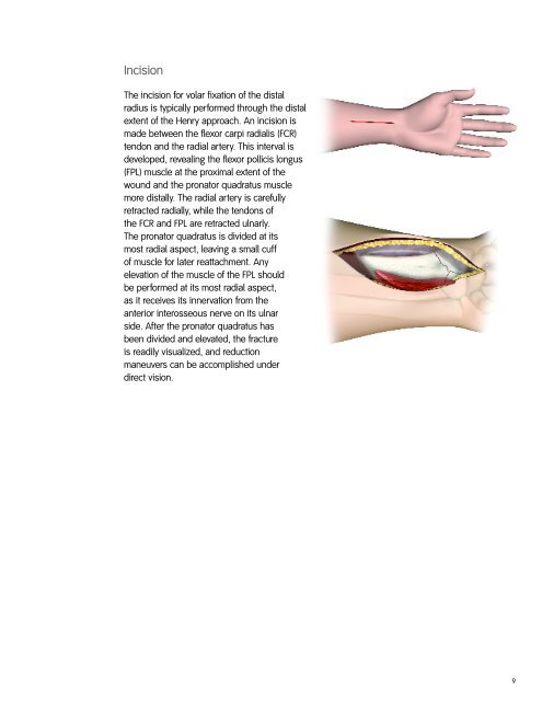
Incision<br />
The incision for volar fixation <strong>of</strong> the distal<br />
radius is typically performed through the distal<br />
extent <strong>of</strong> the Henry approach. An incision is<br />
made between the flexor carpi radialis (FCR)<br />
tendon and the radial artery. This interval is<br />
developed, revealing the flexor pollicis longus<br />
(FPL) muscle at the proximal extent <strong>of</strong> the<br />
wound and the pronator quadratus muscle<br />
more distally. The radial artery is carefully<br />
retracted radially, while the tendons <strong>of</strong><br />
the FCR and FPL are retracted ulnarly.<br />
The pronator quadratus is divided at its<br />
most radial aspect, leaving a small cuff<br />
<strong>of</strong> muscle for later reattachment. Any<br />
elevation <strong>of</strong> the muscle <strong>of</strong> the FPL should<br />
be performed at its most radial aspect,<br />
as it receives its innervation from the<br />
anterior interosseous nerve on its ulnar<br />
side. After the pronator quadratus has<br />
been divided and elevated, the fracture<br />
is readily visualized, and reduction<br />
maneuvers can be accomplished under<br />
direct vision.<br />
9