A sebaceous cyst with a difference: Dermatobia hominis - Journal of ...
A sebaceous cyst with a difference: Dermatobia hominis - Journal of ...
A sebaceous cyst with a difference: Dermatobia hominis - Journal of ...

You also want an ePaper? Increase the reach of your titles
YUMPU automatically turns print PDFs into web optimized ePapers that Google loves.
798<br />
LETTER TO JCP<br />
A <strong>sebaceous</strong> <strong>cyst</strong> <strong>with</strong> a <strong>difference</strong>: <strong>Dermatobia</strong> <strong>hominis</strong><br />
L J Harbin, M Khan, E M Thompson, R D Goldin<br />
.............................................................................................................................<br />
<strong>Dermatobia</strong> <strong>hominis</strong> causes furuncular myiasis and is<br />
endemic to South America. This report describes a case in<br />
a young woman who had recently visited Belize, highlighting<br />
the importance <strong>of</strong> clinical history (including travel<br />
history) and close liaison between pathologist and<br />
surgeon.<br />
A 39<br />
year old American woman was referred by her<br />
general practitioner to the day case surgery unit at St<br />
Mary’s Hospital, London for excision <strong>of</strong> a scalp<br />
“<strong>sebaceous</strong> <strong>cyst</strong>” that had been present for two months. This<br />
had been increasing in size, bleeding intermittently, and was<br />
associated <strong>with</strong> cervical lymphadenopathy. On examination a<br />
3 cm firm, non-mobile <strong>cyst</strong> <strong>with</strong> central punctum was present.<br />
On excision, movement was noted <strong>with</strong>in the <strong>cyst</strong> cavity,<br />
and further dissection revealed the presence <strong>of</strong> a live larva,<br />
which crawled across the surgical trolley after removal. The<br />
<strong>cyst</strong> was removed in its entirety, and the wound debrided and<br />
cleaned <strong>with</strong> strict aseptic technique, <strong>with</strong> closure in the usual<br />
manner. Both theatre staff and patient were understandably<br />
alarmed by the nature <strong>of</strong> the <strong>cyst</strong> contents, and after the<br />
patient had been calmed, it was established that she had visited<br />
Belize four months previously, where she was bitten on<br />
the scalp.<br />
The histopathology department received two specimens: a<br />
skin ellipse <strong>with</strong> part <strong>of</strong> a <strong>cyst</strong> wall attached to the inferior<br />
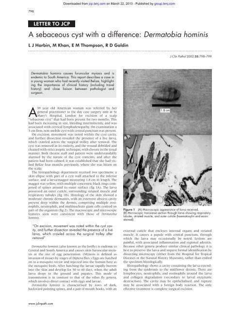
surface, and a larva/maggot measuring 1.8 cm in length. The<br />
maggot was yellow, <strong>with</strong> multiple concentric black rings composed<br />
<strong>of</strong> spines around its outer surface (fig 1A). The larva<br />
possessed an outer cuticle, surrounding striated muscle and<br />
respiratory tubules (fig 1B). Histology <strong>of</strong> the skin showed a<br />
moderate chronic dermatitis, <strong>with</strong> an extensive abscess cavity<br />
present deep <strong>with</strong>in the dermis, comprising multiple eosinophils,<br />
neutrophils, and multinucleate giant cells centred on<br />
part <strong>of</strong> the organism (fig 2). The macroscopic and microscopic<br />
features seen were consistent <strong>with</strong> those <strong>of</strong> <strong>Dermatobia</strong><br />
<strong>hominis</strong>.<br />
“On excision, movement was noted <strong>with</strong>in the <strong>cyst</strong> cavity,<br />
and further dissection revealed the presence <strong>of</strong> a live<br />
larva, which crawled across the surgical trolley after<br />
removal”<br />
<strong>Dermatobia</strong> <strong>hominis</strong> (also known as the botfly) is endemic to<br />
Central and South America and causes skin furuncular myiasis<br />
at the site <strong>of</strong> egg penetration. (Myiasis is defined as<br />
invasion <strong>of</strong> tissues by stages <strong>of</strong> Diptera flies.) Eggs are hatched<br />
on to a mosquito vector and injected into the human host as<br />
the mosquito feeds. After hatching the larvae rapidly burrow<br />
into the skin and develop for 50 to 60 days, when the adult<br />
larva drops to the ground and pupates. This mode <strong>of</strong><br />
transmission is in contrast to that <strong>of</strong> the other fly genera,<br />
which involves direct contact <strong>with</strong> eggs and larvae.<br />
<strong>Dermatobia</strong> <strong>hominis</strong> is characterised by rows <strong>of</strong> dark,<br />
backward pointing spines, and a pair <strong>of</strong> mouth hooks, <strong>with</strong> an<br />
www.jclinpath.com<br />
Downloaded from<br />
jcp.bmj.com on March 22, 2013 - Published by group.bmj.com<br />
J Clin Pathol 2002;55:798–799<br />
Figure 1 (A) Macroscopic appearance <strong>of</strong> larva received.<br />
(B) Microscopic transverse section through larva showing respiratory<br />
tubules, striated muscle, and outer cuticle (haematoxylin and eosin<br />
stained).<br />
external cuticle that encloses internal organs and striated<br />
muscle. It causes a papule <strong>with</strong> central punctum, through<br />
which the larva may occasionally be noted. Lesions are<br />
painful, <strong>with</strong> associated inflammation and regional adenitis.<br />
Because other genera produce similar clinical pathology it is<br />
best to preserve the larva and request formal identification by<br />
dissecting microscopy (either from the Hospital for Tropical<br />
Diseases or the Natural History Museum), rather than embed<br />
the specimen histologically.<br />
Histopathology shows a cavity containing the larva extending<br />
from the epidermis to the mid/lower dermis. There are<br />
lymphocytes, neutrophils, and eosinophils around the larva<br />
and collagen degradation (secondary to larval enzymatic<br />
destruction). The cavity may be epithelialised, and rupture<br />
may be associated <strong>with</strong> a foreign body reaction. The only<br />
effective treatment is complete surgical excision.
Downloaded from<br />
jcp.bmj.com on March 22, 2013 - Published by group.bmj.com<br />
Letter to JCP 799<br />
Figure 2 Intradermal abscess centred on organism (haematoxylin<br />
and eosin stained).<br />
A review <strong>of</strong> the literature revealed several reports <strong>of</strong> D <strong>hominis</strong><br />
from Europe, Scandinavia, Australia, and the Americas,<br />
reflecting the widening experience <strong>of</strong> infectious disease as<br />
foreign travel increases. In a review <strong>of</strong> 13 cases in Munich,<br />
Germany, all cases <strong>of</strong> D <strong>hominis</strong> infection were related to travel<br />
to the Central American tropics. 1 The most frequent differential<br />
diagnoses are infected <strong>sebaceous</strong> <strong>cyst</strong>, or a furuncle <strong>with</strong><br />
associated lymphadenopathy. Almost all cases are present on<br />
limbs, although two papers detailed D <strong>hominis</strong> infection <strong>of</strong> the<br />
eye: both <strong>of</strong> which caused palpebral swelling, 23 and one report<br />
described a woman <strong>with</strong> a long standing breast mass, excision<br />
biopsy <strong>of</strong> which revealed granulomatous inflammation<br />
centred around a fly larva. 4<br />
This case highlights the importance <strong>of</strong> the clinical history<br />
(including travel history), meticulous surgical technique to<br />
<br />
<br />
Take home messages<br />
• We report a case <strong>of</strong> <strong>Dermatobia</strong> <strong>hominis</strong> infection causing<br />
furuncular myiasis in a young woman who had<br />
recently visited Belize<br />
• This case highlights the importance <strong>of</strong> clinical history<br />
(including travel history) and close liaison between<br />
pathologist and surgeon<br />
effect complete removal <strong>of</strong> the fly larva, and essential communication<br />
between surgeon and the pathologist to achieve<br />
prompt diagnosis.<br />
.....................<br />
Authors’ affiliations<br />
L J Harbin, E M Thompson, R D Goldin, Departments <strong>of</strong> Pathology<br />
and Surgery, St Mary’s Hospital, London W2 1NY, UK<br />
M Khan, Department <strong>of</strong> Surgery, St Mary’s Hospital<br />
Correspondence to: Dr R D Goldin, Department <strong>of</strong> Histopathology,<br />
St Mary’s Hospital, London, W2 1NY, UK; r.goldin@ic.ac.uk<br />
Accepted for publication 9 May 2002<br />
REFERENCES<br />
1 Jelinek T, Nothdurft HD, Rieder N, et al. Cutaneous myiasis: review <strong>of</strong><br />
13 cases in travellers returning from tropical countries. Int J Dermatol<br />
1995;34:624–6.<br />
2 Bangsgaard R, Holst B, Krogh E, et al. Palpebral myiasis in a Danish<br />
traveller caused by the human bot-fly (<strong>Dermatobia</strong> <strong>hominis</strong>). Acta<br />
Ophthalmol Scand 2000;78:487–9.<br />
3 Goodman RL, Montalvo MA, Reed JB, et al. Photo essay: anterior<br />
orbital myiasis caused by human bot-fly (<strong>Dermatobia</strong> <strong>hominis</strong>). Arch<br />
Ophthalmol 2000;118:1002–3.<br />
4 Kahn DG. Myiasis secondary to <strong>Dermatobia</strong> <strong>hominis</strong> (human botfly)<br />
presenting as a long-standing breast mass. Arch Pathol Lab Med<br />
1999;123:829–31.<br />
<br />
<br />
<br />
<br />
<br />
www.jclinpath.com
References<br />
Email alerting<br />
service<br />
Topic<br />
Collections<br />
Notes<br />
A <strong>sebaceous</strong> <strong>cyst</strong> <strong>with</strong> a <strong>difference</strong>:<br />
<strong>Dermatobia</strong> <strong>hominis</strong><br />
L J Harbin, M Khan, E M Thompson, et al.<br />
J Clin Pathol 2002 55: 798-799<br />
doi: 10.1136/jcp.55.10.798<br />
Updated information and services can be found at:<br />
http://jcp.bmj.com/content/55/10/798.full.html<br />
These include:<br />
This article cites 4 articles<br />
http://jcp.bmj.com/content/55/10/798.full.html#ref-list-1<br />
Article cited in:<br />
http://jcp.bmj.com/content/55/10/798.full.html#related-urls<br />
Receive free email alerts when new articles cite this article. Sign up in the<br />
box at the top right corner <strong>of</strong> the online article.<br />
Articles on similar topics can be found in the following collections<br />
Dermatology (194 articles)<br />
To request permissions go to:<br />
http://group.bmj.com/group/rights-licensing/permissions<br />
To order reprints go to:<br />
http://journals.bmj.com/cgi/reprintform<br />
To subscribe to BMJ go to:<br />
http://group.bmj.com/subscribe/<br />
Downloaded from<br />
jcp.bmj.com on March 22, 2013 - Published by group.bmj.com