Vasitis nodosa and associated clinical findings - Journal of Clinical ...
Vasitis nodosa and associated clinical findings - Journal of Clinical ...
Vasitis nodosa and associated clinical findings - Journal of Clinical ...

You also want an ePaper? Increase the reach of your titles
YUMPU automatically turns print PDFs into web optimized ePapers that Google loves.
Downloaded from jcp.bmj.com on August 23, 2013 - Published by group.bmj.com<br />
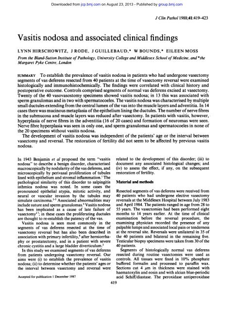
<strong>Vasitis</strong> <strong>nodosa</strong> <strong>and</strong> <strong>associated</strong> <strong>clinical</strong> <strong>findings</strong><br />
J Clin Pathol 1988;41:419-423<br />
LYNN HIRSCHOWITZ, J RODE, J GUILLEBAUD,* W BOUNDS,* EILEEN MOSS<br />
From the Bl<strong>and</strong>-Sutton Institute <strong>of</strong> Pathology, University College <strong>and</strong> Middlesex School <strong>of</strong> Medicine, <strong>and</strong> *the<br />
Margaret Pyke Centre, London<br />
SUMMARY To establish the prevalence <strong>of</strong> vasitis <strong>nodosa</strong> in patients who had undergone vasectomy<br />
segments <strong>of</strong> vas deferens resected from 40 patients at the time <strong>of</strong> vasectomy reversal were examined<br />
histologically <strong>and</strong> immunohistochemically. The <strong>findings</strong> were correlated with <strong>clinical</strong> history <strong>and</strong><br />
postoperative outcome. Controls comprised segments <strong>of</strong> normal vas deferens excised at vasectomy.<br />
Twenty <strong>of</strong> the 40 vasovasostomy specimens showed vasitis <strong>nodosa</strong>; in 13 this was <strong>associated</strong> with<br />
sperm granulomas <strong>and</strong> in two with spermatocoeles. The vasitis <strong>nodosa</strong> was characterised by multiple<br />
small ductules extending from the central lumen <strong>of</strong> the vas into the muscle layers <strong>and</strong> adventitia. In 14<br />
cases there was mucinous metaplasia <strong>of</strong> the epithelium lining the ductules. The number <strong>of</strong> nerve fibres<br />
in the submucosa <strong>and</strong> muscle layers was reduced after vasectomy. In patients with vasitis, however,<br />
hyperplasia <strong>of</strong> nerve fibres in the adventitia (16 <strong>of</strong> 20 cases) <strong>and</strong> formation <strong>of</strong> neuromas were seen.<br />
Nerve fibre hyperplasia was seen in only one, <strong>and</strong> sperm granulomas <strong>and</strong> spermatocoeles in none <strong>of</strong><br />
the 20 specimens without vasitis <strong>nodosa</strong>.<br />
The development <strong>of</strong> vasitis <strong>nodosa</strong> was independent <strong>of</strong> the patients' age or the interval between<br />
vasectomy <strong>and</strong> reversal. The restoration <strong>of</strong> fertility did not seem to be affected by previous vasitis<br />
<strong>nodosa</strong>.<br />
In 1943 Benjamin et al proposed the term "vasitis<br />
<strong>nodosa</strong>" to describe a benign disorder, characterised<br />
macroscopically by nodularity <strong>of</strong> the vas deferens, <strong>and</strong><br />
microscopically by perivasal proliferation <strong>of</strong> tubules<br />
lined with epithelium <strong>and</strong> stromal inflammation.' The<br />
pathological similarity <strong>of</strong> this disorder to salpingitis<br />
isthmica <strong>nodosa</strong> was noted. In some cases the<br />
pronounced epithelial atypia, mitotic activity, <strong>and</strong><br />
neural or vascular invasion by the tubules may<br />
simulate carcinoma.2`5 Associated abnormalities may<br />
include suture <strong>and</strong> sperm granulomas.6 <strong>Vasitis</strong> <strong>nodosa</strong><br />
has been implicated as a cause <strong>of</strong> late failure <strong>of</strong><br />
vasectomy2 7; in these cases the proliferating ductules<br />
are thought to re-establish the patency <strong>of</strong> the vas.<br />
<strong>Vasitis</strong> <strong>nodosa</strong> is seen most commonly in the<br />
segments <strong>of</strong> vas deferens resected at the time <strong>of</strong><br />
vasectomy reversal but has also been described in<br />
association with primary infertility,8 after herniorrhaphy<br />
or prostatectomy, <strong>and</strong> in a patient with severe<br />
chronic cystitis <strong>and</strong> a large bladder diverticulum.2<br />
In this study we examined segments <strong>of</strong> vas deferens<br />
from patients undergoing vasectomy reversal. Our<br />
aims were (i) to establish the prevalence <strong>of</strong> vasitis<br />
<strong>nodosa</strong>; (ii) to determine whether the patients' ages or<br />
the interval between vasectomy <strong>and</strong> reversal were<br />
Accepted for publication I December 1987<br />
related to the development <strong>of</strong> this disorder; (iii) to<br />
document any <strong>associated</strong> histological changes; <strong>and</strong><br />
(iv) to assess the effect, if any, on the subsequent<br />
restoration <strong>of</strong> fertility.<br />
Material <strong>and</strong> methods<br />
Resected segments <strong>of</strong> vas deferens were received from<br />
40 patients who had undergone elective vasectomy<br />
reversals at the Middlesex Hospital between July 1983<br />
<strong>and</strong> April 1984. The patients ranged in age from 28 to<br />
55 years. The vasectomies had been performed eight<br />
months to 14 years earlier. At the time <strong>of</strong> <strong>clinical</strong><br />
examination before the reversal procedure, the<br />
examining physician recorded the presence <strong>of</strong> any<br />
palpable lumps <strong>and</strong> <strong>associated</strong> local pain or tenderness<br />
at the reversal site. Reversals were unilateral in 35 <strong>of</strong><br />
the 40 patients <strong>and</strong> bilateral in the remaining five.<br />
Testicular biopsy specimens were taken from 30 <strong>of</strong> the<br />
40 patients.<br />
Segments <strong>of</strong> histologically normal vas deferens<br />
resected during routine vasectomies were used as<br />
controls. All tissues were fixed in 10% phosphate<br />
buffered formalin <strong>and</strong> processed to paraffin wax.<br />
Sections cut 4 gm in thickness were stained with<br />
haematoxylin <strong>and</strong> eosin <strong>and</strong> with alcian blue-periodic<br />
acid Schiff/diastase. The peroxidase antiperoxidase<br />
419
Downloaded from jcp.bmj.com on August 23, 2013 - Published by group.bmj.com<br />
420<br />
method was used to immunostain sections with polyclonal<br />
rabbit anti-PGP 9 5. This antiserum specifically<br />
labels neurons, neuroendocrine cells, <strong>and</strong> nerve fibres.9<br />
The patients' sperm counts were monitored at three<br />
to four monthly intervals for six to 15 months after<br />
vasovasostomy <strong>and</strong> any pregnancies in their spouses<br />
were recorded.<br />
An unpaired Student's t test was used to compare<br />
the age <strong>of</strong> patients with <strong>and</strong> without vasitis <strong>nodosa</strong>,<br />
<strong>and</strong> also the interval between vasectomy <strong>and</strong> reversal<br />
in these two groups. The significance <strong>of</strong> the associations<br />
between vasitis <strong>nodosa</strong> <strong>and</strong> other pathological<br />
abnormalities was analysed in 2 x 2 contingency tables<br />
by x2 test.<br />
Results<br />
jSff<br />
Hirschowitz, Rode, Guillebaud, Bounds, Moss<br />
stte a\.z,\<br />
s<br />
I6<br />
VASITIS NODOSA<br />
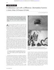
In 20 <strong>of</strong> the 40 specimens varying numbers <strong>of</strong> tubules<br />
lined by columnar or cuboidal epithelium extended<br />
from the vas into the surrounding muscle <strong>and</strong><br />
perivasal connective tissue (table 1, fig 1). In most<br />
cases the lining epithelium <strong>of</strong> the tubules was well<br />
differentiated but in the more florid cases <strong>of</strong> vasitis<br />
<strong>nodosa</strong> the nuclei were hyperchromatic, the nuclear:<br />
cytoplasmic ratio was increased, <strong>and</strong> occasional cells<br />
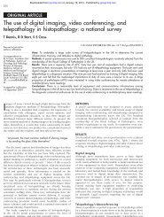
were in mitosis. In eight cases tubules extended into<br />
perivasal nerve fascicles (figs 2a <strong>and</strong> b) but there was<br />
no evidence <strong>of</strong> vascular invasion. In only four <strong>of</strong> the<br />
specimens with vasitis <strong>nodosa</strong> were there no <strong>associated</strong><br />
histological lesions. In the remaining cases vasitis<br />
<strong>nodosa</strong> was accompanied by sperm granulomas,<br />
abnormalities in the innervation <strong>of</strong> the vas, or<br />
mucinous metaplasia in the proliferating ductules.<br />
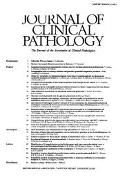
SPERM GRANULOMAS<br />
These were only present in 13 cases with vasitis <strong>nodosa</strong><br />
(table 1, fig 3). The granulomas consisted <strong>of</strong> poorly<br />
circumscribed aggregates <strong>of</strong> chronic inflammatory<br />
cells <strong>and</strong> histiocytes surrounding collections <strong>of</strong><br />
extravasated spermatozoa. In some cases the<br />
aggregates <strong>of</strong> chronic inflammatory cells were<br />
admixed with ceroid-containing histiocytes. Ceroid<br />
pigment in the granulomas <strong>of</strong> vasitis <strong>nodosa</strong> results<br />
Table 1 Histological changes in excised segments <strong>of</strong> vas:<br />
association with vasitis <strong>nodosa</strong><br />
With<br />
Without<br />
vasitis<br />
vasitis<br />
(n= 20) (n= 20) p value<br />
Sperm granuloma 13 0 p
Downloaded from jcp.bmj.com on August 23, 2013 - Published by group.bmj.com<br />
<strong>Vasitis</strong> <strong>nodosa</strong> <strong>and</strong> <strong>associated</strong> <strong>clinical</strong><strong>findings</strong><br />
I ~ ~ . .l^.<br />
421<br />
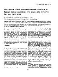
Figs 2a <strong>and</strong> b Perivasal nerve fascicles immunostainedfor PGP 9-5 showing infiltration by ductules (arrows).<br />
(Peroxidase antiperoxidase.)<br />
!~~~~~PA~ ~ ~ '<br />
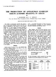
Fig 3 Section <strong>of</strong>sperm granuloma <strong>associated</strong> with vasitis<br />
<strong>nodosa</strong>. Central aggregate <strong>of</strong>spermatozoa is surrounded by<br />
macrophages <strong>and</strong> lymphocytes. (Haematoxylin <strong>and</strong> eosin.)<br />
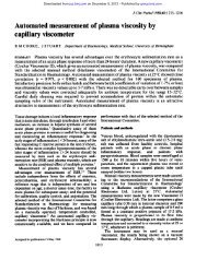
Fig 4 Section through intertwining nervefibres <strong>of</strong>neuroma<br />
in wall <strong>of</strong> vas. Some mononuclear inflammatory-cells are<br />
present. (Haematoxylin <strong>and</strong> eosin.)
Downloaded from jcp.bmj.com on August 23, 2013 - Published by group.bmj.com<br />
422<br />
vasitis <strong>nodosa</strong>, however, there were many enlarged<br />
nerve fascicles in the adventitia <strong>of</strong> the vas. Some <strong>of</strong><br />
these formed neuromas (fig 4) consisting <strong>of</strong> r<strong>and</strong>omly<br />
orientated, intertwining bundles <strong>of</strong> nerve fibres.<br />
Hyperplasia <strong>of</strong> the nerve fibres in the adventitia, with<br />
or without neuroma formation, was present in 16 <strong>of</strong><br />
the specimens with, but in only one <strong>of</strong> those without<br />
vasitis <strong>nodosa</strong> (x2 = 23-02, p < 0-0001).<br />
MUCINOUS METAPLASIA<br />
The columnar epithelial lining <strong>of</strong> the proliferating<br />
tubules was, in part, replaced by well differentiated<br />
mucin-secreting epithelium (table 1, fig 5). This feature<br />
was noted in 14 <strong>of</strong> the cases with vasitis <strong>nodosa</strong>.<br />
Staining <strong>of</strong> the tissues with alcian blue <strong>and</strong> periodic<br />
acid Schiff/diastase confirmed the presence <strong>of</strong> both<br />
intracellular <strong>and</strong> extracellular mucin.<br />
TESTICULAR BIOPSY SPECIMENS<br />
All <strong>of</strong> these showed evidence <strong>of</strong> active spermatogenesis.<br />
The seminiferous tubules were moderately dilated<br />
<strong>and</strong> some <strong>of</strong> the spermatogenic epithelium had<br />
sloughed into the tubular lumena <strong>and</strong> was mixed with<br />
spermatids. This appearance was thought to be due to<br />
obstruction to testicular outflow.'0<br />
The mean age <strong>of</strong> patients with vasitis <strong>nodosa</strong> was<br />
38 4 years (SD 3 5) <strong>and</strong> <strong>of</strong> those without, 38-8 years<br />
(SD 7 3) (table 2). The interval between vasectomy <strong>and</strong><br />
Fig 5 Ductules showing mucinous metaplasia <strong>of</strong> lining<br />
epithelium. (Alcian blue-periodic acid Schif/diastase.)<br />
Table 2<br />
Hirschowitz, Rode, Guillebaud, Bounds, Moss<br />
<strong>Clinical</strong><strong>findings</strong><br />
With Without<br />
vasitis vasitis p value<br />
Age (years) Range 32-45 28-55<br />
Mean (SD) 38-4 (3-5) 38-8 (7-3) p= 0-8<br />
Interval between<br />
vasectomy <strong>and</strong><br />
reversal (years) Range 5-12 07-14<br />
Mean (SD) 7 9 (2.7) 6-9 (4-1) p=0 4<br />
Palpable lump 12/17* 6/18* p = 0 03<br />
Sperm count >2x 10⁶<br />
or impregnation 6/17* 11/20* p=052<br />
Ensuing pregnancies 3 6 p = 0-38<br />
*Denominator indicates number <strong>of</strong> patients examined.<br />
reversal was also similar in the two groups: 7 9 (SD<br />
2.7) <strong>and</strong> 6-9 (SD 4-1) respectively.<br />
Twelve patients with vasitis <strong>nodosa</strong> <strong>and</strong> six without<br />
had palpable lumps at the vasectomy site. (x2 = 4 9,<br />
p = 0 03) (table 2). In none <strong>of</strong> the patients were the<br />
lumps painful or tender to palpation.<br />
After vasectomy reversal the patients were followed<br />
up for six to 15 months. During this time six <strong>of</strong> the<br />
patients without vasitis <strong>nodosa</strong> successfully impregnated<br />
their spouses as did three patients with vasitis<br />
<strong>nodosa</strong> (x2 = 0-76, p = 0-38) (table 2). Sperm counts<br />
between 2 x 10⁶/ml <strong>and</strong> 69 x 106/ml were noted in three<br />
patients with vasitis <strong>and</strong> in five without.<br />
Persistent azoospermia or hypozoospermia (< 2 x 10⁶<br />
spermatozoa/ml) was present in 11 patients with <strong>and</strong><br />
in nine without vasitis <strong>nodosa</strong>.<br />
Discussion<br />
Present <strong>findings</strong> show that vasitis <strong>nodosa</strong> complicates<br />
about 50% <strong>of</strong> vasectomies, irrespective <strong>of</strong> the age <strong>of</strong><br />
the patient or the interval between vasectomy <strong>and</strong><br />
reversal. Associated histological abnormalities include<br />
sperm granulomas, mucinous metaplasia <strong>of</strong> the<br />
proliferating ductules, <strong>and</strong> hyperplasia <strong>of</strong> nerves in the<br />
perivasal tissues. The restoration <strong>of</strong> fertility after<br />
vasovasostomy does not seem to be affected by<br />
previous vasitis <strong>nodosa</strong>.<br />
The prevalence <strong>of</strong> vasitis <strong>nodosa</strong> in the present study<br />
was slightly lower than the 66% reported by Taxy et at<br />
<strong>and</strong> Kiser et al.3 In both <strong>of</strong> these studies the interval<br />
between vasectomy <strong>and</strong> reversal ranged from one to 15<br />
years. The incidence <strong>of</strong> vasitis <strong>nodosa</strong> does not seem<br />
to increase after the first few months following<br />
vasectomy, suggesting that vasitis develops, if at all,<br />
within this period. Civantos et al noted vasitis <strong>nodosa</strong><br />
as early as one month after vasectomy.2<br />
Although several mechanisms have been proposed'<br />
to explain the pathogenesis <strong>of</strong> vasitis <strong>nodosa</strong>, it is<br />
generally believed that raised intravasal pressure, due<br />
to obstruction <strong>of</strong> the vas, causes disruption <strong>of</strong> the<br />
lining epithelium <strong>and</strong> extravasation <strong>of</strong> spermatozoa
Downloaded from jcp.bmj.com on August 23, 2013 - Published by group.bmj.com<br />
<strong>Vasitis</strong> <strong>nodosa</strong> <strong>and</strong> <strong>associated</strong> <strong>clinical</strong><strong>findings</strong> 423<br />
into the surrounding tissue.21' The spermatozoa elicit barrier" which normally shields the spermatozoa from<br />
an intense inflammatory reaction which results in the the immune system.2223<br />
formation <strong>of</strong> sperm granulomas. Ductules are thought<br />
to be formed by growth <strong>of</strong> epithelial cells from We thank Dr R J Thompson for providing antibody to<br />
breaches in the mucosa <strong>of</strong> the vas into the inflamed PGP9.5.<br />
tissue. Distension <strong>of</strong> ductules due to continued<br />
formation <strong>of</strong> spermatozoa probably causes the<br />
References<br />
spermatocoeles. Chronic inflammation <strong>of</strong> the ductal<br />
epithelium may initiate the mucinous metaplasia that<br />
was observed in the present study. We encountered<br />
sperm granulomas only in association with vasitis<br />
<strong>nodosa</strong>, the prevalence being similar to that <strong>of</strong><br />
previous studies.8 2<br />
The cause <strong>and</strong> <strong>clinical</strong> importance <strong>of</strong> the nerve<br />
hyperplasia <strong>and</strong> neuromas are unclear. Although the<br />
literature<br />
neuromas are probably traumatic in origin, their<br />
predominance in those patients who have had vasitis<br />
<strong>nodosa</strong> indicates that factors other than the surgery<br />
alone are likely to have a role. It is conceivable that the<br />
proliferating epithelium elaborates growth factors<br />
which induce hyperplasia <strong>of</strong> the perivasal nerves.<br />
Alternatively, the severe inflammation <strong>and</strong> the infiltration<br />
<strong>of</strong> nerves by proliferating ductules may exacerbate<br />
the surgical trauma <strong>and</strong> thereby promote the<br />
nerve hyperplasia <strong>and</strong> neuroma formation. Although<br />
pain <strong>and</strong> tenderness have been reported in association<br />
with vasitis <strong>nodosa</strong>,'3 none <strong>of</strong> the patients in this series<br />
presented with painful masses. The infiltration <strong>of</strong><br />
nerves'1'7 <strong>and</strong> blood vessels'8 by ductules are features<br />
that may simulate metastatic adenocarcinoma <strong>of</strong> the<br />
prostate, which tends to spread along lymphatics in<br />
the adventitia <strong>of</strong> the vas.2 The typical ductal branching<br />
<strong>of</strong> vasitis <strong>nodosa</strong> <strong>and</strong> the presence <strong>of</strong> spermatozoa<br />
within the ducts <strong>and</strong> in the granulomas may help in<br />
distinguishing this process from carcinoma.3 Rarely,<br />
vasitis <strong>nodosa</strong> may be the site <strong>of</strong> arrest <strong>of</strong> malignant<br />
cells from a primary tumour in the ipsilateral testis.'9<br />
Silber found that the presence <strong>of</strong> sperm granulomas<br />
in the segment <strong>of</strong> vas excised during vasectomy<br />
reversal augured a favourable outcome.'220 He suggested<br />
that the sperm granuloma acted as a "safety<br />
valve", relieving the high pressure in the testicular part<br />
<strong>of</strong> the vas, <strong>and</strong> thereby alleviating the effects <strong>of</strong> high<br />
intravasal pressure on the testis itself. We found no<br />
significant difference between the fertility <strong>of</strong> patients<br />
with, <strong>and</strong> those without, sperm granulomas in the<br />
excised segment <strong>of</strong> vas.<br />
There were several patients in this study, who failed<br />
to impregnate their spouses, despite the recovery <strong>of</strong><br />
sperm counts to normal or near normal levels. These<br />
results may reflect the relatively short period <strong>of</strong> follow<br />
up. Alternatively, the persistent infertility may be due<br />
to the development <strong>of</strong> antisperm antibodies, which can<br />
be detected in 50 to 70% <strong>of</strong> patients after vasectomy.s21<br />
This procedure is believed to disrupt the "blood-testis<br />
1 Benjamin JA, Robertson TD, Cheetham JG. <strong>Vasitis</strong> <strong>nodosa</strong>: a new<br />
<strong>clinical</strong> entity simulating tuberculosis <strong>of</strong> the vas deferens. J Urol<br />
1943;49:575-82.<br />
2 Civantos F, Lubin J, Rywlin AM. <strong>Vasitis</strong> <strong>nodosa</strong>. Archives <strong>of</strong><br />
Pathologyl972;94:355-61.<br />
3 Kiser GC. Fuchs EF, Kessler S. The significance <strong>of</strong> vasitis <strong>nodosa</strong>.<br />
J Urol 1986;136:42-4.<br />
4 Graham JB, O'Connor VJ. Spermatic cord tumors: review <strong>of</strong><br />
<strong>and</strong> a case <strong>of</strong> an unusual vas deferens tumor in an<br />
infertility problem. J Urol 1954;72:946-9.<br />
5 Taxy JB. <strong>Vasitis</strong> <strong>nodosa</strong>. Arch Pathol Lab Med 1978;102:643-7.<br />
6 Chapman ES, Heidger PM. Spermatic granuloma <strong>of</strong> vas deferens<br />
after vasectomy in rhesus monkeys <strong>and</strong> men. Urology<br />
1979;13:629-39.<br />
7 Philp T, Guillebaud J, Budd D. Late failure <strong>of</strong> vasectomy after two<br />
documented analyses showing azoospermic semen. Br Med J<br />
1984;289:77-9.<br />
8 Taxy JB, Marshall FF, Erlichman RJ. Vasectomy. Sub<strong>clinical</strong><br />
pathologic changes. Am J Surg Pathol 1981;5:767-72.<br />
9 Rode J, Dhillon AP,- Doran JF, Jackson P, Thompson RJ. PGP<br />
9.5, a new marker for human neuroendocrine tumours. Histopathology<br />
1985;9:147-58.<br />
10 Levin HS. Testicular biopsy in the study <strong>of</strong> male infertility. Its<br />
current usefulness, histologic techniques, <strong>and</strong> prospects for the<br />
future. Hum Pathol 1979;10:569-84.<br />
11 Olson AL. <strong>Vasitis</strong> <strong>nodosa</strong>. Am J Clin Pathol 1971;55:364-8.<br />
12 Silber SJ. Microscopic vasectomy reversal. Fertil Steril<br />
1977;28:1 191-202.<br />
13 Schmidt SS. Spermatic granuloma: an <strong>of</strong>ten painful lesion. Fertil<br />
Steril 1979;31:178-81.<br />
14 Balogh K, Travis WD. The frequency <strong>of</strong> perineural ductules in<br />
vasitis <strong>nodosa</strong>. Am J Clin Pathol 1984;82:710-3.<br />
15 Zimmerman KG, Johnson PC, Paplanus SH. Nerve invasion by<br />
benign proliferating ductules in vasitis <strong>nodosa</strong>. Cancer<br />
983;51:2066-9.<br />
16 Kovi J, Agbata A. Benign neural invasion in vasitis <strong>nodosa</strong>.<br />
JAMA 1974;228:1519<br />
17 Goldman RL, Azzopardi JG. Benign neural invasion in vasitis<br />
<strong>nodosa</strong>. Histopathology 1982;6:309-15.<br />
18 Balogh K, Travis WD. Benign vascular invasion in vasitis <strong>nodosa</strong>.<br />
Am J Clin Pathol 1985;83:426-30.<br />
19 Heaton JM, Maclennan KA. <strong>Vasitis</strong> <strong>nodosa</strong>-a site <strong>of</strong> arrest <strong>of</strong><br />
malignant germ cells. Histopathology 1986;10:981-9.<br />
20 Silber SJ. Sperm granuloma <strong>and</strong> reversibility <strong>of</strong> vasectomy. Lancet<br />
1977;ii:588-9.<br />
21 Samuel T, Rose NR. The lessons <strong>of</strong> vasectomy-a review. J Clin<br />
Lab Immunol 1980;3:77-83.<br />
22 Anonymous. Safety <strong>of</strong> vasectomy [Editorial]. Lancet 1979;ii:<br />
1057-8.<br />
23 Lepow IH, Crozier R. Vasectomy: immunologic <strong>and</strong> pathophysiologic<br />
effects in animals <strong>and</strong> man. New York: Academic<br />
Press, 1979;581-95.<br />
Requests for reprints to: Dr J Rode, Department <strong>of</strong> Histopathology,<br />
University College <strong>and</strong> Middlesex School <strong>of</strong><br />
Medicine, Rockefeller Building, University Street, London<br />
WC 1 E 6JJ, Engl<strong>and</strong>.
Downloaded from jcp.bmj.com on August 23, 2013 - Published by group.bmj.com<br />
<strong>Vasitis</strong> <strong>nodosa</strong> <strong>and</strong> <strong>associated</strong><br />
<strong>clinical</strong> <strong>findings</strong>.<br />
L Hirschowitz, J Rode, J Guillebaud, et al.<br />
J Clin Pathol 1988 41: 419-423<br />
doi: 10.1136/jcp.41.4.419<br />
Updated information <strong>and</strong> services can be found at:<br />
http://jcp.bmj.com/content/41/4/419<br />
References<br />
Email alerting<br />
service<br />
These include:<br />
Article cited in:<br />
http://jcp.bmj.com/content/41/4/419#related-urls<br />
Receive free email alerts when new articles cite this<br />
article. Sign up in the box at the top right corner <strong>of</strong> the<br />
online article.<br />
Notes<br />
To request permissions go to:<br />
http://group.bmj.com/group/rights-licensing/permissions<br />
To order reprints go to:<br />
http://journals.bmj.com/cgi/reprintform<br />
To subscribe to BMJ go to:<br />
http://group.bmj.com/subscribe/