Diffuse Cirrhosis-like Hepatocellular Carcinoma - Rush University ...
Diffuse Cirrhosis-like Hepatocellular Carcinoma - Rush University ...
Diffuse Cirrhosis-like Hepatocellular Carcinoma - Rush University ...

Create successful ePaper yourself
Turn your PDF publications into a flip-book with our unique Google optimized e-Paper software.
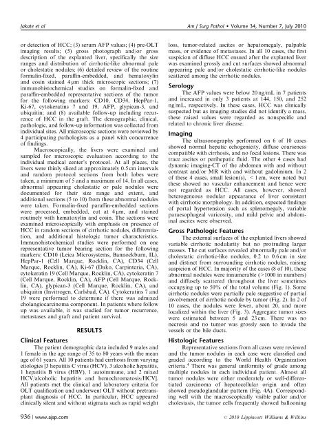
Jakate et al Am J Surg Pathol Volume 34, Number 7, July 2010<br />
or detection of HCC; (3) serum AFP values; (4) pre-OLT<br />
imaging results; (5) gross photograph and/or gross<br />
description of the explanted liver, specifically the size<br />
ranges and distribution of cirrhotic-<strong>like</strong> abnormal pale<br />
or cholestatic nodules; (6) detailed review of the routine<br />
formalin-fixed, paraffin-embedded, and hematoxylin<br />
and eosin stained 4 mm thick microscopic sections; (7)<br />
immunohistochemical studies on formalin-fixed and<br />
paraffin-embedded representative sections of the tumor<br />
for the following markers: CD10, CD34, HepPar-1,<br />
Ki-67, cytokeratins 7 and 19, AFP, glypican-3, and<br />
ubiquitin; and (8) available follow-up including recurrence<br />
of HCC in the graft. The demographic, clinical,<br />
pathologic, and follow-up information was collected from<br />
individual sites. All microscopic sections were reviewed by<br />
4 participating pathologists as a panel with concurrence<br />
of findings.<br />
Macroscopically, the livers were examined and<br />
sampled for microscopic evaluation according to the<br />
individual medical center’s protocol. At all places, the<br />
livers were thinly sliced at approximately 0.5 cm intervals<br />
and random protocol sections from both lobes were<br />
taken, a minimum of 5 and a maximum of 14. In all cases,<br />
abnormal appearing cholestatic or pale nodules were<br />
documented for their size range and extent, and<br />
additional sections (5 to 10) from these abnormal nodules<br />
were taken. Formalin-fixed paraffin-embedded sections<br />
were processed, embedded, cut at 4 mm, and stained<br />
routinely with hematoxylin and eosin. The sections were<br />
examined microscopically with emphasis on presence of<br />
HCC in random sections of cirrhotic nodules, differentiation,<br />
and additional histologic tumor characteristics.<br />
Immunohistochemical studies were performed on one<br />
representative tumor bearing section for the following<br />
markers: CD10 (Leica Microsystems, Bannockburn, IL),<br />
HepPar-1 (Cell Marque, Rocklin, CA), CD34 (Cell<br />
Marque, Rocklin, CA), Ki-67 (Dako, Carpinteria, CA),<br />
cytokeratin 19 (Cell Marque, Rocklin, CA), cytokeratin 7<br />
(Cell Marque, Rocklin, CA), AFP (Cell Marque, Rocklin,<br />
CA), glypican-3 (Cell Marque, Rocklin, CA), and<br />
ubiquitin (Invitrogen, Carlsbad, CA). Cytokeratins 7 and<br />
19 were performed to determine if there was admixed<br />
cholangiocarcinoma component. In patients where follow<br />
up was available, it was studied for tumor recurrence,<br />
metastases and graft and patient survival.<br />
RESULTS<br />
Clinical Features<br />
The patient demographic data included 9 males and<br />
1 female in the age range of 35 to 80 years with the mean<br />
age of 61 years. All 10 patients had cirrhosis from varying<br />
etiologies [3 hepatitis C virus (HCV), 3 alcoholic hepatitis,<br />
1 hepatitis B virus (HBV), 1 autoimmune, and 2 mixed<br />
HCV/alcoholic hepatitis and hemochromatosis/HCV].<br />
All patients met the clinical and laboratory criteria for<br />
OLT qualification and underwent OLT without pretransplant<br />
diagnosis of HCC. In particular, HCC appeared<br />
clinically silent and without stigmata such as rapid weight<br />
936 | www.ajsp.com<br />
loss, tumor-related ascites or hepatomegaly, palpable<br />
mass, or evidence of metastases. In all 10 cases, the first<br />
suspicion of diffuse HCC ensued after the explanted liver<br />
was examined grossly and cut surfaces showed abnormal<br />
appearing pale and/or cholestatic cirrhotic-<strong>like</strong> nodules<br />
scattered among the cirrhotic nodules.<br />
Serology<br />
The AFP values were below 20 ng/mL in 7 patients<br />
and increased in only 3 patients at 144, 150, and 252<br />
ng/mL, respectively. In these cases, HCC was clinically<br />
suspected but as imaging studies did not identify a mass,<br />
these raised values were regarded as nonspecific and<br />
related to chronic liver disease.<br />
Imaging<br />
The ultrasonography performed on 6 of 10 cases<br />
showed normal hepatic echogenicity, diffuse coarseness<br />
compatible with cirrhosis, and no focal lesions. There was<br />
trace ascites or perihepatic fluid. The other 4 cases had<br />
dynamic imaging-CT of the abdomen with and without<br />
contrast and/or MR with and without gadolinium. In 2<br />
of these 4 cases, small lesion(s), 1000 in numbers)<br />
and diffusely scattered throughout the liver sometimes<br />
occupying up to 50% of the total volume (Fig. 1). Some<br />
cirrhotic nodules were partially pale suggestive of partial<br />
involvement of cirrhotic nodule by tumor (Fig. 2). In 2 of<br />
10 cases, the nodules were fewer, about 20, and more<br />
localized within the liver (Fig. 3). Aggregate tumor sizes<br />
were estimated between 5 and 23 cm. There was no<br />
necrosis and no tumor was grossly seen to invade the<br />
vessels or the bile ducts.<br />
Histologic Features<br />
Representative sections from all cases were reviewed<br />
and the tumor nodules in each case were classified and<br />
graded according to the World Health Organization<br />
criteria. 4 There was general uniformity of grade among<br />
multiple nodules in each individual patient. Almost all<br />
tumor nodules were either moderately or well-differentiated<br />
carcinoma of hepatocellular origin and often<br />
showed pseudoglandular pattern (Fig. 4A). Corresponding<br />
well with the macroscopically visible pallor and/or<br />
cholestasis, the tumor cells frequently showed ballooning<br />
r 2010 Lippincott Williams & Wilkins