Diffuse Cirrhosis-like Hepatocellular Carcinoma - Rush University ...
Diffuse Cirrhosis-like Hepatocellular Carcinoma - Rush University ...
Diffuse Cirrhosis-like Hepatocellular Carcinoma - Rush University ...

You also want an ePaper? Increase the reach of your titles
YUMPU automatically turns print PDFs into web optimized ePapers that Google loves.
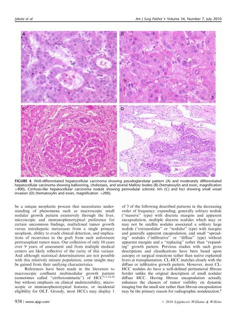
Jakate et al Am J Surg Pathol Volume 34, Number 7, July 2010<br />
FIGURE 4. Well-differentiated hepatocellular carcinoma showing pseudoglandular pattern (A) and moderately differentiated<br />
hepatocellular carcinoma showing ballooning, cholestasis, and several Mallory bodies (B) (hematoxylin and eosin, magnification<br />
400). <strong>Cirrhosis</strong>-<strong>like</strong> hepatocellular carcinoma nodule showing perinodular sclerotic rim (C) and foci showing small vessel<br />
invasion (D) (hematoxylin and eosin, magnification 200).<br />
be a unique neoplastic process that necessitates understanding<br />
of phenomena such as macroscopic small<br />
nodular growth pattern extensively through the liver,<br />
microscopic and immunophenotypical preference for<br />
certain uncommon findings, multiclonal tumor growth<br />
versus intrahepatic metastases from a single primary<br />
neoplasm, ability to evade clinical detection, and implications<br />
of recurrence in the graft from such unforeseen<br />
pretransplant tumor mass. Our collection of only 10 cases<br />
over 9 years of assessment and from multiple medical<br />
centers are <strong>like</strong>ly reflective of the rarity of this variant.<br />
And although statistical determinations are not possible<br />
with this relatively minute population, some insight may<br />
be gained from their unifying characteristics.<br />
References have been made in the literature to<br />
macroscopic confluent multinodular growth pattern<br />
(sometimes called ‘‘cirrhotomimetic’’) of HCC 3,5,14,16<br />
but without emphasis on clinical undetectability, microscopic<br />
or immunophenotypical features, or incidental<br />
eligibility for OLT. Grossly, most HCCs may display 1<br />
938 | www.ajsp.com<br />
of 3 of the following described patterns in the decreasing<br />
order of frequency: expanding, generally solitary nodule<br />
(‘‘massive’’ type) with discrete margins and apparent<br />
encapsulation, multiple discrete nodules which may or<br />
may not be satellite nodules associated a solitary large<br />
nodule (‘‘extranodular’’ or ‘‘nodular’’ type) with margins<br />
and generally apparent encapsulation, and small ‘‘spreading’’<br />
nodules (‘‘infiltrative’’ or ‘‘diffuse’’ type) without<br />
apparent margins and a ‘‘replacing’’ rather than ‘‘expanding’’<br />
growth pattern. Previous studies with such gross<br />
descriptions and classifications have been based upon<br />
autopsy or surgical resections rather than native explanted<br />
livers at transplantation. CL-HCC matches closely with the<br />
diffuse or infiltrative growth pattern. However, most CL-<br />
HCC nodules do have a well-defined peritumoral fibrous<br />
border un<strong>like</strong> the original description of small nodular<br />
diffuse HCC. Having fibrous encapsulation actually<br />
enhances the chances of tumor visibility on dynamic<br />
imaging but the small size rather than fibrous encapsulation<br />
may be the primary reason for radiographic nondetection. 6<br />
r 2010 Lippincott Williams & Wilkins